
Even the simplest cell may house thousands of individual chemical reactions. But the number of different kinds of chemical reaction is surprisingly small. In fact, we saw the same underlying pattern in our discussion of genetic regulation. While the specific details of the vast array of regulatory events may be complicated, only a fairly small number of different principles are needed to explain the essential mechanisms at work in an organism. There is often a surprising degree of simplicity underlying the seemingly complex events of life. As we will see in this chapter, the genetic control of metabolism is no different. But we can never lose sight of the fact that it is the understanding of the specific processes, in all their complexity, that actually makes the difference for an individual patient.
The study of metabolism focuses on function. Metabolism is made up of the biochemical reactions with which a living system obtains energy from the environment and stores it or uses it for growth and other biological activity. As we saw in Chapter 2, the study of so-called “inborn errors of metabolism” gave Garrod and other early researchers their first insight into how our genetic makeup controls life processes. One of the earliest recognized human metabolic diseases, phenylketonuria (PKU; Figure 8-1), provides a good prototype of the way our current understanding of the genetic control of metabolism is applied in practice.
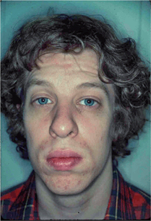
Figure 8-1. Adult male with PKU. This patient was born before the advent of newborn screening. His diagnosis was not made until he was 23 years old. He has severe cognitive impairments.
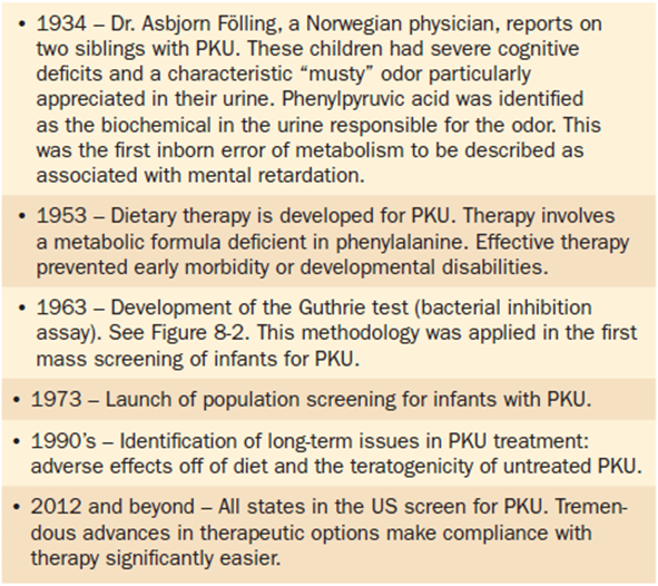
The recessive condition PKU was first described in 1934 by A. Fölling, based on his study of two siblings. This brother and sister showed mental retardation. They also had a characteristic musty odor to their urine, where Fölling discovered phenylpyruvic acid. A special diet low in phenylalanine, adopted in 1955, was the first effective therapy for this condition. Soon after that, a blood screening test was developed, and states began to adopt laws requiring newborn children to be tested before they were discharged from hospitals. Today, about 300 newborn children are diagnosed with PKU each year, and mental retardation is prevented by early implementation of a low-phenylalanine diet. As microarrays and other biochemical assays enter the field, designing better-tailored treatment regimens can benefit future patients. But few will probably be as directly treatable as this dietary approach to a comparatively simple metabolic disorder.
As we learn more about individual metabolic disorders, the conditions that were once grouped together as examples of the same defect are often found to be functionally related but biochemically separable. After all, metabolism is a sequential process of reaction steps toward a shared outcome. But even when the underlying biochemistry is well-understood, the treatment regimen may not be easy to implement. Even in the relatively straightforward example of PKU, a low phenylalanine diet is not one that affected individuals willingly follow for the rest of their lives. In this chapter we will focus on examples of metabolic disorders and on how to think about the processes at work. But understanding the impact of these conditions and their treatment options on the lifestyles of affected individuals is also important.
Part 1: Background and Systems Integration
The Basic Chemistry of Metabolism
Enzymes are protein-based catalysts that accelerate chemical reactions. These reactions define the changing structure and biochemistry of events that occur at all levels of biological activity. Enzymes work by lowering the activation energy needed for a targeted chemical reaction to occur. By definition, an enzyme speeds up a chemical reaction without being permanently changed by the reaction. In other words, enzymes are not chemical reactants. That means that a small amount of a given enzyme can have a potentially large effect on the biochemical reaction it catalyzes. For most biological reactions, the amount of enzyme is produced in excess. This serves to buffer critical developmental steps against the effects of environmental changes and other potential sources of functional variation. For that reason, a clinically diagnosable disease is often not apparent until the level of the key enzyme is at 5% or less of its normal concentration.
Often a cofactor will help an enzyme work more efficiently. A coenzyme is an organic molecule that acts as a donor or acceptor of molecular groups added to or taken from a substrate molecule. For example, water-soluble vitamins like members of the vitamin B complex and vitamin C act in this way.
Metabolism is a network of chemical reactions. When viewed from the perspective of genetics, our focus may be upon one specific mutation. But we should never forget the overall context of its function. With that in mind, there are several recurring patterns that can help create the landscape of metabolic interactions. First, the activity of a relevant enzyme is generally more important than the level of its substrate. Certain steps in a biochemical pathway, especially steps that are not reversible, can serve as regulatory control sites. Allosteric control at these steps is the key to their regulation. Allosteric control refers to the reversible changes in protein shape, and thus functions, of active sites that can be produced when the enzyme binds to a small molecule. The small molecule thus acts as an allosteric effector. Allosteric interactions can either enhance or inhibit the enzyme’s catalytic ability and thus induce or repress its activity temporarily.
In addition to allosteric interactions, some proteins are also affected by covalent modification. Examples include phosphorylation and adenylation at key steps in a metabolic pathway. The pathways can, therefore, be rapidly activated or inhibited by small changes. Changes in enzyme amounts and levels of activity can also affect metabolism, as can the partitioning of key metabolic processes in different cellular compartments, like the cytosol or the mitochondrial matrix.
The Logic of Mapping Biochemical Pathways
A mutation can cause a problem in a metabolic pathway in at least two ways. The mutation can cause a missing or deficient product, or it can cause the buildup of an earlier precursor. Both mechanisms may have serious phenotypic consequences. Knowledge of the effect of each mutation will ultimately allow researchers to map the relationship between steps in a biochemical or developmental pathway and the gene, and thus the active enzyme, responsible for each catalytic reaction. George Beadle and Edward Tatum shared a Nobel Prize for their work with the simple bread mold, Neurospora crassa. In their studies, mutations became powerful tools with which to dissect a biochemical pathway. A short description of their approach will illustrate the logic that is applied today in a wide array of organisms. Even though the specific techniques now employed can be quite diverse, the underlying logic has the same roots.
As we have seen, an enzyme catalyzes the conversion of one molecule into another, with a series of enzymes carrying out the sequential steps to form an ultimate product. Using Neurospora, for example, colonies can be grown by replicate plating of cells onto a series of petri dishes (Figure 8-2a). If the petri dish medium is “complete,” it contains all of the basic nutrients needed for both normal and defective cells to survive. Minimal medium, however, has only those basic precursor nutrients from which all other nutritional requirements can be produced by a normal cell. Thus, if a cell can survive on complete medium but not on minimal medium, it must have a mutation in some critical biochemical pathway. In 1963, Robert Guthrie’s Bacterial Inhibition Assay for Phenylketonuria (PKU) was the first application of this screening technique for disorders in human newborns (Figure 8-2b). Within a decade it was adopted as the first newborn screen in large populations.
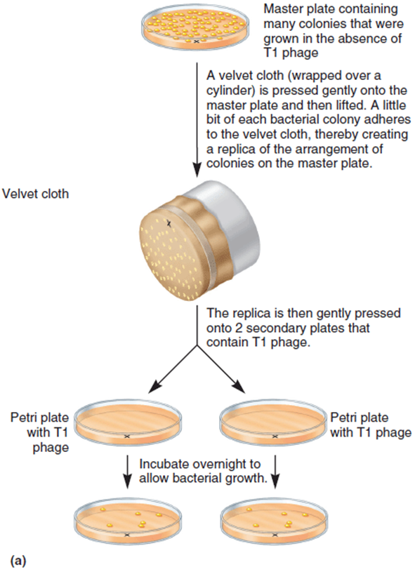
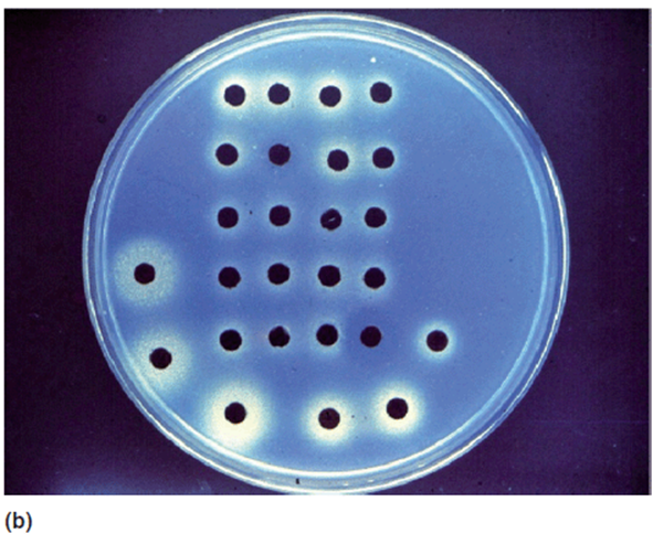
Figure 8-2. Microbial inhibition assay for metabolic disorders. (a) Schematic of process using the fungus Neurospora. (b) Photo of a plate used for the “Guthrie test.” This was a bacterial inhibition assay for phenylketonuria. This methodology allowed for the mass screening of newborns for PKU. (Reprinted with permission from Brooker RJ: Genetics: Analysis & Principles, 3rd ed. New York: McGraw-Hill, 2008.)
To dissect the biochemistry of a specific pathway, the first step is to collect a series of mutations with a shared nutritional deficiency. These cells can grow on complete medium and on medium supplemented with a specific end product, such as the amino acid phenylalanine. But they cannot grow on minimal medium. Such nutritional mutants are called auxotrophs; genetically normal cells are called prototrophs. The next step is to replica plate auxotrophs onto a series of petri dishes containing minimal medium that has been supplemented with specific chemicals in the biosynthetic pathway. In the sample of growth results shown in Table 8-1, a plus sign indicates that the plated cells are able to grow and form a colony, while a minus sign indicates lack of growth. Strains and intermediates are presented at random in this first table, as would be the case for a collection of initial laboratory data.
Table 8-1. Growth (+) or Absence of Growth (−) of Six Different Auxotroph Mutations for a Hypothetical “End Product” in a Biochemical Pathway. By Evaluating the Ability of Each Mutant Strain to Grow on Minimal Medium (Min), or Minimal Medium Supplemented With an Intermediate Molecule in the Pathway, the Mutants Can Be Mapped in Terms of Their Role in the Pathway
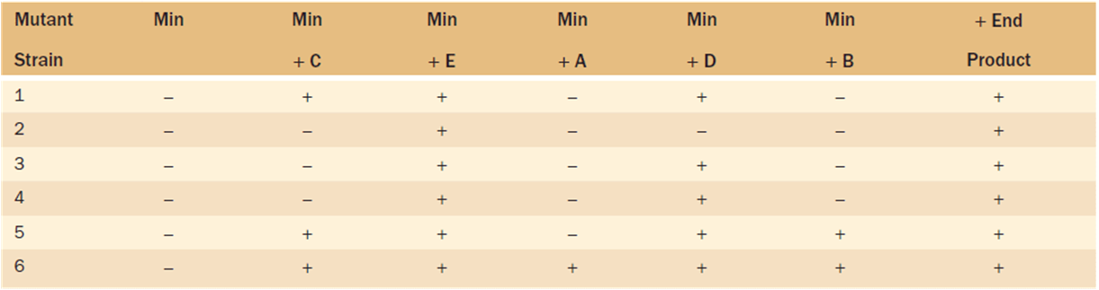
The logic for interpreting these data is straightforward. Consider one sample step. If a specific mutation produces a defective enzyme incapable of converting intermediate molecule B into C (shown by the broken arrowhead in the following sequence), then it cannot grow if given supplements of B or any other molecules that occur earlier in the pathway. But it can grow if given supplements of C or of molecular intermediates that occur later in the sequence of biochemical steps.
Earlier Precursor → → → B ↵ C → → → End Product
These results are reflected in the first row of Table 8-1. Cells plated onto petri dishes supplemented with chemicals A or B cannot convert those into the next molecule in the sequence. But supplementation with any later molecule can allow the sequence of reactions to continue normally. By arranging the sequence of precursor molecules and mutant growth characteristics so that the plusses are grouped at the right end of each line and the mutant strains are ranged from most to least successful in supplementation, the pathway can be read directly from the table. The example of randomly-collected data in Table 8-1 is followed by an organized sequence of mutation steps and intermediate molecules in Table 8-2. In this hypothetical case the sequence of intermediate molecular steps is:
Table 8-2. Data From Table 8.1 Reordered in Terms of the Sequence of Functional Intermediates and the Mutant Blocks in This Biochemical Pathway. (Min = Minimal Medium)
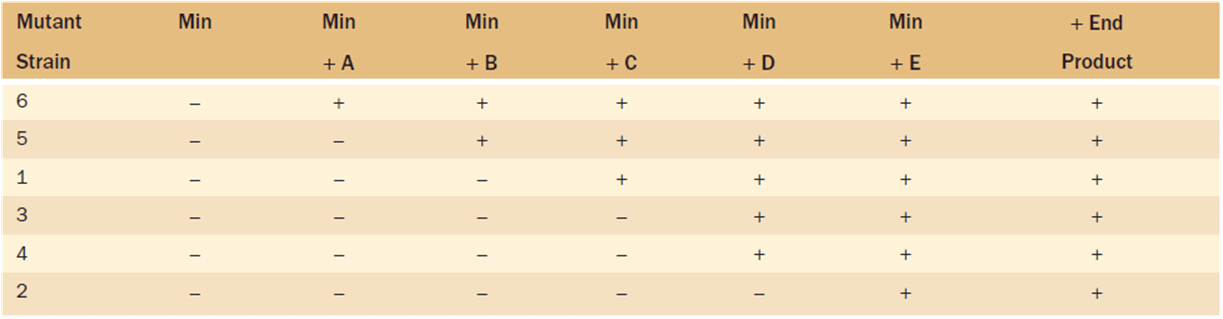
Earlier Precursor → A → B → C → D → E → End Product
In the case of the conversion of molecule C into D, either there are two separate molecular steps controlled by enzymes #3 and #4 with an unknown intermediate, or these two mutants affect the same gene and are allelic.
Although this classical example using Neurospora focuses on a specific experimental design, the logic can be applied broadly. In the next section we will take an overview of the evaluation of inborn errors of metabolism in humans, and Part 2: Medical Genetics will discuss important clinical examples.
Mutations and the Level of Phenotypic Discernment
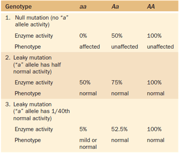
In classical studies of metabolic defects, the enzymatic activity was completely absent. A null allele is one in which the enzyme is nonfunctional. But we now know many examples in which the enzyme is functional, but kinetically weakened. Such so-called “leaky” mutations yield a protein with lowered enzyme activity or which has an activity that is conditionally affected by changes in some environmental variable.
Differences in the level of enzyme activity can, but will not always, cause a phenotypic change. Such variation is expressed in terms of phenotypic discernment (Table 8-3), that is, the “ability to detect.” If the mutation has no activity in homozygotes, the phenotype of the individual is expected to show the consequences of that absence. But as noted earlier, there is a large excess production of many enzymes, and a leaky mutant with only 5% of the normal enzyme activity may be phenotypically normal or have a mild to severe phenotypic expression.
Table 8-3. Sample Levels of Phenotypic Discernment
An increasing number of metabolic diseases can be diagnosed by screening newborns. Gas chromatography or mass spectrometry assays, DNA microarrays, and other techniques can be used for screening. But screening tests that have been in common use for decades often draw attention to cases where these more precise techniques can be employed efficiently. This is clearly an area in which pediatric genetics will change rapidly as new techniques become increasingly available.
Part 2: Medical Genetics
Introduction
In 1902 Archibald Garrod defined a category of genetic disorders that he referred to as “inborn errors of metabolism.” These conditions have their origin in human biochemistry. Inborn errors of metabolism (IBEMs) occur when there is a block in a metabolic pathway due to an inherited defect in an enzymatic protein. They thus represent the clinical sequelae of abnormalities of normal metabolic pathways. They are of great clinical importance for several reasons:
1. They were the first conditions in which a specific genetic change could be linked to an altered protein (i.e., to an enzyme) and the change in the protein could be linked to associated pathophysiological abnormalities.
2. The first large scale population screening efforts for genetic conditions were screening newborns for IBEMs. Screening newborns for selected genetic disorders remains one of the great success stories in public health. This will be discussed in detail in Chapter 11 on Genetic Testing and Screening.
3. Effective therapies exist for many IBEMs. The success of therapy is highly dependent on the early recognition and intervention of the disorder.
4. Although these conditions are individually somewhat rare, collectively they are encountered regularly in the general practice of medicine.
5. The range of clinical signs and symptoms seen in IBEMs is extremely broad—so much so that they should be considered in the differential diagnosis of almost any clinical feature. The presentations can range from prenatal (hiccups) to adult psychiatric problems.
The topic of inborn errors of metabolism is extremely broad. Various large multivolume texts provide comprehensive lists of conditions with detailed discussion of the biochemistry, pathophysiology, molecular genetics, and clinical aspects of these disorders. For this chapter, we have selected the salient features of the more common conditions. We provide general information about the disorders and outline clinically important features. For more detailed discussions, the reader is referred to any one of several excellent books listed at the end of this chapter in the Resources section.
Phenylketonuria (PKU) is an inborn error of metabolism that results from a problem with converting the amino acid phenylalanine to tyrosine. More is known about this condition than any other IBEM. As such we will use PKU as our example frequently throughout the rest of this chapter to highlight the unifying principles. In addition, the Clinical Correlation section of this chapter focuses on the medical problems and treatment issues in PKU.
Basic Principles of Inborn Errors of Metabolism
The primary understanding of IBEMs requires a review of human biochemistry. Chemical reactions in living organisms are typically facilitated by enzymes. Enzymes are simply biochemical catalysts. These chemical reactions are such that kinetically they would occur spontaneously, albeit slower (Figure 8-3a). Enzymes (catalysts) “push” natural reactions along at an accelerated rate (Figure 8-3b). Some enzymes may also have their activity enhanced by the presence of cofactors (Figure 8-3c). Cofactors (sometimes called coenzymes) act in concert with an enzyme. Cofactors are typically non-protein compounds that bind to the enzymatic protein. They may be organic or inorganic compounds. Vitamins are typically organic cofactors. The enzyme by itself is referred to as an apoenzyme. The enzyme coupled with the cofactor is the holoenzyme. For some enzymes, a deficiency of cofactor will reduce its activity, for others, the enzyme may not function at all without it. Some enzymes will require multiple cofactors for effective functioning.
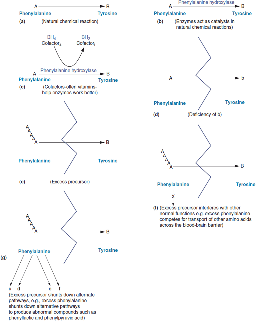
Figure 8-3. (a) Naturally occurring chemical reaction. Enzyme kinetics favor the spontaneous conversion given the right substrates. The example here is the conversion of phenylalanine (phe) to tyrosine (tyr). (b) The enzyme phenylalanine hydroxylase acts as a catalyst in the conversion of phe to tyr. (c) Biopterin (BH4) acts as a cofactor that enhances phenylalanine hydroxylase activity. (d) Deficiency in an enzyme slows down the enzymatic process. This results in the decreased production of the product (tyr) of the reaction. (e) Deficiency in an enzyme slows down the enzymatic process. Another result is the buildup of the precursor (phe) in the reaction. (f) One result of an excess of precursor can be the interference with other normal processes. Excess phenylalanine will interfere with the transport of other large neutral amino acids across the blood-brain-barrier. (g) Another result of an excess of precursor is shutting down alternative (not typical) pathways. The end result is the generation of compounds not usually present in significant quantities. These compounds can themselves produce physiological problems.
An interesting biological phenomenon was mentioned earlier but is important to emphasize. For most human enzyme systems there is apparently a tremendous excess of enzyme above what is needed for normal functioning. In fact, the excess is so significant, that for many conditions, an abnormality or disease may not occur until the enzyme activity falls below 5% of “normal.” Thus, most IBEMs are inherited in a recessive manner with close to null allele function for both copies. The majority of IBEMs are autosomal recessive with a few being X-linked recessive.
Pathophysiology of Inborn Errors of Metabolism: How Do Enzymatic Blocks Cause Disease?
An understanding of the pathophysiology of a disorder is central to developing appropriate therapies and interventions. It is not enough simply to say that a mutation changes an enzyme and causes the enzyme not to work. More importantly, the question is what happens when the enzyme does not work? What are the metabolic consequences of this abnormality? How does this translate into actual symptoms for the patient?
The intuitive answer to these questions is that an enzymatic block will result in a deficiency of the product of the enzymatic reaction. This is an important consideration. In our example of PKU, genetic changes that disable the enzyme phenylalanine hydroxylase disrupt the conversion of phenylalanine to its related amino acid tyrosine. This leads to decreased amounts of tyrosine, which in turn produces several problems (Figure 8-3d). In PKU, deficiency of tyrosine can lead to impaired protein production in general, due to the fact that there is not enough tyrosine for the synthesis of any protein. In addition, tyrosine is a precursor in the synthesis of the neurotransmitters l-dopa, dopamine, norepinephrine, and epinephrine. Low tyrosine levels will thus result in decreased production of these important chemicals with subsequent neurological problems. Also, tyrosine is in the metabolic pathway of melanin synthesis. This correlates with a long-standing observation that people with PKU tend to be lightly pigmented (Figure 8-4).
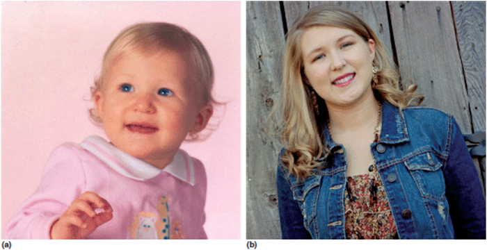
Figure 8-4. Young lady with phenylketonuria. (a) Infancy (b) Early adolescence. Note lighter pigmentation.
Equally important, there are other pathophysiological consequences of an enzymatic block. Besides a deficiency of the product of the reaction, a block will also lead to an accumulation of excess precursor(Figure 8-3e). Intuitively, one might wonder what possible problems might result from having “extra” phenylalanine in the system. The answer is actually “quite a bit.” The large neutral amino acid transporter (LAT1) is a membrane transport protein that preferentially transports neutral branched amino acids (valine, leucine, isoleucine) and aromatic amino acids (tryptophan, tyrosine) across the blood-brain barrier. In PKU, excess amounts of phenylalanine compete with these other amino acids for transport across the blood-brain barrier resulting in deficiency of these other amino acids in the brain (Figure 8-3f). It is this deficiency that is thought to be one of the primary sources of problems in untreated PKU in older individuals. Another problem with having an excess of a precursor is that the something has to be done with the extra. Often, excess precursor shunts down alternate biochemical pathways in ways that would not naturally occur (Figure 8-3g). In PKU, excess phenylalanine is shunted into other pathways with resultant excess of phenyllactic acid, phenylpyruvic acid, and phosphoethanolamine. The excess secretion of these compounds and their metabolites in the urine is responsible for the “phenyl-ketones” in the urine from which the condition gets its name. The increased amount of phenylpyruvic acid is the source of musky smell associated with untreated patients. Finally, excess phosphoethanolamine has a variety of effects, one of which may be CNS excitation, which has been postulated to be associated with the hyperactivity seen in persons with poor control.
Presenting Features of IBEMs
As mentioned earlier, IBEMs may show a plethora of presenting symptoms—so much so that they can appropriately be placed in the differential diagnosis of almost any human malady. For instance, there are solid data that suggest that upward of 25% of neonatal deaths attributed to overwhelming infection (septicemia) in which an infectious agent cannot be identified are actually due to undiagnosed IBEMs. With advances in newborn screening (see Chapter 11), it is hoped that this number will decrease. At the other end of the spectrum, there are many adults with primary psychiatric diagnoses who in reality have a metabolic disorder as the cause of their mental illness. The mantra is “if you don’t think about it, you will miss it.”
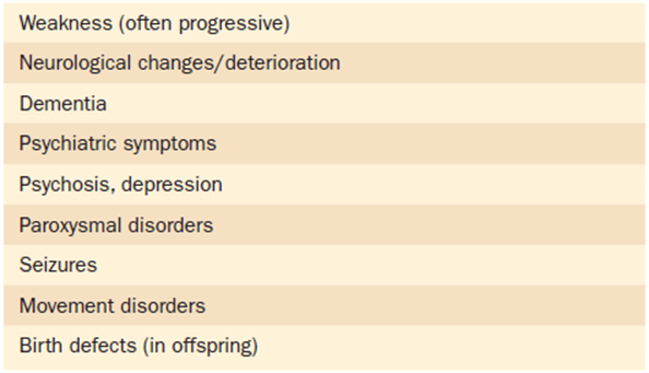
It is helpful to think about the presenting features of metabolic disorders in terms of where in the life cycle they may present. But it is also important to remember that these groupings are not absolute. Most metabolic disorders have a spectrum of presentations depending upon the severity of the metabolic defect. As such, many IBEMs have clinically defined subtypes (i.e., infantile, juvenile, adult). These designations are helpful in the clinical realm in that they provide the family with some general prognostic information. Tables 8-4 to 8-7 provide lists of common symptoms that could suggest an IBEM listed by periods of the life cycle when they would likely occur. Still, it is important to remember that these are actually artificial designations of what is in fact a continuum of clinical outcomes.
Table 8-4. Presenting Clinical Symptoms of Metabolic Disorders: Prenatal
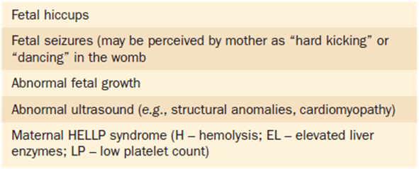
Table 8-5. Presenting Clinical Symptoms of Metabolic Disorders: Neonate
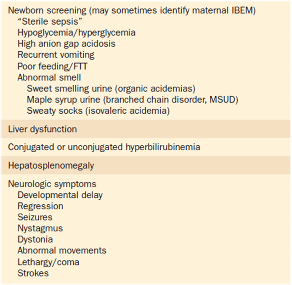
Table 8-6. Presenting Clinical Symptoms of Metabolic Disorders: Older Infant/Child
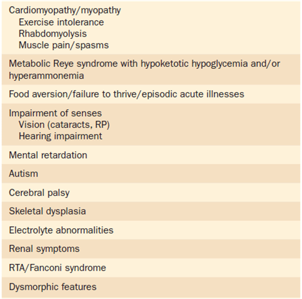
Table 8-7. Presenting Clinical Symptoms of Metabolic Disorders: Adult
One special category of presentation of IBEMs worth mentioning is structural congenital anomalies. There are several described multiple anomaly syndromes that ultimately have been shown to have an IBEM as the basic etiology. Table 8-8 lists many of these. One example is Smith-Lemli-Opitz syndrome. Smith-Lemli-Opitz syndrome (SLO) is a well-described multiple anomaly syndrome characterized by facial, digital, and genital abnormalities (Figure 8-5).
Table 8-8. Examples of Metabolic Disorders Associated With Structural Congenital Anomalies
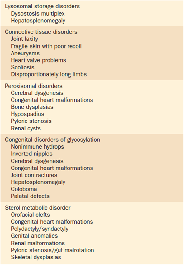
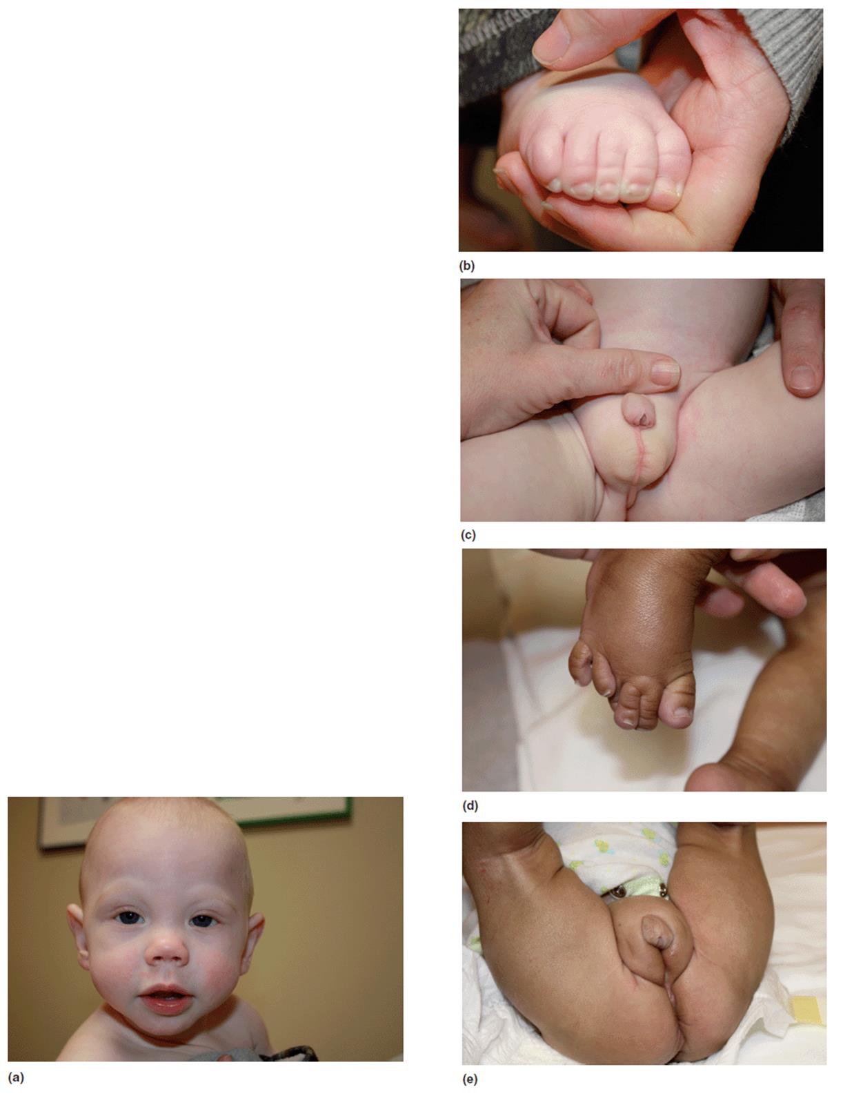
Figure 8-5. Two children with Smith-Lemli-Opitz syndrome demonstrating the key facial, digital, and genital findings of this condition.
These individuals also have marked neurodevelopmental disabilities and autistic behaviors (over half will meet standardized diagnostic criteria for autism). Years after the description of this syndrome, several clinicians noticed an unexpected finding on general laboratory testing. They noted that many of the patients with SLO had significantly lower serum cholesterol levels. Prompted by this finding, subsequent investigations demonstrated that the cause of SLO was an enzymatic defect of an enzyme called 7-dehydrocholesterol reductase (Figure 8-6). It is now known that deficiency of this enzyme results in a deficit of cholesterol production and an increase in the precursor 7-dehydrocholesterol. The pathophysiology of SLO is in part due to an overall deficiency in cholesterol and all of the many metabolic derivatives of it.
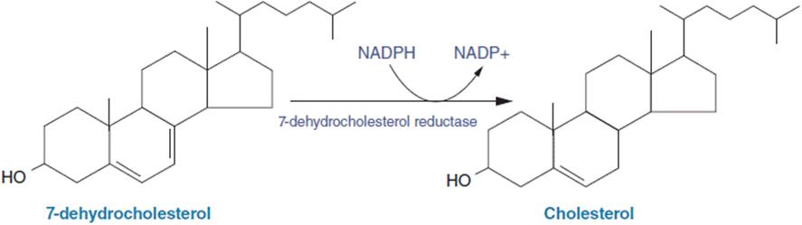
Figure 8-6. The terminal step in cholesterol synthesis is the conversion of 7-dehydrocholesterol to cholesterol. The block of this step due to reduction of the activity of the 7-dehydrocholesterol reductase enzyme results in Smith-Lemli-Opitz syndrome.
Diagnosis of IBEMs
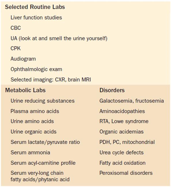
Within the realm of clinical medicine, metabolic disorders will often have a mystical folklore about them. Nongenetic physicians typically do not go back and review their notes from biochemistry class. Referrals are often made with the request “Something is not right, could it be one of those metabolic things.” Although the vast majority of readers of this text will not go on to careers in metabolic genetics, it is still important that you be aware of such conditions. Recognition and intervention—at least at the general level—of a metabolic condition can be crucial to a better outcome in the time before a metabolic consultant arrives. The astute clinician should always be alert to the possibility of an IBEM particularly since there are effective treatments available for many of them. They should also be very familiar with the symptom and associations listed in Tables 8-4 to 8-7. If a metabolic disorder is suspected, appropriate laboratory tests can be obtained immediately. Table 8-9 lists important selected routine laboratories and specific metabolic laboratories that will be helpful in a diagnostic evaluation.
Table 8-9. Testing for Metabolic Disorders
Key Metabolic Disorders
As noted earlier, detailed discussions of all known metabolic disorders require several volumes of quite large books (see Supplementary Readings list at the end of this chapter). And information is growing. Still, there are certain clinical “pearls” that are important for all physicians to be aware of. As such we have selected several conditions for brief discussions. Those selected below were chosen for any of several reasons: conditions that are relatively common, conditions that require quick recognition and intervention to prevent morbidity and mortality, and a few that simply show up frequently on the board examinations. Hopefully, much of this has been covered in a prior medical biochemistry course. Table 8-10 provides a frame work for thinking about IBEMs by category.
Table 8-10. Major Inborn Errors of Metabolism—Organized by Category
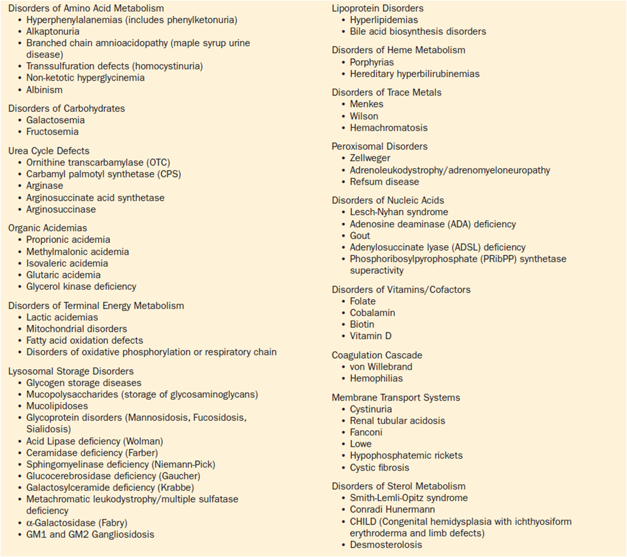
While there are many different ways to subgroup these conditions, we prefer to think along the lines of chemical “families.” We have organized the conditions to be discussed later according to these categories. While these discussions may seem cursory, they at least provide a frame of reference for critically important issues related to the IBEMs. For detailed discussions, the reader is referred to the Reference section of this chapter for a listing of key metabolic texts.
1. Disorders of amino acid metabolism. Historically, the disorders of amino acid metabolism have received more attention than probably any other category of IBEMs.
Alkaptonuria was one of the first metabolic conditions described—being one of Garrod’s original four conditions described in his Croonian Lectures of 1902. It is caused by a defect in phenylalanine and tyrosine catabolism. It results in the accumulation of homogentisic acid, an intermediary metabolite in the degradation of tyrosine. The hallmark diagnostic feature of this disorder is that homogentistic acid in the urine darkens upon standing exposed to air. The black appearing urine is striking and often heralds the diagnosis prior to the appearance of symptoms (i.e., pigment staining of connective tissue causing a disorder known as onchronosis, coronary artery disease, and kidney stones).
Homocystinuria is a disorder of sulfated amino acids (methionine) due to cystathionine beta synthase (CBS) deficiency. Affected individuals have physical features suggestive of a connective tissue disorder. One such feature is spontaneous dislocation of the lenses of the eyes. It is fascinating to note that when this happens, the dislocation is always in the inferior and medial directions, in contrast to Marfan syndrome where the dislocation is superior and laterally (Figure 8-7). This metabolic block produces an increased homocystine level, which has the effect of increasing the cohesiveness of platelets. The tendency for “sticky platelets” leads to an increased risk for pathologic thrombotic events. This can contribute to cognitive changes and can cause significant medical problems and reduced longevity. While homocystinuria is an autosomal recessive disorder, carriers (heterozygotes) for the condition also appear to have an increased risk of pathologic vascular occlusion.
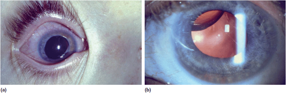
Figure 8.7. (a) Lens dislocation (medial and inferior) in the eye of a patient with homocystinuria. (b) For comparison note the superior and lateral lens dislocation in a patient with Marfan syndrome.
A deficiency of an enzyme complex known as branched-chain ketoacid dehydrogenase causes a buildup of the branched-chain amino acids (leucine, isoleucine, and valine) and their metabolites. The excess metabolites of this disorder are organic acids, which have a distinctive odor that can be identified in an affected infant’s urine. This odor truly smells like maple syrup, with the condition thus being called maple syrup urine disease (MSUD). Patients with MSUD typically present as seriously ill infants with vomiting, lethargy, metabolic acidosis, and neurological compromise.
Disruption of the catabolism of other amino acids results the accumulation of other organic acids. Isovaleric academia is due to abnormal leucine metabolism. Affected infants have presenting symptoms similar to other organic acidemias such as vomiting, metabolic acidosis, and neurological compromise (seizures, stupor, and coma). Infants with isovaleric academia are reported to have an odor reminiscent of “sweaty socks.”
2. Disorders of carbohydrate metabolism. Galactosemia and fructosemia are disorders of the metabolism of the sugars galactose and fructose, respectively. Infants with these conditions have similar symptoms that include vomiting, liver dysfunction, renal failure, and overall systemic collapse if not treated. Infants with galactosemia are at high risk for Escherichia coli sepsis. Both conditions are caused by enzymatic defects that render the person incapable of adequately metabolizing the respective sugar. The onset of the conditions differs with the introduction of the sugar in the diet. Galactose, which is derived from lactose, is often present in the infant diet in the first few days of life. Fructose is usually introduced later (4-6 months) with the introduction of fruits into the diet or with the first dose of a sucrose containing medication. Careful attention to the diet history and notice of the timing of dietary changes can be invaluable in arriving at a rapid diagnosis. The mainstay of treatment for both conditions is the elimination of the offending sugar from the diet.
The glycogen storage disorders (GSDs) are a group of conditions that share in common some disturbance of glycogen metabolism—either synthesis or degradation. GSDs are mostly characterized by a fasting hypoglycemia. Thus, infants typically do not present until their diet is modified toward times of more extended fasting. Some may have impressive hepatomegaly (Figure 8-8). Pompe disease is somewhat unique in this group as it presents with early severe infantile hypotonia and cardiomyopathy (Figure 8-9). The defective enzyme, acid maltase (acid alpha-glucosidase), is a scavenger enzyme in the lysozymes that rapidly degrade glycogen in a non-directed fashion. Physiologically it is much more like other lysosomal storage disorders (see below).
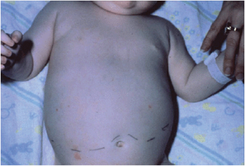
Figure 8-8. One-year-old child with glycogen storage disease type III. The line drawn on the abdomen delineates the lower margin of the liver.
3. Urea cycle disorders. The catabolism of protein ultimately results in the generation of ammonia, an extremely toxic compound. Excess levels of ammonia will result in severe and irreversible neurological damage. Patients may present with seizures, stupor, or coma. In less severe cases, symptoms may include fluctuating sensorium and/or psychiatric disturbances. The urea cycle exists to eliminate ammonia quickly from the body. Inborn errors of any of the five reactions of the cycle will lead to varying degrees of hyperammonemia and the resultant symptoms. Because ammonia is so toxic and the damage is irreversible, it is crucial that clinicians consider a hyper-ammonemic disorder in any patient with unexplained neurological changes.
Ornithine transcarbamylase (OTC) deficiency is an X-linked disorder. It is due to an error in the second reaction of ammonia detoxification. Affected males usually present with an early severe neonatal encephalopathy. Rapid intervention is critical in preventing morbidity and mortality. Female heterozygotes of OTC deficiency will present with a wide range of symptoms depending upon the degree of inactivation (Lyonization) of the X chromosome carrying the mutation. Symptoms in carrier females can range from completely unaffected to self-assigned protein restriction to fluctuating sensorium and/or psychiatric symptoms.
4. Disorders of fatty acid oxidation. Fatty acid oxidation is a complex process of mobilizing stored fat to meet increased energy demands. The major components of this process are the mobilization of free fatty acids via the lipolysis of diacylglycerols in the adipocytes mediated by lipases, uptake of the free fatty acids by the cells, activation of the fatty acids into acyl-CoA derivatives by fatty acyl-CoA ligase, transport of the fatty acyl-CoA complex into the mitochondria facilitated by carnitine palmitoyltransferases, and beta-oxidation of the fatty acids within the mitochondria. Different types of fatty acid molecules are processed by different enzymes specific to the type and size of the particles. Interruption of any portion of this process will result in significant reductions in energy production.
Medium chain acyl-CoA dehydrogenase (MCAD) deficiency is an autosomal recessive disorder that results in abnormalities of the beta-oxidation of medium-sized fatty acids. MCAD is one of the most common IBEMs. In fact it is the most common metabolic condition on the typical newborn screening panel. The major clinical symptom is hypoglycemia. It is important to note that it is one of the specific causes of hypoketotic hypoglycemia. Other symptoms include lethargy and seizures. The presentation of MCAD varies greatly from a sudden infant death (SIDs) picture in infants, to a metabolic Reye syndrome in children, to episodic unexplained weakness in adults.
5. Lysosomal storage disorders (LSDs). The lysosomes are subcellular organelles that carry on a variety of catabolic processes via acid hydrolases. Various acid hydrolases are specific to their own category of biochemicals. The typical presentation of LSD is a person who is normal as an infant, but at some later point in life begins to experience progressive problems related to the accumulation of uncleared biochemicals. The LSDs comprise about 40 different disorders categorized by the type of biochemical that accumulates (e.g., mucopolysaccharides, complex proteo-lipids, mucolipids, and glycoproteins). As noted earlier, GSD type II (Pompe disease) is also LSD (Figure 8-9).
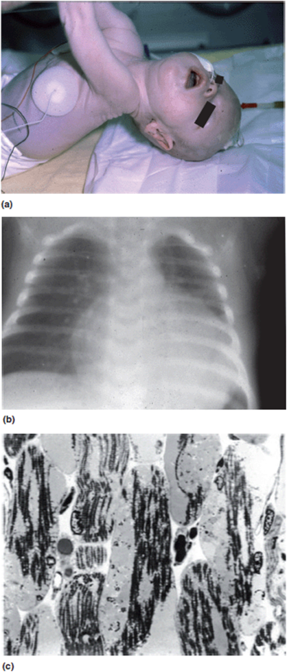
Figure 8-9. Newborn with Pompe disease. (a) Note severe hypotonia. (b) Striking cardiomegaly. (c) Muscle biopsy at 2 months old. Note tremendous glycogen stores disrupting muscle fibers. (c: Reproduced, with permission, from Amalfitano A, Bengur AR, Morse RP, Majure JM, Case LE, Veerling DL, Mackey J, Kishnani P, Smith W, McVie-Wylie A, Sullivan JA, Hoganson GE, Phillips JA 3rd, Schaefer GB, Charrow J, Ware RE, Bossen EH, Chen YT. Recombinant human acid alpha-glucosidase enzyme therapy for infantile glycogen storage disease type II: results of a phase I/II clinical trial. Genet Med. 2001 Mar-Apr;3(2):132-138.)
An increased awareness of the LSDs and their early recognition has occurred over the past several years. This has been prompted by the advent of targeted therapies. Enzyme replacement either by direct infusion or transplanted cells has recently been developed for several LSDs. Clinically available therapies for Pompe disease, Hurler syndrome, Hunter syndrome (Figure 8-10), Fabry disease, Maroteaux-Lamy disease, and Gaucher disease exist. Therapies are involved and quite expensive, but they still represent the first wave of hope for patients with these devastating progressive conditions. Given the possibility of treatment, there is a heightened emphasis on early detection.
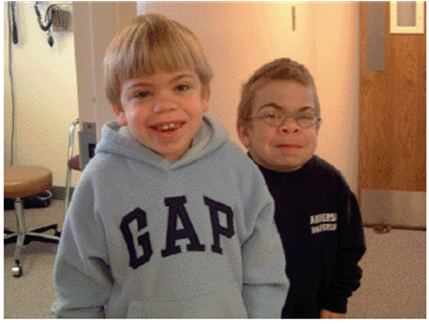
Figure 8-10. Brothers with Hunter syndrome (mucopolysaccharidosis type II).
Tay-Sachs disease is a lysosomal storage disorder characterized by the buildup of GM2 ganglioside. It is an autosomal recessive disorder. Clinically, infants with Tay-Sachs are normal as infants. At around 4 to 5 months of age, they begin to have neurological regression due to neuronal loss from the accumulation of the gangliosides. Infants will begin to lose skills and their hearing, begin to have seizures, and many develop a characteristic exaggerated “startle response.” Fundoscopic examination often reveals a “cherry red macule” (Figure 8-11), which is typical of this condition but not unique—being seen also in a handful of other storage disorders. Tay-Sachs is also notable for being one of several conditions that occur much more frequently in persons of Eastern European (Ashkenazi) Jewish descent (this will be discussed more in Chapter 15 on Population Genetics).
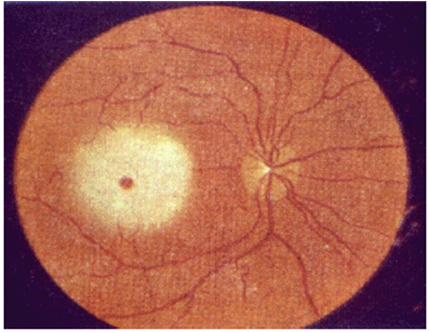
Figure 8-11. Retinal photograph in a patient with Tay-Sachs disease demonstrating the finding described as a “cherry red macule.”(de Aragão REM, Ramos RMG, Pereira FBA, et al: “Cherry red spot” in a patient with Tay-Sachs disease: case report. Arquivos Brasileiros de Oftalmologia. 2009;72(4):537-539.)
6. Disorders of trace metals. Trace metals (iron, copper, zinc, manganese, cadmium, and so forth), as the name implies, occur in minute quantities (parts per million) in the body, and yet they play significant roles in the overall health of the individual. Trace metals typically function as cofactors for enzymes. Nutritional deficits of these metals produce well-recognized symptom complexes (e.g., acrodermatitis enteropathica with zinc deficiency). In addition there are known IBEMs of trace metal metabolism that are expressed as their own clinical entity.
Menkes disease is a disorder of copper transport. The primary pathological consequence is poor delivery of copper to the subcellular compartments where it is needed (deficiency). It is an X-linked disorder. The condition is characterized by neurological dysfunction/degeneration. Because of the copper deficiency, connective tissue processing is impaired. Thus, patients with Menkes disease will have connective tissue disorder-like findings, such as tortuous blood vessels, bony abnormalities, and “sagging facies” (Figure 8-12a). Earlier descriptions called the condition “Menkes kinky hair disease” describing the abnormal hair of these patients. Because of the copper deficiency, the hair is hypopigmented, brittle, and quite curly. Under the microscope the hairs shafts have distinct angulations called “pili torti” (Figure 8-12b).
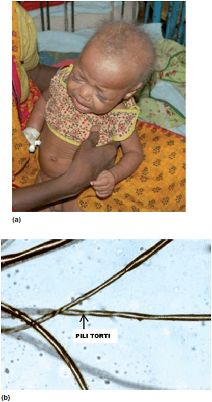
Figure 8-12. (a) Infant with Menkes syndrome—a disorder of copper metabolism. (b) Hair shaft from a patient with Menkes disease showing the “kinked” lesion in the shaft (pili torti). (Reprinted with permission from Datta AK, Ghosh T, Nayak K, et al: Menkes kinky hair disease: A case report. Cases J. 2008;1:158.)
Wilson disease (also known as hepatolenticular degeneration) is another disorder of copper metabolism. It is an autosomal recessive condition. The responsible gene has been identified, but the exact pathophysiological mechanism has yet to be completely worked out. The observable end result, however, is copper overload, which seems responsible for the symptoms. One observable feature of this is brown circumferential rings on the periphery of the iris called Kayser–Fleisher ring (Figure 8-13).
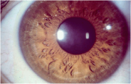
Figure 8-13. Kayser-Fleischer ring in a patient with Wilson disease.
Hemochromatosis is a disorder of iron metabolism. It is one of the most common known human genetic disorders. It is estimated that about 1 in 250 people in the United States have this condition. It is an autosomal recessive that has genetic heterogeneity. The “classic” form of hemochromatosis is caused by changes in a gene called HFE at chromosome locus 6p21.3. Although the exact pathogenetic mechanism is not known, it is clear that the responsible genes play critical roles in iron transport. The end results are a variety of symptoms due to iron overload. This includes liver problems (cirrhosis and tumors), diabetes, gonadal dysfunction, joint pains, and cardiomyopathy. Treatment is designed to decrease iron intake and to chelate excess iron from the body. Hemochromatosis is a condition that demonstrates a sex-influenced phenotype. As an autosomal recessive disorder, it occurs at an equal frequency in males in females. Males, however, are more severely affected. It is presumed that this is in part due to the female menstrual cycle, which naturally provides a mechanism for eliminating iron from the system.
7. Disorders of nucleic acids. There are several reported disorders of nucleic acid metabolism. The most well-known condition is Lesch-Nyhan syndrome. Lesch-Nyhan syndrome is characterized by a phenotype of severe neurobehavioral symptoms. The most striking features are cognitive deficits, movement disorders, and dramatic self-injurious behaviors (severe biting with mutilation of the lips and fingers). It is an X-linked recessive disorder. It is caused by changes in the hypoxanthine-guanine phosphoribosyl transferase enzyme (HgPRT), which plays a central role in purine metabolism. Another metabolic defect of nucleotide metabolism is adenosine deaminase (ADA) deficiency, which produces severe combined immune deficiency (SCID). Adenylosuccinate lyase (ADSL) deficiency and phosphoribosylpyrophosphate synthetase (PRibPP) superactivity are defects of nucleotide metabolism that have been seen in patients with non-syndromic mental retardation and/or autism.
8. Vitamins and cofactors. Many metabolic disorders have been reported due to abnormalities of the vitamins or cofactors associated with specific enzymatic reactions. Biotinidase deficiency is a disorder of the recycling of the cofactor biotin. This results in biotin deficiency. Patients with this disorder will have progressive symptoms of dystonia, eczema-like rash, cognitive deficits, and seizures. The treatment is easy and effective–simply supplement biotin.
Clinically, the cofactor abnormality often mimics the actual enzyme deficiency. Still, there are typically significant differences in the implications for therapy. Such is the case for the disorders of biopterin metabolism, which clinically resembles phenylketonuria (with BH4 being the cofactor for PKU) but has significant differences in outcome and therapy.
It is also important to note that some cofactors operate with multiple enzyme systems. Thus, a defect in cobalamin metabolism produces characteristic abnormalities of two different enzymatic defects: methylmalonic acidemia and homocystinuria.
9. Disorders of sterol metabolism. Despite all of the bad press that cholesterol receives, it is a critical compound. It is a major component of cell membranes and a key precursor in the synthesis of biochemicals such as steroids, vitamin D, bile acids, and so forth. The synthesis of cholesterol starts with acetyl-CoA, which then proceeds through more than 15 enzymatic steps until the end product of cholesterol is achieved. Blocks in many of these steps have been described in association with recognizable clinical syndromes, such as Antley-Bixler syndrome and CHILD syndrome. The best characterized of these disorders is Smith-Lemli-Opitz syndrome, which is a disorder of the terminal step of cholesterol synthesis as described earlier.
Treatment of Inborn Errors of Metabolism
Most major advances in medical genetics over the past few decades have been in the areas of diagnostics and the understanding of the pathogenesis of genetic conditions. There is much to be said for advances like these. They provide invaluable insight into the central issues of these conditions, and their worth should not be downplayed. Still, in the realm of clinical medicine, patients are much more interested in treatments than diagnoses. In general, genetic therapies are lagging far behind genetic diagnoses. This idea will be covered in detail in Chapter 14, Genetic Therapeutics. As a group, advances in therapies for inborn errors of metabolism are far ahead of those for most other genetic conditions. This is largely due to the pathogenic nature of these disorders. Biochemical processes are ongoing and continuous, in contrast to, say, structural processes. Thus, IBEMs have a greater potential for therapy. In the following Clinical Correlation section, we will discuss the treatment of one such disorder, phenylketonuria, in detail.
Part 3: Clinical Correlation
Phenylketonuria (PKU) is an IBEM of phenylalanine metabolism. It is by far the best understood metabolic disorder in humans. Metabolic geneticists have had many decades of experience in dealing with patients with this disorder (Table 8-11). It is caused by a deficiency in the enzyme phenylalanine hydroxylase or one of the cofactor systems in this enzymatic reaction. The end result is impairment of the conversion of phenylalanine to tyrosine. With this metabolic block, there are multiple secondary metabolic abnormalities (Figure 8-3a-f).
Table 8-11. Major Events in the History of Phenyketonuria (PKU)
Prior to newborn screening and effective therapies, children born with PKU had a severe phenotype (Figure 8-1). Many children died in the first few months of life. For those that survived, there were notable symptoms of mental retardation, seizures, microcephaly, an eczematous rash, hypo-pigmentation, musty smell to urine, discoordination, and autistic-like behaviors. Autopsy and imaging studies demonstrated severely disrupted myelination.
With the advent of public health programs in newborn screening, infants with PKU could be identified in the first few days of life. As such, therapies could be instituted prior to the onset of serious symptoms. The treatment of PKU is a classic example of restoring a normal phenotype without modification of a mutant genotype. The mainstay of treatment for persons with PKU is dietary modification. They are prescribed a semisynthetic diet, low in phenylalanine but adequate in other nutrients. They are provided just enough phenylalanine in this diet to allow for the needs of endogenous protein synthesis. It is notable how low this level actually is. The typical human diet (especially in the United States) has a marked excess of protein intake as compared to the actual metabolic needs. In fact, a patient with PKU can be maintained on a diet that has a minimum protein intake of about 1.5 g/kg/day. This diet is started as soon as the diagnosis is made, hopefully within the first 10 days of life. Patients are monitored regularly for phenylalanine and tyrosine levels. An interdisciplinary team of metabolic geneticists, nutritionists, and pediatricians follow these patients closely with the expectation of normal growth and neural development.
PKU was the first metabolic disorder that was successfully treated. As such there is a long history of experience and new discoveries along the path of treatment and intervention. Over time, many unexpected long-term sequelae have been identified. For instance, in the early days of PKU treatment, a presumption was made that therapy could be stopped after age five. This was based on the conclusion that most of the neurological abnormalities were due to aberrant myelin formation. Since the majority of myelination occurs in the first few years of life, it was assumed that therapy could stop after this window of time was complete. The short-term outcomes were impressive. Children that would have died or had severe neurological compromise survived and developed normally. Clearly, this was an amazing success story. Fortunately, a committed group of metabolic specialists banded together in what was known as the National Collaborative PKU study. Much of the effort on this project was unfunded volunteer work provided by specialists who simply wanted to know what the long-term outcomes of this never-before-seen entity (long-term normally developing persons with PKU) might look like. This collaborative identified several fascinating phenomena in this group.
First, although the treated children survived and had normal early neurodevelopment, over time a definite loss in IQ points was observed. Analysis of the study data demonstrated small, but definite, losses of IQ points for each year off of diet. This cognitive loss also was shown to be cumulative. The mechanism was shown not to be problems with myelin formation, but rather to problems with the transport of amino acids across the blood-brain barrier (BBB). Phenylalanine shares with other similar amino acids a common transport mechanism, called the large neutral amino acid transporter. In the case of PKU in which treatment is stopped in childhood, the devastating effects of dysmyelination are avoided. But, the extremely high circulating concentrations of phenylalanine compete with the other amino acids which share the same transport mechanism. The relative deficiency of transport of these other amino acids into the CNS results in problems with overall function, hence the loss in IQ. If PKU continues untreated, other findings are also seen in the adults. White matter changes are noted in the brain, and changes are seen in the cerebellum. These individuals demonstrate decreased performance on measures of attention, coordination, and information processing.
Another novel situation emerged with the successful treatment of children with PKU. As these children grew up as healthy, typically-developing young adults, the first group reached childbearing age in the early 1990s. As women with PKU but who as a group had not been treated since childhood began to have children of their own, another unexpected outcome was seen. Hyper-phenylalaninemia in the mothers was found to be teratogenic to the infants. Infants born to mothers with untreated PKU have essentially a 100% incidence of microcephaly and neurodevelopmental delays. The infants also often have dysmorphic features and congenital heart disease. Further work demonstrated that this adverse effect on the developing fetus could be avoided with reinstitution of the diet. In order to obtain maximum benefit, the diet must be rigid and begin prior to conception.
The logical conclusion from both of these later developing outcomes was straightforward: diet for life is now recommended. It is important to point out, that it is rather simple for us, the physicians, to recommend diet for life. But in practice, what this is asking of the patients is something quite involved. The metabolic formulas that they are asked to drink to provide nutrients without phenylalanine have an unpleasant taste. The newer formulas are significantly better than the early ones, which tasted surprisingly like quinine, but are nonetheless not great tasting. Also, the amount of protein that can be eaten is minimal. Meats are not allowed. Many vegetables, such as potatoes, are not “free foods” as they contain enough protein to be significant. Imagine having to count out 10 potato chips as your total allowance for lunch.
To help patients with compliance, several recent advances in therapy have emerged. As mentioned the newer formulas have a better taste. Sapropterin (trade name Kuvan) functions exactly like BH4, the cofactor in the phenylalanine hydroxylase enzyme. The addition of sapropterin to the dietary therapy in PKU will further reduce blood phenylalanine levels and allow liberalization of the diet. Another addition to the therapy can be to supplement the large neutral amino acids. This will improve the ratio of phenylalanine to other amino acids that share the same BBB transporter. This also allows liberalization of diet. Phenylalanine ammonia lyase (PEG-PAL) converts phenylalanine to a nontoxic derivative. At this time, it is an investigational enzyme substitution therapy. Other therapeutic strategies are being explored. Enzyme replacement therapy (ERT) in theory should work. The major obstacle here is delivery of the enzyme to the appropriate site. ERT is currently not available for PKU. Also, without further advances in ERT, the risk to benefit ratio probably makes this therapeutic mode unlikely to be developed, given other more benign therapies. Placing a normal phenylalanine hydroxylase gene in place of the mutant gene in the patient (i.e., gene therapy) is a possible mode of therapy, but it is unlikely in the near future for both technical and ethical reasons.
In the end, what is the final outcome? Dietary treatment is clearly effective at ameliorating the severe effects of PKU. But early-treated children have mean IQ scores about one half a standard deviation lower than scores for their unaffected siblings and the corresponding population norms. Women with PKU can have normal children with early and rigorous therapy. Clearly, continued studies and advances in therapy are needed. Most importantly, the “PKU story” highlights the need for ongoing evaluation of patients with early identified metabolic disorders. Analogous to the National Collaborative PKU study, effective Long term Follow-Up programs are needed, especially given the rapid expansion of the recommended newborn screening panels (see Chapter 11 Genetic Testing and Screening).
 Board-Format Practice Questions
1. In regards to inborn errors of metabolism:
A. the original description was by Aborigines with a urine taste test.
B. these are most commonly inherited as dominant disorders.
C. disease can be caused by a deficiency of product upstream from the enzymatic block.
D. disease can be caused by a surplus of metabolites downstream from the enzymatic block.
E. deficiency of downstream product can cause disease.
2. A 10-day-old infant presents with poor feeding, vomiting, and lethargy. Sepsis (overwhelming bacterial infection in the blood) is suspected. A look at the prior history reveals normal pregnancy and delivery. The child was discharged after 48 hours of life. Over the next week, the baby became progressively more ill. If a genetic disease such as inborn errors of metabolism is suspected, which of the following is least informative?
A. Family history of neonatal deaths.
B. Family history of consanguinity.
C. Infant’s feeding/dietary history information.
D. Any unusual odors.
E. Chromosomal analysis.
3. Inborn errors of metabolism may have a plethora of presentations. Which one of the following is one such presentation?
A. Overgrowth.
B. Jaundice/high bilirubin levels.
C. Tumor formation.
D. Enlarged muscles.
E. Enhanced intellectual functioning.
4. Inborn errors of metabolism:
A. may cause structural congenital anomalies.
B. have been described since the 16th century.
C. because they are rare and have no effective therapies, are of limited clinical interest.
D. early treatment and intervention almost guarantees no long-term problems.
E. are distinct in their presentation from most other medical conditions.
5. In regards to phenylketonuria (PKU):
A. with the advent of newborn screening, it is quite rare to see an untreated child in the United States.
B. dietary therapy is effective and easy to implement.
C. effective therapy prevents all complications of the disorder.
D. high levels of phenylalanine in fathers with PKU results in birth defects in their infants (such as microcephaly and congenital heart disease).
E. excess tyrosine leads to disorders of neurotransmitter concentrations.
Supplementary Readings
Clarke, Joe T. R. A Clinical Guide to Inherited Metabolic Diseases.
Hoffmann, Georg F., Johannes Zschocke, and William L. Nyhan. Inherited Metabolic Diseases: A Clinical Approach.
Nyhan, William L., and Pinar T. Ozand. Atlas of Metabolic Diseases.
Saudubray, Jean-Marie, Georges van den Berghe, and John H. Walter. Inborn Metabolic Diseases: Diagnosis and Treatment.
Scriver, Charles R., William S. Sly, Barton Childs, Arthur L. Beaudet, David Valle, Kenneth W. Kinzler, and Bert Vogelstein. The Metabolic and Molecular Bases of Inherited Disease, 4 volumes.