
Shobha H. Mehta, MD
Robert J. Sokol, MD
ESSENTIALS OF DIAGNOSIS
 A careful history to reveal specific risk factors
A maternal physical examination organized to identify or exclude risk factors
Routine maternal laboratory screening for common disorders
Special maternal laboratory evaluations for disorders suggested by any evaluative process
Comprehensive fetal assessment by an assortment of techniques over the entire course of the pregnancy
OVERVIEW
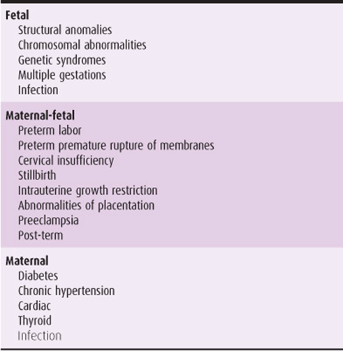
High-risk pregnancy is broadly defined as one in which the mother, fetus, or newborn is, or may possibly be, at increased risk of morbidity or mortality before, during, or after delivery. Factors that may lead to this increased risk include maternal health, obstetric abnormalities, and fetal disease. Table 12–1 provides an overview of some major categories that comprise a high-risk pregnancy.
Table 12–1. Major categories of high-risk pregnancies.
The purpose of this chapter is to outline basic and essential aspects of diagnostic modalities available for determination of pregnancies at risk that can be used in practice in a rational manner.
The incidence of high-risk pregnancy varies according to the criteria used to define it. A great many factors are involved, and the effects of any given factor differ from patient to patient. Outcomes can include mortality of the mother and/or the fetus/neonate. Leading causes of maternal death include thromboembolic disease, hypertensive disease, hemorrhage, infection, and ectopic pregnancy. The leading causes of infant mortality (death from birth to 1 year of age) are congenital malformations and prematurity-related conditions. Although there is variation in definition depending on the resource, a perinatal death is one that occurs at any time after 28 weeks’ gestation through the first 7 days after delivery. The perinatal mortality rate is the number of perinatal deaths per 1000 live births. Preterm birth is the leading cause of perinatal morbidity and neonatal mortality.
In assessing pregnancies to determine risk, several key concepts may offer tremendous insight. Human reproduction is a complex social, biochemical, and physiologic process that is not as successful as once thought. Approximately half of all conceptions are lost before pregnancy is even recognized. Another 15–20% are lost in the first trimester. Of this latter group, more than half have abnormal karyotypes and defy current methodologies for prevention of loss. However, many other causes of reproductive loss are amenable to diagnosis and treatment. In this chapter we discuss the indications and justifications for antepartum care and intrapartum management.
PRECONCEPTION CARE
Preconception evaluation and counseling of women of reproductive age has gained increasing acceptance as an important component of women’s health. Care given in family planning and gynecology centers provides a potential opportunity to maximize maternal and fetal health benefits before conception. Issues of potential consequence to a pregnancy, such as medical problems, lifestyle (eg, substance abuse, weight, exercise), or genetic issues should be investigated and interventions devised before pregnancy. Specific recommendations include folic acid for the prevention of fetal neural tube defects (0.4 mg/d), strict blood sugar control in diabetic women, general management of any medical problems in the mother, avoidance of known teratogenic medications, and smoking cessation.
PRENATAL PERIOD
Initial Screening
The initial prenatal visit is important in evaluation and assessment of risk during the pregnancy and should take place as early in the pregnancy as possible, preferably in the first trimester. Information of vital importance includes maternal medical and obstetric history, physical examination, and key laboratory findings.
A. Maternal Age
Extremes of maternal age increase risks of maternal or fetal morbidity and mortality. Adolescents are at increased risk for preeclampsia–eclampsia, intrauterine growth restriction, and maternal malnutrition.
Women of increasing age at the time of delivery are at higher risk for preeclampsia, diabetes, and obesity, as well as other medical conditions. An increased risk of caesarean section, stillbirth, and placenta accreta are noted in women with advanced maternal age.
The risk of fetal aneuploidy increases with increasing age; the American College of Obstetricians and Gynecologists (ACOG), however, has recommended that maternal age of 35 years no longer be used as a cutoff to determine who is offered screening and who is offered invasive testing. Instead, patient counseling regarding options followed by maternal serum screening, ultrasound, and/or invasive testing, depending on patient’s wishes, should occur.
B. Modality of Conception
It is important to differentiate spontaneous pregnancy from that resulting from assisted reproductive technologies (ART). Use of ART increases the risks of perinatal mortality (both stillbirths and early neonatal deaths), multifetal gestation, preterm birth (both singletons and multiples), congenital anomalies, and low birth weight.
C. Past Medical History
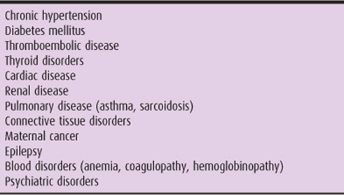
Many medical disorders can complicate the pregnancy course for the mother and thus the fetus. It is important that these diseases and their severity be addressed before conception if possible. During pregnancy, the patient may require aggressive management and additional visits and testing to follow the course of the disease, in addition to possible consultation or management of a high-risk specialist. Table 12–2 lists some of the most important disorders that may complicate pregnancy.
Table 12–2. Some maternal diseases, disorders, and other complications of pregnancy.
D. Family History
A detailed family history is helpful in determining any increased risk of heritable disease states (eg, Tay-Sachs, cystic fibrosis, sickle cell disease) that may affect the mother or fetus during the pregnancy or the fetus after delivery. Other relevant findings includes family history of thromboembolism, birth defects (particularly cardiac anomalies), and medical history of first-degree relatives (particularly diabetes).
E. Ethnic Background
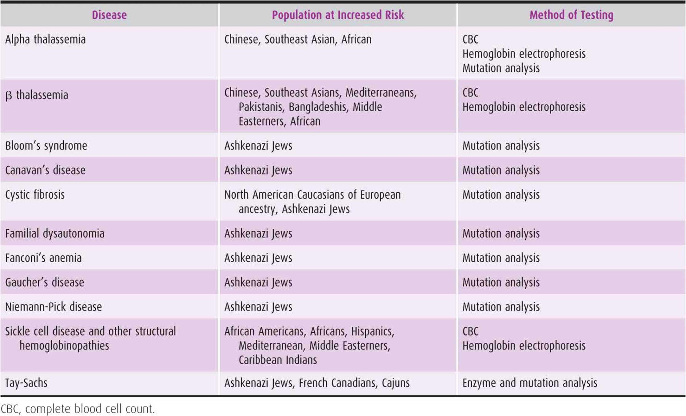
Population screening for certain inheritable genetic diseases is not cost effective because of the relative rarity of those genes in the general population. However, many genetic diseases affect certain ethnicities in disproportionate amounts, allowing cost-effective screening of those particular groups. Table 12–3 lists several common inheritable genetic diseases for which screening is possible. It includes the group at risk as well as the method of screening.
Table 12–3. Common inheritable genetic diseases.
F. Past Obstetric History
1. Recurrent abortion—A diagnosis of recurrent abortion is made after 3 or more consecutive spontaneous losses of a pregnancy before 20 weeks’ gestation. Recurrent abortion is best investigated before another pregnancy occurs; workup can be initiated after 2 losses. If the patient is currently pregnant, however, as much of the workup as possible should be performed.
• Karyotype of abortus specimen
• Parental karyotype
• Survey for cervical and uterine anomalies
• Screening for hormonal abnormalities (ie, hypothyroidism)
• Infectious disease evaluation of the genital tract
The association between inherited thrombophilias and recurrent pregnancy loss is unclear; thus the testing for these is currently not recommended in the clinical setting. Screening for antiphospholipid antibodies (acquired thrombophilia) may be appropriate.
2. Previous stillbirth or neonatal death—A history of previous stillbirth or neonatal death should trigger an immediate investigation regarding the conditions or circumstances surrounding the event. If the demise was the result of a nonrecurring event, such as cord prolapse or traumatic injury, then the present pregnancy has a risk approaching the background risk. However, stillbirth or neonatal death may suggest a cytogenetic abnormality, structural malformation syndrome, or fetomaternal hemorrhage. Review of records, including autopsy, placental pathology, and karyotype if obtained, is vital. As with fetal loss, the association of unexplained stillbirth with inherited thrombophilias is unclear and testing is not recommended, although a maternal thrombophilia workup may be considered with stillbirth in the setting of severe placental thrombosis or infarcts, significant fetal growth restriction, or in the patient with a history of thrombosis.
3. Previous preterm delivery—A history of preterm birth confers an increased risk of early delivery in subsequent pregnancies. Furthermore, the risk of a subsequent pre-term birth increases as the number of prior preterm births increases, and the risk decreases with each subsequent birth that is not preterm. The recurrence risk also rises as the gestational age of the previous preterm delivery decreases. Despite intense investigation, the incidence of preterm delivery has slightly increased in the United States, due in large measure to medical intervention producing indicated preterm deliveries. Eighty-five percent of preterm deliveries occur between 32 and 36 6/7 weeks, and they carry minimal fetal or neonatal morbidity. The remaining 15% of preterm deliveries, however, account for nearly all of the perinatal morbidity and mortality. Common causes of perinatal morbidity in premature infants include respiratory distress syndrome, intraventricular hemorrhage, bronchopulmonary dysplasia, necrotizing enterocolitis, sepsis, apnea, retinopathy of prematurity, and hyperbilirubinemia. Preterm deliveries can be divided into 2 types: spontaneous and indicated, with indicated preterm deliveries caused by medical or obstetric disorders that place the mother and/or fetus at risk. The clinical risk factors most often associated with spontaneous preterm birth include history of previous preterm birth, genital tract infection, nonwhite race, multiple gestation, bleeding in the second trimester, and low prepregnancy weight. Recent multicenter trials have shown that progesterone in the form of 17α-hydroxyprogesterone caproate, given as weekly injections of 250 mg beginning in the second trimester, can decrease the risk of preterm delivery in patients with a history of prior spontaneous preterm delivery.
4. Rh alloimmunization or ABO incompatibility—All pregnant patients should undergo an antibody screen at the first prenatal visit. Those patients who are Rh (D)–negative with no evidence of anti-D alloimmunization should receive Rh (D) immunoglobulin (RhoGAM; Ortho-Clinical Diagnostics, Rochester, NY) 300 μg at 28 weeks of gestation. Patients who are Rh (D) sensitized can be followed with maternal titers and/or amniocentesis for fetal blood typing, followed by either serial amniocentesis for ΔOD450 or serial middle cerebral artery peak systolic velocity measurements, as well as fetal blood sampling via cordocentesis.
5. Previous preeclampsia–eclampsia—Previous preeclampsia–eclampsia increases the risk for hypertension in the current pregnancy, especially if underlying chronic hypertension or renal disease is present.
6. Previous infant with genetic disorder or congenital anomaly—A woman with a previous history of a fetus with a chromosomal abnormality is a frequent indication of cytogenetic testing, although this may be preceded by first- or second-trimester screening and anatomy ultrasound (US). The rate of recurrence depends on the abnormality.
7. Teratogen exposure—A teratogen is any substance, agent, or environmental factor that has an adverse effect on the developing fetus. Whereas malformations caused by teratogen exposure are relatively rare, knowledge of exposure can aid in the diagnosis and management.
A. DRUGS—Alcohol, antiseizure medications (phenytoin, valproic acid, etc.), lithium, mercury, thalidomide, diethylstilbestrol (DES), warfarin, isotretinoin, and so forth.
B. INFECTIOUS AGENTS—Cytomegalovirus, Listeria, rubella, toxoplasmosis, varicella, Mycoplasma, and so forth.
C. RADIATION—It is believed that medical diagnostic radiation delivering less than 0.05 Gy (5 rad) to the fetus has no teratogenic risk.
Physical Examination
Physical examination is important not only during the initial visit, but also throughout the pregnancy. Collection of maternal height and weight information allows for calculation of maternal body mass index, useful in risk assessment for many pregnancy abnormalities. In addition, weight gain is followed throughout the pregnancy, also a useful parameter for several risk factors in pregnancy. Vital sign abnormalities can lead to the diagnosis of many key obstetric complications. Fever, defined as a temperature of 100.4°F or greater, can be a sign of chorioamnionitis. Signs or symptoms of chorioamnionitis should be assessed, and, if chorioamnionitis is suspected, amniocentesis for microscopy and culture should be considered. Depending on clinical correlation, delivery may be necessary. Maternal tachycardia can be a sign of infection, anemia, or both. Isolated mild tachycardia (>100 beats/min) should be evaluated and followed up, as should maternal tachyarrhythmias. Maternal heart rate is noted to increase normally in pregnancy, however. The normal pattern of maternal blood pressure readings is a decrease from baseline during the first trimester, reaching its nadir in the second trimester, and slightly rising in the third trimester, although not as high as the baseline levels. Repeated blood pressure readings of 140/90 mm Hg taken 6 hours apart should be considered evidence of preeclampsia or gestational hypertension. Increases in systolic and diastolic blood pressure, although no longer part of the definition, may also be an indication of development of pregnancy-related hypertensive disease. The rest of the physical examination should be performed during the initial visit and focused examination during each visit. Fundal height measurements and fetal heart tone checks should also be performed.
Urinalysis
At the first prenatal visit, a clean-catch urine culture and sensitivity should be performed. Any growth should be treated with the appropriate antibiotics. At all subsequent visits, urine dipstick testing to screen for protein, glucose, leukocyte esterase, blood, or any combination of markers is useful in identifying patients with a change in baseline urinary composition.
Screening Tests
Screening tests during the initial visit include testing for rubella, rapid plasma reagin, hepatitis B, blood type, HIV, gonorrhea, and Chlamydia, and Pap smear.
American College of Obstetricians and Gynecologists. Inherited Thrombophilias in Pregnancy. ACOG Practice Bulletin No. 113. Washington, DC: American College of Obstetricians and Gynecologists; 2010.
American College of Obstetricians and Gynecologists. Screening for Fetal Chromosomal Anomalies. ACOG Practice Bulletin No. 77. Washington, DC: American College of Obstetricians and Gynecologists; 2007 (Reaffirmed 2008).
Branch DW, Gibson M, Silver RM. Clinical practice. Recurrent miscarriage. N Engl J Med 2010;363:1740–1747. PMID: 20979474.
Goldenberg RL, Culhane JF, Iams JD, Romero R. Epidemiology and causes of preterm birth. Lancet 2008;371:75–84. PMID: 18177778.
Mari G, Deter RL, Carpenter RL, et al. Noninvasive diagnosis by Doppler ultrasonography of fetal anemia due to maternal red cell alloimmunization. Collaborative group for Doppler assessment of the blood velocity in anemic fetuses. N Engl J Med 2000;342:9–14. PMID: 10620643.
Meis PJ, Klebanoff M, Thom E, et al. Prevention of recurrent preterm delivery by 17 alpha-hydroxyprogesterone caproate. N Engl J Med 2003;348:2479–2485. PMID: 12802023.
Mercer BM, Macpherson CA, Goldenberg RL, et al. Are women with recurrent spontaneous preterm births different from those without such history? Am J Obstet Gynecol 2006;194: 1176–1184. PMID: 16580328.
Reddy UM. Prediction and prevention of recurrent stillbirth. Obstet Gynecol 2007;110:1151–1164. PMID: 17978132.
Schieve LA, Ferre C, Peterson HB, Macaluso M, Reynolds MA, Wright VC. Perinatal outcome among singleton infants conceived through assisted reproductive technology in the United States. Obstet Gynecol2004;103:1144–1153. PMID: 15172846.
ANTEPARTUM MANAGEMENT
Genetic Testing
A. First-Trimester Screening
Nuchal translucency, measured between 11(0/7) and 13(6/7) weeks, combined with maternal serum free β-human chorionic gonadotropin (β-hCG) and pregnancy-associated plasma protein-A levels, has been found to have 87.0% sensitivity for detection of trisomy 21, with a 5% false-positive rate. In the absence of chromosomal abnormalities, an increased nuchal translucency is associated with an increased risk of structural cardiac abnormalities and skeletal dysplasias, and so forth. Further US findings in the first trimester, including absence of nasal bone and abnormal ductus venosus Doppler findings, may further improve the detection rate for aneuploidy but require a high level of sonographic skill. Patients with an abnormal screening result can be offered invasive testing such as chorionic villus sampling for more accurate detection of fetal aneuploidy. The advantage of the first-trimester screen (as opposed to second-trimester maternal screening, discussed next) is that it allows for earlier detection of aneuploidy.
B. Second-Trimester Maternal Serum Screening
Frequently known as the “triple screen,” this test includes maternal serum α-fetoprotein (MSAFP), β-hCG, and estriol. In some institutions, only the MSAFP is used, whereas in other institutions, a fourth test for inhibin is included, making it a “quad test.” The usefulness of this screen is its ability to identify pregnancies at increased risk for open neural tube defects, as well as for certain chromosomal abnormalities, especially trisomy 21 (70% sensitivity for Down syndrome detection). This test is effective at 15–22 weeks’ gestation and therefore can identify an at-risk pregnancy in time to pursue more definitive diagnosis, if desired. It is important to note, however, that maternal serum screening is not a definitive test and must be followed by invasive testing (discussed later) for karyotype determination.
The first- and second-trimester screen should not both be ordered independently during a pregnancy; this approach leads to unacceptably high false-positive rates for aneuploidy. Approaches have been developed, however, that allow for both tests to be used in combination to determine aneuploidy risk. One such approach is integrated aneuploidy screening, in which a single risk assessment is calculated using all 6 analytes after completion of both tests. Integrated screening has a Down syndrome detection rate of 95%, with a 5% false-positive rate. One major drawback is the withholding of information until the second trimester, precluding the benefits of early diagnosis and chorionic villus sampling. Another approach is stepwise sequential screening, which presents patients with risk-assessment results after completion of the first-trimester component and then again after the second-trimester blood draw. With this test, the detection rate for Down syndrome is 95%, with a 5% false-positive rate. Combined sequential screening is similar, but patients with a very low first-trimester risk do not have second-trimester analysis performed.
C. Screening for Carriers of Genetic Disease
Screening for sickle cell disease should be offered to individuals of African and African American descent and those from the Mediterranean basin, the Middle East, and India. Hemoglobin electrophoresis is the definitive test to determine the carrier status of sickle cell disease as well as other hemoglobinopathies.
Cystic fibrosis carrier screening should be discussed with all patients. Carrier rates, however, are highest in Caucasians, including parents of Eastern European Jewish (or Ashkenazi Jewish) descent. Furthermore, the detection rate of known mutations is highest in these groups and is lower in other groups. For instance, Asian Americans have a cystic fibrosis carrier rate of 1 in 94, and the detection rate of testing in this population is 49%. Current guidelines recommend offering information regarding testing to all groups but counseling lower-risk groups of the limitations of the testing.
Given the higher prevalence of other recessive genetic diseases among individuals of Ashkenazi Jewish descent, carrier screening should be offered for Tay-Sachs disease, Canavan’s disease, and familial dysautonomia. Screening is also available for mucolipidosis IV, Niemann-Pick disease type A, Fanconi’s anemia group C, Bloom’s syndrome, and Gaucher’s disease and may be considered in this population.
Preterm Labor Detection
Many patients present throughout pregnancy with signs and symptoms of preterm labor, specifically uterine contractions. Although the cost of missing true preterm labor is high, many patients are not in true labor, and the financial cost of aggressive management of these patients is also high. The accurate diagnosis of preterm labor may be aided by 2 screening tests: cervical length measurement and fetal fibronectin.
Gestational Diabetes Screening
Although recent consensus groups have recommended screening for gestational diabetes based on risk factors, many studies have shown this to be inadequate for detecting patients with gestational screening versus universal screening.
Routine screening consists of performing a glucose challenge test between 24 and 28 weeks. The test consists of a 50-g oral glucose load with a plasma glucose level drawn exactly 1 hour after. If the value is 140 mg/dL or over, a more specific glucose tolerance test (GTT) should be performed (the cutoff may be lowered to 130 mg/dL to improve sensitivity). The GTT involves obtaining a fasting plasma glucose level, giving a 100-g oral glucose load, then drawing plasma levels at 1 hour, 2 hours, and 3 hours after the glucose load. A test is considered positive for gestational diabetes if 2 of the 4 values are elevated. The thresholds proposed by Carpenter and Coustan are currently favored (fasting >95 mg/dL, 1 hour >180 mg/dL, 2 hour >155 mg/dL, 3 hour >140 mg/dL).
Group B Streptococcus
Group B streptococcus (GBS) asymptomatically colonizes between 10% and 30% of pregnant women, but perinatal transmission can result in a severe and potentially fatal neonatal infection. The current GBS testing protocol emphasizes the importance of culture screening and treatment over risk factor–based screening. For this reason, patients should be screened with a rectovaginal culture at 35–37 weeks. If the culture is positive, patients should be treated with intrapartum antibiotics. Intrapartum antibiotic prophylaxis has been shown to decrease the risk of perinatal GBS transmission. If the culture result is unknown, patients should be treated if in preterm labor, with rupture of membranes greater than or equal to 18 hours, or maternal fever greater than 100.4°F during labor. All patients with GBS bacteriuria during the pregnancy or a previous neonate with GBS sepsis should be treated with intrapartum antibiotics.
American College of Obstetricians and Gynecologists. Prevention of Early-Onset Group B Streptococcal Disease in Newborns. ACOG Committee Opinion No. 279. Washington, DC: American College of Obstetricians and Gynecologists; 2002.
American College of Obstetricians and Gynecologists. Screening for Fetal Chromosomal Anomalies. ACOG Practice Bulletin No. 77. Washington, DC: American College of Obstetricians and Gynecologists; 2007.
Gabbe SG, Graves CR. Management of diabetes mellitus complicating pregnancy. Obstet Gynecol 2003;102:857–868. PMID: 14551019.
Iams JD, Romero R, Culhane JF, Goldenberg RL. Primary, secondary, and tertiary interventions to reduce the morbidity and mortality of preterm birth. Lancet 2008; 371:164–175. PMID: 18191687.
Wapner RJ, Jenkins TM, Khalek N. Prenatal diagnosis of congenital disorders. In: Creasy RK, Resnik R (eds): Maternal-Fetal Medicine: Principles and Practice. Philadelphia, PA: Saunders Elsevier; 2009.
American College of Obstetricians and Gynecologists. Preconception and Prenatal Carrier Screening for Genetic Diseases in Individuals of Eastern European Jewish Descent. ACOG Committee Opinion No. 442. Washington, DC: American College of Obstetricians and Gynecologists; 2009.
FETAL ASSESSMENT
Performed during all trimesters, the techniques used are diverse, and the information obtained varies according to the quality of imaging, depth of investigation, and gestational age of pregnancy.
Ultrasound
Ultrasound (US) has evolved continuously over the last 30 years, with better equipment produced each year. Real-time sonography allows a 2-dimensional (2-D) image to demonstrate fetal anatomy, as well as characteristics such as fetal weight, movement, volume of amniotic fluid, and structural anomalies, such as myomas or placenta previa that may affect the pregnancy. Three-dimensional sonography allows volume to be ascertained, creating a 3-D–appearing image on the 2-D screen, which assists in identifying and clarifying certain anatomic anomalies. Most recently, 4-D machines have been developed, which produce 3-D video in real time. As the machines become more technically advanced and the computers that run them become faster, the images obtained will continue to improve and push the boundaries of sonographic prenatal diagnosis.
Diagnostic US is widely used in the assessment of the pregnancy and the fetus and offers a more than 80% detection rate of anomalies in experienced centers. The benefits and limitations of US should be discussed with all patients, and the decision to perform a US rests jointly with the physician and the patient.
A standard US examination should provide information such as fetal number, presentation, documentation of fetal viability, assessment of gestational age, amniotic fluid volume, placental location, fetal biometry, and an anatomic survey of the fetus(es). A limited US examination is a goal-directed search for a suspected problem or finding. A limited US can be used for guidance during procedures such as amniocentesis or external cephalic version, assessment of fetal well-being, or documentation of presentation or placental location intrapartum. A specialized US examination is performed when an anomaly is suspected based on history, biochemical abnormalities, or results of either the limited or standard scan. Other specialized examinations include fetal Doppler, biophysical profile (BPP), fetal echo-cardiogram, or additional biometric studies.
US evaluation of fetal anatomy may detect major structural anomalies. Gross malformations such as anencephaly and hydrocephaly are commonly diagnosed and rarely missed; however, more subtle anomalies such as facial clefts, diaphragmatic hernias, and cardiac defects are more commonly reported to have been missed by US. The basic fetal anatomy survey should include visualization of the cerebral ventricles, 4-chamber view of the heart, and examination of the spine, stomach, urinary bladder, umbilical cord insertion site, and renal region. Any indication of an anomaly should be followed by a more comprehensive sonogram. Typically, the fetal anatomic survey is performed at 17–20 weeks; however, there is controversy regarding the potential benefits of an earlier sonogram at 14–16 weeks using the transvaginal probe. The earlier scan allows earlier detection of anomalies that are almost always present by the second trimester, as well as allowing greater detailed viewing of the fetal anatomy by using the higher resolution vaginal transducers.
Aneuploidy Screening
Multiple second-trimester sonographic findings associated with aneuploidy or “markers” of aneuploidy have been identified. The presence of single or multiple markers adjusts the patient’s age-related risk of aneuploidy based on the particular markers present. Such sonographic findings include, but are not limited to:
• Echogenic intracardiac focus
• Choroid plexus cysts
• Pyelectasis
• Echogenic bowel
• Short femur
• Increased nuchal fold
Chorionic Villus Sampling
Chorionic villus sampling (CVS) is an invasive test performed between 9 and 13 weeks’ gestation and can be performed either transcervically or transabdominally. CVS is performed under sonographic guidance with the passing of a sterile catheter or needle into the placental site. Chorionic villi are aspirated and undergo cytogenetic analysis. The benefit of CVS over amniocentesis is its availability earlier in pregnancy. The overall pregnancy loss rate is higher than that of midtrimester amniocentesis, likely as a result of the increased background rate of spontaneous pregnancy loss between 9 and 16 weeks; limited data suggest that procedure-related loss rate for CVS appears to approach the rate of midtrimester amniocentesis. One disadvantage of CVS is that, unlike amniocentesis, it does not allow diagnosis of neural tube defects.
Amniocentesis
Amniocentesis is also performed under the guidance of ultrasonography. A needle is inserted transcutaneously through the maternal abdominal wall into the amniotic cavity, and fluid is removed. There are many uses for this amniotic fluid. In the early second trimester, these include AFP evaluation for neural tube defect assessment and the most common indication of cytogenetic analysis. In this case, amniocentesis is often performed between 15 and 20 weeks’ gestation, and fetal cells from the amniotic fluid are obtained. Risks associated with the procedure are considered to be very low, with the risk of abortion as a result of amniocentesis considered to be between 1 in 200 to 1 in 450 amniocenteses.
Amniocentesis also provides a useful tool later in pregnancy, at low risk, for diagnosis of intra-amniotic inflammation and infection as a risk factor for preterm labor and adverse outcome, as well as documentation of fetal lung maturity.
Fetal Blood Sampling
Also referred to as cordocentesis or percutaneous umbilical blood sampling, fetal blood sampling is an option for chromosomal or metabolic analysis of the fetus. Benefits of the procedure include a rapid result turnaround and the ability to perform the procedure in the second and third trimesters. Intravascular access to the fetus is useful for assessment and treatment of certain fetal conditions such as Rh sensitization and alloimmune thrombocytopenia. However, there is a higher risk of fetal death compared with the other methods. Fetal loss rates are approximately 2%, but can vary depending on the fetal condition involved.
American College of Obstetricians and Gynecologists. Invasive Prenatal Testing for Aneuploidy. ACOG Practice Bulletin No. 88. Washington, DC: American College of Obstetricians and Gynecologists; 2007.
American College of Obstetricians and Gynecologists. Ultrasonography in Pregnancy. ACOG Practice Bulletin No. 101. Washington, DC: American College of Obstetricians and Gynecologists; 2009.
American Institute of Ultrasound in Medicine. AIUM practice guideline for the performance of an antepartum obstetric ultrasound examination. J Ultrasound Med 2010;29:157–166. PMID: 20040791.
Nicolaides KH. Nuchal translucency and other first-trimester sonographic markers of chromosomal abnormalities. Am J Obstet Gynecol 2004;191:45–67. PMID: 15295343.
Sonek JD, Cicero S, Neiger R, et al. Nasal bone assessment in prenatal screening for trisomy 21. Am J Obstet Gynecol 2006;195: 1219–1230. PMID: 16615922.
ANTEPARTUM FETAL TESTING
Fetal Movement Assessment
A decrease in maternal perception of fetal movement can precede fetal death, sometimes by several days. Perception of 10 distinct movements in a period of up to 2 hours is considered reassuring; if fewer, patients are often advised to undergo further testing.
Nonstress Test
Fetal movements associated with accelerations of fetal heart rate (FHR) provide reassurance that the fetus is not acidotic or neurologically depressed. A reactive and therefore reassuring nonstress test (NST) is defined as 2 or more FHR accelerations, at least 15 beats/min above the baseline and lasting at least 15 seconds within a 20-minute period. Vibroacoustic stimulation can elicit FHR accelerations that can reduce overall testing time without compromising detection of an acidotic fetus. In the case of a nonreassuring NST, further evaluation or delivery depends on the clinical context. In a patient at term, delivery is warranted. Pregnancy remote from term poses a more challenging dilemma to the clinician. If resuscitative efforts are not successful in restoring reactivity to the NST, then ancillary tests or testing techniques may prove useful in avoiding a premature iatrogenic delivery for nonreassuring FHR patterns, as the false-positive rate may be as high as 50–60%.
Biophysical Profile
The biophysical profile (BPP) is another way to assess fetal well-being. The BPP is composed of 5 components: NST, fetal breathing movements (30 seconds or more in 30 minutes), fetal movement (3 or more in 30 minutes), fetal tone (extension/flexion of an extremity), and amniotic fluid volume (vertical pocket of 2 cm or more). Each component is worth 2 points; a score of 8 or 10 is normal, 6 is equivocal, and 4 or less is abnormal. A BPP score of 10/10 or 8/10 with normal amniotic fluid volume is associated with a low risk of fetal asphyxia in the following week (approximately 1 per 1000).
Modified Biophysical Profile
Modified BPP combines NST, a short-term indicator of fetal acid–base status, with amniotic fluid index (AFI), a long-term indicator of placental function. AFI is measured by dividing the uterus into 4 equal quadrants and measuring the largest vertical pocket in each quadrant; the results are then summed and expressed in millimeters. The modified BPP has become a primary mode of antepartum fetal surveillance; a nonreactive NST or an AFI less than 50 mm (oligohydramnios) requires further fetal assessment or intervention.
Contraction Stress Test
The contraction stress test (CST) is based on the response of FHR to uterine contractions, with the premise that fetal oxygenation will be worsened during contractions. This can result in late decelerations in an already suboptimally oxygenated fetus. The test requires 3 contractions in 10 minutes to be adequate for interpretation. A positive or abnormal test is defined as late decelerations occurring with more than half of the contractions. A test is considered to be suspicious or equivocal if any late decelerations are seen (with fewer than 50% of contractions) and negative with no late decelerations. Contraindications to CST include any contraindications to labor, such as placenta previa or prior classic caesarean section. This test is now rarely used.
Growth Ultrasound
Growth US studies, performed every 3–4 weeks, are useful for assessment of fetuses that may be at risk for growth restriction secondary to medical conditions of pregnancy or fetal abnormalities.
Doppler Studies
Fetal Doppler studies initially were used to assess the placenta by evaluation of umbilical artery outflow. They have since evolved to a more comprehensive multivessel evaluation of fetal status. Doppler studies can be used to assess a compromised fetus (particularly the growth-restricted fetus) and may function as a diagnostic tool that alerts the clinician to the need for further intervention, including BPP, continuous fetal monitoring, or possibly delivery. In addition, the peak systolic velocity of the middle cerebral artery is used to assess fetal anemia in cases of alloimmunization and parvo-virus infection.
Fetal Maturity Tests
A. Indications for Assessing Fetal Lung Maturity
ACOG has recommended that fetal pulmonary maturity should be confirmed before elective delivery before 39 weeks’ gestation unless fetal maturity can be inferred from any of these criteria: fetal heart tones have been documented for 20 weeks by nonelectronic fetoscope or for 30 weeks by Doppler; 36 weeks have elapsed since a serum or urine hCG-based pregnancy test was reported to be positive; US measurement of crown–rump length at 6–11 weeks of gestation or measurements at 12–20 weeks support a gestational age equal to or greater than 39 weeks. For those patients for whom delivery is mandated by fetal or maternal indications, testing of fetal lung maturity should not be performed, nor does a mature fetal lung maturity result before 39 weeks’ gestation indicate delivery in the absence of appropriate clinical circumstances
1. Lecithin/sphingomyelin ratio—The lecithin/sphingomyelin (L/S) ratio for assessment of fetal pulmonary maturity was first introduced by Gluck and colleagues in 1971. The test depends on outward flow of pulmonary secretions from the lungs into the amniotic fluid, thereby changing the phospholipid composition of the latter and permitting measurement of the L/S ratio in a sample of amniotic fluid. In the absence of complications, the ratio of these 2 components reaches 2.0 at approximately 35 weeks. The presence of blood or meconium may interfere with test interpretation.
2. Phosphatidylglycerol—Phosphatidylglycerol (PG) is a minor constituent of surfactant. It begins to increase appreciably in amniotic fluid several weeks after the rise in lecithin. Its presence is more indicative of fetal lung maturity because PG enhances the spread of phospholipids on the alveoli.
3. Fluorescence polarization—The fluorescence polarization test, currently the most widely used test, uses polarized light to quantitate the competitive binding of a probe to both albumin and surfactant in amniotic fluid; thus it is a true direct measurement of surfactant concentration. It reflects the ratio of surfactant to albumin and is measured by an automatic analyzer, such as the TDx-FLM. An elevated ratio has been correlated with the presence of fetal lung maturity; the threshold for maturity is 55 mg of surfactant per gram of albumin.
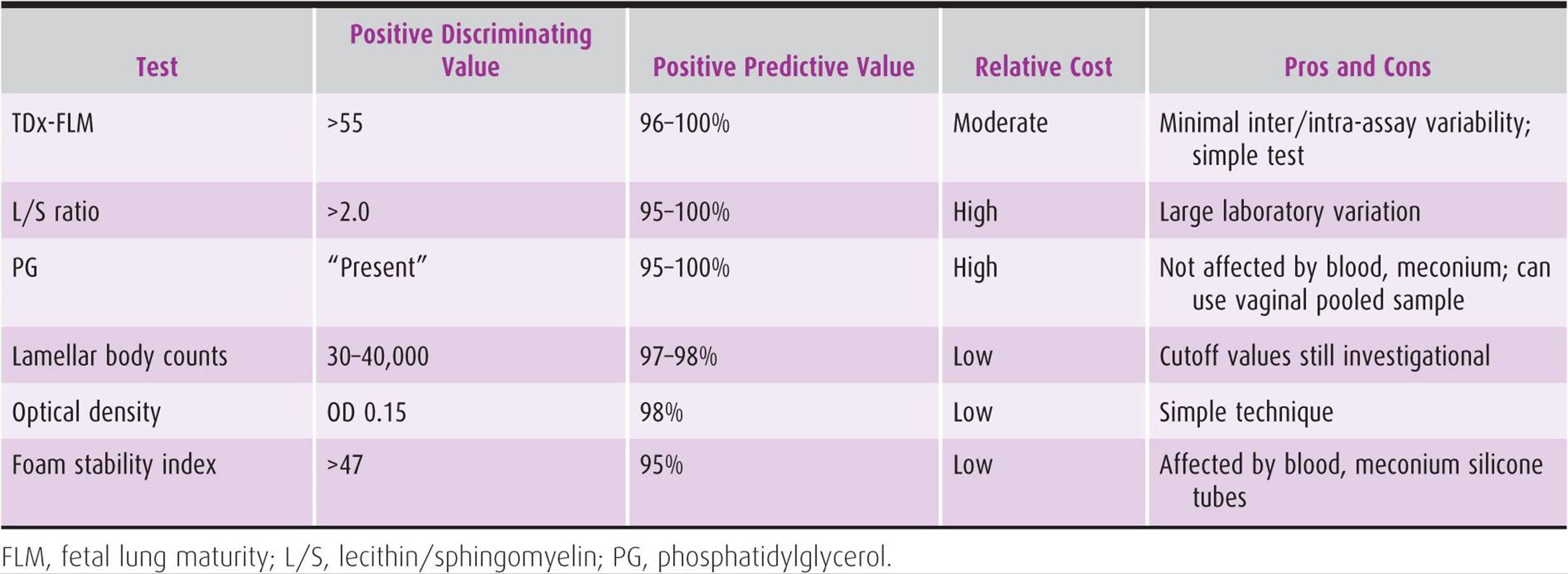
Table 12–4 lists all fetal maturity tests available, discriminating levels, and specific characteristics of the tests.
Table 12–4. Fetal maturity tests.
American College of Obstetricians and Gynecologists. Antepartum Fetal Surveillance. ACOG Practice Bulletin No. 9. Washington, DC: American College of Obstetricians and Gynecologists; 1999 (Reaffirmed 2009).
American College of Obstetricians and Gynecologists. Fetal Lung Maturity. ACOG Practice Bulletin No. 97. Washington, DC: American College of Obstetricians and Gynecologists; 2008.
Baschat AA. Fetal growth restriction—from observation to intervention. J Perinat Med 2010;38:239–246. PMID: 20205623.
INTRAPARTUM MANAGEMENT
Fetal Heart Rate Monitoring
Use of electronic fetal monitoring (EFM) has been increasing over the last several decades, up to 85% in 2002. No randomized controlled trials have compared the benefits of EFM versus no form of monitoring during labor. Randomized clinical trials comparing EFM with intermittent auscultation showed an increase in caesarean section rate, caesarean section rate for fetal distress, and operative vaginal delivery rate, with no reduction in overall perinatal mortality, but possibly a decrease in perinatal mortality from fetal hypoxia. Given these findings, ACOG has stated that either EFM or intermittent auscultation is acceptable, given that guidelines for intermittent auscultation are met. Intermittent auscultation may not, however, be acceptable in high-risk patients.
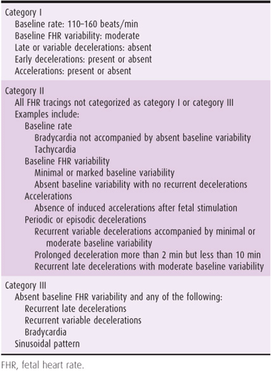
Despite its widespread use, EFM suffers from poor interobserver and intraobserver reliability, uncertain efficacy, and a high false-positive rate. In 2008, a workshop sponsored by ACOG, the Eunice Kennedy Shriver National Institute of Child Health and Human Development (NICHD), and the Society of Maternal-Fetal Medicine recommended an EFM interpretive system based on a three-tiered approach (Table 12–5). Utility of this approach remains to be seen.
Table 12–5. Three-tiered fetal heart rate interpretation system.
Fetal Heart Rate Definitions
The baseline is the mean FHR rounded to increments of 5 beats/min, at least 2 minutes in any 10-minute segment. Normal baseline is between 110 and 160 beats/min. Baseline FHR below 110 is defined as bradycardia; baseline FHR above 160 is defined as tachycardia. Either bradycardia (particularly when the new baseline is less than 80 beats/min) or tachycardia (particularly when associated with a decrease in variability or repetitive late or severe variable decelerations) suggests a nonreassuring fetal status. Accelerations (at 32 weeks or greater) are defined as elevations above the baseline of 15 beats/min lasting 15 seconds or longer; less than 32 weeks it is defined by elevations of 10 beats/min lasting at least 10 seconds. Two or more accelerations in a 20-minute interval are reassuring; this defines reactivity in an NST. Variability is defined by fluctuations in the FHR of 2 cycles per minute or greater and can range from absent to marked. Decelerations are categorized as early, late, and variable. Early decelerationsgenerally mirror contractions in timing and shape and are generally not ominous, often representing head compression. Late decelerations are smooth falls in the FHR beginning after the contraction has started and ending after the contraction has ended. They are associated with fetal hypoxemia and a potential for perinatal morbidity and mortality. Variable decelerations are abrupt in decline and return to baseline, vary in timing with the contraction, and usually represent cord compression. These are most ominous when repetitive and severe (below 60 beats/min). A prolonged deceleration is a decrease of 15 beats/min below the baseline lasting between 2 and 10 minutes.
Ancillary Tests
A. Fetal Scalp Blood Sampling
In the presence of a nonreassuring FHR pattern, a scalp blood sample for determination of pH or lactate can be considered. Although the specificity is high (normal values rule out asphyxia), the sensitivity and positive predictive value of a low scalp pH in identifying a newborn with hypoxic–ischemic encephalopathy is low. For these reasons, in addition to the technical skill and expense of the procedure, fetal scalp pH is no longer used in many institutions.
B. Vibroacoustic Stimulation/Scalp Stimulation
The presence of an acceleration after a vaginal examination in which the examiner stimulates the fetal vertex with the examining finger or after vibroacoustic stimulation (as described earlier under “Nonstress Test”) confirms the absence of acidosis (pH >7.2). Some clinicians prefer these methods over fetal scalp blood sampling because they are less invasive.
C. Fetal Pulse Oximetry
Fetal pulse oximetry, the measure of the fetus’ oxygenation during labor, was developed with the goal of improving the specificity of FHR monitoring and decreasing the number of caesarean sections secondary to nonreassuring fetal status. It has not been demonstrated to be a clinically useful test in evaluating fetal status and is not recommended.
D. ST-Segment Analysis During Labor
ST-segment analysis via computerized real-time analysis of the ST-segment interval of the fetal electrocardiogram has been shown in initial studies to, in combination with intrapartum monitoring with EFM, increase the ability of obstetricians to identify compromised fetuses and to intervene more appropriately. A large multicenter randomized controlled trial is currently underway in the United States.
CONCLUSION
Assessing pregnancy to determine risk, as well as careful monitoring of pregnancies with a recognized risk, begins early in the gestation. Preconception counseling of patients with known medical or genetic disorders helps to optimize outcomes. Early and frequent prenatal care allows the care provider to screen his or her patient population to identify pregnancies at risk and act accordingly. Additionally, pregnancies identified as complicated by 1 or more issues can be followed by assortments of maternal and fetal surveillance techniques to maximize therapeutic treatment.
As technology advances and our ability to both diagnose and treat improve, the methods for assessment and care of the pregnancy at risk will be a constantly changing field.
American College of Obstetricians and Gynecologists. Intrapartum Fetal Heart Rate Monitoring: Nomenclature, Interpretation, and General Management Principles. ACOG Practice Bulletin No. 106. Washington, DC: American College of Obstetricians and Gynecologists; 2009.
American College of Obstetricians and Gynecologists. Management of Intrapartum Fetal Heart Rate Tracings. ACOG Practice Bulletin No.116. Washington, DC: American College of Obstetricians and Gynecologists; 2010.
Macones GA, Hankins GD, Spong CY, Hauth J, Moore T. The 2008 National Institute of Child Health and Human Development workshop report on electronic fetal monitoring: Update on definitions, interpretation, and research guidelines. Obstet Gynecol 2008;112:661–666. PMID: 18757666.
Mires G, Williams F, Howie P. Randomised controlled trial of cardiotocography versus Doppler auscultation of fetal heart at admission in labour in low risk obstetric population. BMJ 2001; 322:1457–1460. PMID: 11408301.
Neilson JP. Fetal electrocardiogram (ECG) for fetal monitoring during labour. Cochrane Database Sys Rev 2006; 3:CD000116. PMID: 16855950.