
Jacob Bornstein MD, MPA
Disorders of the vulva and vagina are very common and cause considerable discomfort. Until recently, however, our understanding of vulvar conditions has been scant due to the lack of communication between gynecologists, dermatologists, pathologists, and sex therapists, each with his or her own ideas of the natural history, mode of diagnosis, and preferred therapy. An obvious consequence is the propagation of terms for the same disorders. The establishment in 1970 of the International Society for the Study of Vulvovaginal Disease (ISSVD) fostered exchange of ideas, collective discussion, and understanding of the natural history and modern treatment of vulvar diseases. Common terminology was established. The terminology of benign vulvar and vaginal disorders used in this chapter is based on the guidelines of the ISSVD. The morphologic and functional approach is accessible to the novice in vulvar and vaginal disease. At the same time it emphasizes breakthroughs in the understanding of the different vulvar pain syndromes, the influence of the vaccine against human papillomavirus (HPV) on vulvar diseases, and the modern treatment of the vulvar dermatoses. The premalignant and malignant vulvar and vaginal disorders are discussed in Chapter 47.
ANATOMY & PHYSIOLOGY
The anatomy of the vagina and vulva is described in Chapter 1. In recent years the vulvar vestibule, the site of origin of vestibulodynia, the “provoked” vulvar pain of dyspareunia, has been a focus of attention. Although the vulvar skin is devoid of estrogen receptors, the development of vaginal disorders is influenced by the presence or absence of endogenous or exogenous estrogen. Estrogen thickens the vaginal epithelium, which leads to the accumulation of glycogen in the epithelial cells. The intraepithelial glycogen metabolizes to lactic acid. The resultant vaginal pH of 3.5–4.0 promotes the growth of normal vaginal flora, chiefly lactobacilli and acidogenic corynebacteria. Asymptomatic Candida organisms may be present in small quantities.
ESSENTIALS OF DIAGNOSIS
Evaluation of a patient with vulvar and/or vaginal symptoms requires the following:
 A meticulous review of physiologic systems to reveal underlying medical conditions that may lead to vulvar symptoms.
• Diabetes mellitus may be associated with vulvar pruritus or pain as a consequence of vulvovaginal candidiasis or, in advanced cases, as a result of neuropathic pain.
• Elevated serum levels of biliary salts, such as in biliary stasis or primary biliary cirrhosis, may cause vulvar pruritus.
• Hematologic disorders such as polycythemia or lymphoma may be associated with systemic symptoms, including vulvar pruritus.
• A complete history of potential causes of vulvar irritation, including creams, powders, soaps, type of underwear, and cleansing techniques, should be reviewed.
• Assessment of compliance to previous prescriptions may help determine whether failure of past treatments is attributable to incorrect diagnosis or to inadequate treatment.
• Patients sometimes refrain from the use of common medications for vulvar disease due to their high potency or potential for side effects. Typical examples are systemic or potent topical steroids such as clobetasol propionate (Dermovate) and tricyclic antidepressants such as amitriptyline.
• Information regarding previous infections should be elicited.
• Sexual activity, although sensitive to both patient and healthcare provider, needs evaluation.
• The use of feminine hygiene products (eg, douching, soaps, perfumes) and medications (eg, pessaries, diaphragms, oral contraceptive pills, antibiotics) can alter the normal vaginal flora.
• Overlying garments made of synthetic fabrics that retain heat and moisture can exacerbate vulvovaginal symptoms.
Active inquiry about vulvar pain, discharge, and pruritus, as patients may fail to disclose these intimate matters.
A physical examination including inspection of all mucosal and skin surfaces, because many skin conditions, such as psoriasis, seborrheic dermatitis, pemphigus, and lichen planus, can affect the vulva.
A vaginal examination including evaluation of the physical, chemical, and microbiologic properties of vaginal discharge. The perianal region should be inspected, as many vulvar disorders affect it. Specimens and cultures may include vaginal wet prep and culture for yeast and bacteria. HPV DNA determination may be required.
Vulvar examination: although examination of the vulva and vagina may be completed using a magnifying glass, a colposcopic examination (ie, “vulvoscopy,” “vaginoscopy” is preferred, especially if a biopsy is to be taken. Two processes are important in the diagnosis of vulvar and vaginal lesions:
1. Try to assess to which of the 6 morphologic types (Table 39–1) the lesion belongs. Then mentally browse the list of possible causes for that lesion.
Table 39–1. Morphologic classification of vulvovaginal disease.

2. Biopsy liberally any suspicious lesion, because in most cases the final diagnosis is based on histopathologic findings. Thus the vulvar biopsy is almost a universal requirement.
PRINCIPLES OF OBTAINING A VULVAR BIOPSY
A satisfactory full-thickness sample of the skin and tumor can be obtained with a dermatologic punch biopsy under local anesthesia. Many vulva experts endeavor not to compress the sample tissue so as to preserve the original morphology of the lesion. Inclusion of the lesion margins helps identify abnormal features. This is also important in case of an ulcer, as its center may be necrotic and noninformative. Hospitalization is not required in most cases. Bleeding can be controlled by local pressure, argentum nitrate application, or, in rare cases, by applying a stitch.
VULVAR DISORDERS
The color of vulvar epithelium or lesions depends principally on the width of keratin layer, vascularity of the dermis, thickness of the overlying epidermis, and the amount of intervening pigment, either melanin or blood pigment.
WHITE LESION
Pathogenesis
The white appearance of a lichenoid lesion of the vulva is primarily due to the maceration of a thickened keratin layer resulting from increased moisture in the vulvar area. The epidermal thickening of neoplasia obscures the underlying vasculature and, in conjunction with the macerating effects of the moist environment, usually produces a hyperplastic white lesion. A diffuse white lesion of the vulva may also occur with the loss or absence of melanin pigmentation as with vitiligo, a hereditary disorder. Leukoderma is a localized white lesion resulting from transient loss of pigment in a residual scar formed after healing of an ulcer.
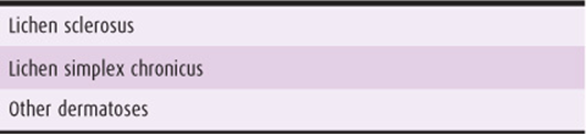
Formerly, a white lichenoid lesion was termed leukoplakia, kraurosis vulva, and senile vulvitis. In 1976 Jeffcoate introduced the term dystrophy, but in 1987 the ISSVD changed the term to nonneoplastic epithelial disorders of vulvar skin and mucosa (Table 39–2). This term was coined to emphasize that excision procedures such as vulvectomy are not required, as this condition is not neoplastic. Implied by the term is the need of a biopsy for definitive diagnosis. The use of the term dystrophy with atypia has been abandoned, and lesions that contain atypia are now called vulvar intraepithelial neoplasia (VIN). They are described in Chapter 47.
Table 39–2. Classification of nonneoplastic epithelial disorders of vulvar skin and mucosa.
LICHEN SCLEROSUS
ESSENTIALS OF DIAGNOSIS
Lichen sclerosus is the most common nonneoplastic epithelial vulvar disorder.
Intense pruritus occurs, usually in women older than 60 years.
The vulvar skin is thin, wrinkled, and white, with areas of lichenification and hyperkeratosis (see Table 39–3 for definitions).
Table 39–3. Dictionary of vulvar terms.

The anterior parts of the labia minora of both sides agglutinate.
Erosions, fissures, subepithelial hemorrhages, and ulcerations result from scratching.
Biopsy is required.
Pathogenesis
Lichen sclerosus, a benign, chronic, inflammatory process, is the most common vulvar dermatologic disorder. Possible etiologic factors of this multifactorial condition include vitamin A deficiency, an autoimmune process, excess of the enzyme elastase, and decreased activity of 5-alpha reductase enzyme, which prevents the conversion of testosterone to dihydrotestosterone (the trophic hormone of the skin) and results in thinning of the skin. The effectiveness of treatment with topical testosterone cream supports the latter hypothesis.
Clinical Findings
A. Symptoms & Signs
This disease usually appears in women older than 60 years. Of the rare appearances in childhood, spontaneous resolution at adolescence occurs in approximately half. Most patients present with pruritus. Some complain of vulvar pain or dyspareunia and/or present with asymptomatic white lesions.
The progression and typical clinical characteristics of acute lichen sclerosus include the following:
1. Erythema and edema of vulvar skin
2. Development of white plaques representing lichenification and hyperkeratosis
3. Uniting of white plaques
4. Intense pruritus leading to scratch–itch cycle
5. Telangiectasias and subepithelial hemorrhages resulting from scratching
6. Erosions, fissures, and ulcerations
The progression and typical clinical characteristics of chronic lichen sclerosus include the following:
1. Thin, wrinkled, and white skin with a cigarette-paper appearance
2. Agglutination of the anterior parts of the labia minora of both sides to cover the clitoris and create phimosis (Fig. 39–1)
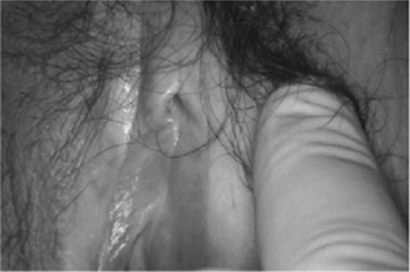
Figure 39–1. Advanced lesion of lichen sclerosus. The labia minora and prepuce of the clitoris have blended into the labial skin.
3. Contraction of the vulvar structures with resultant introital stenosis, previously termed kraurosis
4. Involvement of the perianal region in the form of 8: around the vulva and around the anus.
5. Development in some women of islands of hyperplastic epithelia within the atrophic lichen sclerosus epithelium
Histologic Findings
Definitive diagnosis depends on identification of the following 5 histologic features in the biopsy (Fig. 39–2):
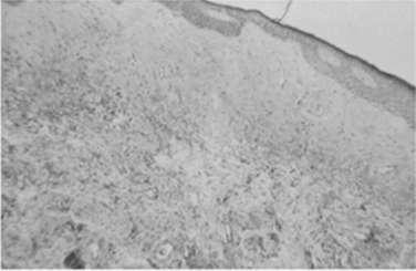
Figure 39–2. Microscopic appearance of lichen sclerosus, characterized by hyperkeratosis, flattened epidermis, and hyalinization of the dermis.
1. Thin hyperkeratotic layer
2. Thinning of the epithelial layer
3. Flattening of the papillae (Rete pegs)
4. Homogenization of the stroma
5. Deep lymphocytic infiltration
Differential Diagnosis
Other causes of a white lesion in the vulva are vitiligo, lichen simplex chronicus, and other dermatoses, such as psoriasis.
Complications
The high rate of squamous cell cancer in women with lichen sclerosus (3–5%) prompts biopsy of all new lesions. Cancer develops mainly in women who continue to suffer from vulvar itch or neglect treatment. It is expected that the introduction of clobetasole, an effective topical treatment for lichen sclerosus, may decrease the incidence of vulvar carcinoma developing from lichen sclerosus lesions.
Treatment
The first step is to stop the itch–scratch cycle and minimize the dermal inflammation. General measures of vulvar hygiene should be applied: avoiding tight undergarments, cleansing daily with mild soap, and drying the vulvar skin with a hair dryer.
A. Medications
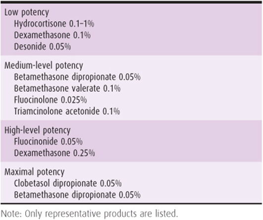
An oral antihistamine agent can be taken at bedtime. Although clobetasole dipropionate 0.05% (Dermovate) is a superpotent topical steroid (Table 39–4), it is recommended at the start to obtain immediate relief, stop the itch–scratch cycle, and reinstitute the patient’s belief in the health care provider. To prevent or minimize the side effects of this steroid, topical application should be in a small amount, similar to that of toothpaste on a toothbrush, twice daily for 2 weeks, then once daily for 2 weeks, then twice weekly for 2 weeks, and then as needed for the rest of the woman’s life. Some recommend tapering to a lower-potency topical steroid for treatment maintenance. Atrophic degeneration of the skin secondary to the steroid is rare.
Table 39–4. Potency of steroids.
Some recommend tacrolimus cream, retinoid, antimalarial agents, or photodynamic therapy for these who do not respond to clobetasole. Surgical therapy should be limited to treatment of introital narrowing leading to dyspareunia or associated intraepithelial or invasive squamous cell neoplasia.
When nothing else relieves the itch, either of the following may be tried: intralesional injection of steroids or surgical undermining of the affected skin without excision, with the intention of undercutting the nerve fibers (Mering procedure).
The following 3 treatments, popular in the past, have been discontinued:
1. Topical application of 2% testosterone propionate ointment; less effective than clobetasole, it leads to virilization.
2. Intralesional injection of alcohol, a painful procedure that leads to sloughing of the vulvar tissues.
3. Vulvectomy; after this unnecessary mutilation, the disease tends to recur at the adjacent tissues.
Prognosis
The disease is chronic and usually recurs with cessation of treatment. The introduction of clobetasole dipropionate leads to resolution of symptoms in most patients and reversal of skin changes in approximately half of patients.
VULVAR LICHEN SIMPLEX CHRONICUS
Vulvar lichen simplex chronicus is the current term for the condition that includes the previously designated hyperplastic dystrophy, squamous cell hyperplasia, atopic dermatitis, atopic eczema, and neurodermatitis.
Clinical Findings
A. Symptoms & Signs
Vulvar lichen simplex chronicus is characterized by benign epithelial thickening and hyperkeratosis resulting from chronic irritation, such as from the use of perfumed pads or chronic vulvovaginal infections. The accompanying pruritus leads to rubbing and scratching, which becomes involuntary over time. As epithelial thickening develops, the humid environment of the vulva causes maceration, and a raised white lesion may become diffuse and even involve the adjacent thighs, perineum, or perianal skin.
Histopathologic Findings
Biopsy is necessary to exclude intraepithelial neoplasia and invasive tumor. Histologic examination demonstrates hyperkeratosis and acanthosis (see Table 39–3 for definitions), resulting in thickening of the epithelium and elongation of the rete pegs. In contrast to lichen sclerosus, there is no dermal inflammatory infiltrate.
Differential Diagnosis
Differential diagnosis includes the other nonneoplastic epithelial disorders such as lichen sclerosus, flat condyloma acuminatum, psoriasis, and vulvar intraepithelial neoplasia.
Treatment
Treatment of squamous cell hyperplasia starts with general measures of vulvar hygiene. Sitz baths and lubricants can help restore moisture to cells and reconstruct the epithelial barrier. Oral antihistamines may help relieve pruritus. In addition, topical application of medium-potency steroids twice daily can decrease the inflammation and pruritus. Vulvar epithelium takes at least 6 weeks to heal. For intractable cases, antidepressants or subcutaneous intralesional injection of steroids can be considered.
LICHEN PLANUS
Clinical Findings
Lichen planus rarely affects the vulva. It is a mucocutaneous dermatosis characterized by the presence of sharply marginated flat-topped papules on the skin and less sharply marginated white plaques on oral and genital mucous membranes. The pathogenesis is unknown. In the vulva 2 clinical aspects can be observed: classic leukoplastic lesions and erosive lesions. Vulvar erosive lichen planus seems to be more frequent but is often ignored. The clinical appearance of vaginal erosive lichen planus is similar to that of desquamative inflammatory vaginitis.
Treatment
Treatment of lichen planus is mainly topical, starting with hydrocortisone foam for the vagina (Colifoam). If unsuccessful, fluorinated corticosteroids, ultrapotent corticosteroids, or topical treatment with tacrolimus 0.1% can be tried. Careful and frequent examination of the vagina for formation of adhesions is important. In cases of severe pruritus and intensive mucocutaneous involvement, systemic steroids should be used. Introital stenosis and vaginal adhesions can be treated by use of vaginal dilators in graduated sizes or from surgical release of scars.
RED LESION
The red color of these lesions results from thinning epidermis, revealing capillary vasculature. Other causes are vasodilatation associated with inflammation and neovascularization of a neoplasia. Although they may manifest any acute dermatitis, red lesions are mainly associated with acute candidal vulvovaginitis. Vulvovaginitis is discussed later in this chapter, under Vaginal Disorders. Paget’s disease, a nonsquamous intraepithelial neoplasia characterized by an eczematous-like red lesion spreading over the vulvar skin, is discussed in Chapter 47. Other red lesions include seborrheic dermatitis, lupus erythematosus, and some cases of VIN. VIN also presents as a white or dark lesion and as an ulcer or tumor.
PSORIASIS
Psoriasis is a chronic relapsing dermatosis that affects the scalp, the extensor surfaces of the extremities, the trunk, and the vulva. Sometimes the vulvar skin is the only body surface affected. Primary lesions are raised and appear typically erythematous, resembling a candidal infection. Most lesions are sharply demarcated. The silver scaly crusts that characterize psoriasis on other parts of the body are usually absent; hence the lesion is red. Treatment includes topical corticosteroids.
DARK LESION
Dark lesions result from an increased quantity or concentration of melanin or hemosiderin pigments, sometimes subsequent to trauma. A persistent dark lesion on the vulva skin likely represents a nevus or a melanoma.
MELANOSIS OR LENTIGO
Melanosis or lentigo is a benign darkly pigmented flat lesion that may be mistaken for a melanoma. A nevus on the vulvar skin may be flat, slightly elevated, papillomatous, dome-shaped, or pedunculated. Melanomas of the vulva are uncommon neoplasms constituting only 1–3% of vulvar cancers. They are extremely aggressive malignant lesions and may arise from pigmented nevi of the vulva.
CAPILLARY HEMANGIOMA
Senile (cherry) hemangiomas are usually multiple, small, dark blue, asymptomatic papules that are discovered incidentally during examination of the older patient. Excision biopsy is needed only if the hemangiomas bleed repeatedly. A cryosurgical probe or carbon dioxide laser can also be used.
Childhood hemangiomas are usually diagnosed in the first few months of life. They may vary in size from small strawberry hemangiomas to large cavernous ones. They tend to be elevated and bright red or dark, depending on their size and the thickness of the overlying skin. Although tending to increase in size during the first few months of life, they often become static or regress without therapy after age 18 months. Although most of these hemangiomas only require observation, and not therapy, larger ones may require treatment with cryosurgery, argon laser therapy, or sclerosing solutions.
OTHER DARK LESIONS
In some VIN, melanin pigment that is not contained in atypical squamous cells concentrates in local macrophages, causing dark coloration of the tumor. Vulvar epithelium may darken after use of estrogen cream or oral contraceptive pills. Kaposi’s sarcoma, dermatofibroma, and seborrheic keratoses are examples of dark lesions. Biopsy of a dark lesion should include the whole lesion, as incomplete removal of melanoma has been suggested as a cause of accelerated spread.
ULCER
The most common cause of ulcerative lesion is a sexually transmitted disease (STD). The most common STD causing vulvar ulcer or erosion is herpes genitalis.
HERPES GENITALIS
ESSENTIALS OF DIAGNOSIS
Frequently preceded by a prodrome: burning, itching and flu-like symptoms.
Vesicles develop but erode rapidly, resulting in painful erosions or ulcer.
Each erosion is surrounded by a red halo (Fig. 39–3).

Figure 39–3. Ulcers of herpes genitalis. Each is surrounded by a red halo. The lesions are spread in a serpentine-like fashion.
Lesions are spread in a serpentine-like fashion.
Recurrences are common.
Only 20% of affected patients are diagnosed correctly.
The gold-standard of diagnosis is viral culture. Other reliable tests are glycoprotein-G–based specific serology and polymerase chain reaction.
Herpesvirus hominis (herpes simplex virus [HSV], herpes genitalis) is responsible for recurrent and disabling symptomatic disease, venereal transmission, and infection to the neonate (ie, herpes encephalitis). HSV type 1 and type 2 are the variants that affect the vulva and vagina. Serologic prior exposure to HSV type 2 is evident in 20–25% of women. Approximately 83% of patients develop antibodies to HSV type 2 within 21 days of a primary infection. Approximately 60% of primary genital infections are by herpesvirus simplex type 2, and the remainder by type 1.
Pathogenesis
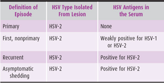
Infection occurs through intimate contact, mainly sexual intercourse. The virus contaminates secretions and mucosal surfaces, entering the skin and mucosa through cracks and other lesions. In turn, the erosions and ulcers of herpes simplex provide a port of entry to other sexually transmitted infections, such as HIV. The virus initially replicates in the dermis and epidermis and then stays latent in a nearby nerve ganglion. Incubation time is 2–7 days. Periods of viral shedding without any symptoms may occur (Table 39–5). This asymptomatic shedding is of particular concern. When unrecognized, the patient continues to have unprotected sexual intercourse, unknowingly transmitting the virus to her partner. This phenomenon is extremely common. For approximately half of the afflicted, asymptomatic viral shedding is identifiable within 1 year of the primary outbreak. Later, decreased viral shedding frequency, down to 2% at 10 years, makes detection difficult.
Table 39–5. Definition of episode of genital herpes infection.
Prevention
Avoidance of direct contact with active lesions prevents spread of the disease. Recommendations for prevention of dissemination are as follows:
• Precautions even in the absence of active lesions, due to asymptomatic shedding of the virus. The most frequent shedding occurs during the year after the first episode.
• Constant use of condoms. However, the condom does not prevent all infections, as lesions may develop in the area outside the tissue covered by the condom.
• The carrier in a serologically discordant couple should consider taking suppressive antiherpetic medication for prolonged periods.
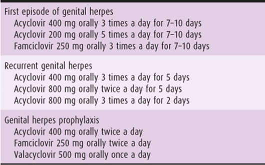
• Administration of a prolonged suppressive therapy for individuals with 6 or more recurrences per year, patients who suffer from distressing prodromes or outbreaks, and men with lesions outside the area that can be protected by a condom (Table 39–6).
Table 39–6. Oral treatment of herpes genitalis.
Clinical Findings
A. Symptoms & Signs
Prodromal symptoms of tingling, burning, or itching, as well as flu-like feeling with fever, malaise, headaches, and myalgia, may present shortly before the appearance of vesicular eruptions. The vesicles erode rapidly, resulting in painful erosions or ulcers (see Table 39–3 for definitions) distributed in small patches or involving most of the vulvar surfaces. Each lesion is surrounded by a red halo. The lesions appear in a serpentine-like fashion on the vulva, hence the name of the disease: herpes (from Latin, “serpentine, snake-like”). In contrast to the common myth, the typical herpes simplex skin lesion is not a vesicle but rather an erosion or ulcer. Bilateral inguinal adenopathy may be present. Urinary symptoms such as dysuria and urinary retention may develop, necessitating hospitalization and placement of a urinary catheter. In 20% of the cases, the primary infection is asymptomatic (Table 39–5). In 60% of the affected, herpesvirus infection is incorrectly diagnosed as a recurrent yeast infection. Lesions may persist for 2–6 weeks with no subsequent scarring.
Diagnosis
Due to the far-reaching implications on a woman and her relationship with her partner, genital herpes should be carefully diagnosed using the appropriate tests and after all other conditions in the differential diagnosis of vulvar ulcers are excluded.
The gold standard of diagnosis is viral culture on fibro-blasts. The virus can be cultured from vesicle fluid or a scraping from an erosion or an ulcer during the acute phase. However, organisms cannot usually be cultured after the primary lesions heal, which occurs within 2 weeks. Although polymerase chain reaction is a sensitive test to detect HSV DNA, its detection in a lesion may indicate a previous outbreak rather than a current event.
A smear scraped from the lesion and stained by Papanicolaou stain or Giemsa (Tzanck test) yields fast results, but is less sensitive and less specific than a culture. Cytologic characteristics of genital herpes include the following:
1. Giant cells
2. Multiple nuclei
3. Molding of the nuclei (compression of one into another)
4. Ground-glass appearance
A. Serologic Tests
Approximately 85% of individuals develop immunoglobulin (Ig) M antibodies to HSV 2 virus within 21 days of exposure. In the past, serologic tests were unreliable because they could not differentiate between herpes virus types 1 and 2, which share approximately 80% of their antigens. New type-specific serologic tests for herpes simplex virus are now available. To distinguish between herpes simplex types 1 and 2, the IgG and IgM antibodies of the type-specific glycoprotein G–based assays should be specifically requested.
Differential Diagnosis
Sometimes the first referral is for a vulvar ulcer of unknown etiology. Although genital herpes is the most frequent cause of vulvar ulcer, other causes, mainly sexually transmitted infections such as syphilis, chancroid, and lymphogranuloma venereum, exist as well (Table 39–7). Noninfectious causes include Behçet’s syndrome and desquamative inflammatory vaginitis (DIV), which are discussed later in this chapter. Of particular significance in the differential diagnosis is vulvovaginal candidiasis, recognized as the “great imitator” of genital herpes. Itching and considerable erythema usually present in vulvar and vaginal candidiasis. Persistent scratching can lead to small ulcers or excoriations (see Table 39–3 for definition) that resemble herpetic lesions. Many physicians diagnose any burning, itching, and erythematous lesion in the vulva as vulvar candidiasis and treat with antifungal medications. As a result, many cases of genital herpes are misdiagnosed. Suspicion for genital herpes should increase when a lesion is particularly painful; when there is a complaint about ulcer or erosions, burning vulvar pain, nonspecific influenza-like symptoms, or referred pain to the legs; or when presumed candidiasis does not heal after 1 course of therapy.
Table 39–7. The differential diagnosis of vulvar ulcers.

Complications
In addition to pain and discomfort, herpes genitalis entails social and psychologic implications, such as stigmatization and apprehension of recurrent outbreaks during which sexual activity should be abstained. Furthermore, there is a moral obligation to forewarn every partner, before sexual intercourse, of the possibility of infection. The embarassment of self-disclosure may lead to reluctance to start a new relationship. To ease the stress, the health care provider should inform the patient that administration of acyclovir or its valine analog valcyclovir in a continuous prophylactic manner, as suppressive treatment, prevents outbreaks and reduces viral transmission.
A. Neonatal Herpes
The incidence of neonatal herpes simplex virus infection ranges from 1 in 1800 live births in California to 1 in 60,000 in England. Infection of the newborn is associated with a 60% mortality rate, and at least half of the survivors have significant neurologic and/or ocular sequelae. The risk of infection to an infant born vaginally to a woman with active primary genital infection is 40–50% and to one with recurrent infection, 5%. However, most infants who develop herpetic infection are born to women who have no history or clinical evidence of infection during pregnancy. Therefore, identification of women whose infants may be in jeopardy is difficult. All pregnant women should be asked whether they or their partners have had genital herpetic lesions. Women with a history of herpes can deliver vaginally if no clinical signs or symptoms of infection are present. Obtaining routine weekly vaginal cultures to detect herpes is no longer standard procedure. However, some physicians initiate suppressive antiviral therapy at 36 weeks to decrease the need for caesarean section in women with frequent outbreaks.
Treatment
The lesions of herpesvirus infection are self-limiting and heal spontaneously unless they become infected secondarily. Symptomatic treatment includes good genital hygiene, loose-fitting undergarments, cool compresses or sitz baths, and oral analgesics. Indications for hospitalization for a severe primary infection include urinary retention, severe headache or other systemic symptoms, and body temperature exceeding 38.3 °C (101 °F). Immunosuppressed patients are more prone to systemic dissemination and should be carefully managed. Treatment includes intravenous acyclovir for hospitalized patients and oral and/or topical antivirals for ambulatory patients. Recurrent herpes should be treated at the onset of prodromal symptoms or vesicle formation. If initiated early, 1-day treatment may suffice. Once-daily continuous prophylactic (suppressive) dosing for many years may be considered for frequent recurrent outbreaks, with 40–70% of patients free of recurrence at 1 year (Table 39–6).
Prognosis
Despite measurable humoral and cell-mediated immunity, reactivation of the virus occurs. After replication in the skin, the viral particles are transported along the peripheral sensory nerve fibers to the dorsal root ganglion, where latent infection is established. Exogenous factors known to contribute to activation of herpesvirus include fever, emotional stress, and menstruation. Immunocompromised patients are prone to develop extensive local disease and systemic dissemination. Whether frequent coitus promotes recurrent disease is unknown. Type 2 virus is more likely than type 1 to recur. Approximately 50% of patients have a recurrence within 6 months of the primary infection. The recurrent ulcers tend to be smaller, fewer in number, and confined to a constant area in the vulva, cervix, or vagina. Healing is generally complete in 1–3 weeks. The virus is not recoverable within 7 days of healing of recurrent lesions. Inguinal adenopathy and systemic symptoms generally do not occur with recurrent outbreaks. Primary infections can generally be distinguished from secondary infections based on clinical findings. Extragenital sites, such as the fingers (herpetic whitlow), buttocks, and trunk (eczema herpeticum, see Table 39–3), have been described.
BEHÇET’S SYNDROME
Behçet’s syndrome is a rare inflammatory disorder characterized by a classic symptom triad: (1) recurrent oral aphthae or ulcers, (2) recurrent genital aphtae or ulcerations, and (3) uveitis. The painful genital ulcers are preceded by small vesicles or papules and last for variable periods. Their borders are irregular. After healing, deep ulcerations may result in scarring or fenestration of the labia. Ocular lesions begin as superficial inflammation and may proceed to iridocyclitis and even blindness. In addition to the classic symptoms, the disease may cause thrombophlebitis or involve the joints in a form of monoarticular arthritis. Central nervous system symptoms manifest in severe disease. Susceptibility to Behçet’s disease is strongly associated with the HLA-B51 allele. Prevalence is highest in Eastern Europe and the Mediterranean. Although the exact etiology is unknown, the disease likely represents an underlying autoimmune process.
Behçet’s syndrome, together with disseminated lupus erythematosus and pemphigus, should be included in the differential diagnosis of recurrent aphthous ulcers of the oral and vaginal mucosa. Ophthalmic examination and human leukocyte antigen typing can aid in diagnosis. Treatment starts with colchicine tablets. Topical and systemic corticosteroids provide immediate relief.
INVESTIGATION OF A VULVAR ULCER
Patient History
A thorough patient history should be taken, including the general health condition, because debilitating or chronic diseases such as AIDS can lead to chronic infections that generate ulcers. The patient should be asked about outbreaks of vulvar ulcers, as well as previous evaluations and their results. Recurrent lesions are typical of both genital herpes and Behçet’s syndrome. Finally, the patient should be asked about any medications she takes. Severe allergic reactions, such as manifested by Stevens-Johnson syndrome, may also cause large ulcers in the vulva and vagina.
Physical Examination
A general physical checkup should include assessment of dermatologic diseases, such as lichen planus, which, in addition to the characteristic dark plaques in the back and limb areas, may appear as extensively desquamated skin and ulcers in the vulva. Pemphigus vulgaris, an autoimmune dermatologic disease with antibodies directed against intercellular sites of stratified squamous epithelium, may present as vesicobullous lesions in the vulva. The oral mucosae should be checked for aphthae or ulcers. Behçet’s syndrome, Crohn’s disease, lichen planus, and pemphigus vulgaris present as ulcerative lesions in both oral and genital skin and mucosa.
Specific Tests
Using dark-field microscopy, a sample from the base of the ulcer should be investigated for the presence of Treponema pallidum, the causative agent of syphilis. Cultures for HSV and serologic tests to exclude chlamydia, systemic lupus erythematosus, HIV, and syphilis should be taken. The absence of a clear diagnosis prompts a biopsy to enable visualization of granulomas and vasculitis apparent in Crohn’s disease and Behçet’s syndrome and to exclude malignant and pre-malignant tumors.
SMALL TUMORS
CONDYLOMA ACUMINATUM
ESSENTIALS OF DIAGNOSIS
Asymptomatic white papillary growths, small at first, tend to coalesce (Fig. 39–4).

Figure 39–4. Vulvar and perineal condylomata acuminata.
Affects the vulva, vagina, and cervix in women; the penis and scrotum in men; and the pubis, perineum, perianal, and oropharyngeal areas in both sexes.
A colposcope is necessary to identify small and flat lesions.
Biopsy may be needed to rule out neoplasia.
Recurrent respiratory papillomatosis, characterized by laryngeal papillomas on the vocal cords, may develop in infants delivered through an infected vaginal canal.
Pathogenesis
The incubation period ranges from a few weeks to months, and sometimes years. Hence it is impossible to determine the day the viral infection was contracted. Condylomata acuminata (genital warts) are caused by the human papilloma virus (HPV), mainly types 6 and 11. Other types of HPV, particularly 16, 18, 45, 31, and 52, are responsible for intraepithelial and invasive neoplasia in the vagina, cervix, vulva, oropharynx, perineum, and perianal areas. The rate of HPV infection is high and rising. Worldwide, 30 million cases of genital warts (condylomata acuminata) are diagnosed annually. It is estimated that 30–60% of the population has been infected with HPV at some point in their lives. Clinical symptoms, however, present in fewer than 1%. The virus is small and contains all its genetic material on a single double-stranded molecule of DNA. Viral DNA probes have identified more than 35 types of HPV that infect the genital tract. Most HPV types cause asymptomatic infections. The viruses are sexually transmitted and infect both partners.
Prevention
Two vaccines against HPV are available: a bivalent vaccine against high-risk HPV types 16 and 18 and a quadrivalent vaccine against types 6, 11, 16, and 18. Administration is recommended before sexual debut. Both vaccines are intended to protect against cervical cancer and high-grade cervical intraepithelial neoplasia (CIN) caused by HPV 16 and HPV 18. Only the quadrivalent vaccine is designed to prevent condylomata acuminata and low-grade CIN caused by HPV 6 and HPV 11. The quadrivalent vaccine has been approved by the US Food and Drug Administration for women aged 9–45 years and men aged 9–26 years. Both prophylactic vaccines are aimed to prevent primary persistent infection and are targeted against the L1 gene product, which is the major protein of the HPV capsid. The vaccines are produced by inserting the HPV L1 gene into the DNA of the yeast Saccharomyces cerevisiae and creating a recombinant DNA. The yeast expresses the L1 capsid protein that spontaneously assembles into a virus-like particle (VLP). The VLP resembles the native HPV virus, but lacks the DNA core. Therefore, it does not carry any infectious or carcinogenic risk. The human immune system recognizes the VLP as if it were HPV itself, thus producing a neutralizing antibody response. The commercial vaccine contains the 97% purified VLP adsorbed onto an aluminum adjuvant, which varies between pharmaceutical companies. The adjuvant system 04 (AS04) of the bivalent vaccine more significantly accelerates an immune reaction.
Both vaccines demonstrate cross-protection, although against different HPV types. Both vaccines are safe and effective.
Clinical Findings
A. Symptoms & Signs
The typical condyloma is a white, exophytic, or papillomatous growth (Fig. 39–4). Papillary growths, small at first, tend to coalesce and form large cauliflower-like masses that may proliferate profusely. Condyloma acuminata may affect the vagina, cervix, vulva, oropharynx, perineum, and perianal areas.
The florid, papillomatous condyloma is a raised white lesion with fingerlike projections often containing capillaries. Although large lesions can be seen with the naked eye, the colposcope is necessary to identify smaller lesions. Colposcopic examination also permits identification of flat, spiked, and inverted condyloma. Flat condyloma appear as white lesions with somewhat granular surfaces. A mosaic pattern and punctation may also be present, suggesting VIN, which must be excluded by biopsy. Hyperkeratotic lesions present as spiked lesions, with surface projections and prominent capillary tips.
Differential Diagnosis
Other small tumors and cysts of the vulva, to be discussed later in this chapter, should be ruled out before diagnosing condylomata acuminata. In particular, molluscum contagiosum and epidermal and keratin cysts are look-alikes. Condyloma lata, a variation of secondary syphilis, should also be considered in differential diagnosis. Syphilis infection is discussed in Chapter 43.
Definitive diagnosis has medicolegal significance, because condylomata acuminata are sexually transmitted. In children they have been implicated as signs of sexual abuse.
Complications
Condylomatous warts may grow rapidly during pregnancy. Warts at the vaginal introitus may bleed during delivery and predispose the newborn to genital warts or recurrent respiratory papillomatosis (RRP). RRPs are laryngeal papilloma on the vocal cords that may, in rare cases, descend to the pulmonary parenchyma, present as recurrent pneumonia, and become fatal. However, unlike herpetic lesions, vulvar, vaginal, and cervical HPV lesions are not contraindications to a vaginal delivery, but rather require treatment during pregnancy. Condylomata that are recognized early in pregnancy should be treated at 30–32 gestational weeks to allow healing before delivery. When treatment is not successful, or the condylomata cover considerable vulvar area or tend to bleed, delivery by caesarean section should be considered.
Treatment
Before treatment is initiated, the entire lower genital tract should be examined with the colposcope and a cytologic smear taken from the cervix. Lesions may extend to the anal canal or urethral meatus. Treating condylomata only in the vulva, while disregarding others in the vagina or cervix, may result in frequent recurrences. Some recommend testing for syphilis, hepatitis B and C, chlamydia, and HIV due to comorbidity of sexually transmitted diseases. A biopsy may be indicated to rule out intraepithelial or invasive neoplasia. Natural infection after an outbreak produces a low-level, ineffective immune response. Recurrences after treatment may represent reinfection or clinical manifestation of latent disease. The virus presents in normal cells as well as in those with condylomatous changes; therefore, recurrence is common. Biopsy should be considered, especially if the cervix is involved, the condyloma does not respond to standard treatment, or the lesion is pigmented, indurated, fixed, and/or ulcerated. Normal micropapillae of the inner labia minora (vestibular micropapillomatosis) are often confused with papillary HPV and lead to unnecessary therapy. True HPV disease is patchy, with koilocytes, and with more intense acetowhite changes.
During treatment, the patient should keep the area as clean as possible and abstain from sexual intercourse or have her partner use a condom. If clinical disease recurs, then the sexual partner should be examined and treated as necessary. Penile, urethral, and perianal warts in the male may be overlooked.
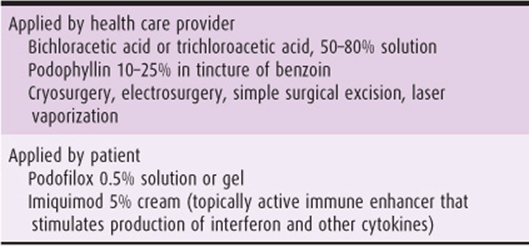
Whether treatment actually affects the natural progression or eradicates HPV infection is unclear. Concomitant vulvovaginitis should be treated initially. Treatment should be based on patient preference and convenience. Table 39–8 details treatments of condylomata acuminata. If treatment fails with an initial regimen, a different agent can be used. Patients should be informed that, though rare, complications of treatment can result in adhesions, scarring, and loss of pigmentation.
Treatment may be self-administered by the patient or applied by the health care provider (Table 39–8). Clinical disease may appear on only a small area of the infected surface. Hence some specialists recommend CO2 laser ablation of all visible lesions, plus a low-dose treatment (brushing), under colposcopic guidance, of a 1-cm margin of normal adjacent skin in all areas where subclinical HPV infection may coexist. Intralesional or systemic interferon has demonstrated effectiveness in refractory cases. Chemotherapeutic agents such as fluorouracil ointment or bleomycin in the form of intralesional injections can also be used as second-line therapies.
Table 39–8. Treatment of condyloma acuminata.
During pregnancy, electrocoagulation, cryotherapy, or CO2 laser therapy should be administered at approximately 32 weeks to avoid, on one hand, post-treatment necrosis, which may last as long as 4–6 weeks, and to prevent, on the other hand, recurrence if treated too early. Podophyllin, podofilox, and imiquimod should not be used during pregnancy.
Prognosis
Recurrences are frequent with all treatment modalities. Prevention of recurrence is particularly difficult in patients who are immunosuppressed or currently receiving long-term corticosteroid therapy. Examination of sexual partners is not necessary because most partners are likely to have subclinical infection. The use of condoms for a few months after treatment may help in reducing “ping-pong” transmission to and from partners who may be infected.
MOLLUSCUM CONTAGIOSUM
These benign epithelial poxvirus-induced tumors are dome-shaped, with a typical umbilicus. Size varies, up to 1 cm. Lesions are often multiple and are mildly contagious. As mentioned above, they are look-alikes of condylomata acuminata. Under the microscope they appear as numerous inclusion bodies (molluscum bodies) in the cytoplasm of cells. Each lesion can be treated by desiccation, freezing, or curettage and chemical cauterization of the base. Topical imiquimod is an alternative therapy. Scarring is frequent.
EPIDERMAL CYSTS
Previously named keratin cysts, these cysts are of epidermal origin. They are lined with squamous epithelium and filled with oily material and desquamated epithelial cells. Epidermal inclusion cysts may result from suturing of skin fragments during closure of the vulvar mucosa and skin after trauma or episiotomy. However, most epidermal cysts arise from occlusion of pilosebaceous ducts. These cysts are usually small, solitary, and asymptomatic, but rarely become irritated or infected.
SEBACEOUS CYSTS
Sebaceous cysts develop when sebaceous gland ducts become occluded and sebaceous material accumulates. These cysts are frequently multiple and almost always involve the labia majora. Although generally asymptomatic, acutely infected cysts may require incision and drainage.
APOCRINE SWEAT GLAND CYSTS
Apocrine sweat glands, abundant in the skin of the labia majora and the mons pubis, become active after puberty. Occlusion of the ducts with keratin results in the extremely pruritic, microcystic Fox-Fordyce disease. This disease should be suspected in patients with consistent vulvar pruritus.
ACROCHORDON
An acrochordon is a flesh-colored, soft polypoid tumor of the vulvar skin, also called a fibroepithelial polyp or simply a skin tag. The tumor does not become malignant and is of no clinical importance, unless it becomes traumatized, causing bleeding. Simple excisional biopsy in the office is ordinarily adequate therapy.
LARGE TUMORS
BARTHOLIN’S DUCT CYST AND ABSCESS
Clinical Findings
Obstruction of the main duct of Bartholin’s gland results in retention of secretions and cystic dilatation. Infection is an important cause of obstruction; however, other causes include inspissated mucus and congenital narrowing of the duct. Secondary infection may result in recurrent abscess formation.
The gland and duct are located deep in the posterior third of each labium major, with the duct opening into the vestibule. Enlargement in the postmenopausal patient may reflect a malignant process (although the incidence is <1%), and biopsy should be considered.
Acute symptoms generally result from infection, which leads to pain, tenderness, dyspareunia, and even difficulty in walking with adducted thighs. The surrounding tissues become edematous and inflamed. A fluctuant, tender mass is usually palpable. Unless an extensive inflammatory process is present, systemic symptoms or signs of infection are unlikely.
Treatment
Primary treatment consists of drainage of the infected cyst or abscess, preferably by marsupialization or by insertion of a Word catheter (an inflatable bulb-tipped catheter). The incision should be made in the vestibule, close to the original orifice of the Bartholin’s gland duct. Simple needle aspiration, or incision and drainage, may provide only temporary relief, as recurrent cystic dilatation may recur. Appropriate antibiotics should be administered if considerable inflammation develops. Excision of the cyst may be required in recurrent cases or in the postmenopausal patient.
LEIOMYOMA, FIBROMA, & LIPOMA
Tumors of mesodermal origin present infrequently on the vulva. However, they can become extremely large. Leiomyomas, arising from muscle in the round ligament, appear as firm, symmetric, freely mobile tumors deep in the substance of the labium majus. Fibromas, arising from proliferation of fibroblasts, vary in size from small subcutaneous nodules revealed incidentally to large polypoid tumors. Large tumors often undergo myxomatous degeneration and are very soft and cystic to palpation. Lipomas consist of a combination of mature fat cells and connective tissue. They can be differentiated from degenerated fibromas only by histopathologic examination. Small tumors can be removed under local anesthesia in the office. Large tumors require general anesthesia and operating room facilities. The diagnosis of sarcoma is based on histologic assessment.
NEUROFIBROMA
Neurofibromas are fleshy polypoid lesions that may manifest as solitary, solid tumors of the vulva or be associated with generalized neurofibromatosis (Recklinghausen’s disease). Arising from the neural sheath, they are usually small lesions of no consequence. Multiple disfiguring tumors of the vulva may interfere with sexual function and require excision or vulvectomy.
GRANULAR CELL MYOBLASTOMA (SCHWANNOMA)
Granular cell myoblastoma is usually a solitary, painless, slow-growing, infiltrating but benign tumor of neural sheath origin, most commonly found in the tongue or integument. Approximately 7% involve the vulva. The infected area consists of small subcutaneous nodules 1–4 cm in diameter. With increasing size, they erode through the surface and result in ulcerations that may be confused with cancer. The margins of the tumor are indistinct, and wide local excision is necessary to completely excise the cells extending into contiguous tissues. The area of resection must be periodically re-examined and secondary excision performed promptly if recurrence is suspected.
VARICOSITIES
Clinical Findings
Varicosities of the vulva involve 1 or more veins. Severe varicosities of the legs and vulva may be aggravated during pregnancy. Symptomatic vulvar varices in a woman who is not pregnant are uncommon and may signify an underlying vascular disease, either primary or secondary to a tumor in the pelvis. Rupture of a vulvar varicosity during pregnancy may cause profuse hemorrhage. Pain and tenderness may be caused by acute phlebitis or thrombosis.
Treatment
Treatment of vulvar and vaginal varicosities is seldom necessary. Surgical intervention is generally required only in rare cases of rupture and hemorrhage. Persistent postpartum cases may be alleviated by injection of a sclerosing agent.
HEMATOMA
The vulva has a rich blood supply arising predominately from the pudendal vessels. A ruptured vessel, especially in a pregnant woman, can cause significant bleeding and hematoma formation due to the distensible nature of the vulvar tissue. In rare cases, intercourse leads to laceration with external bleeding or to hematoma that may dissect the tissues in the labia majora or the rectovaginal septum. After trauma, an ice pack should be applied. If the hematoma continues to expand, embolization of the vessel, which is usually a branch of the pudendal artery, is a new, noninvasive approach to control the bleeding. Alternatively, the area should be incised and any bleeders (which may be multiple) ligated. The wound can be packed and left open or closed with a drain in place, if appropriate. Antibiotics should be administered on an individual basis, depending on the initiating event and contamination in the area.
EDEMA
Clinical Findings
The loose integument of the vulva predisposes to the development of edema. Causes of vulvar edema include vascular or lymphatic obstruction resulting from an underlying neoplasm or infection such as lymphogranuloma venereum (LGV), vaginal delivery accompanied by frequent vaginal examinations, trauma from a bicycle accident (saddle injury) in a young girl, and a kick to the pudendum. Severe generalized vulvar edema may represent an underlying systemic illness such as congestive heart failure, nephrotic syndrome, preeclampsia, or eclampsia. Acute edema may result from a systemic or local allergic reaction, immobilization, or ovarian hyperstimulation syndrome.
Treatment
An ice pack applied to the perineum after an acute trauma tends to retard the development of severe edema. Ice application should be restricted to 15 minutes every hour to prevent cold burns. Warm packs or warm sitz baths may then be applied after 1–2 days to help resolve the associated inflammation and/or hematoma.
LYMPHANGIOMA
Lymphangiomas are tumors of the lymphatic vessels. They may be difficult to differentiate from hemangiomas microscopically unless blood cells are present within the blood vessels. Lymphangioma cavernosum may cause a diffuse enlargement of 1 side of the vulva and extend over the remainder of the vulva and perineum. A tumor that is sufficiently enlarged should be surgically excised. Lymphangioma simplex tumors (circumscription tumors) are usually small, soft, white, or purple nodules or small wartlike lesions most commonly seen on the labia majora. Usually asymptomatic, they do not require excision unless intense pruritus and excoriation present. Lymphangioma simplex tumors are not alleviated with topical measures.
OTHER VULVAR TUMORS
A number of other uncommon cystic vulvar tumors must be considered in the differential diagnosis of vulvar tumors. Anteriorly Skene’s duct cyst should be differentiated from urethral diverticulum, as careless incision of the latter may damage the urethra and result in urinary incontinence. An inguinal hernia may extend into the labium majus, causing a large cystic dilatation. Suspicion of an inguinal hernia is indication for ultrasound examination of the contents of the labial tumor. The appearance of peristaltic waves is evident that a hernia is in fact present. Occlusion of a persistent processus vaginalis (canal of Nuck) may cause a cystic tumor or hydrocele. Gartner’s duct cyst is a dilatation of the mesonephric duct vestiges, always at the lateral vaginal wall. Supernumerary mammary tissue that persists in the labia majora may form a cystic or solid tumor, or even an adenocarcinoma. Engorgement of such tissue in the pregnant patient can be symptomatic.
VULVAR MANIFESTATION OF SYSTEMIC DISEASES
LEUKEMIA
Rarely, nodular infiltration and ulceration of the vulva and rectovaginal septum occur with acute leukemia.
DERMATOLOGIC DISORDERS
Recurrent ulcerations of the mucous membranes of the mouth and vagina may be manifestations of disseminated lupus erythematosus. Bullous eruptions of apparently normal skin and mucous membrane surfaces of the vulva may be early signs of pemphigus vulgaris (a rare, chronic vesicobullous disease, associated with the autoimmune disease, keratinolysis). Contact dermatitis is an inflammatory response of the vulvar tissue to agents that may be either locally irritating or inductive of sensitivity on contact. The local reaction to a systemically administered drug is called dermatitis medicamentosa.
OBESITY
Acanthosis nigricans is a hyperpigmented lesion associated with obesity and is characterized by papillomatous hypertrophy. Although generally benign, it may be associated with an underlying adenocarcinoma. Pseudoacanthosis nigricans is a benign process that may appear on the skin of the vulva and inner thighs in obese and darkly pigmented women. Glucose intolerance, insulin resistance, chronic anovulation, and androgen disorders may be associated.
Intertrigo is an inflammatory reaction involving the genitocrural folds or the skin under the abdominal panniculus. Common in obese individuals, it results from persistent moistness of the skin surfaces. A superficial fungal or bacterial infection may be concomitant. The area may be either erythematous or white from maceration. Maintaining dryness by wearing absorbent cotton undergarments and dusting with cornstarch powder may be helpful.
DIABETES MELLITUS
Diabetes mellitus is the systemic disease most commonly associated with chronic pruritus vulvae. Diabetic vulvitis is caused by chronic vulvovaginal candidiasis. The diagnosis of diabetes should be considered in any patient who responds poorly to antifungal treatment or who has recurrent fungal infections. Such patients should undergo glucose tolerance testing. In rare cases of long-term diabetes mellitus, the associated neuropathy may present as vulvar pruritus or burning. In uncontrolled diabetes, the vulvar epithelium often undergoes lichenification and secondary bacterial infection. Occasionally, vulvar abscesses, chronic subcutaneous abscesses, and draining sinuses develop from a bacterial infection. Treatment should include controlling the underlying diabetes and specific therapy for the bacterial or fungal infection. Suppressive antifungal therapy using fluconazole should be initiated in diabetic patients with recurrent vulvovaginal candidiasis.
Necrotizing fasciitis presents most commonly in diabetics. It is an uncommon, acute, rapidly spreading, sometimes fatal polymicrobial infection of the superficial fascia and subcutaneous fascia. It may appear after a surgical procedure or after minor trauma. It presents as an extremely painful, tender, edematous, and indurated region with central necrosis and peripheral purplish erythema. Treatment requires incision and debridement of involved tissue and broad-spectrum antibiotics.
INFESTATIONS OF THE VULVA
PEDICULOSIS PUBIS
Pathogenesis
The crab louse (Phthirus pubis) is transmitted through sexual contact or from shared infected bedding or clothing. The louse eggs are laid at the base of a hair shaft near the skin. The eggs hatch in 7–9 days, and the louse must attach to the skin of the host to survive. The result is intense pubic and anogenital itching.
Clinical Findings
Minute pale-brown insects and their ova may be seen attached to terminal hair shafts.
Treatment
Treatment consists of permethrin 1% cream, lindane 1% shampoo, or pyrethrins with piperonyl butoxide. Lindane is not recommended for pregnant or lactating women or for children younger than 2 years. It is important to treat all contacts and sterilize clothing that was in contact with the infested area.
SCABIES
Pathogenesis
Sarcoptes scabiei causes intractable itching and excoriation (see Table 39–3 for definition) of skin surfaces in the vicinity of minute skin burrows where parasites deposited ova. The scabies mite is transmitted, often directly, from infected persons.
Treatment
The patient should take a hot soapy bath, scrubbing the burrows and encrusted areas thoroughly. Treatment consists of application of permethrin cream (5%) to the entire body from the neck down, with particular attention to the hands, wrists, axillae, breasts, and anogenital region. It should be washed off after 8–14 hours. Alternatively, lindane (1%) in lotion or cream form can be applied in a thin layer to all areas of the body and washed off after 8 hours. All potentially infected clothing or bedding should be washed or dry-cleaned. All people who were in contact must be treated as described to prevent reinfection. Therapy should be repeated in 10–14 days if new lesions develop.
ENTEROBIASIS (PINWORM, SEATWORM)
Clinical Findings
Enterobius vermicularis infection is common in children. Symptoms are nocturnal perineal itching and perianal excoriation. To diagnose, apply adhesive cellulose tape to the anal region, stick the tape to a glass slide, and examine under the microscope for ova.
Treatment
Patients should wash their hands and scrub their nails after defecation. Underclothes must be boiled. Application of ammoniated mercury ointment to the perianal region twice daily relieves itching. Pinworms succumb to systemic treatment with pyrantel pamoate, mebendazole, or pyrvinium pamoate.
MYCOTIC INFECTIONS OF THE VULVA
FUNGAL DERMATITIS (DERMATOPHYTOSES)
Clinical Findings
Tinea cruris is a superficial fungal infection of the genitocrural area that is more common in men than in women. The most common carriers are Trichophyton mentagrophytes and Trichophyton rubrum. The initial lesions are usually located on the upper inner thighs and are well circumscribed, erythematous, dry, scaly areas that coalesce. Scratching causes lichenification and a gross appearance similar to neurodermatitis. Diagnosis depends on microscopic examination (as for Candida) (Fig. 39–5). Culture on Sabouraud’s medium confirms diagnosis.

Figure 39–5. Potassium hydroxide preparation showing branched and budding Candida albicans.
Treatment
Treatment with 1% haloprogin, tolnaftate, or a similar agent is effective. Twice-daily application of topical imidazole preparation for 2–3 weeks is also highly effective.
Tinea versicolor generally involves the skin of the trunk, although vulvar skin is occasionally involved. The lesions are usually multiple and may have a red, brown, or yellowish appearance. Diagnosis is the same as for other fungal infections. Treatment with selenium sulfide suspension daily for 5–7 days is usually effective, as are topical imidazole preparations applied for 4 weeks. Ketoconazole has been used in recalcitrant cases.
DEEP CELLULITIS CAUSED BY FUNGI
Blastomycosis and actinomycosis are examples of deep mycoses that usually affect internal organs but may also involve the skin. Involvement of the vulvar epithelium in these diseases is rare in the United States. Diagnosis is usually by laboratory exclusion of the granulomatous sexually transmitted diseases, tuberculosis, and other causes of chronic infection. Treatment of blastomycosis with amphotericin B or hydroxystilbamidine is not very effective. Penicillin successfully treats actinomycosis in most cases.
OTHER INFECTIONS OF THE VULVA
IMPETIGO
Impetigo is caused by the hemolytic Staphylococcus aureus or by streptococci. This autoinoculable disease spreads quickly throughout the body, also to the vulva. The thin-walled vesicles and bullae that develop display reddened edges and crusted surfaces after rupture. The disease is common in children, particularly on the face, hands, and vulva.
The patient must be isolated and the blebs incised or crusts removed aseptically. Neomycin or bacitracin should be applied topically twice daily for 1 week. Bathing with an antibacterial soap is recommended.
FURUNCULOSIS
Vulvar folliculitis is caused by a staphylococcal infection of hair follicles. Furunculosis occurs if the infection spreads into the perifollicular tissues, producing localized cellulitis. Some follicular lesions are palpable as tender subcutaneous nodules that resolve without suppuration. A furuncle begins as a hard, tender subcutaneous nodule that ruptures through the skin, discharging blood and purulent material. After expulsion of a core of necrotic tissue, the lesion heals. New furuncles may appear sporadically over time.
Applications of topical antibiotic lotions effectively treat minor infections. Deeper infections can be treated with hot soaks, followed by incision and drainage of the pustules. Appropriate systemic antibiotics are warranted when extensive furunculosis is present.
ERYSIPELAS
Erysipelas is a rapidly spreading erythematous lesion of the skin caused by invasion of the superficial lymphatics by β-hemolytic streptococci. Erysipelas of the vulva is exceedingly rare and is most commonly seen after trauma to the vulva or a surgical procedure. Systemic symptoms of chills, fever, and malaise associated with an erythematous vulvitis should raise suspicion of this infection. Vesicles and bullae may appear, and erythematous streaks leading to the regional lymph nodes are typical.
Treatment consists of systemic (preferably parenteral) penicillin or large doses of orally administered tetracycline.
HIDRADENITIS SUPPURATIVA
Hidradenitis suppurativa is a refractory process of the apocrine sweat glands, usually associated with staphylococci or streptococci. Inspissation of secretory material and secondary infection occlude ducts of these glands. Multiple pruritic subcutaneous nodules appear and eventually develop into abscesses and then rupture. The process generally involves the skin of the entire vulva, resulting in multiple abscesses and subsequent chronic draining of sinuses and scars. Treatment at an early stage consists of drainage and administration of antibiotics based on organism-sensitivity testing. Long-term therapy with isotretinoin may be considered. Antiandrogen therapy with cyproterone acetate or ethinyl estradiol is an effective alternative treatment. When severe chronic infections do not respond to medical therapy, the involved skin and subcutaneous tissues, down to the deep fascia, must be removed. The area will generally not heal after a primary closure. The wound may therefore be left open and allowed to heal by secondary intention or a split-thickness graft may be placed. Squamous cell carcinoma is rarely associated with hidradenitis suppurativa.
VESTIBULAR DISEASE
VULVAR PAIN SYNDROME
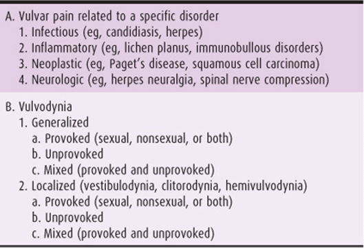
Vulvar pain in the absence of relevant visible physical findings is termed vulvodynia (Table 39–9). Women suffering from vulvodynia describe their symptoms as burning, rawness, irritation, dryness, and hyperpathia (pain provoked by very light touch). Approximately 16% of the female population has experienced vulvodynia at some time. The ISSVD has classified vulvodynia by localized vulvodynia (provoked or unprovoked) and generalized vulvodynia (provoked or unprovoked) (Table 39–9).
Table 39–9. Terminology of vulvar pain (ISSVD 2003).
LOCALIZED PROVOKED VULVODYNIA, OR VESTIBULODYNIA
ESSENTIALS OF DIAGNOSIS
Affects mostly young women: 20–30 years old.
Introital pain on vestibular or vaginal entry (entry dyspareunia).
Vestibular tenderness—pressure from a cotton-tipped applicator at the vestibule reproduces the pain.
Erythema is seldom seen. No other lesions are present.
Pathogenesis
Localized provoked vulvodynia, or provoked vestibulodynia (PVD), was formerly known as vulvar vestibulitis and clitorodynia. The vestibule is the nonkeratinized squamous epithelium of the vulva between the labia minora. The “Hart” line is the external perimeter including the hymen (Fig. 39–6). In the vulvar vestibule of PVD, mast cell proliferation and degranulation, hyperinnervation (Fig. 39–7), decreased natural killer cell activity, and enhanced heparanase activity have been detected. Inflammation of the vestibule is related to both mast cell proliferation and hyperinnervation, which act reciprocally, and ultimately increase local inflammation. Mast cells secrete mediators, such as nerve growth factor (NGF), histamine, and serotonin, which have been found to sensitize and induce the proliferation of C-afferent nerve fibers. These nerve fibers release neuropeptides, including NGF, which increase the proliferation and degranulation of mast cells, cause hyperesthesia, and enhance the inflammatory response. Although mast cells are activated by inflammation, they themselves increase inflammation. This, in turn, increases the density of nerve fibers, leading to further activation of mast cells, and contributing to inflammation. In this way, both inflammatory hyperinnervation and neurogenic inflammation play significant roles in the cycle, resulting in chronic vulvar pain.
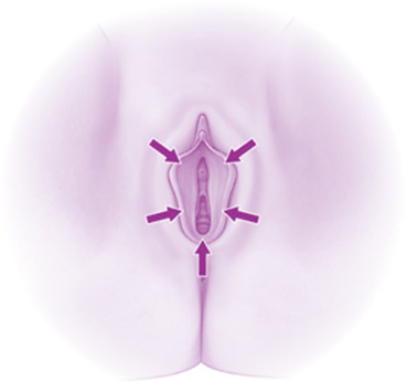
Figure 39–6. Hart line is the outer perimeter of the vestibule.
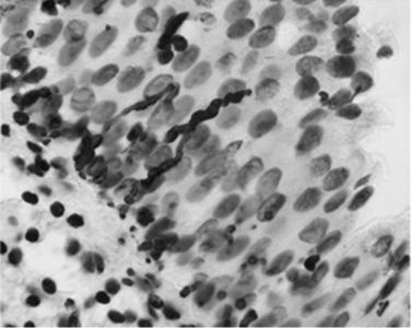
Figure 39–7. Hyperinnervation of the vestibular epithelium. Stain by PGP 9,13.
Prevention
There are currently no prophylactic measures.
Clinical Findings
A. Signs & Symptoms
Two clinical criteria only are needed for diagnosis: (1) introital pain on vestibular or vaginal entry (entry dyspareunia), (2) vestibular tenderness—pressure from a cotton-tipped applicator at the vestibule, which reproduces the pain. Originally, a third criterion, vestibular erythema, was used by Friedrich. However, erythema is rarely seen in LPV. Biopsy is not required for diagnosing LPV.
This condition may affect women of all ages, but mainly those between 20 and 30 years of age, who complain of introital dyspareunia (severe pain or burning on vaginal penetration by their partner). However, some women, due to shyness or despair, complain of persistent vaginal discharge. The clue to diagnosis in these cases may be extreme sensitivity at bimanual pelvic examination and expression of fear from speculum insertion. The woman should be questioned in regard to several factors that may be associated with vestibulodynia: prior surgery to the vestibule (ie, episiotomy, vaginal surgery, CO2 laser treatment), prior infections (eg, HPV, herpes, Candida), and associated urologic disorders, mainly interstitial cystitis. Interstitial cystitis and vestibulodynia have similar pathogeneses, including increased mast cell count in the subepithelial tissue.
B. Different Clinical Presentations of Vestibulodynia
• Primary (pain since first attempt at sexual intercourse) or secondary (pain is experienced after an initial period of pain-free intercourse)
• Pure (without concomitant vulvovaginitis) or complicated (with recurrent vulvovaginitis)
• With or without continuous vulvar pain
Differential Diagnosis
Vaginismus is an involuntary contraction of the muscles at the introitus. It is usually secondary to vestibulodynia, but in many cases is erroneously the only diagnosis received by a patient with dyspareunia. Vulvovaginitis should be excluded. Vaginal pH and microscopic examination of vaginal secretions with KOH and normal saline are effective for evaluating vaginitis. Acetowhite changes with application of 5% acetic acid, as well as any distinct lesions, should be biopsied to evaluate for an underlying dermatosis, infection, or neoplastic process. Other causes of vestibular sensitivity should be assessed (Table 39–9).
Complications
Intractable dyspareunia may have a detrimental affect on intimate relationships. Secondary depression is common.
Treatment
Some women, often after consultation with a number of physicians, administer antifungal or antibacterial medications for long periods, to no avail. Frustrated, they become apprehensive of additional treatment failure. Implementation of the following 3-level treatment plan is therefore suggested. Starting with simple measures, this plan should be reevaluated every 3 months.
1. The initial 3 months should focus on pelvic floor physical therapy with biofeedback. The patient should maintain vulvar hygiene, including the use of cotton underwear, drying of the skin, and avoidance of constrictive garments and irritating agents. Topical application of 5% lidocaine cream once daily, and of soothing oils, such as nut oil and tea-tree oil, twice daily, are recommended. In women with a poorly estrogenized vagina due to menopause or to consumption of birth control pills, a daily application of topical estrogenic preparation may be effective. A low-oxalate diet with supplementation of daily calcium citrate may relieve symptoms by decreasing the urinary oxalate crystal concentration that irritates the vulvar vestibule.
2. If, after 3 months, the condition does not improve, oral treatment with the tricyclic antidepressant amitriptyline (Elatrolet) 10–75 mg daily, pregabalin, or gabapentin may be initiated for 3 months.
3. For women who continue to complain of severe dyspareunia after these 2 periods, surgical therapy by vulvar vestibulectomy (Fig. 39–8) with vaginal advancement is the most effective treatment. Vaginal advancement covers the tissue defect and places the mucous-skin junction, which may be sensitive, outside the introitus. The combined sum of complete and partial responses to surgery from 38 studies of surgical treatment is 89%. A high proportion, 93%, of women who underwent vestibulectomy expressed satisfaction with the surgery, stating they would recommend it to another woman experiencing similar symptoms.
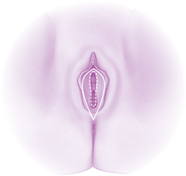
Figure 39–8. The outline of vestibulectomy for localized provoked vulvodynia.
Treatments that were proposed in the past but abandoned due to lack of efficacy include intralesional or systemic interferon injection to treat possible HPV, trigger point injections with long-acting injectable anesthetics or steroids, and CO2 laser vaporization.
New therapies that are currently being evaluated include injections of botulism toxin, topical application of nitroglycerine or nifedipine to treat vaginal muscle spasm as the source of vulvodynia, and topical application of tricyclic antidepressant amitriptyline.
Prognosis
Available treatments cure as many as two-thirds of those affected. In recalcitrant cases, women continue to suffer from dyspareunia, even subsequent to surgery. For them, referral to a pain treatment center may be an option.
GENERALIZED UNPROVOKED VULVODYNIA
The etiology of this condition is unknown. The pain involves a larger surface area than does that of localized provoked vulvodynia. The average patient is in her 60s and suffers from hypertension, sometimes receiving various treatments. The pain or burning is usually constant, sometimes with periods of relief and flares. For definitive diagnosis, the following conditions must be excluded: localized provoked vulvodynia, infections and dermatoses, pudendal nerve entrapment, pudendal nerve injury due to childbirth, referred pain from ruptured disk, neuropathic viruses such as herpes simplex or varicella-zoster, and neurologic disease such as multiple sclerosis. A test for allodynia and hyperalgesia using a cotton-tipped swab is usually negative. Therefore, diagnosis of this neuropathic pain is by exclusion.
The most effective treatment for generalized unprovoked vulvodynia is tricyclic antidepressants, mainly amitriptyline (Elatrolet) 10–75 mg daily. Initial relief is expected after a few weeks. In the meantime, topical local anesthetics may be applied. The patient should be counseled on elimination of irritants. If symptoms are not relieved after 3 months, initiation of anticonvulsants such as gabapentin should be considered. If the patient is refractory to such treatment, the next step is referral to a pain clinic, where epidural, other regional blocks, or narcotics may be used.
ACOG Practice Bulletin Number 93, 2008. Diagnosis and management of vulvar skin disorders. Obstet Gynecol 2008;111: 1243–1253. PMID: 18448767.
Bornstein J, Cohen V, Zarfati D, Sela S, Ophir E. Involvement of heparanase in the pathogenesis of localized vulvodynia. J Gynecol Pathol 2008;27:136–141. PMID: 18156988.
Bornstein J, Goldshmid N, Sabo E. Hyperinnervation and mast cell activation may be used as histopathologic diagnostic criteria for vulvar vestibulitis. Obstet Gynecol Invest 2004; 58:171–178. PMID: 15249746.
Edwards L. New concepts in vulvodynia. Am J Obstet Gynecol 2003;189:S24–S30. PMID: 14532900.
Goldstein AT, Klingman D, Christopher K, Johnson C, Marinoff SC. Surgical treatment of vulvar vestibulitis syndrome: outcome assessment derived from a postoperative questionnaire. J Sex Med2006;3:923–931. PMID: 16942537.
Gunter J. Vulvodynia: New thoughts on a devastating condition. Obstet Gynecol Surv 2007;62:812–819. PMID: 18005458.
Moyal-Barracco M, Lynch PJ. 2003 ISSVD terminology and classification of vulvodynia: A historical perspective. J Reprod Med 2004;49:772–777. PMID: 15568398.
Nyirjesy P. Vulvovaginal candidiasis and bacterial vaginosis. Infect Dis Clin North Am 2008;22:637–652. PMID: 18954756.
VAGINAL DISORDERS
VAGINITIS
CANDIDIASIS
ESSENTIALS OF DIAGNOSIS
Intense vulvar pruritus
A white vaginal discharge
Vulvar erythema
Filaments and spores in vaginal discharge can be seen in saline (“wet prep”) and KOH preparations
The gold standard for diagnosis is a vaginal culture
Pathogenesis
Approximately 75% of women experience an episode of vulvovaginal candidiasis during their lifetime. Candida albicans, the most common Candida species, causes symptomatic vulvovaginitis in approximately 90% of the cases. C albicans frequently inhabits the mouth, throat, large intestine, and vagina. Clinical infection is dependent on considerable growth and colonization and may be associated with a systemic disorder (diabetes mellitus, HIV, obesity), pregnancy, medication (antibiotics, corticosteroids, oral contraceptives), and chronic debilitation.
Prevention
Nonabsorbent undergarments should be avoided. The vulva and vaginal area should be kept dry. Controlling any underlying metabolic illnesses, especially diabetes, can prevent candidal growth. Even when diabetes is not present, a low-sugar diet is recommended, as the glucose in a vaginal discharge may promote the growth of the yeast. Complicating medications, especially antibiotics, estrogen, or oral contraceptive, should be discontinued if possible. Some experts recommend administering a prophylactic dose of an antifungal medication simultaneous to every antibiotic administration.
Clinical Findings
A. Symptoms & Signs
Vulvovaginal candidiasis presents with intense vulvar pruritus; a white, cheesy vaginal discharge; and vulvar erythema. A burning sensation may follow urination, particularly if there is excoriation of the skin from scratching. Widespread involvement of the skin adjacent to the labia may suggest an underlying systemic illness. The labia minora may be erythematous and edematous.
B. Wet Prep Evaluation
Diagnosis is based on a normal vaginal pH ≤4.5 and microscopic evaluation of vaginal secretions both in a saline preparation (wet prep) and mixed with 10% KOH solution. Identification of C albicansrequires detections of filamentous forms (pseudohyphae) of the organism (Fig. 39–5). Spores may be present as well, but the presence of spores alone may indicate a Candida glabrata infection. The gold standard for diagnosis is a vaginal culture.
Differential Diagnosis
Genital herpes and localized provoked vulvodynia should be included in the differential diagnosis. Other causes of vaginal discharge are discussed later in this chapter.
Complications
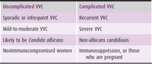
Complications include an entity called complicated vulvovaginal candidiasis, described in Table 39–10.
Table 39–10. Classification of vulvovaginal candidiasis (VVC).
Treatment
The current medical treatment of candidal infection is by imidazoles, fungistatic agents that interfere with the production of the sterol of the cell wall (Table 39–11). These are available as topical creams, vaginal suppositories, and oral agents. Application of a topical steroid may be beneficial to the patient with severe vulvar itch or edema. In evaluating the patient with complicated candidal vulvovaginitis, underlying predisposing disease processes should be addressed. Additionally, cultures of the vagina should be taken to identify resistant strains. C glabrata and Candida tropicalis, which are detected with increasing frequency, require prolonged periods of treatment.
Table 39–11. Imidazole medications used in the treatment of noncomplicated vulvovaginal candidiasis.
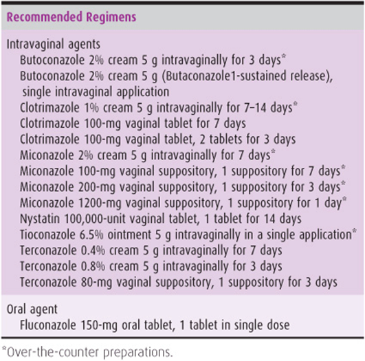
Treatment regimens for complicated candidal vulvovaginitis include prolonging antifungal therapy for at least 2 weeks, consistent with the life cycle of yeast; self-medication for 3–5 days upon first evidence of symptoms; and prophylactic treatment for several days before menstruation or during antibiotic therapy. Oral administration of fluconazole 150 mg weekly for 6 months or itraconazole 100 mg daily for 6 months may reduce the frequency of recurrence to 10% during maintenance therapy. Liver function should be monitored during prolonged oral therapy. Treatment of the partner may be considered in cases of symptomatic balanitis. Gentian violet 1%, an aniline dye, has demonstrated effectiveness against C albicans and C glabrata when painted over vaginal surfaces once weekly. Boric acid compounded in a 600-mg suppository form, administered daily for 6 weeks, is also effective treatment for candidiasis and yeast infestation. Polyenes, such as nystatin, which is not absorbed in the gastrointestinal tract, may be taken orally to reduce intestinal colonization. Flucytosine may be administered in resistant cases.
Prognosis
Recurrent disease may result from insufficient duration of therapy, recontamination, or resistant strains. Unfortunately, in 57% of patients, recurrences present within 6 months of discontinuation of prophylactic treatment.
BACTERIAL VAGINOSIS
ESSENTIALS OF DIAGNOSIS
Homogeneous vaginal discharge
Amine (fishy) odor when potassium hydroxide solution is added to vaginal secretions (commonly called the “whiff test”)
Presence of clue cells (more than 20% of epithelial cells) on microscopy (Fig. 39–9)
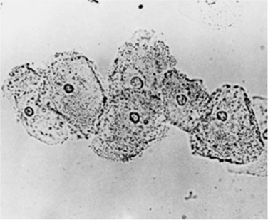
Figure 39–9. Bacterial vaginosis. Saline wet mount of clue cells. Note the absence of inflammatory cells.
Vaginal pH >4.5
Decrease in lactobacillus, small gram-variable rods, or curved gram-variable rods in gram-stained smear
Pathogenesis
Bacterial vaginosis (BV), previously referred to as Gardnerella vaginitis, Haemophilus vaginitis, or nonspecific vaginitis, is the most common cause of symptomatic bacterial infection in reproductive-aged women in many countries. This condition is characterized by an alteration in the normal vaginal flora. The concentration of the hydrogen peroxide–producing lactobacillus decreases, and there is overgrowth of Gardnerella vaginalis, Mobiluncus spp., anaerobic gramnegative rods (Prevotella spp., Porphyromonas spp., Bacteroides spp.), and Peptostreptococcus spp. Whether bacterial vaginosis is a true sexually transmitted disease is controversial, although women who are not sexually active are rarely affected.
Prevention
Maintaining vaginal pH at a normal range may prevent recurrences. The potential benefit of lactobacillus intravaginal suppositories in restoring normal flora, and of acidifying vaginal douching, is being studied.
Clinical Findings
A. Symptoms & signs
Bacterial vaginosis presents as a “fishy” vaginal discharge, which is more noticeable after unprotected intercourse, due to the increased pH caused by the ejaculate. The patient complains of a milky, homogenous, malodorous, usually nonirritating discharge. The term vaginosis, rather than vaginitis, is used due to the absence of vaginal mucosal inflammation, such as presents in candidal infections.
B. Diagnostic Scales
Two diagnostic scales are often used to diagnose bacterial vaginosis: Amsel’s criteria and Nugent’s score. According to Amsel’s criteria, which establishes accurate diagnosis of bacterial vaginosis in 90% of affected women, 3 of the following 4 criteria must be met:
1. Homogeneous vaginal discharge (color and amount may vary).
2. Amine (fishy) odor when potassium hydroxide solution is added to vaginal secretions (whiff test).
3. Presence of clue cells (>20% of epithelial cells) on microscopy. Clue cells are identified as numerous stippled or granulated epithelial cells (Fig. 39–9). This appearance is caused by the adherence of G vaginalis organisms to the edges of the vaginal epithelial cells.
4. Vaginal pH >4.5.
Nugent’s score is a Gram stain scoring system that provides a more sensitive (93%) and specific (70%) diagnosis than does the wet mount. The score is calculated by assessing for the presence of the following:
1. Large gram-positive rods (lactobacillus morphotypes; decrease in lactobacillus, scored 0–4)
2. Small gram-variable rods (G vaginalis morphotypes, scored 0–4)
3. Curved gram-variable rods (Mobiluncus spp. morphotypes, scored 0–2)
The total score ranges from 0 to 10. A score of 7–10 is consistent with bacterial vaginosis.
C. Other Diagnostic Tests
A culture of G vaginalis is not recommended as a diagnostic tool due to low specificity. Cervical Papanicolaou tests have low sensitivity. However, a DNA probe-based test may be clinically useful. Other commercially available tests for the diagnosis of BV include a card test for the detection of elevated pH and trimethylamine and proline aminopeptidase. The home-use of the VI-Sense panty liner has recently demonstrated effectiveness in the early detection of BV and of its recurrences after medical treatment.
Differential Diagnosis
Cervicitis and cervical neoplasia should be considered in the differential diagnosis of bacterial vaginosis.
Complications
BV is reported to increase the risk of preterm delivery. It is unclear whether metronidazole treatment of asymptomatic pregnant women reduces the rates of preterm delivery and adverse pregnancy outcomes. In nonpregnant women, BV is associated with posthysterectomy vaginal cuff cellulitis, postabortion infection, and pelvic inflammatory disease.
Treatment
Treatment should be administered to symptomatic patients and considered in asymptomatic patients. Several treatment regimens exist (Table 39–12). Of importance, intravaginal administration of oil-based clindamycin reduces the effectiveness of condoms and diaphragms. For pregnant women, metronidazole 250 mg orally 3 times daily is recommended for 7 days or, alternatively, clindamycin 300 mg orally twice daily for 7 days. There is no evidence supporting the use of topical agents during pregnancy. Management strategies for recurrent vaginosis include use of condoms, longer treatment periods, prophylactic maintenance therapy, oral or vaginal application of yogurt containing lactobacillus acidophilus, intravaginal planting of other exogenous lacto-bacilli, and acidification of the vagina. Treatment of the male rarely helps in preventing recurrence in the female.
Table 39–12. Treatment of bacterial vaginosis.
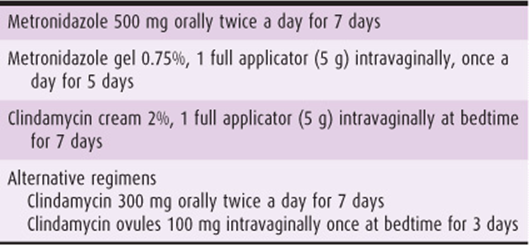
Prognosis
Recurrence is frequent. Overgrowth of candida albicans after antibiotic treatment of bacterial vaginosis may be misinterpreted as recurrent bacterial vaginosis. A “universal” treatment of vulvovaginitis using a combination of clotrimazole and metronidazole in a single vaginal suppository has demonstrated effectiveness. Its use may prevent candida overgrowth.
TRICHOMONAS VAGINITIS
ESSENTIALS OF DIAGNOSIS
Profuse, frothy, greenish, and foul-smelling discharge
pH of the vagina usually exceeding 5.0
Vaginal erythema with multiple small petechiae (strawberry spots)
Wet mount reveals an increase in polymorphonuclear cells and motile flagellates in 50–70% of zculture-confirmed cases
Pathogenesis
Trichomonas vaginalis is a unicellular flagellate protozoan (Fig. 39–10) that is larger than polymorphonuclear leukocytes but smaller than mature epithelial cells. T vaginalis infects the lower urinary tract in both women and men. It is the most prevalent nonviral sexually transmitted disease in the United States. Nonsexual transmission is infrequent because large numbers of organisms are required to produce symptoms.

Figure 39–10. Trichomonas vaginalis as found in vaginal and prostatic secretions. A: Normal trophozoite. B: Round form after division. C: Common form seen in stained preparation. Cysts not found. (Reproduced, with permission, from Brooks GF, Butel JS, Ornston LN. Jawetz, Melinick, & Adelberg’s Medical Microbiology. 19th ed. Appleton & Lange; 1991.)
Clinical Findings
A. Symptoms & Signs
A persistent vaginal discharge is the principal symptom with or without secondary vulvar pruritus. The discharge is profuse, extremely frothy, greenish, and at times foul-smelling. The pH of the vagina usually exceeds 5.0. Involvement of the vulva may be limited to the vestibule and labia minora. The labia minora may become edematous and tender. Urinary symptoms may occur; however, burning with urination is most often associated with severe vulvitis. Examination of the vaginal epithelium and cervix shows generalized vaginal erythema with multiple small petechiae, the so-called strawberry spots, which may be confused with epithelial punctation. Wet mount with normal saline reveals an increase in polymorphonuclear cells and characteristic motile flagellates in 50–70% of culture-confirmed cases.
B. Wet Mount Diagnosis
Vaginal trichomoniasis is usually diagnosed by microscopy of a wet mount preparation of vaginal secretions. Sensitivity is only 60–70%. Immediate evaluation is required, as the heat generated by the microscope light source causes the T vaginalis to discontinue its typical movements.
C. Other Diagnostic Tests
Other tests for trichomoniasis include immunochromato-graphic capillary flow dipstick technology and nucleic acid probing. Sensitivity exceeds 83%, and specificity is 97%. Results of the immunochromatographic Trichomonas Rapid Test are available in 10 minutes, and those of the nucleic acid probe test within 45. False-positive results do occur. Papanicolaou smears have a sensitivity of approximately 60% and also yield false-positive results. Culture is the most sensitive and specific method of diagnosis. In women in whom trichomoniasis is suspected but not confirmed by microscopy, vaginal secretions should be cultured for T vaginalis.
Treatment
Systemic therapy with metronidazole is the treatment of choice, because trichomonads sometimes present in the urinary tract system. Partners should be treated simultaneously, with intercourse avoided or a condom used until treatment is completed. US Centers for Disease Control and Prevention recommendations are presented in Table 39–13. If such treatments are not effective, sensitivity of a culture of T vaginalis to metronidazole and tinidazole should be determined. Side effects of metronidazole include nausea or emesis with alcohol consumption. Contraindications include certain blood dyscrasias (neutropenia) and central nervous system diseases. An oncogenic effect has been demonstrated in animals but not in humans. Resistance to metronidazole therapy is rare but is rising and can be confirmed in vitro.
Table 39–13. Treatment of Trichomonas vaginitis.
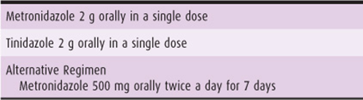
Trichomoniasis is associated with a number of perinatal complications and increased incidence in the transmission of HIV. Women with trichomoniasis should be evaluated for other sexually transmitted diseases, including Neisseria gonorrhoeae, Chlamydia trachomatis, syphilis, and HIV.
NEISSERIA GONORRHOEAE
Of women infected by N gonorrhoeae, 85% are asymptomatic. The glandular structures of the cervix, urethra, vulva, perineum, and anus are most commonly infected. In acute disease, patients present with a copious mucopurulent discharge and gramnegative diplococci within leukocytes. However, diagnosis should be confirmed with nucleic acid amplification or a culture from the endocervix, urethra, rectum, or mouth. An estimated 15–20% of women with lower tract disease develop upper genital tract disease with salpingitis, tubo-ovarian abscess, and peritonitis. Ectopic pregnancy and infertility are classic long-term consequences. If active infection is present during vaginal delivery, the newborn may develop conjunctivitis by contamination. Uncomplicated gonococcal infections of the cervix are treated with ceftriaxone 125 mg administered intramuscularly (IM) in a single dose. Single oral doses of cefixime 400 mg, ciprofloxacin 500 mg, ofloxacin 400 mg, or levofloxacin 250 mg are other recommended regimens. Quinolones are no longer recommended because some strains of N gonorrhoeae are quinolone-resistant. Spectinomycin 2 g IM in a single dose is an option for patients sensitive to cephalosporins. Empirical Treatment of C trachomatis should be considered, as this infection often coexists.
CHLAMYDIA TRACHOMATIS
The screening of sexually active young women for C trachomatis is important because some infections are asymptomatic, and some present with a mucopurulent cervicitis, dysuria, and/or postcoital bleeding. C trachomatis can be identified by culture (50–90% sensitivity), a direct fluorescent antibody (50–80% sensitivity), enzyme immunoassay (40–60% sensitivity), or, most recently, by using nucleic acid amplification tests (polymerase chain reaction or ligase chain reaction, 60–100% sensitivity). All tests have a specificity >99%. C trachomatis causes atypical cytologic findings on Papanicolaou smear and an ascending infection, salpingitis, in 20–40% of untreated patients. More than 50% of upper tract infections may be caused by C trachomatis, leading to tubal occlusion, ectopic pregnancy, or infertility. Untreated C trachomatis can also cause neonatal conjunctivitis. C trachomatis may present as LGV, which most commonly affects the vulvar tissues. Retroperitoneal lymphadenopathy may present. The initial lesion in LGV presents as a transient, painless vesicular lesion or shallow ulcer at the inoculation site. More advanced disease is characterized by anal or genital fistulas, stricture, or rectal stenosis. The disease is uncommon in the United States, but endemic in Southeast Asia and Africa.
If C trachomatis is suspected or diagnosed, both the patient and partner should be treated. They should also be evaluated for concurrent gonococcal infections. Recommended therapy includes azithromycin 1 g orally in a single dose or doxycycline 100 mg orally twice daily for 7 days. Erythromycin base 500 mg orally 4 times daily for 7 days, ofloxacin 300 mg orally twice daily, and levofloxacin 50 mg once daily for 7 days are alternative regimens. Doxycycline, levofloxacin, and ofloxacin should be avoided in pregnancy and during lactation. Patients should abstain from intercourse for 7 days. Test of cure is required in cases of possible reinfection or persistent symptoms and during pregnancy. Repeat testing should be considered 3 weeks after treatment with erythromycin. Rescreening is recommended 3–4 months after treatment. For LGV, the recommended regimen is doxycycline 100 mg twice daily for 21 days.
OTHER INFECTIONS
Mycoplasma hominis and Ureaplasma urealyticum also cause genital disease. Polymerase chain reaction is more sensitive than culture. Mycoplasma infections can cause infertility, spontaneous abortion, postpartum fever, salpingitis, and pelvic abscess, as well as nongonococcal urethritis in men. The most effective treatment is doxycycline 100 mg orally twice daily for 10 days.
CONDITIONS MIMICKING VAGINITIS
Cervicitis due to chlamydial infection, cervical polyps, or cervical or vaginal cancer can cause a mucopurulent discharge and bleeding. Adenocarcinoma of the cervix may be missed by cytologic cervical screening and by colposcopy, becasue it generally develops in the endocervical canal rather than at the squamocolumnar junction. Excessive cervical ectropion may cause excessive discharge of cervical mucus from normal endocervical cells. Vaginal adenosis may cause the same type of clear, mucoid-type discharge without associated symptoms. Excessive desquamation of the vaginal epithelium may produce a diffuse gray-white pasty vaginal discharge, which may be confused with candidiasis. Vaginal pH is normal. Microscopic evaluation shows normal bacterial flora, mature vaginal squamae, and no increase in the number of leukocytes. Excessive but normal vaginal discharge should be treated with reassurance and, if required, with cryosurgery, carbon dioxide treatment, or loop conization of the cervix. Continuous use of a tampon should be avoided.
DESQUAMATIVE INFLAMMATORY VAGINITIS
This rare condition of vaginitis should be considered in a patient with hard-to-treat vaginitis. The cause is unknown. Patients complain of a profuse purulent vaginal discharge, burning and pain upon urinating (dysuria) or intercourse (dyspareunia), and occasional spotting. Adherence of both vaginal walls, with gradual stenosis, is a common complication. The disease is a variant of the dermatologic disease lichen planus. In many cases, typical lichen planus layers are found on the skin, oral mucosa, and gums. Upon inspection, the vagina is found to be erythematous, inflamed, and desquamated. A thick discharge and a white membrane cover the vagina. The purulent discharge contains many immature epithelial and pus cells without any identifiable cause. Vaginal erythema is present, and synechiae may develop in the upper vagina, causing partial occlusion. Vaginal pH may be elevated. Wet mount and Gram’s stain demonstrate an increased number of parabasal cells, an absence of gram-positive bacilli, and the presence of gram-positive cocci.
The recommended therapy is intravaginal administration of 2% clindamycin cream 5 g daily for 7 days or clindamycin pessaries followed by a foam containing hydrocortisone and pramoxine into the vaginal mucosa to create a protective layer. A second line of therapy is vaginal insertion of corticosteroids in the form of suppository or cream. Recently, application of tacrolimus cream, as an immunosuppressor agent, has also been suggested.
CHEMICAL VAGINITIS
Chemical vaginitis secondary to multiple irritating offenders, including topical irritants (sanitary supplies, spermicides, feminine hygiene supplies, soaps, perfumes); allergens (latex, antimycotic creams), and possibly excessive sexual activity, can cause pruritus, irritation, burning, and vaginal discharge. The etiology may be confused with vulvovaginal candidiasis. Treatment consists of removal of the offending agent. A short course of corticosteroid treatment may be used along with sodium bicarbonate sitz baths and topical vegetable oils.
ATROPHIC VAGINITIS
Clinical Findings
Prepubertal, lactating, and postmenopausal women lack the vaginal effects of estrogen production. The pH of the vagina is abnormally high, and the normally acidogenic flora of the vagina may be replaced by mixed flora. The vaginal epithelium is thinned and more susceptible to infection and trauma. Although most patients are asymptomatic, many postmenopausal women report vaginal dryness, spotting, presence of a serosanguineous or watery discharge, and/or dyspareunia. Some of the symptoms of irritation are caused by a secondary infection. On examination, the vaginal mucosa is thin, with few or absent vaginal folds. The pH is 5.0–7.0. The wet mount shows small, rounded parabasal epithelial cells and an increased number of polymorphonuclear cells.
Treatment
Treatment includes intravaginal application of estrogen cream. Because approximately one-third of the vaginal estrogen is systemically absorbed, this treatment may be contraindicated in women with a history of breast or endometrial cancer. The estradiol vaginal ring, which is changed every 90 days, may provide a preferable route of administration for some women. Estradiol hemihydrate (Vagifem) 1 tablet intravaginally daily for 2 weeks and then twice a week for at least 3–6 months may be more convenient. Systemic estrogen therapy should be considered if there are no contraindications.
FOREIGN BODIES
Pathogenesis
Foreign bodies commonly cause vaginal discharge and infection in preadolescent girls. Paper, cotton, or other materials may be placed in the vagina and cause secondary infection. Children may require vaginoscopy using a small-caliber hysteroscope or vaginal examination under anesthesia to identify or rule out a foreign body or tumor high in the vaginal vault. The vaginal canal can be flushed in the office using a small catheter in an attempt to remove a foreign body. In adults, a forgotten menstrual tampon, a contraceptive device, or a pessary may cause a malodorous discharge. The diagnosis can usually be made by pelvic examination.
Clinical Findings
Clinical symptoms associated with foreign bodies include abnormal malodorous vaginal discharge and intermenstrual spotting. Symptoms are generally secondary to drying of the vaginal epithelium and micro-ulcerations, which can be detected by colposcopy. Ulcerative lesions, particularly associated with tampon use, are typically located in the vaginal fornices and have rolled, irregular edges with a red granulation tissue base (Fig. 39–3). Regenerating epithelium at the ulcer edge may shed cells that may be interpreted as atypical, suggesting dysplasia. The lesions heal spontaneously once tampon use is discontinued. A foreign body retained in the vagina for a prolonged period may erode into the bladder or rectum.
Treatment
Treatment involves removal of the foreign body. Rarely, antibiotics are required for ulcerations or cellulitis of the vulva or vagina. Dryness or ulceration of the vagina secondary to use of menstrual tampons is transient and heals spontaneously.
Toxic shock syndrome is the most serious complication associated with the use of vaginal tampons. It may develop also without tampon use. The syndrome has been linked to staphylococcal vaginal infection in healthy young women who use high-absorbency tampons continuously throughout the menstrual period. Some of the clinical manifestations are secondary to the release of staphylococcal exotoxins. Symptoms consist of a high fever (≥38.9 °C [102 °F]), possibly accompanied by severe headache, sore throat, myalgia, vomiting, and diarrhea. The disease may resemble meningitis or viremia. Palmar erythema and a diffuse sunburn-like rash have been described. The skin rash usually disappears within 24–48 hours, but occasionally a patient has a recurrent maculopapular, morbilliform eruption between days 6 and 10. Superficial desquamation of the palms and soles often follows within 2–3 weeks. Progressive hypotension may occur and proceed to shock levels within 48 hours. Multisystem organ failure may occur, including renal and cardiac dysfunction. The incidence of toxic shock syndrome was 1 in 100,000 among females 15–44 years of age in 1986. Any menstruating woman who presents with sudden onset of a febrile illness should be evaluated and treated for toxic shock syndrome. The tampon should be removed, cultures sent, and the vagina cleansed to decrease the organism inoculum. Appropriate supportive measures should be provided and β-lactamase–resistant penicillin or vancomycin (if the patient is allergic to penicillin) administered. Women who have been treated for toxic shock syndrome are at considerable risk for recurrence. Therefore, these women should avoid tampon use.
VIRAL INFECTIONS
The viruses that affect the vagina are the herpesvirus (herpes simplex, varicella-zoster, and cytomegalovirus), poxvirus (molluscum contagiosum), and papillomavirus types. The main features of these infections have been discussed under Vulvar Diseases.
HERPESVIRUS
The herpesvirus (HSV) may cause erosions, ulcerations, or an exophytic necrotic mass involving the vagina or cervix and causing a profuse vaginal discharge. The cervix may be tender to manipulation and bleed easily. The primary lesion lasts approximately 2 weeks and heals without scarring. Recurrent infections may cause cervical lesions. The virus may be cultured from ulcers or ruptured vesicles. Cervical cytologic examination may reveal multinucleated giant cells with intranuclear inclusions.
HUMAN PAPILLOMAVIRUS INFECTION
As discussed in the section on Vulvar Diseases, condylomata may affect the vagina and cervix as well. Condylomatous vaginitis causes a rough vaginal surface, manifesting white projections from the pink vaginal mucosa. Vaginal discharge resulting from a secondary yeast or bacterial infection is the most common symptom of florid condylomas. Postcoital bleeding may occur. No specific symptoms are related to the other types of condylomas. States of immunosuppression (pregnancy, HIV infection, diabetes, renal transplant) are associated with massive proliferation of condyloma and are often difficult to treat.
PARASITIC INFECTION
Less common causes of vaginitis are parasitic infections with pinworms (Enterobius vermicularis) and Entamoeba histolytica. Pinworm infection is generally seen in children. Fecal contamination at the introitus is the source of infection. The perineal area is extremely pruritic. The parasite is generally detected by pressing a strip of adhesive cellulose tape to the perineum. The tape is then adhered to a slide, allowing the double-walled ova to be identified under the microscope. E histolytica infection of the vagina and cervix is rare in the United States but is quite common in developing countries. Severe infection may resemble cervical cancer, but symptoms are generally due to vulvar involvement. Trophozoites of E histolytica may be demonstrated on wet-mount preparations or occasionally on a Papanicolaou smear.
Ahmed AM, Madkan V, Tyring SK. Human papillomaviruses and genital disease. Dermatol Clin 2006;24:157–165. PMID: 16677964.
Bornstein J. the HPV vaccines—which to prefer? Obstet Gynecol Surv 2009;64:345–350. PMID: 19386141.
Bornstein J. Human papillomavirus vaccine: The beginning of the end for cervical cancer. Isr Med Assoc J 2007; 9:156–158. PMID: 17402325.
Geva A, Bornstein J, Dan M, Shoham HK, Sobel JD. The VI-Sense-vaginal discharge self-test to facilitate management of vaginal symptoms. Am J Obstet Gynecol 2006;195:1351–1356. PMID: 16769019.
Greer L, Wendel GD. Rapid diagnostic methods in sexually transmitted infections. Infect Dis Clin North Am 2008;22: 601–617. PMID: 18954754.
ACOG Practice Bulletin Number 61, 2005. Human papillomavirus. Obstet Gynecol 2005;105:905–918. PMID: 15802436.
Moyal-Barracco M, Edwards L. Diagnosis and therapy of anogenital lichen planus. Dermatol Ther 2004;17:38–46. PMID: 14756889.
O’Mahony C. Genital warts: Current and future management options. Am J Clin Dermatol 2005;6:239–243. PMID: 16060711.
ACOG Practice Bulletin Number 72, 2006. Vaginitis. Obstet Gynecol 2006;107:1195–1206. PMID: 16648432.
Val I, Almeida G. An overview of lichen sclerosus. Clin Obstet Gynecol 2005;48:808–817. PMID: 16286827.
Centers for Disease Control and Prevention, Workowski KA, Berman SM. Sexually transmitted diseases treatment guidelines, 2006. MMWR Recomm Rep 2006;55(rr-11):1–94. PMID: 16888612.
The author thanks Dr. Doron Zarfati for assistance in obtaining some of the figures and Ms Cindy Cohen for assistance in preparing the manuscript.