Michael A. Belfort
The enthusiasm for operative vaginal delivery peaked in the early 20th century when almost 50% of all deliveries were with forceps. Over the last three decades this has changed and worldwide there has been a decrease in the use of operative techniques. This has been accompanied by a relative increase in abdominal delivery. There are various explanations for this trend including the development of more effective anesthesia techniques, the introduction of antibiotics, the effective use of blood products, and the development of improved surgical techniques. Thus, cesarean section has become the most commonly performed surgical procedure of our time and operative vaginal delivery rates have significantly decreased. When operative vaginal delivery is performed now, it is more commonly carried out with a vacuum extractor than with forceps and in some parts of the world (Scandinavia), forceps are hardly ever used. In the United States and increasingly in Europe, medicolegal concerns have reduced enthusiasm for operative delivery and the teaching thereof. This is an unfortunate situation since, for the correct indication, and in well-trained hands, operative vaginal delivery is safe and effective and reduces the incidence of abdominal delivery and its attendant complications. The objectives of this chapter are two-fold: first, to outline the basic knowledge that should be required of those wishing to perform operative vaginal deliveries, and second to discuss selected important topical issues that have recently been highlighted in the literature.
APPLIED MATERNAL PELVIC ANATOMY
The art of ante- and intrapartum clinical assessment of the maternal pelvis is slowly disappearing from many training programs as this aspect of obstetric evaluation continually receives less emphasis. In many instances, the argument that labor is the best test of the cephalopelvic relationship is a reasonable one. The counter argument, however, is that accurate clinical maternal pelvimetry may reveal findings that would prevent an unnecessarily difficult labor in a case of obvious cephalopelvic disproportion.
A knowledge of the female pelvis and its different configurations is important. Figure 23.1 shows the four parent types of maternal pelvis, defines their major characteristics as described in the Caldwell-Moloy classification, and lists the percentage of women with each type of pelvis. Table 23.1 lists some important diameters of the maternal pelvis. Detailed clinical knowledge of the type of pelvis and the relative dimensions of that pelvis and the fetal presenting part attempting passage through that pelvis, should form the foundation of any decision to undertake operative vaginal delivery. To this end, the following sections present applied anatomy of the maternal pelvis and fetal head.
|
|
|
FIG. 23.1. The four major types of female pelvis. |
|
|
|
TABLE 23.1. Diameters of the female pelvis |
PERFORMING CLINICAL PELVIMETRY
The pelvis is usually assessed in terms of the inlet, midcavity, and outlet.
Inlet
The inlet can only be adequately evaluated prior to engagement of the fetal head, since once the head occupies the midcavity the posterior inlet cannot be accessed. The examiner first assesses the anteroposterior (AP) diameter of the inlet by measuring the diagonal conjugate (distance from the undersurface of the symphysis pubis to the sacral promontory). The obstetric conjugate (the narrowest AP diameter of the inlet) is estimated by subtracting 2 cm from the diagonal conjugate. The transverse diameter of the inlet cannot be measured clinically. An idea of the shape and the extent of the circumference of the inlet can be gained by sweeping the examining fingers laterally along the pelvic brim. If more than two thirds of the brim can be easily palpated, and especially if the posterior portions of the brim can be felt, there may be a contracted inlet.
Midcavity (Midpelvis)
The midcavity of the pelvis is evaluated by assessing the shape of the sacrum (curved or straight), the width of the sacrosciatic notch, and the prominence of, and distance between, the ischial spines. A contracted midpelvis characteristically shows a flattened forward projecting sacrum, prominent ischial spines with a narrowed interspinous distance, and a shortened sacrospinous ligament which is less than two fingerbreadths long.
Outlet
Evaluation of the outlet consists of:
· determining the distance between the ischial tuberosities (normally approximately 10 cm)
· palpating the coccyx to determine its orientation and mobility (normally mobile and not protruding into the pelvic cavity)
· evaluating the subpubic angle (>90 degrees) and retropubic angle (flattened in a platypelloid pelvis and sharply angulated in an android pelvis)
· determining the convergence or divergence of the pelvic sidewalls (see Fig. 23.1).
During labor, clinical pelvimetry can be combined with fetal parameters such as head position, molding, caput succedaneum, asynclitism, maternal and fetal soft tissue edema, presence of meconium, descent of the head, and fetal heart rate response, with contractions or pushing. By assimilating the information from repeated examinations an idea of how the fetal head is progressing through the pelvis can be gained. In this way the operator is afforded a more complete estimation of the chances of a successful vaginal delivery using forceps.
The use of the term “adequate pelvis” is to be discouraged since it is very nonspecific. Clinical assessment of the maternal pelvis prior to an operative vaginal delivery is a prerequisite and the findings should be documented in more definite terms with mention being made of the nature of the pelvic inlet, the midpelvis, and the outlet. Since each delivery is a separate event, and applies to a specific fetus, the best time to assess the adequacy of a particular patient's pelvis is during labor and delivery. In this way the relationship between the pelvis and the fetal head can be better evaluated.
Some important definitions of clinical pelvimetry are given in Table 23.2.
|
|
|
TABLE 23.2. Important definitions of clinical pelvimetry |
American College of Obstetricians and Gynecologists' Classification of Station
In 1988 forceps operations were redefined by the American College of Obstetricians and Gynecologists (ACOG). The new classification uses the level of the leading bony point of the fetal head in centimeters at or below the level of the maternal ischial spines (0+ to 5+) to define station instead of the previously used method of describing the birth canal in terms of thirds (0+ to 3+).
APPLIED FETAL CEPHALIC ANATOMY
A knowledge of term fetal skull anatomy is important for anyone performing operative vaginal deliveries since different head positions present different diameters which significantly impact the rate of success in terms of delivery. Figure 23.2 shows important landmarks, and Figure 23.3 presents relevant diameters of the fetal skull. Table 23.3 shows the diameters and circumferences of the fetal head that will need to negotiate the maternal pelvis in specific fetal head positions.
|
|
|
FIG. 23.2. Important landmarks of the fetal skull. |
|
|
|
FIG. 23.3. Important diameters of the fetal skull. |
|
|
|
TABLE 23.3. Diameters and circumferences of the fetal head that will need to negotiate the pelvis with the head in specific positions |
A number of terms are applied to swellings seen on the neonate's head. Some such as caput succedaneum are expected and do not pose a risk to the neonate, and some are pathologic and need immediate attention. Figure 23.4 shows these swellings schematically.
|
|
|
FIG. 23.4. Swellings and bleeds associated with normal and operative vaginal delivery. |
Caput Succedaneum
Commonly referred to as caput, caput succedaneum describes edema of the scalp (serous effusion between aponeurosis and periosteum) overlying the leading part of the skull. This is a normal occurrence, the result of pressure from the cervix interrupting venous and lymphatic drainage from the scalp during labor. There are varying degrees of caput and these are described subsequently. Caput usually disappears within hours after birth.
Caput succedaneum should be differentiated from two life-threatening bleeding differential diagnoses: cephalhematoma and subgaleal (subaponeurotic) hemorrhage.
Cephalhematoma
A cephalhematoma is a collection of blood between the periosteum and the skull bone. The extent of the bleed is limited by the periosteal attachments at the suture lines but can still be significant. Cephalhematomas usually take hours to develop and weeks to reabsorb.
Subgaleal (Subaponeurotic) Hemorrhage
This occurs when emissary veins are damaged and blood accumulates in the potential space between the galea aponeurotica (epicranial aponeurosis) and the periosteum of the skull (pericranium). Since the subaponeurotic space has neither containing membranes nor boundaries, the subgaleal hematoma may extend from the orbital ridges to the nape of the neck. This condition is dangerous because of the large potential space for blood accumulation and the possibility of life-threatening hemorrhage. Subgaleal bleeding presents with diffuse swelling of the head and signs of hypovolemic shock (e.g., pallor, hypotension, tachycardia, and increased respiration rate). The signs may be present at delivery or may not become clinically apparent until several hours or up to a few days following delivery. The swelling is usually diffuse, shifts dependently when the infant's head is repositioned, and indents easily on palpation. However, in some cases the swelling is difficult to distinguish from the edema of the scalp. On occasion, the hypotension and pallor are the dominant signs while the cranial findings are unremarkable.
Molding
Molding is an expected deformation of the fetal head that occurs during the delivery process. A small amount of occipitoparietal molding is normal during the second stage of labor but excessive skull bone molding (particularly parieto-parietal molding) is abnormal during the first stage of labor. Molding can be classified in the following way (Fig. 23.5):
|
|
|
FIG. 23.5. Degrees of molding of the fetal head. |
· 0+ = suture easily felt between the two bones
· 1+ = no suture felt but the bones can be separated easily with minimal digital pressure
· 2+ = overlap of bones but they can be separated with digital pressure
· 3+ = overlap of bones that cannot be separated with digital pressure.
FETAL HEAD POSITION AND ITS DETERMINATION
Knowledge of the position of the fetal head is essential in order to safely perform an operative vaginal delivery. The obstetrician should be able to determine the relationship of the fetal cranial bones and fontanelles to one another, and using this information, plan and execute the procedure. It should be remembered that caput succedaneum, molding, asynclitism, and maternal and fetal soft tissue edema can all combine to mask the true fetal head position. However, an attempt at determining the position of the fetal head should become a reflex action during a vaginal examination in labor. In difficult cases, ultrasound can be used, either translabially or transabdominally. It is axiomatic that until the exact head position is known, forceps should not be applied.
Abdominal Examination as Part of the Routine Assessment Prior to Operative Vaginal Delivery
Examination of the abdomen should be included in the assessment of a patient prior to vaginal examination and any attempt at operative vaginal delivery. This evaluation will help in a number of ways. First, it will confirm the lie and give an idea of where the fetal back is in relation to the uterine midline. If the fetal back is not felt or is palpated laterally, there is a higher likelihood of the baby being in an occipitoposterior or transverse position. Often this knowledge will help the examiner make sense of an otherwise difficult vaginal examination. Second, fetal weight can be assessed and, in experienced hands, this has been shown as to be accurate as intrapartum ultrasound evaluation. Third, and possibly most important, the amount of fetal head palpated above the pelvic brim can be assessed (Fig. 23.6). This is an important observation, and one that is more frequently used in Europe than in the United States. Crichton described a method of assessing the level of the fetal head clinically in terms of “fifths of the fetal head palpable above the maternal pubic symphysis” (1) (Fig. 23.7). No more than two-fifths of an unmolded fetal head should be palpated abdominally once the occiput is felt at the ischial spines. If a significant proportion of the fetal head (more than three fifths) is still palpable above the pubic symphysis, regardless of the fact that scalp is felt at the ischial spines, the head should be regarded as unengaged and an operative delivery should be avoided.
|
|
|
FIG. 23.6. Abdominal palpation and the determination of the amount of fetal head still above the pelvic brim. |
|
|
|
FIG. 23.7. The rule of three's. See text for complete description. |
PREREQUISITES FOR OPERATIVE VAGINAL DELIVERY
Prior to embarking on any operative vaginal delivery it is important to ensure that the procedure will meet with the greatest possible chance for success. To this end a number of prerequisites have been suggested (2). Table 23.4 presents a mnemonic that I have found useful in ensuring the best chance of success for an operative vaginal delivery. Some of the most important prerequisites for a non-rotational operative vaginal delivery can be easily remembered with the use of this mnemonic (F-O-R-C-E-P-S).
|
|
|
TABLE 23.4. Prerequisites for an operative vaginal delivery |
PREPARATION OF THE PATIENT
Preparation for a forceps delivery is very important and can influence the delivery outcome significantly. Essential aspects include:
· proper positioning
· adequate anesthesia
· understanding of the process
· recheck that prerequisites have been satisfied
· “ghosting”
Proper Positioning
Proper positioning will usually be in a modified lithotomy position with the patient sitting up in a way that she can view and remain actively involved in the delivery. In special cases (such as cardiac disease) this should be modified with the patient in a Fowler position with her feet on small stools below the level of her hips. The legs should be abducted but not to the point where excessive tension is placed on the perineal body since spontaneous tearing or unanticipated extension of an episiotomy may occur during delivery. In cases where a direct application of the Kielland forceps is being attempted, the patient's buttocks should overhang the edge of the table (Fig. 23.8). This will allow the shank and handle of the anterior branch to be perpendicular to the floor during the initial application of the blade which may be essential to obtain correct positioning (discussed later in this section). Care should be taken not to overflex the hips, although sufficient flexion to approximate a McRoberts position is useful during the delivery itself. Once the delivery is accomplished the patient should be repositioned to prevent pressure on or stretching of the nerves.
|
|
|
FIG. 23.8. Positioning for an operative vaginal delivery. |
The most common nerve injuries noted following vaginal delivery with prolonged lithotomy positioning and their putative mechanisms are listed in Table 23.5. The patient's hands should be positioned on handles attached to the table during the delivery to allow her to support herself and maximize maternal pushing efforts during traction.
|
|
|
TABLE 23.5. Common nerve injuries, presentation, and etiology |
Adequate Anesthesia
Optimal analgesia for forceps operations is provided by epidural or spinal conduction anesthesia. Frequently, however, the situation does not allow this, and in such cases a pudendal block and perineal infiltration should be instituted if possible. General anesthesia may occasionally be indicated.
Understanding of the Process
Full understanding of the process will help the patient participate and cooperate in the delivery. Explaining each step as it happens, encouraging her, and communicating one-on-one with her as the operation is performed will allay the patient's anxiety and allow her to feel part of the delivery. This is important since patients often feel quite apprehensive about the procedure.
Recheck that Prerequisites Have Been Satisfied
One of the most important prerequisites to recheck just before placing the forceps is the fetal head station and position. This may change during the preparation of the patient and in some cases forceps may no longer be required.
“Ghosting”
Assembling the forceps or vacuum extractor prior to use will enable the operator to check that, in the case of the forceps the two branches are from the same set and that they articulate well, and that in the case of a vacuum extractor all parts fit together and function. The instrument can then be held in a way that simulates the intended application.
THE VACUUM EXTRACTOR
The vacuum extractor is a device made up of a vacuum cup (of various shapes and designs), a method of developing a vacuum within the cup, and a handle used to apply traction to the cup (and via the cup to the fetal head) (Fig. 23.9). It is beyond the scope of this chapter to describe all of these devices in detail but some description of the more commonly seen vacuum extractors is appropriate.
|
|
|
FIG. 23.9. Vacuum extractor cup designs. |
Malstrom designed the stainless steel vacuum cup in the 1950s and this revolutionized the practice of operative vaginal delivery. The device works on the principle of creating an artificial caput (chignon) within the suction cup that enables the cup to hold firmly onto the fetal scalp allowing traction thereon. In the U.S. the stainless steel cups have almost completely been replaced by plastic and Silastic soft cup extractors (Kiwi extractor, Silastic cup, Mityvac M-cup, and plastic versions of the original Malstrom design, see Fig. 23.9). Suction is developed using either an electrical pump system or a handheld device.
There have been arguments about the relative success rates and trauma incidence of the different vacuum extractor models. Johanson and Menon reviewed nine trials involving 1,375 women for the Cochrane Database (3). They report that soft cups are significantly more likely to fail to achieve vaginal delivery (odds ratio [OR] 1.65, 95% confidence interval [CI] 1.19–2.29). However, the soft cups were associated with less scalp injury (OR = 0.45, 95% CI = 0.15–0.60). There was no difference between the two groups in terms of maternal injury. The authors concluded that metal cups appear to be more suitable for “occipitoposterior,” transverse, and difficult “occipitoanterior” position deliveries. The soft cups seem to be more appropriate for straightforward deliveries. There are some design issues that may be important in the choice of device for specific situations. The relatively rigid rod connecting the handle and cup in most plastic extractor designs precludes accurate placement of the instrument on deflexed or posteriorly positioned heads; this contributes to failure. However, high success rates with a rigid but plastic “M” (mushroom) cup (similar to the original Malmstrom stainless steel cup) have been reported in operations involving these common cranial malpresentations.
CHOICE OF INSTRUMENT FOR CEPHALIC PRESENTATIONS
Outlet Operations/Low-pelvic Operations (Rotation 45 Degrees)
With adequate analgesia, the vacuum extractor and forceps are equivalent instruments. If a vacuum extractor is chosen, any of the previously mentioned instruments can successfully be applied.
Low-pelvic Operations with Rotation More Than 45 Degrees and Midpelvic Operations
Procedures that do not involve significant cranial asynclitism are equally well managed by any of the vacuum instruments discussed. For cases in which there is an asynclitic head in a transverse position, the appropriate cup choices include an occipitoposterior (OP) metal cup (e.g., Bird design, O'Neil design) and a plastic OP design (e.g., Kiwi, Mityvac “M”).
If vacuum extraction is performed using a direct OP cup, appropriate choices include an OP metal cup (e.g., Bird design, O'Neil design) or a plastic OP design (e.g., Kiwi, Mityvac “M”) (see Fig. 23.9). Rotational delivery with a vacuum extractor, or delivery of the fetal head in an OP position, can prove difficult and there is a significant risk of maternal soft tissue injury. These types of procedure should be restricted to the experienced surgeon only.
It should always be remembered that high-pressure vacuum generates large amounts of force and that inexpert use will result in poor outcome regardless of the instrument used.
INDICATIONS AND CONTRAINDICATIONS FOR VACUUM EXTRACTION
Maternal and fetal indications for vacuum-assisted delivery are listed in Table 23.6. Relative contraindications for vacuum extraction are given in Table 23.7. The precise gestational age at which a fetus is considered too premature for vacuum extraction delivery is controversial. Bofill and colleagues (4) use a gestational age of 34 weeks as the cutoff point. Morales and colleagues, however, found no significant difference in neonatal morbidity rates following vacuum extraction of preterm infants (birth weights of 1,500 to 2,499 g [3 lbs, 4 oz to 5 lbs, 6 oz]) and full-term infants.
|
|
|
TABLE 23.6. Indications for vacuum-assisted delivery |
|
|
|
TABLE 23.7. Relative contraindications for vacuum extraction |
Placement of the Soft-Cup Vacuum Extractor
A proper vacuum extraction depends upon (a) the accuracy of cup application, (b) the traction technique, (c) the fetal cranial position and station at the time of application, (d) the cup design, and (e) the fetopelvic relationship.
Prior to use of the vacuum extractor, the prerequisites for any operative vaginal delivery should be satisfied (see Table 23.4).
Cup Application
Correct cup placement is paramount and determines the likelihood of success to great extent. The patient is usually placed in the dorsal lithotomy position and adequate anesthesia can usually be provided with an epidural, spinal, or pudendal block. Prior to cup placement the fetal presentation, amount of fetal head palpable above the symphysis pubis, fetal head position, and station should again be confirmed. In cases where fetal head position is difficult to determine, transabdominal or transperineal ultrasound can be of benefit.
The soft cup is introduced by spreading the patient's labia, compressing the cup, and inserting it gently by pressing inward and downward with the inferior edge over the posterior fourchette. Some operators use surgical soap or lubricant to moisten the edge of the cup to help form an effective seal. When correctly applied, the vacuum cup is positioned centrally over the point of cranial flexion, or the pivot point (Fig. 23.10). When the vector of traction force is directed through this pivot point, the fetal head is flexed and not twisted obliquely or extended as the extraction proceeds. Anatomically, the pivot point is an imaginary spot over the sagittal suture of the fetal skull, located approximately 6 cm posterior to the center of the anterior fontanelle or 1 to 2 cm anterior to the posterior fontanelle. When a standard 60-mm cup is used this translates into a position where its edge is approximately 3 cm or 2 fingerbreadths behind the center of the anterior fontanelle in the midline over the sagittal suture. Thus, in vacuum extraction operations, the anterior fontanelle becomes the reference point for checking the instrument application because access to the posterior fontanelle is partially blocked once the extractor cup is in place, rendering this familiar landmark unusable. The farther the cup is placed from the midsagittal position on the fetal head over the cranial pivot or flexion point, the greater the failure rate. Oblique applications result in cranial deflexion or asymmetry as traction is applied. This increases the work of extraction by presenting a larger cranial diameter to the birth canal than would occur had the application been correct.
|
|
|
FIG. 23.10. Optimum placement of the vacuum extractor cup on the fetal head. |
Correct cup placement maintains flexion of the fetal head and avoids traction over the anterior fontanelle (which aggravates fetal neck extension). After positioning the cup, but before inducing the vacuum, the edge of the cup should be palpated circumferentially to detect any entrapment of maternal soft tissue between the cup and the fetal head. Apart from causing significant vaginal or cervical tears with resultant maternal hemorrhage, entrapment of maternal tissue will cause “pop-off” and a failed procedure. Once the surgeon is convinced of an appropriate placement, full vacuum is applied (i.e., 550–600 mm Hg) and traction follows. The degree of vacuum determines the traction forces (see Fig. 23.3). Effective traction usually requires a pressure of at least 0.6 kg per cm2 (440 mm Hg). Although more negative pressures reduce the risk of cup detachment, lowering the pressure beyond 0.8 kg per cm2 (588 mm Hg) increases the risk of fetal scalp and cerebrocranial trauma.
During traction, the operator places the nondominant hand within the vagina, positioning the thumb on the extractor cup and one or more fingers on the fetal scalp. The dominant hand is used to grasp the handle of the instrument. When positioned like this, the surgeon follows the descent of the presenting part and can better judge the appropriate angle for traction while gauging the relative position of the cup edge to the scalp. This helps to detect cup separation while preventing rocking motions or torque (which should be avoided). Once vacuum is applied, the cup should not be twisted. Twisting the cup may lead to “cookie-cutter” or semi-circumferential laceration of the fetal scalp, although this is less likely to occur with the soft cup than with the metal cup. Traction should be in line with the pelvic axis and coordinated with maternal expulsive efforts. In some situations the vacuum extractor can be used to aid rotation of the fetal head during traction efforts (discussed subsequently).
Descent must begin with the initial traction effort; failure to achieve a change in station mandates prompt reassessment of the procedure.
Two common vacuum extraction traction techniques exist: continuous vacuum with maintained traction efforts throughout the procedure versus intermittent vacuum with the vacuum and the traction efforts released between contractions. These techniques have been studied in a randomized controlled trial. No differences were detected between the two groups with regard to the rapidity of delivery, rate of instrument failure, or maternal or fetal outcomes. Thus, the use of either technique is acceptable. Some authors suggest that continued moderate traction may aid in maintaining the progress made in descent of the fetal vertex. If the cup is dislodged, it is reapplied only after careful inspection of the fetal scalp for injury.
Traction is continued until the head is crowned. As the head clears the pubic symphysis, the vertex is pulled upward at an approximate angle of 45 degrees to the floor. Once the head is delivered, suction is released, and the cup is removed. Delivery then proceeds as usual.
Although no “standard of care” currently exists regarding the duration of a vacuum extraction attempt, the number of traction attempts permitted, or the number of “pop-offs” that should be tolerated, common sense decrees certain limits. Most experts suggest that vacuum extraction should not be attempted for more than 20 to 30 minutes, that traction attempts should be limited to four to five, and that the procedure should be abandoned after three cup detachments. Approximately 85% of births requiring vacuum extraction are delivered with four or fewer than four pulls. Thus, if a prolonged extraction with any instrument occurs, the obstetrician must carefully consider if progress truly is taking place. Procedures leading to multiple cup pop-offs or procedures requiring a large number of traction efforts are best abandoned because it is believed that this practice risks injury as the obstetrician strains to overcome disproportion. Finally, the procedure should also be abandoned if there is any evidence of fetal scalp trauma.
No data exist dealing with how long one should persist in the face of fetal bradycardia. I would advise that unless delivery is assured and imminent, operative vaginal procedures should not be performed or should be abandoned in favor of a cesarean section when there is persistent bradycardia. It is very reasonable to attempt an operative vaginal delivery while preparations are being made for cesarean section if it seems highly likely that the procedure will be successful.
Detailed documentation of the operative delivery in the medical record is very important and is discussed in the following section.
Advisory Letter
On May 21, 1998 the U.S. Food and Drug Administration (FDA) released an advisory letter to alert individuals who use vacuum extractors and inform them that these devices may cause serious or fatal complications such as subgaleal (subaponeurotic) hematoma and intracranial hemorrhage. This letter was in response to an increased incidence of reported vacuum-related fetal injuries. The FDA also wanted to ensure that those responsible for the care of neonates are alerted when a vacuum-assisted device is used so that they may adequately monitor for the signs and symptoms of device-related injuries (5). The letter made the following points as to technique:
· The vacuum extractor should only be used when a specific indication exists.
· The operator should be versed in its use and aware of indications, contraindications, and precautions.
· The operator should follow manufacturers' recommendations regarding cup placement, vacuum strength, cumulative duration of applications, and number of extraction attempts.
· Rocking movements or torque should not be applied to the device and only steady traction in the line of the birth canal should be used.
· Neonatal care providers should be alerted that a vacuum has been used.
· Neonatal staff should be educated about the specific complications associated with vacuum devices.
· Adverse events and complications associated with vacuum-assisted devices should be reported to the FDA under the auspices of the Safe Medical Devices Act of 1990.
Following publication of the advisory there was significant discussion at both the organizational and lay level. In some instances draconian restrictions were placed on the use of the vacuum extractor and, in response, ACOG published a committee opinion expressing specific concerns that (a) the advisory letter was based on a very low risk of adverse events, and that (b) reaction to the advisory may result in a higher cesarean section rate, or alternatively in the increased use of forceps by those not trained to use them. The ACOG committee strongly recommended that there be continued use of vacuum-assisted delivery devices in appropriate clinical settings (6).
Subsequent to the ACOG committee opinion, Ross and colleagues studied the effect of the FDA advisory letter on the reporting of vacuum-related injuries (7). They noted a 22-fold increase in reported events within the first 6 months after publication, when compared with the rate prior to the advisory letter—5 per year before versus 110 per year after the advisory letter. There were 10 neonatal deaths, 30 life-threatening events, 12 non–life-threatening events, and 3 equipment failure reports. Of note is that the increased number of reports was due to a significant increase in reports from vacuum device manufacturers (8%–43%). There was a decrease in the number of “voluntary” reports (56%–20%). The authors believe that the sudden increase in reports represents better compliance with reporting, increased awareness of the dangers, and a continued increase in the use of the vacuum extractor. They suggest that that the device may not be as innocuous as was once thought and that many adverse events were going unreported prior to the advisory letter.
In support of this contention is a retrospective chart review from the University of Miami conducted on patients delivered between 1995 and 1999. There were 355 patients in each group who were matched for age, ethnicity, parity, estimated gestational age, and birth weight. Vacuum extraction was 600% more likely to have occurred when the patient had an epidural anesthetic (OR = 6.6 [range, 2.6–16.6]). Following delivery with a vacuum extractor neonates were almost four times more likely to go to a neonatal intensive care unit (OR = 3.7 [range, 1.3–9.6]). There were no differences in the rates of maternal lacerations or meconium-stained fluid. Whether or not the babies were admitted for observation or whether the increased rate of admission truly represented increased risk of injury is unclear.
OBSTETRIC FORCEPS
Anatomy of Forceps
Forceps are a paired instrument made up of two branches that are usually attached to each other in some way by a locking mechanism. In general, the branches are side-specific and the left branch is applied to the left side of the maternal pelvis using the left hand. The right branch is applied to the right side of the maternal pelvis using the right hand. One exception to this rule is the Salinas forceps which has branches that may be applied without concern to side specificity. Application techniques are described subsequently.
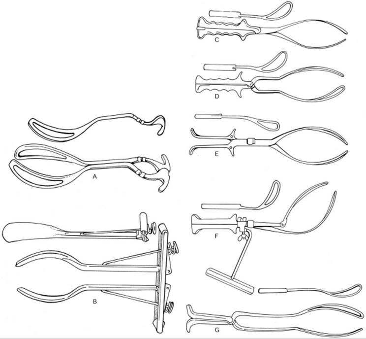
Classic forceps have branches that are made up of a handle, shank, lock, and blade (Fig. 23.11). The tip of the blade is referred to as the toe, the base of the handle is referred to as the heel, and the portion where the shank runs into the blade is termed the shoulder. In some cases an additional part is included to aid in traction and this is termed a traction bar (De Lee forceps) (Fig. 23.11).
|
|
|
FIG. 23.11. Common types of obstetric forceps. |
Handle
Most forceps in current usage have some form of handle that incorporates finger grips or lateral flanges. In some cases, the handle may be very small (Laufe divergent forceps and Wrigley forceps, see Fig. 23.11) or rather bulky as in the Salinas forceps. Classic Elliot forceps have a screw and wheel built into the heel of the instrument that allows adjustment of the distance between the heels in order to limit head compression during use.
Shank
The shank connects the blade to the handle and usually incorporates the locking mechanism. The shanks may be parallel, overlapping, or divergent, and vary in length from very short (Laufe) to rather elongated (Piper, Kielland, Bailey-Williamson forceps). They may also be straight or have varying degrees of curvature (Piper forceps).
Lock
The lock (Fig. 23.12) is usually built into the left-sided shank (or at the junction of the shank and handle) except in the case of the Salinas forceps where the lock forms part of the handle itself. The lock may be:
|
|
|
FIG. 23.12. Types of locks: Sliding, English, French, handle. |
· sliding (only found on the left shank and allows sliding of the shanks while locked—Kielland forceps)
· English (which is formed by the mating of a tongue and slot mechanism—Simpson, Elliot, Neville-Barnes, Anderson forceps)
· French (which has a nut that is tightened to fix the lock)
· pivot (forms part of the finger grip and tends to force the blades apart as traction is applied—Laufe forceps)
· handle (Salinas forceps).
Blade
The blade is the portion of the forceps that grasps the fetal head. Blades may be fenestrated (Simpson, Elliot, Neville-Barnes), pseudo-fenestrated (Luikart-Simpson), or solid (Tucker-McLane, Salinas). Fenestrated blades are purported to cause more maternal tissue damage during rotation. Solid blades are thought to reduce maternal injury by reducing friction but they are associated with increased fetal head slippage. Most forceps have a rigid blade that does not deform; the Salinas forceps however does allow some deformation of its shape because of its minimal thickness. Most forceps have two curvatures: A pelvic curvature which is usually concave upward to approximate the maternal pelvic curve of Carus, with the tip of the blade usually about 3 inches above the level of the handles; and a cephalic curvature designed to fit snugly around the fetal head with a diameter of no less than 7.5 cm at its widest portion. Blades best suited to a molded fetal head tend to be those that are elongated and have a more shallow cephalic curvature (Simpson, De Lee forceps). In cases where the fetal head is less molded a shorter blade with a more rounded cephalic curvature may be more suitable, although either type of forceps may be safely used under both circumstances.
Kielland and other rotational forceps, including the Salinas forceps, have almost no pelvic curvature, and this allows safe placement and rotation of the blades in transverse head positions. Rotation of a fetal head using a forceps with a pelvic curvature mandates use of the Scanzoni maneuver (whereby the heel follows the circumference of a large circle during the rotation in order to negate the pelvic curvature of the blade).
Some forceps have a reverse pelvic curvature (Piper and Hawkes-Dennen forceps) in order to allow use during vaginal breech delivery.
Traction Bar
Some forceps (Hawkes-Dennen) have a built-in device to allow the handles to rotate as the head descends through the birth canal. The idea is to optimize the outward and downward vectors so that the head follows the curve of Carus with the least soft and hard tissue friction. This principle of axis–traction can also be obtained using a traction bar (see Fig. 23.11) which fits onto the handle flange of some forceps.
Mechanical Aspects of Obstetric Forceps
From a mechanical point of view, forceps have four potential actions:
1. Correction of deflexion, asynclitism, or positional abnormalities of the fetal head that impedes or retards descent, rotation, and accommodation within the pelvis.
2. Extraction, which includes augmenting or replacing the expulsive force generated by uterine contractions and voluntary pushing.
3. Reduction of the friction between the fetal head and the birth canal.
4. Transient, artificial enlargement of the soft tissue of the birth canal which reduces the resistance of the outlet.
An understanding of the mechanical forces and principles involved in a forceps delivery is important since it allows the operator to better judge the chances of success in each individual case.
Type of Forceps Delivery
Two main parameters determine the successful outcome and associated morbidity of operative vaginal delivery. These are (a) the station at which the procedure is performed, and (b) the degree of rotation required. Based on the new definition of station, the types of forceps delivery were reclassified by ACOG in 1988 (Table 23.8). This new classification has been validated in a comparative study of 357 forceps deliveries classified using both systems. Allowing for up to 45 degrees of rotation in an outlet forceps delivery, and classifying any delivery from 0 to 2+ station as midforceps, regardless of rotation, did not increase morbidity.
|
|
|
TABLE 23.8. Classification of forceps deliveries |
Indications
As the specialty has evolved, so have the uses and indications for forceps. We no longer accept stimulation of contractions, artificial molding of the fetal head, forceful dilation of the cervix, or delivery from a high station as indications for forceps.
The indications for operative vaginal delivery may be divided into standard and special indications (Table 23.9). It must be understood that regional and international practice differences wilS apply and that no indication for operative vaginal delivery is absolute.
|
|
|
TABLE 23.9. Indications for operative vaginal delivery |
Contraindications
Under certain circumstances operative vaginal delivery should be avoided, or at least carefully considered in terms of relative maternal and fetal risk. The specific reason for the procedure should be carefully documented. Accepted contraindications to forceps delivery are listed in Table 23.10.
|
|
|
TABLE 23.10. Contraindication to forceps delivery |
Choice of Instrument
There are approximately 700 different types of forceps described. Most authors subscribe to a classification system that divides forceps into classic and specialized subtypes.
The classic subtype includes such forceps as the Simpson, Elliot, Neville-Barnes, Anderson, Tucker-McLane, and Naegle. These forceps would be suitable for most general prophylactic indications (outlet forceps).
The specialized subtype, which includes the Kielland (see Fig. 23.3), Barton (see Fig. 23.4), and Piper (see Fig. 23.5) forceps, is employed for specific tasks such as rotational deliveries (Kielland and Barton), correction of asynclitism (Kielland), and delivery of the aftercoming head in a breech delivery (Piper).
A novel type of forceps has been introduced into the U.S. from Mexico. The Salinas forceps has two identically shaped, nonfenestrated blades with minimal pelvic curvature which can be used interchangeably (see Fig. 23.11). The Salinas forceps also has a lock at the heel of the forceps and the blades are convergent. The blades are kept in place by maternal tissue and no compressive force is generated by the forceps. The operator holds the locked base of the forceps in both hands and applies gentle traction to effect delivery. The blades will slip off if excessive force is applied. The Salinas forceps can also be used for rotation of the fetal head.
The choice of one instrument over another depends on the indication, the training of the obstetrician, the instruments available, and the situation at hand. A comparison of the perceived advantages/disadvantages of forceps versus vacuum extraction is shown in Table 23.11. There are some who choose their forceps based on the condition of the maternal soft tissue (nonfenestrated blade when there is a lot of edema and potential for tearing; overlapping rather than parallel shanks for a patient with a narrow introitus), fetal head molding (Simpson forceps for a molded head and an Elliot-type forceps for a nonmolded head to allow a better fit of the forceps blade to the fetal head shape), or fetal presentation (Piper forceps for the aftercoming head). The need for a rotation or correction of asynclitism also may dictate the type of forceps used (i.e., Kielland, Luikart-Simpson, Barton, or Salinas forceps). Delivery of the aftercoming head in a breech delivery using Piper or Laufe-Piper forceps will permit flexion of the head without excess traction on the fetal spine and reduce the incidence of spinal cord injuries. Face presentation is another unique situation and, in general, mentum-anterior presentations can be delivered using Kielland or Simpson forceps. Rotation of the mentum- posterior/mentum-transverse face presentation to a mentum-anterior presentation has been described but few choose to perform these maneuvers.
|
|
|
TABLE 23.11. Comparative advantages of vacuum extractors and forceps |
Application of Forceps
Correct insertion of the forceps into the vaginal canal and application to the fetal head is paramount to a successful procedure. The forceps should be placed between contractions. In almost all cases one of two methods will allow good application of the forceps: direct and wandering. Application of the forceps blades should be a gentle and smooth process requiring minimal force. In most cases, if the blade is correctly inserted and positioned, gravity should be sufficient to allow the forceps blade to fall into place. During training it is a good idea to learn to apply the forceps using only the index finger, middle finger and thumb to grasp the shank (Fig. 23.13). The blade should be positioned using the index and middle fingers placed under or on the edges depending on the method employed. No force should be required and if there is resistance the blade should be removed and reapplied. In this way maternal and fetal injury will be minimized. A lubricant such as K-Y jelly placed on the blade may reduce friction and facilitate placement.
|
|
|
FIG. 23.13. Grasp of forceps. |
Direct Placement
With this method, the left blade (i.e., the blade that lies on the maternal left) is conventionally placed first. The operator holds the shank at the level of the lock, and the blade is placed into the vagina in the approximate position that it will ultimately lie. The right middle finger is used to support the edge of the blade at the introitus and the right index finger is used to gently apply the upper border of the blade onto the fetal head. The fingers or hand are not placed deep into the vagina. The practice of placing a hand into the vagina to “protect” the vaginal wall is probably not beneficial since this reduces the available space and increases the force required to place the blade. Once the left blade is correctly positioned, minimal force with the left hand will be required to slide it further into the birth canal and into its correct final position. The index and middle fingers of the right hand “walk” along the upper and lower borders of the blade as it enters the birth canal gently pushing it in and adjusting its position. When correctly placed the branch should be firmly held by the fetal head and maternal tissue (not immovable) and the shank and handle should be in the position expected from the “ghosting.” If the position is different from that anticipated the branch should be gently removed by reversing the movements required to initially place it, and the fetal head position should be checked (if necessary, by a more experienced operator). If the anticipated shank and handle angle are incorrect, attempting to place the right hand branch and forcing the two together is absolutely contraindicated. This will only result in maternal and fetal injury.
Once the left branch is placed, an assistant can be asked to hold the branch in position or it can be briefly left unattended while the right branch is placed. Holding the right branch with the right index finger, middle finger and thumb, the right branch is placed into the vagina in the approximate position that it will ultimately lie. The right branch should be above the left in order to ensure correct locking. The left middle finger is used to support the edge of the blade at the introitus and the left index finger is used to apply slight pressure onto the fetal head. Once the right blade is correctly positioned, minimal force with the right hand will be required to slide it further into the birth canal and into its correct final position. The index and middle fingers of the left hand “walk” along the upper and lower borders of the blade as it enters the birth canal gently pushing it in and adjusting its position to complement that of the left branch. No force should be used to lock the instrument and the final shank and handle position should be the same as that expected from the “ghosting”. Once both branches are placed, the fetal head position and the application of the forceps should be checked prior to any traction efforts.
Wandering Placement
With this method the left branch is also placed first. The operator holds the shank at the level of the lock, and the blade is placed into the vagina with the toe of the blade at the posterior fourchette and the branch held straight upward. The right index and middle fingers are used to support the edge of the blade (Fig. 23.14) at the introitus and to apply slight pressure onto the fetal head. The fingers or hand are not placed deep into the vagina. Once the left blade is correctly positioned, minimal force with the left hand will be required to begin sliding it further into the birth canal. Usually gravity and the weight of the branch is all that is required. As the blade moves into the birth canal the right index and middle fingers (which are held on the maternal left and right edges of the blade, respectively) gently push the blade laterally (maternal left) and “wander” it into position using successive small finger movements and without placing the fingers deep into the vagina. The handle will move in an arc from an initial vertical position to a horizontal position. Once learned, this is a smooth and seamless process. The position is checked as previously described. Once the left branch is placed, an assistant can be asked to hold the branch in position or it can be briefly left unattended while the right branch is placed. Holding the right branch with the right index, middle finger and thumb, the right branch is placed into the vagina in the same way as was the left branch. Once correctly positioned, minimal force with the right hand will cause the blade to move into the birth canal. The left index and middle fingers (which are held on the maternal right and left edges of the blade, respectively) gently push the blade laterally (maternal right) and “wander” it into position in the same way as was done with the left blade. The right branch should be kept above the left during this process to enable correct locking. No force should be used to lock the instrument and the final shank and handle position should be the same as that expected from the “ghosting”.
|
|
|
FIG. 23.14. Wandering placement of forceps. |
Once the blades are placed, their positioning should be checked. The blades are correctly placed when they are situated against the sides of the fetal head in the spaces between the orbits and the ears (Fig. 23.15). The toe of the blade should reach slightly beyond the malar eminences in order to allow even pressure distribution and to concentrate traction and compression forces on one of the less vulnerable regions of the fetal head. Prior to applying rotational or traction forces the operator should check the following:
|
|
|
FIG. 23.15. Correct placement of the forceps blades on the fetal head. |
· The forceps have not accidentally penetrated the side wall of the vagina (any sudden vaginal bleeding after forceps placement should prompt immediate assessment).
· The sagittal suture should be palpated symmetrically and perpendicularly between the two blades.
· The posterior fontanelle should be palpable close to (about 2 cm) the plane of the shanks but not within the borders of the blade.
· The handles, which should not require any more than minimal pressure to close completely, should be pointing straight out and not to either side.
· No significant part of the fenestration of either blade should be palpable in front of the fetal head.
Occipitoanterior Positions
Placement of the forceps in a direct occipitoanterior (OA) position can be accomplished as described above. Minor modifications are required when the head is in a left or right OA position. The exact location of the sagittal suture and posterior fontanelle should be known and the final arrangement of the forceps branches should be “ghosted” prior to insertion.
In left occipitoanterior (LOA) positions the left blade should be placed first using either a direct or a wandering approach. Once the left blade is correctly aligned, the right blade can also be placed using either approach. Under most circumstances the wandering approach will work well. The operator must remember that the blade will need to be maneuvered around the sinciput. Careful digital manipulation with the left index and middle fingers will be required to avoid injury to maternal and fetal soft tissue. If the handles do not lock easily the application is incorrect and should be checked. When this occurs the most common cause is that the blade has not been wandered far enough to clear the fetal brow, or has not advanced enough to lie flat against the fetal cheek. Gentle manipulation with index and middle fingers can be tried after unlocking the blades, but frequently the best action is to remove the blade and reintroduce it.
In an upright occipitoanterior (ROA) position, either blade can be placed first using the direct or wandering approach. If the right blade is placed first, once the left blade is positioned the handles will need to be separated and crossed over in order to lock the branches. If the left blade is placed first, the operator will need to manipulate it to a greater extent than in an LOA placement. In either case, care should be taken to prevent soft tissue injury. Rotation from ROA or LOA to direct OA is usually automatic as traction is gently applied and no deliberate elevation of the fetal head is needed.
Traction
Once the forceps have been applied and the position of the blades checked, traction is usually initiated with maternal contractions. The birth canal has a definite curve and this should be kept in mind when applying traction during a forceps delivery. In order to minimize friction, the traction forces on the fetal head should be maintained perpendicular to the axis of the pelvis. This necessitates following of the curve of Carus. The higher the forceps are applied, the greater the initial downward pull required to negotiate the pelvic curve. As the head descends in the birth canal the angle of traction moves forward (in the supine patient), ultimately turning upward at the outlet.
In order to optimize traction efforts the operator should stand slightly to the side of the patient. It is recommended that a right-handed individual stand to the maternal right with the right hand facing upward and the shanks of the closed forceps between the index and middle fingers (Fig. 23.16). The index and middle fingers are curved around the flange of the handle loosely in a forceps with crossed shanks or a sliding lock. In a Simpson-type instrument the middle finger can be placed in the space between the shanks with the index and ring fingers being placed on the handle flange. This hand will apply the outward traction force. Because the traction occurs at the level of the lock (as opposed to the end of the handles) compression of the fetal head will be limited. By angling the body sideways the major muscle group involved is the deltoid. This acts as a natural limited to the amount of traction that is applied. If the operator was standing directly in front of the patient the much more powerful biceps group would be supplying the majority of the traction force. By only using two fingers the operator will further limit the amount of force applied.
|
|
|
FIG. 23.16. Methods of hand placement and stance. |
The left hand is placed palm down on the shanks of the forceps and exerts a downward force. This is known as the Pajot maneuver. The combined outward and downward forces can be adjusted to produce a vector that follows the curve of Carus (axis–traction). Similar axis–traction can be applied using the Saxtorph maneuver (see Fig. 23.16) in which the operator is seated and uses the fingers of the left hand to pull the locked shanks downward. Simply pulling in the direction of the handles with a classic-type forceps (Simpson or Elliott) will cause the fetal head to be pulled up under the symphysis pubis. This is because the pelvic curvature of the forceps results in the handles being in a plane that is anterior to the plane of the pelvis in which the fetal head is present. When using an instrument with no pelvic curvature, or a reverse pelvic curvature, it is important not to elevate the handles more than 45 degrees above the horizontal in order to reduce the potential for sulcus tears from the toe of the blade.
A large amount of research has gone into determining what forces constitute safe levels for traction and compression. It has been calculated that during normal spontaneous delivery of the fetal head, second stage expulsive forces range between 8.6 kg and 15 kg. During forceps operations traction forces have been estimated to range from 16 kg to 23 kg. The traction forces are reduced by 25% and 50% by the use of forceps with crossed shanks.
Delivery of the fetal head in an OP position has been associated with increased traction and compression forces probably due to the larger presenting diameters of the fetal head.
Traction should be released between contractions in order to reduce the intracranial pressure differential to which the fetus is exposed. In addition, by releasing the pressure between contractions the vagally induced bradycardia that often accompanies traction efforts will be somewhat alleviated. Because of the potential for fetal heart rate abnormalities during delivery, the fetal heart rate should be continuously monitored during forceps deliveries.
Once the head crowns the handles of the classic-type forceps they can be elevated until they are almost perpendicular to the floor. The hand on top of the shanks will now be pulling almost directly outward. At this point the delivery can be completed with the forceps, or the branches can be disarticulated and the delivery can be completed using a modified Ritgen maneuver.
Occipitotransverse Positions
Occipitotransverse (OT) head position frequently results in a deep transverse arrest and resultant cephalopelvic disproportion. In most cases, this requires rotation of the fetal head to OA or OP to allow vaginal delivery, but occasionally, patients with a platypelloid pelvis will spontaneously deliver a baby with a transverse head position. The options open to the modern obstetrician faced with a deep transverse arrest in an OT position include:
· digital or manual rotation
· rotation with traction using a vacuum extractor (see Chapter XX)
· rotation with forceps
· cesarean section
Unless the operator has had sufficient training and experience with rotational delivery using forceps, cesarean section is frequently the most prudent choice.
DIGITAL OR MANUAL ROTATION
In some cases, particularly if the operator has a small hand, digital or manual rotation can be successfully used to rotate the fetal head from an OT position to an OA position. The digital method entails placing the tips of the index and middle fingers onto the edge of that part of the anterior parietal bone that overlaps the occipital bone in the area of the posterior fontanelle. Pressure is then exerted with the tips of the fingers in an attempt to rotate the posterior fontanelle upward and toward the symphysis pubis (see Fig. 23.6). In a manual rotation the whole hand is introduced into the birth canal and the fingers are placed under the posterior parietal bone. The thumb is positioned on the anterior parietal bone and the head is rotated (Fig. 23.17). Care should be taken not to completely disengage the fetal head. These maneuvers are difficult for people with large hands; instrumental rotation may be preferred in these circumstances.
|
|
|
FIG. 23.17. Digital and manual rotation. |
If digital or manual rotation is successful, the patient should be encouraged to push in order to increase descent of the fetal head and to stabilize the fetal head position. If the delivery is to be completed using forceps, the fingers should be kept on the fetal head while the forceps are placed (see Fig. 23.17).
ROTATION WITH TRACTION DURING VACUUM EXTRACTION
The vacuum extractor has been promulgated as an instrument with which to effect rotation of the fetal head. While this is true to some extent, it must be remembered that such rotation should only be accomplished by traction in the axis of the pelvis and not by twisting or rotational motions with the vacuum cup. By applying traction in the correct plane, the head is forced to rotate by virtue of the architecture of the soft tissue and bones of the pelvis. The operator should assess the position of the fetal head and take into account the malposition and the attitude and asynclitism of the presenting part. By applying the traction force in an appropriate direction to correct the flexion and asynclitism of the head, rotation will occur automatically as the head descends. The use of a twisting motion is contraindicated because of the risk of cephalhematoma and scalp laceration imposed by the significant shearing force that can be delivered to the soft tissue of the head. This effect is more pronounced when using the metal cup systems than with the Silastic cup. In most situations where rotation is required, choice of an appropriate forceps method will be more likely to result in successful rotation than use of the vacuum extractor.
ROTATION WITH FORCEPS
Rotation of the fetal head from a transverse position to an OA position is one of the most demanding (and satisfying) obstetric operative procedures. It is also one of the most technically demanding. Forceps rotation can be accomplished using classic forceps (i.e., Elliott, Tucker-McLean, Neville-Barnes), specific rotational forceps (Kielland, Barton), or Salinas forceps. The blades may be applied using the classic method, the wandering method, or the direct method.
When using a forceps with no, or minimal, pelvic curvature (Kielland, Salinas), I prefer using the direct method to apply the blades to a head in a deep transverse arrest (Fig. 23.18). In my experience this method involves the least manipulation. If performed correctly, the blades will be correctly positioned with the minimum potential for fetal and maternal injury. The wandering method is a better choice when using a classic-type forceps. I do not believe that a forceps with a pronounced pelvic curvature should be applied using the classic method and I therefore only describe the classic method using a Kielland forceps. When performing a rotational delivery my preference is to use a forceps designed for rotation rather than a classic-type forceps.
|
|
|
FIG. 23.18. Direct application of Kielland forceps for rotation. |
Occipitoposterior Positions
An occipitoposterior position may be managed by delivery as a direct OP, by digital or manual rotation, by instrumental rotation, or by a combination of these methods. In general, a well-flexed head in an OP position can be delivered as a direct OP. If the head is extended or only minimally flexed then attempting a direct OP delivery may result in an increased risk of maternal and fetal trauma. This is because of the greater traction required to deliver the head due to the larger diameter presented. There is also the chance that traction on a deflexed head will exaggerate the extension and worsen the situation. Under these circumstances I prefer to rotate the head to an OA position, and this will usually help flex the head as well.
Digital or Manual Rotation
This is performed similarly to that described previously for OT positions except that the arc of rotation is greater.
Instrumental Rotation
Forceps or vacuum extraction can be useful for rotating an OP to an OA position. Either classic-type forceps or an instrument designed for rotation (Kielland or Salinas forceps) can be employed.
If a classic-type forceps is used the blades are placed in the same way as for an OA position (described previously). If the head is in a right OP position the forceps are applied as for a left OA and vice versa. Once the branches are locked the posterior fontanelle should be felt approximately 2 cm below the shanks, with the sagittal suture midway, and perpendicular, between the two blades. The head is then rotated into an OA position with the direction of rotation toward the side of the fetal back. As described earlier, the handles of the forceps should describe a wide arc in order to limit the arc of the toes of the blades. Once the rotation is complete, the now upside-down forceps branches should be removed and replaced in the correct orientation. The easiest way to do this is to have two sets of forceps available for use. As the upside-down forceps branch is removed it can be replaced with the appropriate blade from the other set of forceps. The remaining upside-down blade keeps the head in the OA position during the changeover. Once the replacement branch is correctly positioned the remaining upside-down branch can be removed and replaced with the second branch of the replacement set of forceps. The delivery is then completed as previously described.
If Kielland forceps is used, the blades are applied as for an OA (or LOA/ROA) position using the technique described earlier. The buttons on the handles should face the fetal occiput. The rotation is carried out as already described except that the head is rotated through 180 degrees rather than 90 degrees. The handles should rotate on their axis since there is no pelvic curvature to contend with. It may be necessary to elevate the head during the rotation and if this is the case the operator may need to lower the handles toward the floor in order to better the forceps with the birth canal. The same slow, controlled maneuvers and precautions should be used as described previously. There is no need to reverse the blades after the rotation and once the head is OA, traction can be applied for delivery. Alternatively, there are some who place the forceps with the buttons facing the sinciput (upside-down application) for the rotation and then remove and replace the forceps for the delivery (either with the Kielland or with a classic-type forceps).
POST-DELIVERY MANAGEMENT
Following operative vaginal delivery the flexion of the patient's hips should be relieved in order to minimize the risk of nerve injury from prolonged pressure or stretching. The episiotomy repair should be performed as expeditiously as possible to limit blood loss and tissue exposure. The birth canal and cervix should be inspected for lacerations and tissue damage. I prefer not to repair minor lacerations that are not bleeding since they will usually heal very quickly. Even cervical tears that are not bleeding are best left alone under close observation. Care should be taken to inspect the periurethral and perianal/rectal areas after any repairs to exclude perforating sutures. Any complaint of anal pain after a forceps delivery should prompt a careful inspection for a vaginal or vulval hematoma. Any sudden drop in hematocrit should indicate a search for continued bleeding either obvious (vaginal or vulval) or occult (retroperitoneal hemorrhage).
Complications of Operative Vaginal Delivery
Complications arise in two main areas:
1. Inappropriate evaluation and underestimation of dystocia.
2. Misuse of an instrument.
It is important to point out that almost all of the complications seen with operative vaginal delivery have also been reported with spontaneous, uninstrumented vaginal deliveries. This should be kept in mind before attempting to link a particular injury directly to an instrumental delivery.
The risks can be divided into maternal and fetal/neonatal.
Maternal Complications Associated with Operative Vaginal Delivery
Lacerations
The reported incidence of severe laceration (i.e., third-degree and fourth-degree laceration) during vacuum extraction procedures ranges from 10% to 30%. Operative vaginal delivery with forceps is responsible for a higher incidence of third- and fourth-degree perineal lacerations (closer to 50%) than with spontaneous delivery or vacuum extraction.
Meta-analysis of available studies has shown that forceps operations are more likely to specifically result in anal sphincter trauma than vacuum extraction. In pooled data from 8 randomized trials studying maternal delivery trauma, a 6% decrease (95% CI = -.10 to -.02) in anal sphincter trauma occurred if a vacuum extractor, and not forceps, was employed.
Stress Urinary and Anal Incontinence
Delivery trauma is suspected to predispose the patient to subsequent pelvic floor dysfunction; however, data indicate that the long-term effects of operative vaginal delivery on rectal or urinary incontinence are more complex than originally believed. Interpretation of all the available data is not straightforward. There is a suggestion that vaginal delivery is a major contributor to, but not the sole culprit of, long-term perineal dysfunction. It is believed that the greater the degree of perineal trauma, the greater the likelihood that there will be residual sphincter abnormalities.
Arya and colleagues (8) retrospectively studied the incidence of new-onset urinary incontinence after forceps and vacuum delivery compared with spontaneous vaginal delivery. They enrolled primiparous women delivered by forceps (n = 90), vacuum (n = 75), or spontaneous vaginal delivery (n = 150). They then followed these women at 2 weeks, 3 months, and 1 year after delivery and showed that incontinence was similar in the 3 groups at 2 weeks after delivery. The proportion of women developing new-onset urinary incontinence decreased significantly over time in the spontaneous vaginal (P = .003) and vacuum delivery groups (P = .009) but not in the forceps group (P = .2). There was no relationship between urinary incontinence and a history of vaginal lacerations, epidural anesthesia, length of second stage of labor, or infant birth weight. These authors concluded that in primiparous women, urinary incontinence after forceps delivery is more likely to persist compared with spontaneous vaginal or vacuum delivery.
A Swedish study of three large databases involving 1,559 patients who presented for incontinence surgery later in life (20–40 years after delivery) linked the need for surgery to some independent variables. The study included 189 women who had experienced an operative vaginal delivery (almost all were vacuum deliveries). There were positive associations between diabetes, body mass index, parity, birth weight, epidural, and age at first delivery. Interestingly, in this study, operative vaginal delivery, cesarean section, and episiotomy were shown to be protective against incontinence surgery in later life.
Farrell and co-workers (level II-2) studied 690 Canadian primiparas who filled out a questionnaire about urinary incontinence during, and 6 months after, their first pregnancy (9). Twenty-five percent delivered by cesarean section, 19% had an operative vaginal delivery, and 56% had a spontaneous vaginal delivery. The median birth weight was 3,489 g. The rate of urinary incontinence at 6 months was 26%. Vaginal delivery was associated with a higher rate of urinary incontinence than cesarean section. Ten percent of women who had a cesarean section and 22% of those who had a spontaneous vaginal delivery reported postpartum urinary incontinence (OR = 2.1 [range, 1.1–-3.7]). Thirty-three percent of those who had a forceps delivery reported postpartum incontinence (forceps [33%] vs. spontaneous vaginal delivery [22%]; OR = 1.5 [range, 1.0–2.3]) and forceps (33%) vs. cesarean section (10%) (OR = 3.1 [range, 1.7–5.9]). Vacuum extraction showed no higher incidence when compared with spontaneous vaginal delivery, but when compared with cesarean section there was a significantly higher rate of postpartum incontinence (vacuum extraction vs. spontaneous vaginal delivery, no significant difference; vacuum extraction vs. cesarean section (OR = 3.5 [range, 1.3–9.1]). The authors concluded that cesarean section performed before or during labor reduces postpartum urinary incontinence.
Meyer and co-workers studied 107 Swiss primiparas during their pregnancy and at 10 weeks and 10 months after delivery (10). The methods used for collecting data included questionnaire, clinical examination, assessment of bladder neck behavior, urethral sphincter function, and intravaginal/intraanal pressures during pelvic floor contractions. The results did not show any significant differences between the women who had forceps delivery and those who delivered spontaneously in terms of urinary incontinence (20% vs. 15% at 10 months), fecal incontinence (4% vs. 5% at 10 months), and diminished sexual response (12% vs. 18% at 10 months). More women in the forceps group however were shown on objective testing to have a weak pelvic floor (20% vs. 6%; P = .05) and low intraanal pressure (P = .04). The authors concluded that forceps delivery is not responsible for a higher incidence of pelvic floor complaints, or a greater change in bladder neck behavior or urethral sphincter function. However, they do believe that patients who have had a forceps delivery have a significantly greater decrease in intraanal pressure and a greater incidence of a weak pelvic floor.
In terms of fecal incontinence, McArthur and colleagues questioned over 5,000 postpartum women and showed that forceps deliveries are associated with higher risk than vacuum extraction (OR = 1.94; 95% CI = 1.30–2.89) while cesarean section may offer some protection (OR = 0.58; 95% CI = 0.35–0.97). This area of obstetrics remains under-researched and the value of anal manometry and the current methods used in the diagnosis and ascertainment of postpartum anal injury/dysfunction have been questioned.
Long-term follow-up studies controlling for prepartum pelvic support status (e.g., preexisting rectal dysfunction or urinary incontinence, length of labor, anesthesia, clinically observed perineal trauma, and delivery method) are required before changes in current practice can be recommended.
Patients should be counseled that significant vaginal trauma may be related to long-term sphincter dysfunction, but that a relatively atraumatic operative vaginal delivery (i.e., one that does not cause rectal or sphincter rupture) is not known to cause a higher rate of dysfunction than that which follows spontaneous vaginal delivery.
Nerve Injuries Following Operative Vaginal Delivery
An uncommon but distressing complication of vaginal delivery (be it spontaneous or operative) is a nerve injury which is usually the result of incorrect positioning and undue compression or traction on a nerve. Fortunately most of these injuries are apraxias, which recover, but occasionally a neuronotmesis or axonotmesis occurs with long-term disability. The most common cause of these injuries is excessive flexion of the maternal hips at the time of delivery (forced McRoberts position with attendants applying significant pressure to flex the patient's hips) and episiotomy repair (prolonged hyperflexion of the hips). Because the repair of lacerations and the episiotomy may be lengthy, it is advisable that the operator adjust the patient's position after delivery so that there is minimal flexion of the hips. Care should also be directed to pressure points on the patient's legs to avoid nerve compression injuries, and adequate padding and positioning is essential. The common injuries and their causes and presentations are shown in Table 23.5.
Neonatal Injury and Operative Vaginal Delivery
Neonatal Injuries Associated with the Vacuum Extractor
Scalp Bruising/Lacerations
Ecchymoses and, uncommonly, scalp slough or lacerations can follow vacuum extraction. Most of these injuries occur when the recommended limits to total cup applications are exceeded.
Subgaleal hemorrhage is a serious neonatal complication of vacuum extraction. This complication occurred in 1.0% to 3.8% of vacuum extractions in one series but has been much less common in some more recent studies. Approximately half of all subgaleal hemorrhages are related to vacuum extraction. The remainder are associated with forceps operations; less commonly, they occur following spontaneous delivery. This complication has not been reported in recent vacuum extraction meta-analyses, attesting to the rarity of severe scalp injuries, while emphasizing the importance of following strict technical guidelines when performing vacuum extractions/operations. Despite the low incidence of subgaleal bleeding, notification of pediatric personnel whenever an extraction is performed is mandatory, regardless of the immediate condition of the baby because subgaleal hemorrhage may not be clinically apparent until some hours postpartum. Infants with subgaleal hemorrhage present with a boggy scalp, swelling crossing the suture lines, and an expanding head circumference. They may also have signs of hypovolemia, pallor, tachycardia, and a falling hematocrit.
Cephalhematoma is another fetal complication of vacuum extraction. This complication is more common with vacuum extraction (14%–16%) than with forceps delivery (2%).
Intracranial hemorrhage has been found to occur in one of every 860 vacuum-assisted deliveries compared with one of every 1,900 spontaneous deliveries (a statistically significant difference). Yet, the comparative rate of intracranial hemorrhage is not statistically different when vacuum extraction, forceps delivery, and cesarean section during labor are compared. It is possible that the abnormal labor that necessitated the assisted delivery may be an underlying cause for a portion of the morbidity attributed to operative deliveries.
Retinal hemorrhages may be more common in vacuum-assisted deliveries than with forceps (38% vs. 17%) but are most often associated with duration of labor. Because these hemorrhages resolve within several weeks, they are unlikely to be associated with long-term morbidity.
Transient neonatal lateral rectus paralysis has been found to occur more frequently in vacuum-assisted deliveries (3.2%) than in forceps deliveries (2.4%), normal spontaneous vaginal deliveries (0.1%), and cesarean sections (0%). Because the paralysis resolves spontaneously, it is unlikely to be of clinical importance.
Vacuum extraction is associated with a higher rate of neonatal jaundice than is spontaneous delivery or forceps procedures, and this is possibly related to the higher rate of cephalhematoma.
Long-term Neonatal Outcomes
The few available studies evaluating long-term neurologic sequelae of instrumental delivery have found no differences among children delivered spontaneously and children delivered by either vacuum extraction or forceps. Vacuum extraction has not been found to result in significant intellectual or neurologic disability.
Neonatal Injuries Associated with Forceps (Fig. 23.19)
|
|
|
FIG. 23.19. Forceps-associated injury. (Courtesy of Dr. Gerardo Cabrera Meza.) |
Exactly how much neonatal injury can be attributed to a forceps delivery is difficult to assign since it is well known that a protracted spontaneous labor can result in a poor neonatal outcome. The most commonly described neonatal injuries include those described in the following paragraphs.
Superficial Scalp and Facial Markings
These will invariably be present and only occasionally are of significance. Obviously correct placement of the forceps blades will prevent tissue damage to sensitive regions such as the eyelids and face. Inappropriate forceps application (sometimes referred to as the fetal grip) can be easily noted by the bruising on the baby's face in the neonatal period. In general, if the parents are prepared for the markings that they see, and it has been explained to them that these are temporary, their acceptance is good. Abrasions and lacerations are not expected or acceptable, and a careful assessment of one's technique should follow any such occurrence. Excessive pressure on the fetal face can result in vesicle formation, lipoid necrosis, and massive face and head trauma. Pressure localized over the mastoid region can result in facial nerve injury. Most superficial injuries are short-lived and recovery is uncomplicated.
Facial Nerve Injury
Injury to the facial nerve has been reported and is usually the result of compression of the nerve against the rigid cervical spine from a forceps blade at the point where the nerve is superficially placed in the mastoid region. This is usually a transient event, but may result in permanent injury.
Cephalhematoma
Cephalhematoma occasionally occurs with forceps delivery (2%) but is more commonly seen after vacuum extraction (14%–16%). Skull fracture, subgaleal hematoma, and intracranial hemorrhage have all been reported after complicated or difficult forceps deliveries. It is difficult to assess the risk, however, since these problems have also been noted after complicated spontaneous deliveries. In addition, no data are available addressing the incidence of these complications in normal vaginal or abdominal deliveries. If a difficult delivery occurs, the neonatal personnel should be advised and the infant should then be appropriately observed and investigated.
Retinal Hemorrhage
This complication occurs with forceps deliveries (17%) but is more frequently seen after vacuum extraction (38%; P < .05).
Corneal Abrasions/External Ocular Trauma
Forceps delivery is associated with an increased rate of eyelid edema (39%) and minor external ocular trauma (17%) when compared with babies born by spontaneous vaginal delivery (eyelid edema = 10%, and external ocular trauma = 6%). Corneal abrasion is possible and corneal edema has been reported. Long-term sequelae are extremely rare and ophthalmologic screening should be reserved for specific cases.
Intracranial Bleeds
It is the intracranial hemorrhages and infarcts seen following a difficult operative vaginal delivery that are of great concern. Whether or not a specific neonate suffers cerebrovascular injury due to the underlying condition that precipitated the fetal distress that resulted in delivery with the forceps, or whether the damage was caused directly by the forceps at the time of the delivery, is frequently difficult to discern. The decision to attempt a forceps delivery in the face of fetal distress can often be a difficult one. If the operator is experienced and confident that delivery can be achieved with an uncomplicated procedure, appropriate use of forceps in an emergency situation (while preparations for cesarean section are underway) can significantly reduce the time to delivery and reduce potential fetal injury. In most cases with a bad outcome there is no guarantee that cesarean section would have resulted in any different outcome from an appropriately performed operative vaginal delivery. Abdominal delivery in these situations certainly does not remove the threat of litigation. Therefore it is advised that cesarean section be performed for obstetric reasons and not out of fear of litigation.
FAILED OPERATIVE VAGINAL DELIVERY AND SEQUENTIAL USE OF INSTRUMENTS
A failed forceps/vacuum delivery attempt was, until recently, not thought to significantly worsen the neonatal course or increase morbidity or mortality of the neonate. This impression was based on small series. More recent, large, population-based studies have revealed that a failed operative delivery, or the sequential use of different instruments, does significantly increase morbidity and mortality. Towner and colleagues performed a population study of 583,340 live-born, singleton babies delivered from nulliparous women in California between 1992 and 1994 (11). They restricted the study to babies with birth weight between 2.5 and 4.0 kg and breech deliveries were excluded.
Approximately 30% of the deliveries were by operative techniques.
The authors studied the relationship between mode of delivery and the morbidity/mortality of the babies. The study showed that, when compared with spontaneous vaginal delivery, vacuum extraction (OR = 2.7 [range, 1.9 - 3.9]), forceps delivery (OR = 3.4 [range, 1.9 - 5.9]), and cesarean section after abnormal labor (OR = 2.5 [range, 1.8 - 3.4]) resulted in an increased incidence of subdural and cerebral bleeds. Intracranial bleeding occurred with the following frequencies:
· 1:334 failed vacuum extraction or forceps followed by cesarean section
· 1:860 after vacuum extraction
· 1:664 after forceps
· 1:907 after cesarean section during labor
· 1:2,750 after cesarean section without labor
· 1:1,900 normal spontaneous vaginal delivery.
Differences in the rate of intracranial hemorrhage were reported after the use of vacuum extraction or forceps delivery (OR = 1.2 [range, 0.7–2.2]).
One of the surprising findings in this study was that the common risk factor for intracranial bleeding was abnormal labor, rather than simply the mode of delivery.
When compared with successful forceps or vacuum extraction alone, the rate of intracranial bleeding was two to three times higher in babies born after a sequential operative vaginal delivery, or after a failed forceps/vacuum procedure that was followed by a cesarean section. These findings were confirmed by a similar study by Gardella and colleagues who used Washington state birth certificate data linked to hospital discharge records and compared 3,741 vaginal deliveries by both vacuum and forceps, 3,741 vacuum deliveries, and 3,741 forceps deliveries to 11,223 spontaneous vaginal deliveries (12). They showed that compared with spontaneous vaginal deliveries, the sequential use of vacuum and forceps had significantly higher rates of intracranial hemorrhage (RR = 3.9 [range, 1.5–10.1]), brachial plexus (RR = 3.2 [range, 1.6–6.4]), facial nerve injury (RR = 13.3 [range, 4.7–37.7]), seizure (RR = 13.7 [range, 2.1–88]), depressed 5-minute Apgar score (RR = 3.0 [range, 2.2–4.0]), assisted ventilation (RR = 4.8 [range, 2.1–11]), fourth-degree (RR = 11.4 [range, 6.4–20.1] among multiparous women) and other lacerations, hematoma (RR = 6.2 [range, 2.1–18.1] among multiparous women), and postpartum hemorrhage (RR = 1.6 [range, 1.3–2.0]). The relative risk of sequential vacuum and forceps use was greater than the sum of the individual relative risks of each instrument for intracranial hemorrhage, facial nerve injury, seizure, hematoma, and perineal and vaginal lacerations.
Based on these two studies, there is now no question that the sequential use of vacuum and forceps is associated with an increased risk of both neonatal and maternal injury. Unless there is a reasonable likelihood of atraumatic vaginal delivery, a “trial of operative vaginal delivery” is something that should be undertaken with caution. The use of the term “failed forceps” should fall out of use as careful preoperative assessment of mother and baby are applied. A failed forceps suggests that a forceps delivery was attempted and that obvious, repetitive, but futile efforts were made to deliver the baby with at least one genuine attempt at rotation or traction that was without success. When the term “failed forceps” is used it implies that the underlying expectation of the operator was to complete the delivery with the chosen instrument.
In a situation where the correct indications and prerequisites apply, and an appropriate, albeit unsuccessful attempt is made, a better term to use when documenting is “abandoned trial of forceps.” This shows that the attempt was justifiable but stopped when the operator recognized a complication such as an inability to properly apply the instrument, inability to correctly rotate the head, or a lack of descent despite a correct application of the instrument. In this case it is clear to anyone reviewing the chart, that the operator sensed a problem and did not persist.
STRATEGIES TO REDUCE INJURY
One of the most important clues as to the potential for successful operative vaginal delivery can be found in the details of the labor course. Use of labor monitoring graphics such as the Friedman curve or the Philpott and Castle “alert and action lines” will lead to early identification of those patients most likely to have dystocia. The combination of the “history” (labor course) and physical examination will usually allow an accurate assessment of the potential for vaginal delivery. The wise obstetrician will very carefully evaluate a patient who is a candidate for a forceps delivery but who has had a protracted labor. Frequently a slow course, or a second stage arrest, will be associated with an unusually large baby, an unsuitable pelvis, or an unrecognized malposition. These complications may well contraindicate any attempt at instrumental delivery. The fact that a patient can get to complete dilation with a baby whose head appears to be engaged does necessarily mean that the safest route of delivery is vaginal.
Once the decision to proceed with a forceps delivery has been made, the selection of the appropriate instrument, correct positioning of the patient, and satisfaction of the aforementioned prerequisites are important.
The willingness to reassess the decision after application of the forceps blades is also important. Merely applying the blades, or attempting to apply the blades will not cause maternal or fetal damage unless excessive force has been used. At this point, if the application is incorrect or inappropriate, the procedure should be abandoned and the blades gently removed. Whether or not to attempt a second application will again be a judgment call. It is recommended that if this is to occur there should be a careful reevaluation of fetal and maternal condition. If both mother and baby are in good condition, the most experienced individual should be the one to reassess the situation and reapply forceps, if appropriate.
Knowing when to abandon a forceps operation is just as critical as knowing when to attempt one, and reflects the maturity and experience of the operator. This should be an infrequent occurrence since abandoning a procedure almost always indicates incorrect assessment in the first place. However, there will be cases where application of the forceps changes the head position, or results in asynclitism that was not there in the initial evaluation. If, at the traction attempt, there is no descent of the fetal head it is fruitless to continue applying more traction as this is the cause of most fetal injury. The safety of the mother and baby should always be first in the mind of the operator and there should never be a need to complete the operation at all costs. There are no good data addressing the important questions of how many traction attempts are reasonable, or how long the forceps procedure should take. These aspects of obstetrics can only be acquired by clinical training, and are dependent on the experience, skill, and integrity of the operator.
Does Fetal Distress Preclude Operative Vaginal Delivery?
Vintzileos and colleagues demonstrated that in cases where the vacuum extractor was used to deliver babies with suspected fetal distress there were no differences in blood gas parameters when compared with normal spontaneous deliveries. The authors concluded that the use of vacuum extraction is not contraindicated in cases of fetal distress (13).
Documentation and Safety Issues
The application and use of forceps is associated with a definite risk profile that should be explained to the patient prior to their use. If the situation allows, it is my practice to write a short preoperative note detailing the following:
1. Indication for the procedure along with the time and date of the note.
2. The amount of head palpable above the pelvic brim.
3. The position and station of the fetal head.
4. The degree of molding of the parietal bones.
5. The amount of caput succedaneum palpable.
6. The estimated fetal weight.
7. A clinical assessment of the maternal pelvis.
8. A statement regarding the fetal heart rate and character.
9. A statement about the maternal contractions.
10. A statement about maternal condition and type of anesthesia to be used.
11. A statement that the mother understands the reason for the procedure and accepts the risks.
After the delivery an operative note should be written or dictated and should contain as much of the following information as possible:
1. Preoperative and postoperative diagnoses along with the time and date of the note.
2. Reiteration of the physical findings (outlined previously) at the time of the procedure, especially if these were different from those detailed in the preoperative note.
3. Number of applications of the forceps, ease of application, and any complications with the application. If possible have a nurse or assistant document the exact times of application and traction.
4. Duration and force of each traction attempt, as well as the number of traction attempts.
5. Complete explanation of a failed or abandoned procedure with reasons.
6. Detailed description of any maternal or neonatal injuries.
7. Cord blood gases if the 5-minute Apgar is less than 7.
Informed Consent for Forceps Operations?
Although not frequently done, I believe that unless there is an emergency situation, a forceps delivery should be discussed with the patient as one would discuss any operative procedure. In this way informed consent can be obtained and the patient can be prepared for what to expect. The risks and benefits of operative vaginal delivery should be explained (and compared with those of cesarean section) and the patient should be aware of her options. I recommend that this be detailed in the patient's chart prior to the delivery. Most lawsuits are generated as a result of lack of communication between the doctor and the patient. Dissatisfaction with an outcome will always be heightened if the patient feels uninvolved with the decision and perceives that she has been misinformed about the risks. The best time to educate the patient is during her prenatal course or on admission to the hospital. When the patient signs her consent forms during the admission process I believe that a consent form for operative vaginal delivery should be included. The information provided should include a description of what forceps look like and that, despite their shape and size, they are specially designed to protect the mother and baby by gently grasping the fetal head and assisting the infant to negotiate the last portions of the birthing canal. The fact that a timely operative delivery will the time and energy required during the birthing process should be explained. The fact that correct forceps use can be lifesaving should be stressed. The patient should, however, also understand that bad outcomes will still occur due to unforeseen problems, and the obstetrician should be prepared to explain the maternal and fetal/neonatal risks specific to forceps operations. The possibility of facial bruising and possible facial nerve or eye injury should be mentioned before the delivery because of the emotional impact of these injuries after the delivery has occurred, especially when the patient or her family makes accusations that these risks were never mentioned. The detractors of informed consent complain that after being informed of the potential risks of operative vaginal delivery most women will elect to proceed with cesarean section and will forgo vaginal delivery. The argument against this point of view is that since cesarean section is a viable option that will allow minimization of fetal injury (and one that is not associated with a prohibitively high incidence of maternal or neonatal injury) patients have the right to choose to avoid potential trauma to their baby. Many authorities fear that by providing more detailed written informed consent the medicolegal risk increases since it is not possible to cover every eventuality and thus the informed consent will be regarded as incomplete or misleading. This is obviously a controversial matter but at this time there is no standard of care in the United States that demands a written informed consent prior to operative vaginal delivery.
Post-Delivery Responsibilities
It is very important that neonatal caregivers be informed of a vacuum or forceps delivery, and that they monitor the infant for signs of any complications.
CONCLUSIONS
Operative vaginal delivery has been an important part of obstetric practice for nearly 400 years. We must make every effort to retain these skills, to modify and improve them in every possible way, and then to pass them on to those who will follow us. It is in this way that women and children of future generations will benefit from the many years of experience that have gone before them.
SUMMARY POINTS
· Adequate assessment of the “pelvis and passenger” is essential prior to attempting an operative vaginal delivery. Careful assessment of the maternal abdomen prior to attempting operative vaginal delivery forms an important part of this assessment. If the fetal head is not felt to be completely engaged and there is excessive molding of the fetal head, the procedure should be avoided. There are important prerequisites, which if satisfied, will reduce the chances of failed operative vaginal delivery procedures.
· Failed operative vaginal delivery and sequential operative vaginal delivery attempts with different instruments significantly increase the risk of neonatal intracranial complications.
· Both forceps and the vacuum extractor are acceptable and safe instruments for operative vaginal delivery. Operator experience should determine which instrument should be used in a particular situation. The vacuum extractor is associated with an increased incidence of neonatal cephalhematomata, retinal hemorrhages, and jaundice when compared with forceps and the neonatal care provider should be made aware of the mode of delivery in order to exclude these complications.
· Large fetal size and prolonged labor are associated with an increased risk of shoulder dystocia and careful consideration should be given to performing operative vaginal delivery under these circumstances.
· Forceps delivery may be preferable to vacuum extraction in small-for-gestational-age babies since it is associated with a lower incidence of intraocular bleeding.
· Operators should attempt to reduce the time between vacuum application and delivery of the baby since cephalhematoma is more likely if the interval exceeds 5 minutes.
· Midforceps operations, if performed under the correct circumstances by an adequately trained individual, should still be considered an appropriate procedure to be used and taught.
· Efforts should be made to train resident obstetricians in the use of operative vaginal delivery techniques since appropriate and safe employment of forceps and vacuum-assisted delivery still is an important part of modern-day obstetrics.
REFERENCES
1. Crichton D. A reliable method of establishing the level of the fetal head in obstetrics. S Afr Med J 1974;48:784–787.
2. American College of Obstetricians and Gynecologists. ACOG Practice Bulletin No. 17, June 2000.
3. Johanson R, Menon V. Soft versus rigid vacuum extractor cups for assisted vaginal delivery. In: The Cochrane Database of Systemic Reviews. The Cochrane Library 2001; Issue 4. Oxford: Update Software.
4. Bofill JA, Martin JN Jr, Morrison JC. The Mississippi Operative Vaginal Delivery Trial: lessons learned. Contemp Ob Gyn 1998;43:60–79.
5. U.S. Food and Drug Administration Public Health Advisory May 21, 1998.
6. American College of Obstetricians and Gynecologists. ACOG Committee Opinion No. 208, 1998.
7. Ross MG, Fresquez M, El-Haddad MA. Impact of FDA advisory on reported vacuum-assisted delivery and morbidity. J Maternal-Fetal Med 2000;9:321.
8. Arya LA, Jackson ND, Myers DL, et al. Risk of new-onset urinary incontinence after forceps and vacuum delivery in primiparous women. Am J Obstet Gynecol 2001;185:1318–1324.
9. Farrell SA, Allen VM, Baskett TF. Parturition and urinary incontinence in primiparas. Obstet Gynecol 2001;97:350–356.
10. Meyer S, Hohlfeld P, Achtari C, et al. Birth trauma: short and long term effects of forceps delivery compared with spontaneous delivery on various pelvic floor parameters. Br J Obstet Gynecol 2000;107:1360–1365.
11. Towner D, Castro MA, Eby-Wilkens E, et al. Effect of mode of delivery in nulliparous women on neonatal intracranial injury. N Engl J Med1999;341:1709–1714.
12. Gardella C, Taylor M, Benedetti T, et al. The effect of sequential use of vacuum and forceps for assisted vaginal delivery on neonatal and maternal outcomes. Am J Obstet Gynecol 2001;185:896–902.
13. Vintzileos AM, Nochimson DJ, Antsaklis A, et al. Effect of vacuum extraction on umbilical cord blood acid-base measurements. J Maternal-Fetal Med1996;5:11–17.