
Jeffrey L. Hackman
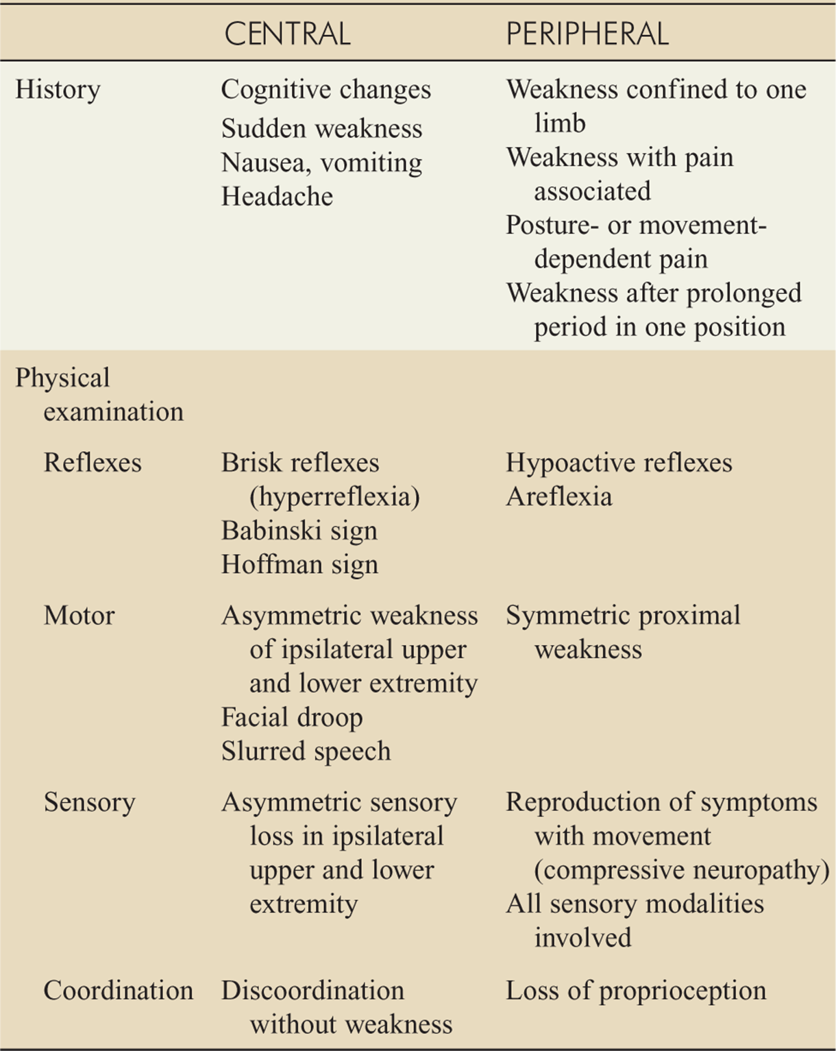
 History and physical examination findings can help distinguish central from peripheral nervous system disorders (Table 148-1).
TABLE 148-1 Differentiating Central from Peripheral Nervous System Disorders
NEUROMUSCULAR JUNCTION DISORDERS
BOTULISM
Botulism is caused by Clostridium botulinum toxin and occurs in three forms: foodborne, wound, and infantile.
In the United States, the principal source is improperly preserved canned foods.
In infantile botulism, organisms arise from ingested spores, often in honey, and produce a systemically absorbed toxin.
Wound botulism should be considered in patients with a wound or a history of injection drug use.
Clinical features appear 6 to 48 hours following ingestion and may be preceded by nausea, vomiting, abdominal cramps, and diarrhea or constipation.
Descending, symmetric paralysis is the classic finding.
The cranial nerves and bulbar muscles are affected first, causing diplopia, dysarthria, and dysphagia.
Dilated nonreactive pupils help distinguish botulism from myasthenia gravis, which does not affect the pupil.
Infants present with constipation, poor feeding, lethargy, and weak cry.
Treatment includes respiratory support, botulinum antitoxin, immunoglobulin, and admission.
TICK PARALYSIS
Tick paralysis or tick toxicosis is caused by a neuro-toxin of multiple tick species.
Symptoms appear 2 to 6 days after tick attachment.
Classic symptoms are ataxia and progressive lower than upper extremity weakness.
Treatment includes removing the tick, local wound care, and supportive care.
ACUTE PERIPHERAL NEUROPATHIES
GUILLAIN–BARRÉ SYNDROME
Guillain–Barré syndrome usually follows a viral or febrile illness, especially Campylobacter jejuni infection, or vaccination.
The classic pattern includes ascending symmetric weakness or paralysis and loss of deep tendon reflexes.
Respiratory failure and autonomic dysfunction may occur.
The Miller Fisher syndrome variant is associated with C. jejuni infection and is characterized by ophthalmoplegia, ataxia, and hyporeflexia.
Cerebrospinal fluid analysis typically show high protein (>45 milligrams/dL) and white cell counts <10 cells/mm3.
Treatment includes respiratory support, immunoglob-ulin and/or plasmapheresis, admission to a monitored or critical care setting, and neurologic consultation.
FOCAL NEUROPATHIES
MEDIAN NEUROPATHY
Carpal tunnel syndrome is the most common form of any focal mononeuropathy.
Pain, paresthesias, and numbness in the median nerve distribution are caused by compression of the median nerve at the wrist.
Tinel sign (performed by tapping the volar surface of the wrist over the median nerve) and Phalen (performed by compressing the opposing dorsal surfaces of the hand with the wrists flexed together) maneuver can confirm the diagnosis, followed by outpatient electrodiagnostic testing.
ED treatment is conservative, with reduction of aggravating factors, splinting with the wrist in neutral position, pain control, and follow-up with a primary care physician or hand specialist.
ULNAR MONONEUROPATHY
Cubital tunnel syndrome is the most common ulnar mononeuroapthy.
Classic symptoms include tingling in the fifth and lateral fourth fingers.
The diagnosis is suggested either by tapping on the cubital tunnel at the elbow, positive elbow flexion sign, or Froment sign. A positive elbow flexion sign is seen when symptoms recur within 3 minutes when the elbow is held in flexion with the wrist in extension. Froment sign may be noted during resistance testing when the thumb intraphalangeal joint flexes to compensate for weakness of the adductor pollicis brevis.
ED treatment is conservative, with reduction of aggravating factors, long arm posterior splint or arm sling to rest the elbow, anti-inflammatories, and surgical referral.
If the nerve compression is acute due to fracture or hematoma, immediate surgical consultation is indicated.
OTHER ENTRAPMENT NEUROPATHIES
Other common nerve entrapments include deep peroneal (causing foot drop and numbness between the first and second toes) and meralgia paresthetica (entrapment of the lateral femoral cutaneous nerve).
Meralgia paresthetica may follow weight loss and pelvic or gynecologic surgery, and causes anterolateral thigh numbness and pain.
These and other entrapments often cause numbness and/or weakness, often respond to conservative management, and may ultimately require referral to a specialist for decompression.
MONONEURITIS MULTIPLEX
Mononeuritis multiplex is the dysfunction of multiple peripheral nerves separated both temporally and anatomically.
Signs and symptoms include weakness, paresthesias, numbness, aches, and spasms of sharp pain.
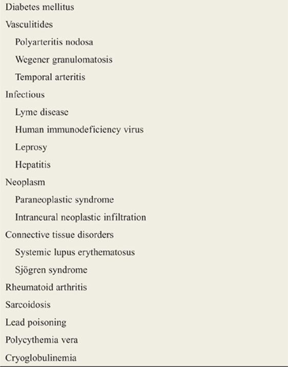
Diabetes is the most common cause, but it is also associated with other systemic diseases (Table 148-2).
TABLE 148-2 Etiologies of Mononeuritis Multiplex
PLEXOPATHIES
BRACHIAL PLEXOPATHY
Brachial plexopathies typically cause weakness, then pain, and paresthesias in the distribution of the affected nerves.
Common causes include trauma (penetrating, humeral neck fracture, or dislocation), shoulder dislocation, neoplasm (Pancoast tumor), radiation, or surgery.
LUMBAR PLEXOPATHY
Plexopathy of the lumbar portion of the plexus causes weakness of hip adduction and flexion and knee extension, decreased sensation at the top and inner thigh, and decreased patellar reflexes.
Lesions of the sacral portion of the plexus cause inability to abduct the thigh, weakness of hip extension and knee flexion, and decreased sensation of the back of the thigh and below the knee.
The most common causes are radiation, diabetic amyo-trophy, aortic aneurysm, retroperitoneal hemorrhage, or compression from arteriovenous malformations.
Imaging may be useful to determine the etiology.
Treatment is directed at the underlying cause.
HIV-ASSOCIATED PERIPHERAL NEUROLOGIC DISEASE
HIV infection and its complications and treatments cause a variety of peripheral nerve disorders.
The most common, drug-induced, and HIV neuropathies are chronic and do not cause acute symptoms.
Patients with HIV have a high rate of mononeuritis multiplex and a myopathy resembling polymyositis.
In early infection, they are more prone to Guillain–Barré syndrome.
In the latter stages of AIDS, they may develop cytomegalovirus (CMV) radiculitis, with acute weakness, primarily lower extremity involvement, and variable bowel or bladder dysfunction.
Primarily, lower extremity weakness and hypore-flexia, as well as sensory deficits, are seen. Rectal tone may be decreased.
MRI (indicated to exclude mass lesion) shows swelling and clumping of the cauda equina.
Admission is required for CMV radiculitis; treatment, which should precede definitive diagnosis, consists of ganciclovir.
DIABETIC PERIPHERAL NEUROPATHY
Half of patients with diabetes have symptoms of neuropathy; 15% require treatment for their symptoms.
The most common manifestation of diabetic peripheral neuropathy is a distal symmetric polyneuropathy with a typical stocking and glove distribution.
Non-healing wounds resulting from impaired sensation due to diabetic peripheral neuropathy are the most common cause of nontraumatic amputation.
Glycemic control and neuropathy are correlated.
ED treatment is focused on management of symptoms.
For further reading in Tintinalli’s Emergency Medicine: A Comprehensive Study Guide, 7th ed., see Chapter 166, “Acute Peripheral Neurologic Lesions,” by Phillip Andrus and Andy Jagoda.