
Approach to Diagnosis
Functional Anatomy of the Somatic Sensory Pathways
History
Sensory Examination
Primary Sensory Modalities
Complex Sensory Functions
Sensory Changes & Their Significance
Peripheral Nerve Lesions
Root Lesions
Cord Lesions
Brainstem Lesions
Thalamic Lesions
Lesions of the Sensory Cortex
Distinction Between Organic & Psychogenic Sensory Disturbances
Peripheral Nerve Lesions
Classification
Mononeuropathy Simplex
Mononeuropathy Multiplex
Polyneuropathy
Clinical Findings
Sensory Disturbances
Motor Deficits
Tendon Reflexes
Autonomic Disturbances
Enlarged Nerves
Evaluation of Patients
Time Course
Age at Onset
Occupational History
Medical History
Drug & Alcohol History
Family History
Differential Diagnosis
Investigative Studies
Treatment
Disease-Specific Treatment
Ventilatory Assistance
Trauma Prevention
Pain Relief
Autonomic Disturbances
Polyneuropathies
Idiopathic Inflammatory Neuropathies
Acute Idiopathic Polyneuropathy (Guillain-Barré Syndrome)
Chronic Inflammatory Demyelinating Polyneuropathy
Metabolic & Nutritional Neuropathies
Diabetes Mellitus
Other Endocrinopathies
Uremia
Liver Disease
Vitamin B12 Deficiency
Infective & Granulomatous Neuropathies
AIDS
Leprosy
Diphtheria
Sarcoidosis
Sepsis & Multiorgan Failure
Neuropathies in Vasculitis & Collagen Vascular Disease
Systemic Necrotizing Vasculitis
Wegener Granulomatosis
Giant Cell Arteritis
Rheumatoid Arthritis
Systemic Lupus Erythematosus
Sjögren Syndrome
Scleroderma & Mixed Connective-Tissue Disease
Neoplastic & Paraproteinemic Neuropathies
Compression & Infiltration by Tumor
Paraneoplastic Syndromes
Paraproteinemias
Amyloidosis
Drug-Induced & Toxic Neuropathies
Alcoholism
Other Drugs
Toxins
Hereditary Neuropathies
Hereditary Motor & Sensory Neuropathies
Hereditary Sensory & Autonomic Neuropathies
Amyloidosis
Friedreich Ataxia
Hereditary Neuropathy with Liability to Pressure Palsies
Metabolic Disorders
Entrapment Neuropathies
Upper Limb Entrapment Syndromes
Median Nerve Compression
Interdigital Neuropathy
Ulnar Neuropathy
Radial Neuropathy
Thoracic Outlet Syndrome
Lower Limb Entrapment Syndromes
Fibular (Peroneal) Neuropathy
Femoral Neuropathy
Saphenous Neuropathy
Lateral Femoral Cutaneous Neuropathy
Obturator Neuropathy
Root & Plexus Lesions
Compressive & Traumatic Lesions
Tabes Dorsalis
Lyme Disease
Myelopathies
Syringomyelia
Subacute Combined Degeneration
Cerebral Disorders
Pain Syndromes
Peripheral Nerve Pain
Radicular Pain
Thalamic Pain
Back & Neck Pain
Low Back Pain
Neck Pain
Herpes Zoster (Shingles)
References
APPROACH TO DIAGNOSIS
An appreciation of the functional anatomy of the sensory components of the nervous system is essential for properly interpreting the history and clinical signs of patients with disorders of somatic sensation.As used here, the term includes sensations of touch or pressure, vibration, joint position, pain, temperature, and more complex functions that rely on these primary sensory modalities (eg, two-point discrimination, stereognosis, graphesthesia); it excludes special senses such as smell, vision, taste, and hearing.
FUNCTIONAL ANATOMY OF THE SOMATIC SENSORY PATHWAYS
The sensory pathway between peripheral tissues (eg, skin or joints) and the cerebral cortex involves three neurons and two central synapses (Figure 10-1).

Figure 10-1. Sensory pathways conveying touch, pressure, vibration, joint position, pain, and temperature sensation. (Reproduced with permission from Barrett KE, Barman SM, Boitano S, Brooks H. Ganong’s Review of Medical Physiology. 23rd ed. New York, NY: McGraw-Hill; 2010.)
First-order sensory neurons from the limbs and trunk have cell bodies in the dorsal root ganglia. Each of these neurons sends a peripheral process that terminates in a free nerve ending or encapsulated sensory receptor and a central process that enters the spinal cord. Sensory receptors are relatively specialized for particular sensations and, in addition to free nerve endings (pain), include Meissner corpuscles, Merkel corpuscles, and hair cells (touch); Krause end-bulbs (cold); and Ruffini corpuscles (heat). First-order sensory neurons synapse centrally at a site that depends on the type of sensation. Fibers mediating touch, pressure, or postural sensation in the limbs and trunk ascend in the posterior columns of the spinal cord to the medulla, where they synapse in the gracile and cuneate nuclei. Other fibers that mediate touch and those subserving pain and temperature appreciation in the limbs and trunk synapse on neurons in the posterior horns of the spinal cord, particularly in the substantia gelatinosa. First-order sensory neurons from the face, which have cell bodies in the trigeminal (gasserian) ganglion, travel in the trigeminal (V) nerve and enter the pons. Fibers mediating facial touch and pressure synapse in the main trigeminal (V) nerve sensory nucleus, whereas those conveying facial pain and temperature synapse in the spinal trigeminal (V) nerve nucleus.
Second-order sensory neurons with cell bodies in the gracile and cuneate nuclei cross the midline and ascend in the medial lemniscus. Second-order sensory neurons that arise in the posterior horns of the spinal cord cross the mid-line and ascend in the anterolateral part of the cord: fibers mediating touch pass upward in the anterior spinothalamic tract, whereas pain and temperature fibers generally travel in the lateral spinothalamic tract. Second-order sensory neurons from the limbs and trunk are joined in the brainstem by fibers from the face: those that mediate facial touch and pressure sensation project from the main trigeminal (V) nerve sensory nucleus via the trigeminal lemniscus, and those that convey facial pain and temperature project from the spinal trigeminal (V) nerve nucleus via the trigeminothalamic tract, to the ipsilateral thalamus. In the thalamus, medial lemniscal and spinothalamic fibers synapse in the ventral posterolateral (VPL) nucleus; spinothalamic fibers also synapse in the ventral posteroinferior (VPI) nucleus and intralaminar (ILa) nuclei; and fibers in the trigeminal lemniscus and trigeminothalamic tract synapse in the ventral posteromedial (VPM) nucleus. In addition, some second-order spinothalamic sensory neurons send collaterals to the reticular formation.
Third-order sensory neurons project from the thalamus to ipsilateral cerebral cortex. Fibers from VPL, VPI, and VPM travel primarily to primary somatosensory cortex in the postcentral gyrus; fibers from ILa also project to striatum, cingulate gyrus, and prefrontal cortex.
HISTORY
Sensory disturbances may consist of loss of sensation, abnormal sensations, or pain. The term paresthesia is used to denote abnormal spontaneous sensations, such as burning, tingling, or pins and needles. The designation dysesthesia denotes any unpleasant sensation produced by a stimulus that is normally painless. The term numbness is often used by patients to describe a sense of heaviness, weakness, or deadness in the affected part of the body—and sometimes to signify any sensory impairment; the meaning must be clarified whenever this word is used.
In obtaining a history of sensory complaints, it is important to determine the location of the symptoms; the mode of onset and progression of the symptoms; whether the symptoms are constant or episodic in nature; whether any factors specifically produce, enhance, or relieve symptoms; and whether there are any accompanying symptoms.
The location of symptoms may provide a clue to their origin. For example, sensory disturbances involving all the limbs suggest peripheral neuropathy, a cervical cord or brainstem lesion, or a metabolic disturbance such as hyperventilation syndrome. Involvement of one entire limb, or of one side of the body, suggests a central (brain or spinal cord) lesion. A hemispheric or brainstem lesion may lead to lateralized sensory symptoms, but the face is also commonly affected. In addition, there may be other symptoms and signs, such as aphasia, apraxia, and visual field defects with hemispheric disease, or dysarthria, weakness, vertigo, diplopia, disequilibrium, and ataxia with brainstem disorders. Involvement of part of a limb or a discrete region of the trunk raises the possibility of a nerve or root lesion, depending on the precise distribution. With a root lesion, symptoms may show some relationship to neck or back movements, and pain is often conspicuous.
The course of sensory complaints provides a guide to their cause. Intermittent or repetitive transient symptoms may represent sensory seizures, ischemic phenomena, or metabolic disturbances such as those accompanying hyper-ventilation. Intermittent localized symptoms that occur at a consistent time may suggest the diagnosis or an exogenous precipitating factor. For example, the pain and paresthesias of carpal tunnel syndrome (median nerve compression at the wrist) characteristically occur at night and awaken the patient from sleep.
SENSORY EXAMINATION
In the investigation of sensory complaints, various modalities are tested in turn, and the distribution of any abnormality is plotted with particular reference to the normal root and peripheral nerve territories. Complete loss of touch appreciation is anesthesia, partial loss is hypesthesia, and increased sensitivity is hyperesthesia. The corresponding terms for pain appreciation are analgesia, hypalgesia, and hyperalgesia or hyperpathia; allodynia refers to the misperception of a trivial tactile sensation as pain.
PRIMARY SENSORY MODALITIES
Light Touch
The appreciation of light touch is evaluated with a wisp of cotton wool, which is brought down carefully on a small region of skin. The patient lies quietly, with the eyes closed, and makes a signal each time the stimulus is felt. The appreciation of light touch depends on fibers that traverse the posterior column of the spinal cord in the gracile (leg) and cuneate (arm) fasciculi ipsilaterally (Figures 10-1 and 10-2), passing to the medial lemniscus of the brainstem (Figure 10-3), and on fibers in the contra-lateral anterior spinothalamic tract.

Figure 10-2. Location and lamination of sensory pathways in the spinal cord. C (cervical), T (thoracic), L (lumbar), and S (sacral) indicate the level of origin of fibers within each tract.


Figure 10-3. Sensory pathways in the brainstem. In the medulla, spinothalamic fibers conveying pain and temperature sensation are widely separated from medial lemniscal fibers mediating touch and pressure; these pathways converge as they ascend in the pons and midbrain.
Pinprick & Temperature
Pinprick appreciation is tested by asking the patient to indicate whether the point of a pin (not a hypodermic needle, which is likely to puncture the skin and draw blood) feels sharp or blunt. The pin should be discarded after use, and it should be handled with care because it is a potential source of infection. Appreciation of pressure or touch by the pinpoint must not be confused with the appreciation of sharpness. Temperature appreciation is evaluated by application to the skin of containers of hot or cold water. For convenience, cold can be tested by application of the side of a metal tuning fork to different bodily regions, and testing of heat is usually omitted from the routine examination. Pinprick and temperature appreciation depend on the integrity of the lateral spinothalamic tracts (Figures 10-1 and 10-2). The afferent fibers cross in front of the central canal after ascending for two or three segments from their level of entry into the cord.
Deep Pressure
Deep pressure sensibility is evaluated by pressure on the tendons, such as the Achilles tendon at the ankle.
Vibration
Vibration sense is evaluated with a tuning fork (128 Hz) that is set in motion and then placed over a bony prominence; the patient is asked to indicate whether vibration, rather than simple pressure, is felt. Many healthy elderly patients have impaired appreciation of vibration below the knees.
Joint Position
Joint position sense is tested by asking the patient to indicate the direction of small passive movements of the terminal interphalangeal joints of the fingers and toes. Patients with severe impairment of joint position sense may exhibit slow, continuous movement of the fingers (pseudoathetoid movement) when attempting to hold the hands outstretched with the eyes closed. For clinical purposes, both joint position sense and the ability to appreciate vibration are considered to depend on fibers carried in the posterior columns of the cord, although there is evidence that this is not strictly true for vibration.
COMPLEX SENSORY FUNCTIONS
Romberg Test
The patient is asked to assume a steady stance with feet together, arms outstretched, and eyes closed and is observed for any tendency to sway or fall. The test is positive (abnormal) if unsteadiness is markedly increased by eye closure—as occurs, for example, in tabes dorsalis. A positive test is indicative of grossly impaired joint position sense in the legs.
Two-Point Discrimination
The ability to distinguish simultaneous touch at two neighboring points depends on the integrity of the central and peripheral nervous system, the degree of separation of the two points, and the part of the body that is stimulated. The patient is required to indicate whether he or she is touched by one or two compass points, while the distance between the points is varied in order to determine the shortest distance at which they are recognized as different points. The threshold for two-point discrimination approximates 4 mm at the fingertips and may be several centimeters on the back. When peripheral sensory function is intact, impaired two-point discrimination suggests a disorder affecting the sensory cortex.
Graphesthesia, Stereognosis, & Barognosis
Agraphesthesia, the inability to identify a number traced on the skin of the palm of the hand despite normal cutaneous sensation, implies a lesion involving the contralateral parietal lobe. The same is true of inability to distinguish between various shapes or textures by touch (astereognosis) or impaired ability to distinguish between different weights (abarognosis).
Bilateral Sensory Discrimination
In some patients with apparently normal sensation, simultaneous stimulation of the two sides of the body reveals an apparent neglect of (or inattention to) sensation from one side, usually because of an underlying contralateral cerebral lesion.
SENSORY CHANGES & THEIR SIGNIFICANCE
It is important to determine the nature and distribution of any sensory change. Failure to find clinical evidence of sensory loss in patients with sensory symptoms must never be taken to imply that the symptoms necessarily have a psychogenic basis. Sensory symptoms often develop well before the onset of sensory signs.
PERIPHERAL NERVE LESIONS
Mononeuropathy
In patients with a lesion of a single peripheral nerve, sensory loss is usually less than would have been predicted on anatomic grounds because of overlap from adjacent nerves. Moreover, depending on the type of lesion, the fibers in a sensory nerve may be affected differently. Compressive lesions, for example, tend to affect preferentially the large fibers that subserve touch.
Polyneuropathy
In patients with polyneuropathies, sensory loss is generally symmetric and is greater distally than proximally—as suggested by the term stocking-and-glove sensory loss or length-dependent neuropathy. As a general rule, the loss will have progressed almost to the knees before the hands are affected. Sensory loss may then be accompanied by a motor deficit and reflex changes. Diabetes mellitus, amyloidosis, and certain other metabolic disorders (eg, Tangier disease, a recessive trait characterized by the near absence of high-density lipoproteins) preferentially involve small nerve fibers that subserve pain and temperature appreciation; in a pure small-fiber sensory neuropathy, the tendon reflexes are unaffected and no motor deficit occurs.
ROOT LESIONS
Nerve root involvement produces impairment of cutaneous sensation in a segmental pattern (Figure 10-4), but because of overlap there is generally no loss of sensation unless two or more adjacent roots are affected. Pain is often a conspicuous feature in patients with compressive root lesions. Depending on the level affected, there may be loss of tendon reflexes (C5-C6, biceps and brachioradialis; C7-C8, triceps; L3-L4, knee; S1, ankle), and if the anterior roots are also involved, there may be weakness and muscle atrophy.


Figure 10-4. A: Cutaneous innervation (anterior view). The segmental or radicular (nerve root) distribution is shown on the left side of the body, and the peripheral nerve distribution on the right side of the body. B: Cutaneous innervation (posterior view). The segmental or radicular (nerve root) distribution is shown on the left side of the body, and the peripheral nerve distribution on the right side of the body. For details of radial, median, ulnar, fibular (peroneal), and femoral nerves, see Appendix.
CORD LESIONS
In patients with a cord lesion, there may be a transverse sensory level. Physiologic areas of increased sensitivity do occur, however, at the costal margin, over the breasts, and in the groin, and these must not be taken as abnormal. Therefore, the level of a sensory deficit affecting the trunk is best determined by careful sensory testing over the back rather than the chest and abdomen.
Central Cord Lesion
With a central cord lesion, such as occurs in syringomyelia, after trauma, and with certain cord tumors, there is characteristically a loss of pain and temperature appreciation with sparing of other modalities. This loss is due to the interruption of fibers conveying pain and temperature that cross from one side of the cord to the spinothalamic tract on the other. Such a loss is usually bilateral, may be asymmetric, and involves only the fibers of the involved segments. It may be accompanied by lower motor neuron weakness in the muscles supplied by the affected segments and sometimes by a pyramidal and posterior column deficit below the lesion (Figure 10-5).

Figure 10-5. Central cord lesions (blue) of moderate (A) or marked (B) extent. Less extensive lesions impair pain and temperature appreciation by interrupting incoming sensory fibers as they cross to the contralateral spinothalamic tract; involvement of anterior horn cells causes lower motor neuron weakness. These deficits are restricted to dermatomes and muscles innervated by the involved spinal cord segments. More extensive lesions also produce disturbances of touch, pressure, vibration, and joint position sense because of involvement of the posterior columns and cause pyramidal signs because of corticospinal tract involvement, especially affecting the arms (see lamination of corticospinal tract in Figure 9-6). These deficits occur below the level of the lesion.
Anterolateral Cord Lesion
Lesions involving the anterolateral portion of the spinal cord (lateral spinothalamic tract) can cause contralateral impairment of pain and temperature appreciation in segments below the level of the lesion. The spinothalamic tract is laminated, with fibers from the sacral segments the outermost. Intrinsic cord (intramedullary) lesions often spare the sacral fibers, whereas extramedullary lesions, which compress the cord, tend to involve these fibers as well as those arising from more rostral levels.
Anterior Cord Lesion
With destructive lesions involving predominantly the anterior portion of the spinal cord, pain and temperature appreciation are impaired below the level of the lesion from lateral spinothalamic tract involvement. In addition, weakness or paralysis of muscles supplied by the involved segments of the cord results from damage to motor neurons in the anterior horn. With more extensive disease, involvement of the corticospinal tracts in the lateral funiculi may cause a pyramidal deficit below the lesion. There is relative preservation of posterior column function (Figure 10-6). Ischemic myelopathies caused by occlusion of the anterior spinal artery take the form of anterior cord lesions.

Figure 10-6. Anterior cord lesion (blue) associated with occlusion of the anterior spinal artery. Clinical features are similar to those seen with severe central cord lesions (Figure 10-5B), except that posterior column sensory functions are spared and the defect in pain and temperature sensation extends to sacral levels.
Posterior Column Lesion
A patient with a posterior column lesion may complain of a tight or bandlike sensation in the regions corresponding to the level of spinal involvement and sometimes also of paresthesias (like electric shocks) radiating down the extremities on neck flexion (Lhermitte sign). There is loss of vibration and joint position sense below the level of the lesion, with preservation of other sensory modalities. The deficit may resemble that resulting from involvement of large fibers in the posterior roots.
Cord Hemisection
Lateral hemisection of the cord leads to Brown-Séquard syndrome. Below the lesion, there is an ipsilateral pyramidal deficit and disturbed appreciation of vibration and joint position sense, with contralateral loss of pain and temperature appreciation that begins two or three segments below the lesion (Figure 10-7). Hyperalgesia and spontaneous pain are sometimes prominent ipsilaterally.

Figure 10-7. Cord lesion (blue) in Brown-Séquard syndrome. Hemisection of the cord causes ipsilateral pyramidal dysfunction and impairment of posterior column sensory function below the level of the lesion, and contralateral impairment of pain and temperature sensation with an upper limit slightly below the level of the lesion.
BRAINSTEM LESIONS
Sensory disturbances may be accompanied by a motor deficit, cerebellar signs, and cranial nerve palsies when the lesion is in the brainstem.
In patients with lesions involving the spinothalamic tract in the dorsolateral medulla and pons, pain and temperature appreciation are lost in the limbs and trunk on the opposite side of the body. When such a lesion is located in the medulla, it also typically involves the spinal trigeminal nucleus, impairing pain and temperature sensation on the same side of the face as the lesion. The result is a crossed sensory deficit that affects the ipsilateral face and contralateral limbs. In contrast, spinothalamic lesions above the spinal trigeminal nucleus affect the face, limbs, and trunk contralateral to the lesion. With lesions affecting the medial lemniscus, there is loss of touch and proprioception on the opposite side of the body. In the upper brainstem, the spinothalamic tract and medial lemniscus run together so that a single lesion may cause loss of all superficial and deep sensation over the contralateral side of the body (Figure 10-3).
THALAMIC LESIONS
Thalamic lesions may lead to loss or impairment of all forms of sensation on the contralateral side of the body, and this may have a distribution that differs from the area of symptomatic involvement. Spontaneous pain, sometimes with a particularly unpleasant quality, may occur on the affected side. Patients may describe it as burning, tearing, knifelike, or stabbing, but often have difficulty characterizing it. Any form of cutaneous stimulation can lead to painful or unpleasant sensations. Such a thalamic syndrome (Dejerine-Roussy syndrome) can also occasionally result from lesions of the white matter of the parietal lobe or from cord lesions, as discussed later.
LESIONS OF THE SENSORY CORTEX
Disease limited to the sensory cortex impairs discriminative sensory function on the opposite side of the body. Thus patients may be unable to localize stimuli on the affected side or to recognize the position of different parts of the body. They may not be able to recognize objects by touch or to estimate their size, weight, consistency, or texture. Cortical sensory disturbances are usually more conspicuous in the hands than in the trunk or proximal portions of the limbs.
DISTINCTION BETWEEN ORGANIC & PSYCHOGENIC SENSORY DISTURBANCES
Psychogenic disturbances of sensation may be associated with such psychiatric disturbances as conversion disorder. They may take any form but most often are restricted to loss of cutaneous sensation. There may be several characteristic features.
Nonorganic sensory loss does not conform in its distribution to any specific neuroanatomic pattern. It may surround a bony landmark or involve an area defined by surface landmarks rather than innervation. Indeed, it is not uncommon for there to be an apparent loss of sensation in one or more extremities, with the margin occurring circumferentially in the axilla or groin; organic sensory loss with such a margin is unusual. Organic peripheral sensory loss over the trunk or face does not usually extend to the midline but stops 3 to 5 cm before it, because of overlap in the innervation on the two sides; with nonorganic disturbances, apparent sensory loss commonly stops precisely at the midline.
There is often a sudden transition between areas of nonorganic sensory loss and areas with normal sensation. By contrast, with organic disturbances, there is usually an area of altered sensation between insensitive areas and adjacent areas with normal sensibility.
In nonorganic disturbances, there may be a dissociated loss that is difficult to interpret on an anatomic basis. For example, there may be a total loss of pinprick appreciation but preserved temperature sensation. Moreover, despite the apparent loss of posterior column function, the patient may be able to walk normally or maintain the arms outstretched without difficulty or pseudoathetoid movements.
In nonorganic sensory disturbances, appreciation of vibration may be impaired on one side but not the other side of a bony midline structure, such as the skull or sternum. The vibrations are in fact conducted to both sides by the bone, so that even if there is a hemisensory disturbance, the vibrations are appreciated on either side in patients with organic sensory disorders.
Finally, it should be noted that sensory disturbances are often suggested to the patient by the examiner’s own expectations. Such findings can be particularly misleading because they may be neuroanatomically correct. One helpful approach is to have the patient outline on the body the extent of any perceived sensory disturbance before formal sensory testing is undertaken.
PERIPHERAL NERVE LESIONS
Sensory symptoms are usually a conspicuous feature in patients with peripheral nerve lesions (Table 10-1). Sensory impairment may be in a distal stocking-and-glove pattern in patients with polyneuropathies or may follow the pattern of individual peripheral nerves in patients with mononeuropathies (Figure 10-4).


Table 10-1. Causes of peripheral neuropathy.
CLASSIFICATION
MONONEUROPATHY SIMPLEX
This term signifies involvement of a single peripheral nerve.
MONONEUROPATHY MULTIPLEX
In this disorder, several individual nerves are affected, usually at random and noncontiguously. Clinical examination reveals a clinical deficit attributable to involvement of one or more isolated peripheral nerves, except when mononeuropathy multiplex is extensive and the resulting deficits become confluent.
POLYNEUROPATHY
The term polyneuropathy denotes a disorder in which the function of numerous peripheral nerves is affected at the same time. This leads to a predominantly distal and symmetric deficit, with loss of tendon reflexes except when small fibers are selectively involved. Polyneuropathies are sometimes subclassified according to the primary site at which the nerve is affected.
In distal axonopathies (axonal neuropathies), the axon is the principal pathologic target; most polyneuropathies fall into this category.
Myelinopathies (demyelinating neuropathies) are conditions that involve the myelin sheath surrounding the axon. These disorders include acute idiopathic polyneuropathy (Guillain-Barré syndrome), chronic inflammatory demyelinating neuropathy, diphtheria, certain paraneoplastic and paraproteinemic states, and various hereditary conditions including metachromatic leukodystrophy, Krabbe disease, and types 1 and 3 Charcot-Marie-Tooth hereditary motor and sensory neuropathy (CMT-1 and 3).
Finally, certain disorders, termed neuronopathies, principally affect nerve cell bodies in the anterior horn of the spinal cord or dorsal root ganglion. Examples are type 2 CMT hereditary motor and sensory neuropathy, pyridoxine-induced neuropathy, and some paraneoplastic syndromes.
CLINICAL FINDINGS
SENSORY DISTURBANCES
Involvement of sensory fibers can lead to numbness and impaired sensation; abnormal spontaneous sensations, such as pain and paresthesias; and perverted sensations such as hyperpathia (amplified response to a normally painful stimulus).
Pain
Pain is a conspicuous feature of certain neuropathies, especially if small fibers within the nerves are affected. The precise mechanism of its genesis is unclear. Polyneuropathies associated with prominent pain include those related to diabetes, alcoholism, porphyria, Fabry disease, amyloidosis, rheumatoid arthritis, and acquired immunodeficiency syndrome (AIDS), as well as dominantly inherited sensory neuropathy and paraneoplastic sensory neuronopathy. Pain is also a feature of many entrapment neuropathies and of idiopathic brachial plexopathy.
Dissociated Sensory Loss
Dissociated sensory loss is impairment of some sensory modalities, such as pain and temperature, with preservation of others, such as light touch, vibration, and joint position sense. Although the presence of a dissociated sensory loss often indicates a spinal cord lesion, it also occurs in peripheral neuropathies when there is selective involvement of peripheral nerve fibers of a certain size, such as occurs in amyloid neuropathy, leprous neuritis, or hereditary sensory neuropathy. Small fiber disease is commonly associated with disproportionate impairment of pain and temperature appreciation, spontaneous pain, and autonomic dysfunction. Large fiber disease, by contrast, results in defective touch, vibration, and joint position sense, early loss of tendon reflexes, and prominent motor symptoms.
MOTOR DEFICITS
The motor deficit that occurs with a peripheral nerve lesion consists of weakness of muscles innervated by the nerve, accompanied in severe cases by wasting and fasciculation. There may be difficulty in the performance of fine tasks; this is compounded by any accompanying sensory loss. The clinical findings reflect a lower motor neuron deficit, and it is the distribution of these signs and the presence of accompanying sensory and reflex changes that suggest they may be due to peripheral nerve involvement.
TENDON REFLEXES
These are impaired or lost if reflex arcs are interrupted on either the afferent or efferent side (C5-C6, biceps and brachioradialis; C7-C8, triceps; L3-L4, knee; S1, ankle). The ankle reflexes are usually the first to be lost in patients with polyneuropathies, but may also be absent in healthy elderly subjects.
AUTONOMIC DISTURBANCES
Autonomic disturbances may be particularly conspicuous in some peripheral neuropathies—especially Guillain-Barré syndrome and neuropathies related to diabetes, renal failure, porphyria, certain paraneoplastic disorders, or amyloidosis. Symptoms include postural hypotension, coldness of the extremities, impaired thermoregulatory sweating, disturbances of bladder and bowel function, and impotence.
ENLARGED NERVES
Palpably enlarged peripheral nerves raise the possibility of leprosy, amyloidosis, hereditary motor and sensory neuropathies, Refsum disease, acromegaly, or chronic inflammatory demyelinating polyneuropathy.
EVALUATION OF PATIENTS
TIME COURSE
Polyneuropathy that develops acutely over a few days usually relates to an inflammatory process, as in the Guillain-Barré syndrome. It may also relate to an underlying neoplasm, to infections such as diphtheria, to metabolic disorders such as acute intermittent porphyria, or to exposure to such toxic substances as thallium or triorthocresyl phosphate. A chronic course with a gradual evolution over several years is typical of many hereditary or metabolic polyneuropathies but also characterizes chronic inflammatory demyelinating polyneuropathy.
Mononeuropathy of acute onset is likely to be traumatic or ischemic in origin, whereas one evolving gradually is more likely to relate to entrapment (ie, compression by neighboring anatomic structures) or to recurrent minor trauma.
AGE AT ONSET
Polyneuropathy that develops during childhood or early adult life often has a hereditary basis, but may also relate to an underlying inflammatory disorder. Polyneuropathy developing in later life is more likely to result from a metabolic, toxic, or inflammatory disorder, or from an underlying neoplasm.
Mononeuropathy presenting in the neonatal period is likely to be developmental in origin or related to birth injury; one developing in later life may relate to entrapment or injury that is often occupationally determined.
OCCUPATIONAL HISTORY
Various industrial toxins can lead to peripheral neuropathy, including carbon disulfide, n-hexane, ethylene oxide, methyl bromide, acrylamide, triorthocresyl phosphate and certain other organophosphates, DDT, arsenic, lead, and thallium. A mononeuropathy is sometimes the first clinical manifestation of an occupationally related polyneuropathy, but it may also develop in response to entrapment or recurrent minor occupational trauma. For example, carpal tunnel syndrome is more common in persons who do heavy manual labor or develop repetitive motion injury as a result of computer terminal use, and a lesion of the deep palmar branch of the ulnar nerve may relate to repeated pressure on the palm of the hand by, for example, punching down heavily on a stapler or using heavy equipment such as a pneumatic road drill.
MEDICAL HISTORY
Metabolic Disorders
Peripheral neuropathy may relate to metabolic disorders such as diabetes mellitus, uremia, liver disease, myxedema, acromegaly, metachromatic leukodystrophy, or Fabry disease. That caused by diabetes is especially important and may take the form of an entrapment mononeuropathy, acute ischemic mononeuropathy, distal sensorimotor polyneuropathy, subacute proximal motor polyradiculopathy (diabetic amyotrophy), thoracoabdominal radiculopathy, or autonomic neuropathy.
Neoplasm
A peripheral neuropathy may also relate to an underlying malignant neoplasm. The peripheral nerves, spinal nerves, and limb plexuses may be compressed or infiltrated by extension of primary tumors or metastatic lymph nodes. Neoplastic disease can also lead to a nonmetastatic (paraneoplastic) sensory or sensorimotor polyneuropathy or to Lambert-Eaton syndrome, a disorder of neuromuscular transmission discussed in Chapter 9.
Connective Tissue Disorders
Certain connective tissue disorders, especially polyarteritis nodosa, rheumatoid arthritis, Churg-Strauss syndrome, and Wegener granulomatosis, may be associated with mononeuropathy multiplex or, less commonly, polyneuropathy, or cranial neuropathy. Polyneuropathy is more common in systemic lupus erythematosus. Patients with rheumatoid arthritis are particularly likely to develop focal entrapment or compressive mononeuropathies in the vicinity of the affected joints.
AIDS
Acquired immune deficiency syndrome (AIDS) is commonly associated with a distal, symmetric, primarily sensory polyneuropathy. Peripheral nerve involvement in AIDS less frequently takes the form of an acute or chronic inflammatory demyelinating polyneuropathy, polyradiculopathy, mononeuropathy multiplex, or autonomic neuropathy. Neuropathies are also seen in patients with asymptomatic human immunodeficiency virus-1 (HIV-1) infection and HIV-1 seroconversion.
DRUG & ALCOHOL HISTORY
Some of the drugs that cause peripheral neuropathy are listed in Table 10-2; there may be selective involvement of motor or sensory fibers with some drugs.

Table 10-2. Selected drugs inducing peripheral neuropathy.
FAMILY HISTORY
Certain polyneuropathies have a hereditary basis. These are discussed later in this chapter in the section on hereditary neuropathies.
DIFFERENTIAL DIAGNOSIS
Peripheral neuropathies can lead to a motor or sensory deficit or both. The preservation of sensation and tendon reflexes distinguishes the motor deficit that results from pure pyramidal lesions or is associated with spinal muscular atrophies, myopathies, or disorders of neuromuscular transmission from that caused by peripheral nerve involvement. Other distinguishing features are discussed in Chapter 9.
Myelopathies are characterized by a pyramidal deficit below the level of the lesion as well as by distal sensory loss. In tabes dorsalis, there is often a history of syphilitic infection, and examination reveals other stigmas of syphilis. In addition, tactile sensation is preserved.
Radiculopathies are distinguished from peripheral neuropathies by the distribution of motor or sensory deficits (Figure 10-4). The presence of neck or back pain that radiates to the extremities in a radicular distribution also suggests a root lesion.
INVESTIGATIVE STUDIES
Laboratory studies in patients with peripheral neuropathy are directed at confirming the diagnosis and revealing any underlying cause.
Electromyography may reveal evidence of denervation in the affected muscles and can be used to determine whether any motor units remain under voluntary control. Nerve conduction studies permit conduction velocity to be measured in motor and sensory fibers. On the basis of electro-diagnostic or histopathologic studies, peripheral neuropathies can be divided into demyelinating or axonal neuropathies. In demyelinating neuropathies, electro-myography typically reveals little or no evidence of denervation, but there is conduction block or marked slowing of maximal conduction velocity in affected nerves. In axonal neuropathies, electromyography shows that denervation has occurred, especially distally in the extremities, but maximal nerve conduction velocity is normal or slowed only slightly.
Laboratory studies that may help to identify the cause of a peripheral nerve disorder include a complete blood count, erythrocyte sedimentation rate, serum urea nitrogen and creatinine, fasting blood glucose, serum vitamin B12, serum protein, protein electrophoresis and immuno-electrophoresis, liver and thyroid function blood tests, serologic test for syphilis (FTA or MHA-TP), rheumatoid factor, antinuclear antibody, and chest x-ray. Depending on the clinical circumstances, serologic tests for Lyme disease, hepatitis, infection with HIV, or paraneoplastic antibodies may be required. Genetic studies may also be necessary after appropriate genetic counseling.
If toxic causes are suspected, a 24-hour urine collection followed by analysis for heavy metals may be necessary, and hair and fingernail clippings can be analyzed for arsenic. Examination of a fresh specimen of urine for porphobilinogen and δ-aminolevulinic acid is necessary if porphyria is suspected.
TREATMENT
DISEASE-SPECIFIC TREATMENT
Treatment of the underlying cause may limit the progression of or even reverse the neuropathy. Disease-specific treatments are discussed later under individual disorders.
VENTILATORY ASSISTANCE
Respiratory function must be monitored carefully—particularly in acute idiopathic polyneuropathy (Guillain-Barré syndrome), chronic inflammatory demyelinating polyneuropathy, and diphtheritic neuropathy—and preparations must be made to assist ventilation if the forced vital capacity reaches 15 mL/kg, the mean inspiratory force reaches –40 mm Hg, dyspnea becomes evident, or the oxygen saturation of arterial blood declines.
TRAUMA PREVENTION
Nursing care is important in patients with severe motor or sensory deficits to prevent decubitus ulcers, joint contractures, and additional compressive peripheral nerve damage. In patients with severe dysesthesia, a cradle (inverted metal bar frame) can be used to keep the bedclothes from touching sensitive areas of the skin.
Extremities with sensory loss must be protected from repeated minor trauma, such as thermal injury, that can destroy tissues. The temperature of hot surfaces should be checked with a part of the body in which sensation is preserved, and the setting of water heaters must be reduced to prevent scalding. The skin and nails must be cared for meticulously.
PAIN RELIEF
Patients with sensory disorders may suffer from especially severe pain, which can dramatically impair quality of life. Treatment of this pain should be a high priority.
Duloxetine (60 mg once daily) or venlafaxine (titrated up to 75 mg two to three times daily in standard formulation or given as the equivalent dose once daily in the extended release formulation) is often helpful; both are selective serotonin and norepinephrine reuptake inhibitors.
Pregabalin (150 mg increasing after 1 week to 300 mg daily in divided doses; maximum, 600 mg daily) is also particularly useful for the relief of neuropathic pain.
Phenytoin 300 mg/d, carbamazepine up to 1,200 mg/d, or mexiletine 600 to 900 mg/d can sometimes relieve the lancinating pain of certain neuropathies. If the pain is more constant, burning, or dysesthetic, amitriptyline 25 to 100 mg at bedtime is often helpful as are other tricyclic agents.
Gabapentin (300 mg three times daily, with subsequent increments depending on response and tolerance) is effective in treating various neuropathic pain disorders; pain relief may similarly occur with lamotrigine, topiramate, or sodium valproate, but this has been documented less well. Lamotrigine requires slow dose titration to avoid skin rashes and other complications. Combined gabapentin and nortriptyline was more effective than either drug given alone for neuropathic pain (diabetic neuropathy or post-herpetic neuralgia) in one study, and the use of this combination has therefore been recommended in patients who show a partial response to either drug given alone and require additional pain relief.
Topical capsaicin is also helpful in neuropathic pain syndromes.
AUTONOMIC DISTURBANCES
Dysautonomic symptoms may be troublesome in several polyneuropathies, but especially in diabetic polyneuropathy. Waist-high elastic hosiery, dietary salt supplementation, and treatment with fludrocortisone 0.1 to 1 mg/d orally may help relieve postural hypotension, but the patient must be monitored carefully to prevent recumbent hypertension. Other medications that may be helpful include clonidine, midodrine, dihydroergotamine, octreotide, or beta-blockers. Instructing the patients to sleep in a semierect rather than a recumbent position is helpful because dysautonomic patients are often unable to conserve salt and water when recumbent at night.
POLYNEUROPATHIES
IDIOPATHIC INFLAMMATORY NEUROPATHIES
ACUTE IDIOPATHIC POLYNEUROPATHY (GUILLAIN-BARRÉ SYNDROME)
Guillain-Barré syndrome is an acute or subacute polyneuropathy that can follow minor infective illnesses, inoculations, or surgical procedures or may occur without obvious precipitants. Clinical and epidemiologic evidence suggests an association with preceding Campylobacter jejuni infection. Its precise cause is unclear, but it appears to have an immunologic basis. Both demyelinating and axonal forms have been recognized, with distinctive clinical and electro-physiologic features. The demyelinative form is more common in the United States, but an axonal variant is encountered occasionally (acute motor sensory axonal neuropathy). In northern China, a related axonal form occurs frequently and has been designated acute motor axonal neuropathy.
Clinical Features
The features useful for diagnosing Guillain-Barré syndrome are summarized in Table 10-3. Patients generally present with ascending weakness that is symmetric, usually begins in the legs, is often more marked proximally than distally, and is sometimes so severe that it is life-threatening, especially if the muscles of respiration or swallowing are involved. Muscle wasting develops if axonal degeneration has occurred. Sensory complaints, although usually less marked than motor symptoms, are also frequent. The deep tendon reflexes are typically absent. There may be marked autonomic dysfunction, with tachycardia, cardiac irregularities, labile blood pressure, disturbed sweating, impaired pulmonary function, sphincter disturbances, paralytic ileus, and other abnormalities.

Table 10-3. Diagnostic criteria for Guillain-Barré syndrome.
Investigative Studies
The cerebrospinal fluid (CSF) often shows a characteristic abnormality, termed cytoalbuminologic dissociation, with increased protein concentration but a normal cell count; abnormalities may not be found in the first week, however. Electrophysiologic studies may reveal marked slowing of motor and sensory conduction velocity or evidence of denervation and axonal loss. The time course of the electrophysiologic changes does not necessarily parallel any clinical developments. When HIV-1 infection is suspected because of the clinical context in which the neuropathy has developed or the presence of high-risk factors, appropriate serologic studies should be performed.
Treatment
Plasmapheresis appears to reduce the time required for recovery and may decrease the likelihood of residual neurologic deficits. It is best instituted early, and it is indicated especially in patients with a severe or rapidly progressive deficit or respiratory compromise. Intravenous immunoglobulin (400 mg/kg/d for 5 days) appears to be equally effective and should be used in preference to plasmapheresis in adults with cardiovascular instability and in children; the two therapies are not additive.
Therapy is otherwise symptomatic, the aim being to prevent such complications as respiratory failure or vascular collapse. For this reason, patients who are severely affected are best managed in intensive care units, where facilities are available for monitoring and assisted respiration if necessary (eg, if the forced vital capacity reaches 15 mL/kg, the mean inspiratory force reaches –40 mm Hg, the patient is short of breath, or the blood oxygen saturation declines). Volume replacement or treatment with pressor agents is sometimes required to counter hypotension, and low-dose heparin may help to prevent pulmonary embolism. Corticosteroids may affect the outcome adversely or delay recovery, and are not indicated.
Prognosis
Symptoms and signs cease to progress by approximately 4 weeks into the illness. The disorder is self-limiting, and improvement occurs over the weeks or months after onset. Approximately 70% of patients recover completely, 25% are left with mild neurologic deficits, and 5% die, usually as a result of respiratory failure. The prognosis is poorer when there is evidence of preceding Campylobacter jejuniinfection, and a more protracted course and less complete recovery are also likely when axonal degeneration rather than demyelination is the primary pathology. Advanced age, the need for ventilatory support, or more rapid onset of symptoms may also predict a poorer prognosis.
CHRONIC INFLAMMATORY DEMYELINATING POLYNEUROPATHY
Chronic inflammatory demyelinating polyneuropathy is clinically similar to Guillain-Barré syndrome except that it follows a chronic progressive course, or a course characterized by relapses, and no improvement is apparent within the 6 months after onset. Its cause is not known. Its clinical features are summarized in Table 10-4.


Table 10-4. Clinical features of chronic inflammatory demyelinating polyneuropathy.
Examination of the CSF reveals findings resembling those in Guillain-Barré syndrome. The electrophysiologic findings indicate a demyelinative neuropathy with superimposed axonal degeneration.
The disorder is often responsive to treatment with corticosteroids (prednisone 60-100 mg/d for 2-4 weeks, then gradually tapered to 5-20 mg every other day), which may have to be continued on a long-term basis. Treatment with intravenous immunoglobulin (1 g/kg daily for 2 days with a single additional infusion at 3 weeks, or 400 mg/kg/d for 5 consecutive days for a total of 2 g, with subsequent courses as needed to maintain benefit) is also effective as initial or later therapy. When used as the initial therapy, it has the advantage of fewer side effects (but greater expense) than prednisone. Its precise mode of action is unknown. Plasma exchange is another effective immuno-modulator therapy, but is more difficult to administer. In nonresponsive patients, treatment with azathioprine or cyclophosphamide may be helpful, but claims of benefit from these agents or from cyclosporine, interferon-beta, or interferon-alpha require confirmation by randomized trials.
METABOLIC & NUTRITIONAL NEUROPATHIES
DIABETES MELLITUS
Peripheral nerve involvement in diabetes is common and may be characterized by polyneuropathy, which is of mixed (sensory, motor, and autonomic) character in approximately 70% of cases and predominantly sensory in approximately 30%; mononeuropathy multiplex; or mononeuropathy simplex (Table 10-5). Such clinical manifestations can occur in isolation or in any combination. The incidence of peripheral nerve involvement may be influenced by the adequacy of diabetes control, which should, in any event, be optimized.

Table 10-5. Neuropathies associated with diabetes.
Clinical Features
The most common manifestation is a distal sensory or mixed polyneuropathy, which is sometimes diagnosed before it becomes symptomatic from the presence of depressed tendon reflexes and impaired appreciation of vibration in the legs. Symptoms are generally more common in the legs than in the arms and consist of numbness, pain, or paresthesias. In severe cases, there is distal sensory loss in all limbs and some accompanying motor disturbance.
Diabetic dysautonomia leads to many symptoms, including postural hypotension, disturbances of cardiac rhythm, impaired thermoregulatory sweating, and disturbances of bladder, bowel, gastric, and sexual function.
Diabetic mononeuropathy multiplex is usually characterized by pain and weakness and often has a vascular basis. The clinical deficit depends on the nerves that are affected.
Diabetic amyotrophy is due to radiculoplexopathy, polyradiculopathy, or polyradiculoneuropathy. Pain, weakness, and atrophy of pelvic girdle and thigh muscles are typical, with absent quadriceps reflexes and little sensory loss.
Diabetic mononeuropathy simplex is typically abrupt in onset and often painful.
CSF protein is typically increased in diabetic polyneuropathy and mononeuropathy multiplex.
Treatment & Prognosis
No specific treatment exists for the peripheral nerve complications of diabetes except when the patient has an entrapment neuropathy and may benefit from a decompressive procedure. The role of growth factors in treatment is currently under study. Pain is troublesome in some patients and responds to the measures outlined earlier. Postural hypotension may respond to treatment with salt supplementation, sleeping in an upright position, wearing waist-high elastic hosiery, fludrocortisone 0.1 to 1 mg/d, and midodrine (an α-adrenergic agonist) 10 mg three times daily. Treatment is otherwise symptomatic. Diabetic amyotrophy usually improves spontaneously. It is important that the control of diabetes is optimized.
OTHER ENDOCRINOPATHIES
Hypothyroidism
This is a rare cause of polyneuropathy. More commonly, hypothyroidism is associated with entrapment neuropathy, especially carpal tunnel syndrome (median nerve entrapment; see later). Polyneuropathy may be mistakenly diagnosed in patients with proximal limb weakness caused by hypothyroid myopathy or in patients with delayed relaxation of tendon reflexes, a classic manifestation of hypothyroidism that is independent of neuropathy. Other neurologic manifestations of hypothyroidism such as acute confusional state (Chapter 4), dementia (Chapter 5), and cerebellar ataxia (Chapter 8) are discussed elsewhere.
Acromegaly
This also frequently produces carpal tunnel syndrome and, less often, polyneuropathy. Because many acromegalic patients are also diabetic, it may be difficult to determine which disorder is primarily responsible for polyneuropathy in a given patient.
UREMIA
A symmetric sensorimotor polyneuropathy, predominantly axonal in type, may occur in uremia. It tends to affect the legs more than the arms and is more marked distally than proximally. Restless legs, muscle cramps, and burning feet have been associated with it. The extent of any disturbance in peripheral nerve function appears to relate to the severity of impaired renal function. The neuropathy itself may improve markedly with renal transplantation. Carpal tunnel syndrome (see later) has also been described in patients with renal disease and may develop distal to the arteriovenous fistulas placed in the forearm for access during hemodialysis. In patients on chronic hemodialysis, it often relates to amyloidosis and the accumulation of β2-microglobulin.
LIVER DISEASE
Primary biliary cirrhosis may lead to a sensory neuropathy that is probably of the axonal type. A predominantly demyelinative polyneuropathy can occur in patients with chronic liver disease. There does not appear to be any correlation between the neurologic findings and the severity of the hepatic dysfunction.
VITAMIN B12 DEFICIENCY
Vitamin B12 deficiency is associated with many features that are characteristic of polyneuropathy, including symmetric distal sensory and mild motor impairment and loss of tendon reflexes. Because controversy exists about the relative importance of polyneuropathy and myelopathy in producing this syndrome, vitamin B12 deficiency is considered in more detail later, in the section on myelopathies. Polyradiculoneuropathy, polyneuropathy, and myelopathy may follow bariatric surgery and relate to nutritional deficiencies, including but not limited to vitamin B12.
INFECTIVE & GRANULOMATOUS NEUROPATHIES
AIDS
Neuropathy is a common complication of HIV-1 infection (Table 10-6); involvement of peripheral nerves is seen at autopsy in approximately 40% of patients with AIDS. It may be a consequence of HIV infection or of secondary infection with other organisms (eg, cytomegalovirus, varicella-zoster virus, Treponema pallidum), have an immunologic basis, or relate to nutritional deficiency or medication.

Table 10-6. Neuropathies associated with AIDS.
Distal symmetric sensorimotor or predominantly sensory polyneuropathy is the most common neuropathy associated with HIV-1 infection. Axons, rather than myelin, are primarily affected. The cause is unknown, but in some patients vitamin B12 deficiency or exposure to neurotoxic drugs may be responsible in part. HIV-1 is rarely identified in the affected nerves. Sensory symptoms predominate and include pain and paresthesias that affect the feet especially. Weakness is a minor or late feature. Ankle and sometimes knee reflexes are absent. The course is typically progressive. Highly active antiretroviral therapy (HAART) may be helpful in improving sensory function. Pain may be controlled pharmacologically, as described earlier. Plasmapheresis is of no benefit.
Inflammatory demyelinating polyneuropathy may occur early in HIV-1 infection and may follow an acute or chronic course. The neuropathy may be immune mediated, but sometimes results from direct, secondary viral infection, as from cytomegalovirus. It is characterized by proximal, and sometimes distal, weakness with less pronounced sensory disturbances, and areflexia or hyporeflexia. The CSF is abnormal, with an elevated protein concentration and often a lymphocytic pleocytosis (unlike the findings in Guillain-Barré syndrome or chronic inflammatory demyelinating polyneuropathy in patients without HIV-1 infection). Some patients improve spontaneously or stabilize, and others may respond to corticosteroids, plasmapheresis, or intravenous immunoglobulin.
Lumbosacral polyradiculopathy occurs late in the course of HIV-1 infection, usually in patients with prior opportunistic infections. Cytomegalovirus infection is thought to be the cause, at least in some instances. Clinical features usually develop over several weeks and include diffuse, progressive leg weakness, back pain, painful paresthesias of the feet and perineum, lower extremity areflexia, and early urinary retention. The course may be fulminant, with ascending paralysis leading to respiratory failure. The course is more benign in some patients, however, especially when the etiology is unclear. CSF findings include mononuclear or polymorphonuclear pleocytosis, elevated protein, and decreased glucose. It is always important to exclude meningeal lymphomatosis, cord compression, or syphilis as the underlying cause, as these require specific treatment and affect the prognosis. Patients with cytomegalovirus infection may respond to ganciclovir 2.5 mg/kg intravenously every 8 hours for 10 days, then 7.5 mg/kg/d 5 days per week. An alternative approach is with foscarnet; in severe cases, both drugs are given. Some worsening in the first 2 weeks of ganciclovir therapy does not indicate treatment failure. The CSF should be reexamined after 3 weeks to determine whether the polymorphonuclear cell count has declined; if it has not, foscarnet should replace ganciclovir.
Mononeuropathy multiplex affects multiple cranial and peripheral nerves, resulting in focal weakness and sensory loss. Some cases may have an autoimmune basis, whereas others result from neoplastic or infectious causes (eg, cytomegalovirus infection) or from vasculopathy. In early HIV-1 infection, mononeuropathy multiplex may be a self-limited disorder restricted to a single limb, with spontaneous stabilization or improvement. Late in AIDS, multiple limbs may be affected in a progressive fashion.
Mononeuropathy simplex tends to occur acutely in early HIV-1 infection and improve spontaneously. A vascular cause is probable.
Autonomic neuropathy tends to occur late in the course of HIV-1 infections and may lead to syncopal episodes, orthostatic hypotension, disturbances of sphincter or sexual function, impaired thermoregulatory sweating, and diarrhea. The dysautonomia may relate to central or peripheral pathology. Treatment is symptomatic (as discussed earlier for diabetic neuropathy).
Medication-related neuropathy may result from treatment with the antiretroviral drugs zalcitabine (ddC), didanosine (ddI), and stavudine (d4T). Such a nucleoside neuropathy develops after approximately 4 months of treatment unless other coexisting conditions make the patient more susceptible. It is a distal, axonal sensory neuropathy, characterized by distal tingling, numbness, and pain. Other drugs that may be associated with a neuropathy in AIDS patients include isoniazid, ethambutol, vincristine, vinblastine, Taxol, thalidomide, and the statins.
LEPROSY
Leprosy is one of the most frequent causes of peripheral neuropathy worldwide. In turn, neuropathy is the most disabling manifestation of leprosy. Mycobacterium leprae affects the skin and peripheral nerves because its growth is facilitated by the cooler temperatures present at the body surface.
In tuberculoid leprosy, the immune response is adequate to confine the infection to one or more small patches of skin and their associated cutaneous and subcutaneous nerves. This produces a hypopigmented macule or papule over which sensation is impaired, with pain and temperature appreciation most affected. Anhidrosis occurs as a result of local involvement of autonomic fibers. Sensory deficits occur most often in the distribution of the digital, sural, radial, and posterior auricular nerves, whereas motor findings usually relate to involvement of the ulnar or fibular (peroneal) nerve. Involved nerves are often enlarged.
Lepromatous leprosy is a more widespread disorder that results in a symmetric, primarily sensory polyneuropathy that disproportionately affects pain and temperature sense. Its distribution is distinctive in that exposed areas of the body—especially the ears; nose; cheeks; dorsal surfaces of the hands, forearms, and feet; and lateral aspects of the legs—are preferentially involved. Unlike most polyneuropathies, that caused by leprosy tends to spare the tendon reflexes. Associated findings include resorption of the digits, trophic ulcers, and cyanosis and anhidrosis of the hands and feet.
Treatment depends on the type of leprosy, but typically involves dapsone, rifampin, and clofazimine. The most recent guidelines of the World Health Organization (http://www.who.int/lep/mdt/regimens/en/index.html) should be followed. In the United States, further information can be obtained from the National Hansen’s Disease Program of the US Department of Health and Human Services (http://www.hrsa.gov/hansensdisease/).
DIPHTHERIA
Corynebacterium diphtheriae infects tissues of the upper respiratory tract and produces a toxin that causes demyeli-nation of peripheral nerves. Within approximately 1 month after infection, patients may develop a cranial motor neuropathy with prominent impairment of ocular accommodation. Blurred vision is the usual presenting complaint. Extraocular muscles and the face, palate, pharynx, and diaphragm may also be affected, but the pupillary light reflex is preserved. Recovery typically occurs after several weeks. A more delayed syndrome that commonly has its onset 2 to 3 months after the primary infection takes the form of a symmetric distal sensorimotor polyneuropathy. Most patients recover completely. Diphtheritic neuropathy is discussed in more detail in Chapter 9.
SARCOIDOSIS
Sarcoidosis can produce mononeuropathy or, rarely, polyneuropathy. The mononeuropathy commonly involves cranial nerves, especially the facial nerve, in which case the resulting syndrome may be indistinguishable from idiopathic facial paralysis (Bell palsy). In some instances, a small-fiber neuropathy leads to pain, dysesthesias, and autonomic involvement. X-rays of the lungs and bones and determination of serum levels of angiotensin-converting enzyme are helpful in establishing the diagnosis of sarcoidosis. Treatment with prednisone, 60 mg/d orally followed by tapering doses, may speed recovery.
SEPSIS & MULTIORGAN FAILURE
Patients with sepsis and multiorgan failure may develop a critical illness polyneuropathy. This manifests primarily by weakness and is therefore discussed in Chapter 9.
NEUROPATHIES IN VASCULITIS & COLLAGEN VASCULAR DISEASE
Systemic vasculitides and collagen vascular diseases can produce polyneuropathy, mononeuropathy simplex, mononeuropathy multiplex, or entrapment neuropathy (Table 10-7).
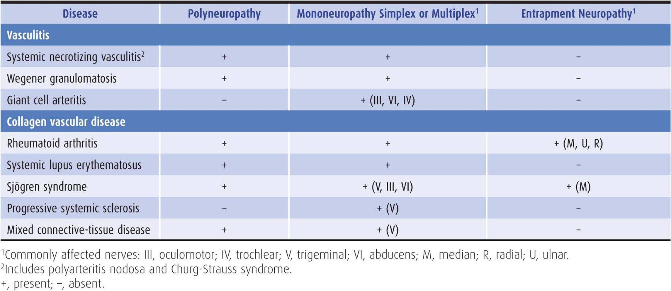
Table 10-7. Neuropathies associated with vasculitis and collagen vascular disease.
SYSTEMIC NECROTIZING VASCULITIS
This includes polyarteritis nodosa and allergic angiitis and granulomatosis (Churg-Strauss syndrome). Neuropathy occurs in approximately 50% of patients, most often as mononeuropathy multiplex, which may manifest itself with the acute onset of pain in one or more cranial or peripheral nerves. Distal symmetric sensorimotor polyneuropathy is less common. Treatment should begin as soon as the diagnosis is made; it includes prednisone 60 to 100 mg/d orally and cyclophosphamide 2 to 3 mg/d orally.
WEGENER GRANULOMATOSIS
Wegener granulomatosis is associated with mononeuropathy multiplex or polyneuropathy in up to 30% of cases. Treatment is the same as for systemic necrotizing vasculitis.
GIANT CELL ARTERITIS
This disorder is considered in detail in Chapter 6. Mononeuropathy affecting cranial nerves innervating the extraocular muscles can occur.
RHEUMATOID ARTHRITIS
Rheumatoid arthritis produces entrapment neuropathy (most commonly involving the median nerve) in approximately 45% of patients and distal symmetric sensorimotor polyneuropathy in about 30%. Mononeuropathy multiplex is a frequent feature in cases complicated by necrotizing vasculitis.
SYSTEMIC LUPUS ERYTHEMATOSUS
This is discussed in Chapter 4 as a cause of acute confusional states. Neuropathy occurs in up to 20% of patients. The most common pattern is a distal, symmetric sensorimotor polyneuropathy. An ascending, predominantly motor polyneuropathy (Guillain-Barré syndrome, see earlier) can also occur, as may mononeuropathy simplex or multiplex, which often affects the ulnar, radial, sciatic, or fibular (peroneal) nerve.
SJÖGREN SYNDROME
Sjögren syndrome involves the peripheral nerves in approximately 20% of cases. Distal symmetric sensorimotor polyneuropathy is most common, entrapment neuropathy (affecting especially the median nerve) is also frequent, and mononeuropathy multiplex can occur.
SCLERODERMA & MIXED CONNECTIVE-TISSUE DISEASE
Progressive systemic sclerosis (scleroderma) and mixed connective-tissue disease may produce cranial mononeuropathy, which most often involves the trigeminal (V) nerve.
NEOPLASTIC & PARAPROTEINEMIC NEUROPATHIES
COMPRESSION & INFILTRATION BY TUMOR
Nerve compression is a common complication of multiple myeloma, lymphoma, and carcinoma. Tumorous invasion of the epineurium may occur with leukemia, lymphoma, and various cancers, particularly carcinoma of the breast or pancreas.
PARANEOPLASTIC SYNDROMES
Carcinoma (especially small-cell cancer of the lung) and lymphoma may be associated with neuropathies that are thought to be immunologically mediated, based on the detection of autoantibodies to neuronal antigens in several cases.
Sensory or Sensorimotor Polyneuropathy
This occurs with both carcinoma and lymphoma. It can be either an acute or chronic disorder, is sometimes asymmetric, and may be accompanied by prominent pain. Among patients with a chronic sensorimotor polyneuropathy of uncertain cause, approximately 10% have a monoclonal gammopathy; many such patients eventually develop a hematologic malignancy, as discussed later. Treatment of the malignancy may result in improvement of the neuropathy.
Sensory Neuronopathy
Carcinoma can also cause sensory neuronopathy, which primarily affects the cell bodies of sensory neurons in the dorsal root ganglion and is associated with the presence of anti-Hu (or ANNA-1) antibodies (Chapter 8). This rare condition may be the presenting manifestation of cancer. Initial symptoms of pain and numbness usually begin distally but sometimes begin proximally or in the face. The disorders often progress over days or several weeks, leading to marked sensory ataxia and impairment of all sensory modalities. Motor involvement is late, and autonomic dysfunction is uncommon. The CSF may have an inflammatory formula. Treatment, even of the underlying tumor, is usually unrewarding.
Motor Neuronopathy & Guillain-Barré Syndrome
Lymphoma may be complicated by motor neuronopathy, a disorder of anterior horn cells. Hodgkin disease and angioimmunoblastic lymphadenopathy are sometimes associated with Guillain-Barré syndrome, which responds to treatment as in patients without malignancy. These and other paraneo-plastic motor disorders (including the Lambert-Eaton myasthenic syndrome, neuromyotonia, and stiff-person syndrome) are discussed in Chapter 9.
Autonomic Neuropathy
An autonomic neuropathy may also occur as a paraneo-plastic disorder, especially in patients with small-cell lung cancer, and relates most often to the presence of anti-Hu antibodies. It is underdiagnosed, often develops in the setting of other paraneoplastic syndromes, and has a poor prognosis. Symptoms may include hypotension, orthostatic hypotension, hypoventilation, abnormal thermoregulatory sweating, sleep apnea, gastroparesis, intestinal pseudo-obstruction, and cardiac arrhythmias, sometimes leading to sudden death. Affected patients do not improve with immunotherapy even after treatment of the underlying tumor.
PARAPROTEINEMIAS
Patients with paraproteinemic demyelinating neuropathy, especially a chronic distal sensory neuropathy, may have a malignant plasma cell dyscrasia. The paraprotein is likely to underlie the neuropathy when it is an immunoglobulin (Ig)M. When it is IgG or IgA, the neuropathy may be clinically and electrophysiologically indistinguishable from chronic inflammatory demyelinating polyradiculoneuropathy and similar in its response to treatment.
Polyneuropathy is a common complication of multiple myeloma. Patients affected by lytic myeloma are usually men. The clinical picture is of a distal symmetric sensorimotor polyneuropathy. All sensory modalities are affected, pain is a frequent feature, and the reflexes are depressed. The disorder is usually progressive and leads to death within 2 years.
Sclerotic myeloma may be accompanied by a chronic demyelinating polyneuropathy. Motor involvement predominates, but vibration and position sense may also be impaired, and the reflexes are depressed. Pain is less common than in the neuropathy of lytic myeloma, and symptoms may improve with treatment of the underlying cancer or by plasmapheresis.
The POEMS syndrome (polyneuropathy, organomegaly, endocrinopathy, M protein, and skin changes) may complicate plasma cell dyscrasias, especially osteosclerotic myeloma. The sensorimotor polyneuropathy may show certain distinctive electrophysiologic features, such as conduction slowing that is more marked in intermediate than distal nerve segments, and often responds to treatment. Local irradiation or resection of an isolated plasmacytoma should be considered, as should melphalan with or without corticosteroids.
A sensorimotor polyneuropathy similar to that observed with lytic myeloma may also occur in Waldenström macroglobulinemia or benign monoclonal gammopathy. Treatment with immunosuppressant drugs and plasma-pheresis is sometimes helpful.
AMYLOIDOSIS
Nonhereditary amyloidosis occurs as an isolated disorder (primary generalized amyloidosis) or in patients with multiple myeloma and may be associated with polyneuropathy. Polyneuropathy is also a feature of hereditary amyloidosis. Amyloid neuropathies are considered later in the section on hereditary neuropathies.
DRUG-INDUCED & TOXIC NEUROPATHIES
ALCOHOLISM
Polyneuropathy is one of the most common neurologic complications of chronic alcoholism; it can occur alone or in combination with other alcohol-related neurologic disorders, such as Wernicke encephalopathy (Chapter 4) or the Korsakoff amnestic syndrome (Chapter 5). Controversy exists concerning the relative contributions of direct neurotoxicity of alcohol and associated nutritional (especially thiamine) deficiency in producing polyneuropathy.
Alcoholic polyneuropathy is typically a symmetric distal sensorimotor neuropathy. The legs are particularly likely to be affected, resulting in defective perception of vibration and touch and depressed or absent ankle reflexes. In some cases, distal weakness is also pronounced, and autonomic dysfunction may occur. When pain is a prominent feature, it may respond to the same treatment described earlier for painful neuropathy.
Abstinence from alcohol and thiamine repletion can halt the progression of symptoms.
OTHER DRUGS
As indicated in Table 10-2, a large number of drugs have been reported to cause neuropathies. A few merit specific comment.
Dapsone, a drug used to treat leprosy, can produce a primarily motor polyneuropathy that is reversible.
Hydralazine, an antihypertensive drug, is associated on rare occasions with a predominantly sensory polyneuropathy that has been attributed to drug-induced pyridoxine deficiency and that resolves after the drug is discontinued.
Isoniazid is a widely used antituberculous agent that interferes with pyridoxine metabolism and produces a polyneuropathy that principally affects the sensory neurons. High doses, hereditary variations in drug metabolism, and malnutrition predispose to this complication. Spontaneous recovery is the rule when administration of the drug is halted. Isoniazid-induced neuropathy can be prevented by concurrent administration of pyridoxine 100 mg/d orally.
Phenytoin is often mentioned as a cause of polyneuropathy, but evidence for phenytoin treatment as a cause of symptomatic neuropathy is sparse.
Pyridoxine (vitamin B6) toxicity has been implicated as the cause of a sensory neuronopathy that disproportionately impairs vibration and position sense. This disorder usually occurs in patients taking at least 200 mg of pyridoxine daily—approximately 100 times the minimum daily requirement. Sensory ataxia, Romberg sign, Lhermitte sign, and ankle areflexia are common findings. Pain is less common, and motor involvement is unusual. Symptoms are usually reversible over months to years if the abuse ceases, but an irreversible syndrome has also been reported after intravenous administration of high doses of pyridoxine.
Vincristine produces a polyneuropathy in most patients who receive the drug for treatment of (usually hemato-logic) cancer. The earliest manifestations are distal sensory symptoms and loss of reflexes. Motor deficits may predominate later in the course, however. Constipation is a common finding and may be due to autonomic involvement. Discontinuing the drug or administering it at a reduced dosage often leads to improvement.
TOXINS
Organic compounds implicated as causes of polyneuropathy include hexacarbons present in solvents and glues (eg, n-hexane, methyl n-butyl ketone) and organophosphates used as plasticizers or insecticides (eg, triorthocresyl phosphate). Sensory involvement is most striking in n-hexane neuropathy, whereas neuropathy caused by triorthocresyl phosphate primarily affects motor nerves. Organophosphate neuropathy is discussed in more detail in Chapter 9.
Heavy metals may also be responsible for polyneuropathy. Neuropathy caused by lead, arsenic, and thallium is discussed in Chapter 9. Gold, which is used to treat rheumatoid arthritis, may cause a symmetric polyneuropathy, and cisplatin (a platinum analogue with anticancer activity) may produce a sensory neuropathy.
HEREDITARY NEUROPATHIES
HEREDITARY MOTOR & SENSORY NEUROPATHIES
These are designated Charcot-Marie-Tooth (CMT) hereditary neuropathies. They constitute a genetically heterogeneous group of disorders having a similar clinical phenotype. There is weakness and wasting of distal muscles in the limbs, with or without sensory loss; pes cavus and reduced or absent tendon reflexes also occur. They are divided into demyelinating (CMT-1) and neuronal (CMT-2) types, the latter sparing sensory neurons and resembling progressive spinal muscular atrophy (Chapter 9). Both types have an autosomal dominant pattern of inheritance, although apparently sporadic cases occur.
CMT-1 has its onset in the first decade, follows a slowly progressive course, and is of variable severity. The nerves are palpably thickened in approximately 50% of cases. Nerve conduction velocities are markedly reduced. CMT-1 is subdivided on the basis of the genetic findings, but the most common form results from duplication of or mutations in the gene for peripheral myelin protein-22 (PMP22).
CMT-2 is generally less severe than CMT-1, is associated with normal or nearly normal nerve conduction velocities, and does not cause nerve enlargement. The most common mutations are in genes for kinesin family member 1B (KIF1B) and mitofusin 2 (MFN2). X-linked dominant (CMT-X) and autosomal recessive (CMT-4) variants have also been described.
Dejerine-Sottas disease (HMSN3) has its onset by 2 years of age with delayed motor milestones and is characterized by a severe sensorimotor neuropathy that frequently extends to the proximal muscles and is associated with skeletal abnormalities such as scoliosis. There is severe demyelination of the nerves. It was previously thought to have an autosomal recessive mode of inheritance, but it is now accepted that it is inherited in an autosomal dominant manner, and the responsible mutations involve the same genes associated with CMT-1.
HEREDITARY SENSORY & AUTONOMIC NEUROPATHIES
These neuropathies also take a variety of forms. In hereditary sensory and autonomic neuropathy (HSAN) type I, there is dominant inheritance, a gradually progressive course from onset in early adulthood, and symmetric loss of distal pain and temperature perception, with relative preservation of light touch. Perforating ulcers over pressure points and painless infections of the extremities are common. The tendon reflexes are depressed, but there is little, if any, motor disturbance. This phenotype is associated with mutations in the genes coding for serine palmitoyltransferase long-chain subunits (SPTLC1 and SPTLC2), the GTPase atlastin-1 (ATL1), or DNA methyltransferase 1 (DNMT1).
In HSAN type II, inheritance is recessive, onset is in infancy or early childhood, all sensory modalities are affected, and tendon reflexes are lost. The affected genes are lysine-deficient protein kinase 1 (WNK1) and family with sequence similarity 134, member B (FAM134B).
HSAN type III (Riley-Day syndrome, familial dysautonomia) is a recessive disorder that commences in infancy and is characterized by conspicuous autonomic dysfunction (absent tearing, labile temperature, and blood pressure), absent taste sensation, impaired pain and temperature sensation, and areflexia. The disorder is linked to mutations in the gene for inhibitor of kappa light polypeptide gene enhancer in B cells, kinase complex-associated protein (IKBKAP).
HSAN type IV is associated with congenital insensitivity to pain and absent sweating and has been related to recessive mutations in the gene encoding a receptor tyrosine kinase for nerve growth factor (NTRK1). Many of these patients have cognitive dysfunction. Death from hyperexia may occur.
HSAN type V resembles type IV, but cognitive abnormalities do not occur. The mutation is in the nerve growth factor β-subunit gene (NGFB).
AMYLOIDOSIS
Polyneuropathy can occur in both hereditary and nonhereditary forms of amyloidosis. Because small-diameter sensory and autonomic nerve fibers are especially likely to be involved, pain and temperaturesensation and autonomic functions are prominently affected. Clinical presentation is commonly with distal paresthesias, dysesthesias, and numbness; postural hypotension; impaired thermo-regulatory sweating; and disturbances of bladder, bowel, or sexual function. Distal weakness and wasting eventually occur. The tendon reflexes are often preserved until a relatively late stage. Entrapment neuropathy, especially carpal tunnel syndrome, may develop as a consequence of amyloid deposits.
In primary amyloidosis, the diagnosis is made by identifying amyloid deposits in the tissues; examination of abdominal fat aspirate is usually the initial step. Treatment has included alkylating agents or autologous peripheral-blood stem-cell transplantation. In hereditary (familial) amyloidosis, genetic testing for transthyretin (TTR) mutations is more useful for establishing the diagnosis than analysis of tissues for amyloid deposition. Treatment by orthotopic liver transplantation is effective.
FRIEDREICH ATAXIA
Friedreich ataxia usually has a recessive mode of inheritance but occasionally occurs with dominant inheritance. It is usually caused by an expanded GAA trinucleotide repeat in a noncoding region of the gene for frataxin(FXN), a mitochondrial protein. However, there is some phenotypic heterogeneity, and the expansion has not been found in all cases, suggesting that other genetic or environmental factors are sometimes responsible. An ataxic gait develops, followed by clumsiness of the hands and other signs of cerebellar dysfunction. Involvement of peripheral sensory fibers lead to sensory deficits of the limbs, with depressed or absent tendon reflexes. There may also be leg weakness and extensor plantar responses from central motor involvement. This condition is considered in detail in Chapter 8.
HEREDITARY NEUROPATHY WITH LIABILITY TO PRESSURE PALSIES
This is a genetically heterogeneous disorder that relates most commonly to deletion in the peripheral myelin protein 22 (PMP22) gene. Inheritance is as an autosomal dominant trait with variable expression. Patients present with simple or multiple mononeuropathies that occur after mild pressure or stretch of nerves, and electrophysiologic studies reveal that abnormalities are more widespread than is evident clinically.
METABOLIC DISORDERS
In acute intermittent porphyria, which is transmitted by recessive inheritance, the initial neurologic manifestation is often a polyneuropathy that (usually) involves motor more than sensory fibers. Sensory symptoms and signs may be predominantly proximal or distal. The peripheral nerves may also be affected in variegate porphyria. Neuropathy caused by porphyria is considered in greater detail in Chapter 9.
Two recessive lipidoses are associated with polyneuropathy with a typical onset in infancy or childhood. These are metachromatic leukodystrophy, which results from deficiency of the enzyme arylsulfatase A, and Krabbe disease, which is due to galactocerebroside β-galactosidase deficiency. Both are inherited in an autosomal recessive fashion.
Lipoprotein deficiencies that cause polyneuropathy include abetalipoproteinemia, which is associated with acanthocytosis, malabsorption, retinitis pigmentosa, and cerebellar ataxia; and Tangier disease, which produces cataract, orange discoloration of the tonsils, and hepatosplenomegaly. These are autosomal recessive conditions.
Refsum disease is an autosomal recessive disorder related to impaired metabolism of phytanic acid. It produces polyneuropathy, cerebellar ataxia, retinitis pigmentosa, and ichthyosis. It can be treated by restricting dietary intake of phytol. Plasmapheresis to reduce body stores of phytanic acid may also be helpful at the initiation of treatment.
Fabry disease is an X-linked recessive deficiency of the enzyme α-galactosidase-A. It results in a painful sensory and autonomic neuropathy, angiokeratomas, renal and cardiac disease, and an increased incidence of stroke. The affected gene is alpha-galactosidase A (GLA). Pharmacologic measures (discussed earlier) may be helpful in treating the pain that characterizes the disorder. Enzyme replacement therapy with agalsidase beta, a recombinant human alpha-galactosidase A enzyme, may also help to reduce pain (as well as stabilize or improve cardiac and renal function), but the relationship of treatment to long-term outcome is unclear.
ENTRAPMENT NEUROPATHIES
Certain peripheral nerves are particularly susceptible to mechanical injury at vulnerable sites. The term entrapment neuropathy is used when the nerve is compressed, stretched, or angulated by adjacent anatomic structures to such an extent that dysfunction occurs. There are numerous entrapment neuropathies, and in many the initial or most conspicuous clinical complaints are of sensory symptoms or pain. Some of the more common syndromes are described next.
UPPER LIMB ENTRAPMENT SYNDROMES
MEDIAN NERVE COMPRESSION
Compression of the median nerve can occur in the carpal tunnel at the wrist. Carpal tunnel syndrome is common during pregnancy and can occur as a complication of trauma, degenerative arthritis, tenosynovitis, diabetes mellitus, myxedema, and acromegaly. Early symptoms are pain and paresthesias confined to a median nerve distribution in the hand, that is, involving primarily the thumb, index, and middle fingers and the lateral half of the ring finger (see Appendix). There may be pain in the forearm and, in occasional patients, even in the upper arm, shoulder, and neck. Symptoms are often particularly troublesome at night and may awaken the patient from sleep. As the neuropathy advances, weakness and atrophy may eventually develop in the thenar muscles.
Examination reveals impaired cutaneous sensation in the median nerve distribution in the hand and, with motor involvement, weakness and wasting of the abductor pollicis brevis and opponens pollicis muscles (see Appendix). There may be a positive Tinel sign (percussion of the nerve at the wrist causes paresthesias in its distribution) or a positive response to the Phalen maneuver (flexion of the wrist for 1 minute exacerbates or reproduces symptoms).
The diagnosis can generally be confirmed by electro-physiologic studies, showing sensory or motor conduction velocity to be slowed at the wrist; there may be signs of chronic partial denervation in median-supplied muscles of the hand.
If the symptoms fail to respond to local corticosteroid injections or simple maneuvers such as wearing a nocturnal wrist splint, surgical decompression of the carpal tunnel may be necessary.
INTERDIGITAL NEUROPATHY
Interdigital neuropathy may lead to pain in one or two fingers, and examination reveals hyperpathia or impaired cutaneous sensation in the appropriate distribution of the affected nerve or nerves. Such a neuropathy may result from entrapment in the intermetacarpal tunnel of the hand, direct trauma, tenosynovitis, or arthritis.
Treatment by local infiltration with corticosteroids is sometimes helpful, but in severe cases neurolysis may be necessary.
ULNAR NEUROPATHY
Ulnar nerve dysfunction at the elbow leads to paresthesias, hypesthesia, and nocturnal pain in the little finger and ulnar border of the hand. Pain may also occur about the elbow. Symptoms are often intensified by elbow flexion or use of the arm.
Examination may reveal sensory loss on the ulnar aspect of the hand and weakness of the adductor pollicis, the deep flexor muscles of the fourth and fifth digits, and the intrinsic hand muscles (see Appendix). The lesion may result from external pressure, from entrapment within the cubital tunnel, or from cubitus valgus deformity causing chronic stretch injury of the nerve. Electrodiagnostic studies may be helpful in localizing the lesion.
Avoiding pressure on or repetitive flexion and extension of the elbow, combined in some instances with splinting the elbow in extension, is sometimes sufficient to arrest progression and alleviate symptoms. Surgical decompression or ulnar nerve transposition to the flexor surface of the arm may also be helpful, depending on the cause and severity of the lesion and the duration of symptoms.
An ulnar nerve lesion may also develop in the wrist or palm of the hand in association with repetitive trauma, arthritis, or compression from ganglia or benign tumors. Involvement of the deep terminal branch in the palm leads to a motor deficit in ulnar-innervated hand muscles other than the hypothenar group, whereas a more proximal palmar lesion affects the latter muscles as well; there is no sensory deficit. With lesions at the wrist involving either the ulnar nerve itself or its deep and superficial branches, both sensory and motor changes occur in the hand. Sensation over the dorsal surface of the hand is unaffected, however, because the cutaneous branch to this region arises proximal to the wrist. Surgical treatment is helpful in relieving compression from a ganglion or benign tumor.
RADIAL NEUROPATHY
The radial nerve may be compressed in the axilla by pressure from crutches or other causes; this is frequently seen in alcoholics and drug addicts who have fallen asleep with an arm draped over some hard surface (so-called “Saturday night palsy”). The resulting deficit is primarily motor, with weakness or paralysis occurring in the muscles supplied by the nerve, but sensory changes may also occur, especially in a small region on the back of the hand between the thumb and index finger (see Appendix).
Treatment involves preventing further compression of the nerve. Recovery usually occurs spontaneously and completely, except when a very severe injury has resulted in axonal degeneration. Physical therapy and a wrist splint may be helpful until recovery occurs.
THORACIC OUTLET SYNDROME
In thoracic outlet syndrome, a cervical rib or band or other anatomic structure may compress the lower part of the brachial plexus. Symptoms include pain, paresthesias, and numbness in a C8 and T1 distribution (Figure 10-4). There may be diffuse weakness of the intrinsic hand muscles, often particularly involving the muscles in the thenar eminence and thereby simulating carpal tunnel syndrome. The section on cervical rib syndrome in Chapter 9 contains further details.
LOWER LIMB ENTRAPMENT SYNDROMES
FIBULAR (PERONEAL) NEUROPATHY
Fibular (peroneal) nerve lesions can occur secondary to trauma or to pressure about the knee at the head of the fibula. The resulting weakness or paralysis of foot and toe extension—and foot eversion—is accompanied by impaired sensation over the dorsum of the foot and the lower anterior aspect of the leg (see Appendix). The ankle reflex is preserved, as is foot inversion.
Treatment is purely supportive. It is important to protect the nerve from further injury or compression. Patients with foot drop may require a brace until recovery occurs. Recovery occurs spontaneously with time and is usually complete unless the injury was severe enough to cause marked axonal degeneration.
Tarsal Tunnel Syndrome
The posterior tibial nerve or its branches can be compressed between the floor and the ligamentous roof of the tarsal tunnel, which is located at the ankle immediately below and behind the medial malleolus. The usual complaint is of burning in the foot, especially at night, sometimes accompanied by weakness of the intrinsic foot muscles. The diagnosis can usually be confirmed electro-physiologically. If treatment with local injection of steroids is not helpful, surgical decompression may be necessary.
FEMORAL NEUROPATHY
Isolated femoral neuropathy may occur in association with diabetes mellitus, vascular disease, bleeding diatheses (eg, hemophilia or treatment with anticoagulant drugs), or retroperitoneal neoplasms. The most conspicuous symptoms and signs relate to weakness of the quadriceps muscle, with reduced or absent knee reflex, but there may also be sensory disturbances in the anterior and medial aspects of the thigh and the medial part of the lower leg. Treatment is of the underlying cause.
SAPHENOUS NEUROPATHY
The saphenous nerve is the terminal sensory branch of the femoral nerve and supplies cutaneous sensation to the medial aspect of the leg about and below the knee (Figure 10-4). Mechanical injury to the nerve can occur at several points along its course; patients then complain of pain or impaired sensation in the distribution of the nerve. Weakness in quadriceps function (ie, extension at the knee; see Appendix) indicates femoral, rather than saphenous, nerve involvement. There is no specific treatment, but the nerve should be protected from further injury.
LATERAL FEMORAL CUTANEOUS NEUROPATHY
The lateral femoral cutaneous nerve supplies sensation to the outer border of the thigh (see Appendix). Its function can be impaired by excessive angulation or compression by neighboring anatomic structures, especially in pregnancy or other conditions that cause exaggerated lumbar lordosis. This leads to pain and paresthesias in the lateral thigh, and examination reveals impaired sensation in this region. This syndrome, known as meralgia paresthetica, is best treated with symptomatic measures such as simple analgesics taken orally. Its course is often self-limited, but in occasional cases it progresses to a patch of permanent, painless numbness.
OBTURATOR NEUROPATHY
Trauma to the obturator nerve—for example, by pelvic fracture or a surgical procedure—can lead to pain radiating from the groin down the inner aspect of the thigh. An obturator hernia or osteitis pubis may cause a similar disorder; there is accompanying weakness of the adductor thigh muscles (see Appendix).
ROOT & PLEXUS LESIONS
COMPRESSIVE & TRAUMATIC LESIONS
The clinical disturbances resulting from acute intervertebral disk prolapse, cervical spondylosis, traumatic plexopathy, cervical rib syndrome, and neuralgic amyotrophy are discussed in Chapter 9. In addition to these conditions, patients with metastatic cancer may develop root or plexus lesions from compression by tumor or as a result of trauma induced by radiation therapy.
Root lesions are typically compressive in nature and usually occur in the setting of neoplastic meningitis, which is discussed in Chapter 4.
Brachial plexopathy can result from tumor infiltration of the brachial plexus, especially by lung or breast cancer, causing severe arm pain and sometimes dysesthesia. Because involvement of the lower trunk of the plexus is most common, symptoms usually occur within the C8 and T1 dermatomes, and Horner syndrome (Chapter 7) is present in approximately 50% of cases. Radiation injury, rather than direct invasion by tumor, should be suspected as the cause when the upper trunk of the brachial plexus (C5 and C6 nerve roots) is involved, weakness is a prominent presenting symptom, arm swelling occurs, or symptoms develop within 1 year after completion of radiation therapy with a total dose of more than 6,000 R.
Lumbosacral plexopathy is usually seen in patients with colorectal, cervical, uterine, or ovarian carcinoma or sarcoma. Clinical features that suggest tumor invasion in this setting include early and severe pain, unilateral involvement, leg swelling, and a palpable rectal mass. Radiation injury is more commonly associated with early prominent leg weakness and bilateral symptoms.
TABES DORSALIS
This type of neurosyphilis, now rare, is characterized mainly by sensory symptoms and signs that indicate marked involvement of the posterior roots, especially in the lumbosacral region, with resulting degeneration in the posterior columns of the spinal cord. Common complaints are of unsteadiness, sudden lancinating somatic pains, and urinary incontinence. Visceral crises characterized by excruciating abdominal pain also occur.
Examination reveals marked impairment of vibration and joint position sense in the legs, together with an ataxic gait and Romberg sign. Deep pain sensation is impaired, but superficial sensation is generally preserved. The bladder is often palpably enlarged; because it is flaccid and insensitive, there is overflow incontinence. Tendon reflexes are lost, and the limbs are hypotonic. Sensory loss and hypotonicity may lead to the occurrence of hypertrophic (Charcot) joints. In many patients there are other signs of neurosyphilis, including Argyll Robertson pupils, optic atrophy, ptosis, a variable ophthalmoplegia, and, in some cases, pyramidal and mental changes from cerebral involvement (taboparesis), as discussed in Chapter 5. Treatment is of the underlying infection.
LYME DISEASE
Lyme disease, like syphilis, is a spirochetal infection that produces both central and peripheral nervous system disease. Central nervous system involvement is manifested by meningitis or meningoencephalitis, as discussed in Chapter 4. Lyme disease is also associated with inflammatory mono-or polyradiculopathy, brachial plexopathy (Chapter 9), mononeuropathy (including facial palsy), and mononeuropathy multiplex. The radiculopathy results in pain, sensory loss, or dysesthesia in affected dermatomes; it also causes focal weakness. One or more cervical, thoracic, or lumbar nerve roots may be involved. Electromyography can confirm the presence of radiculopathy, and serologic testing establishes Lyme disease as the cause. Treatment is described in Chapter 4.
MYELOPATHIES
Myelopathies may present with pain or with a variety of sensory complaints and with motor disturbances. The clinical findings should suggest the level of the lesion, but further investigation is necessary to delineate it more fully and determine its nature. Compressive, ischemic, inflammatory, demyelinative, and traumatic myelopathies are discussed in Chapter 9.
SYRINGOMYELIA
Syringomyelia is cavitation of the spinal cord. Communicating syringomyelia, with communication between the central canal of the cord and the cavity, is a hydrodynamic disorder of the CSF pathways. In noncommunicating syringomyelia, there is cystic dilation of the cord, which is not in communication with the CSF pathways.
The precise clinical disturbance that results depends on the site of cavitation. Typically, there is a dissociated sensory loss at the level of the lesion; pinprick and temperature appreciation are impaired, but light touch sensation is preserved (Figure 10-5). The sensory loss may be reflected by the presence of painless skin ulcers, scars, edema, hyperhidrosis, neuropathic joints, resorption of the terminal phalanges, and other disturbances. Weakness and wasting of muscles occur at the level of the lesion because of the involvement of the anterior horns of the cord (Figure 10-5).
A pyramidal deficit and sphincter disturbances sometimes occur below the level of the lesion because of gliosis or compression of the corticospinal pathways in the lateral columns of the cord. The tendon reflexes may be depressed at the level of the lesion—because of interruption of their afferent, central, or efferent pathways—and increased below it. Scoliosis is a common accompaniment of cord cavitation.
Cavitation commonly occurs in the cervical region; this can cause a capelike distribution of sensory loss over one or both shoulders, diffuse pain in the neck, and radicular pain in the arms; involvement of the T1 segment frequently leads to ipsilateral Horner syndrome. If the cavitation involves the lower brainstem (syringobulbia), there may also be ipsilateral tongue wasting, palatal weakness, vocal cord paralysis, dissociated trigeminal sensory loss, and other evidence of brainstem involvement.
Communicating syringomyelia is often associated with developmental anomalies of the brainstem and foramen magnum region (such as Arnold-Chiari malformation; Chapter 8) or with chronic arachnoiditis of the basal cisterns. Arnold-Chiari malformation can lead to hydro-cephalus, cerebellar ataxia, pyramidal and sensory deficits in the limbs, and abnormalities of the lower cranial nerves, alone or in any combination. CT scans reveal a small posterior fossa and enlarged foramen magnum; other skeletal abnormalities may be present at the base of the skull and upper cervical spine. MRI reveals the syrinx and the Arnold-Chiari malformation, with caudal displacement of the fourth ventricle and herniation of the cerebellar tonsils through the foramen magnum.
Noncommunicating syringomyelia is often due to trauma, intramedullary tumors, or spinal arachnoiditis (Figure 10-8). Posttraumatic syringomyelia generally occurs in patients with preexisting, severe neurologic deficits from spinal trauma after an interval of several years, although rarely it may develop only a few months after the original injury. Presentation is with increase in a previously stable deficit; weakness, impaired sensation, and spasticity are often conspicuous, and radicular pain may be distressing. Focal cord enlargement is found on MRI in patients with cavitation related to previous injury or to intramedullary neoplasms.

Figure 10-8. Sagittal T1-weighted spinal MRI demonstrating a large septated syrinx in the lumbar cord of a patient with disseminated tuberculosis. (Courtesy of A. DiBernardo.)
Treatment depends on the underlying cause. Decompression of a distended syrinx may provide transient benefit. In the case of communicating syringomyelia associated with Arnold-Chiari malformation, removal of the posterior rim of the foramen magnum and amputation of the cerebellar tonsils are sometimes helpful. The cord cavity should be drained, and, if necessary, an outlet should be made for the fourth ventricle. Posttraumatic syringomyelia is treated by surgery if it is causing a progressive neurologic deficit or intolerable pain. A variety of surgical approaches have been used, including various draining procedures from the cord cavity, myelotomy, and formation of surgical meningocele. Radicular pain and sensory disturbances are usually helped, whereas spasticity responds less satisfactorily.
SUBACUTE COMBINED DEGENERATION
Vitamin B12 deficiency (Figure 10-9) may result from impaired absorption by the gastrointestinal tract such as occurs in pernicious anemia or because of gastrointestinal surgery, sprue, or infection with fish tapeworm; it can also be caused by a strictly vegetarian diet. Vitamin B12 deficiency can lead to subacute combined degeneration of the posterior and lateral columns of the spinal cord.

Figure 10-9. Vitamin B12 deficiency myelopathy in a 30-year-old woman, wheelchair-bound owing to an 18-month history of progressive myelopathy. B12 level: 60 pg/mL. Gadolinium-enhanced, T1-weighted cervical and upper thoracic MRI image shows marked enhancement of the posterior cord. (Image courtesy of R. Laureno.)
Onset of subacute combined degeneration is with distal paresthesias and weakness in the extremities (involvement of the hands occurs relatively early), followed by the development of spastic paraparesis, with ataxia from the impairment of postural sensation in the legs. Lhermitte sign may be present, and examination reveals a combined posterior column (vibration and joint position sense) and pyramidal deficit in the legs. Plantar responses are extensor, but tendon reflexes may be increased or depressed, depending on the site and severity of the involvement. Signs of cord involvement can be accompanied by centrocecal scotoma or optic atrophy from optic (II) nerve involvement, by behavioral or psychiatric changes (Chapter 4), or by peripheral neuropathy. The neurologic manifestations are often accompanied by macrocytic megaloblastic anemia, but this is not invariably present.
The serum vitamin B12 level is low in untreated cases. If malabsorption of vitamin B12 is the cause, the Schilling test is abnormal, and there is usually gastric achlorhydria with pernicious anemia. Hematologic findings may be normal, however, especially if folic acid supplements have been given. Electrophysiologic studies may confirm peripheral nerve involvement, and median- or tibial-derived somatosensory evoked potentials may show abnormalities indicative of posterior column dysfunction. Spinal MRI sometimes shows abnormalities in the posterior columns.
Treatment is with vitamin B12 given by intramuscular injection daily (50-1,000 μg) for 2 weeks, then weekly (100 μg) for 2 months, and monthly (100 μg) thereafter. Note that folic acid supplements do not help the neurologic disorder; in addition, they may mask associated anemia.
A similar disorder may result from nitrous oxide abuse, which leads to inactivation of vitamin B12. Management is focused on the prevention of further exposure to nitrous oxide.
Copper deficiency can also lead to subacute combined degeneration. The copper deficiency may follow total parenteral hyperalimentation, copper insufficiency with enteral feeding, malabsorption, gastric surgery, or excessive zinc ingestion, which inhibits the intestinal absorption of copper. Low serum copper and ceruloplasmin levels and urinary copper excretion confirm the diagnosis. Treatment involves copper supplementation and modification of any risk factors, which leads to stabilization of the neurologic deficit.
CEREBRAL DISORDERS
Sensory symptoms may relate to diverse diseases involving the brainstem or cerebral hemispheres (eg, stroke). The clinical features of the sensory deficit have been described earlier in this chapter and, together with the nature and extent of any accompanying neurologic signs, should suggest the probable site of the lesion.
PAIN SYNDROMES
Pain from infective, inflammatory, or neoplastic processes is a feature of many visceral diseases and may be a conspicuous component of certain neurologic or psychiatric diseases. It can also occur with no obvious cause.
In evaluating patients with pain, it is important to determine the level of the nervous system at which the pain arises and whether it has a primary neurologic basis. In taking the history, attention should be focused on the mode of onset, duration, nature, severity, and location of the pain; any associated symptoms; and factors that precipitate or relieve the pain.
Treatment depends on the underlying cause and clinical context of the pain and is discussed later. A brief comment is necessary, however, about stimulation-produced analgesia and, in particular, about spinal cord stimulation (previously known as dorsal column stimulation) and peripheral nerve stimulation. These approaches were based on principles encapsulated by the gate control theory, in which activation of large myelinated fibers was held to interrupt nociceptive transmission in the spinal cord, but their precise mechanism of action is uncertain. Spinal cord stimulation is known to affect certain neurotransmitter systems, particularly substance P and γ-aminobutyric acid (GABAergic) systems.
PERIPHERAL NERVE PAIN
Pain arising from peripheral nerve lesions is usually localized to the site of nerve injury or confined to the territory of the affected nerve. It may have a burning quality, and when mixed (motor and sensory) nerves are involved, there may be an accompanying motor deficit. Painful peripheral neuropathies include those caused by diabetes, polyarteritis, alcoholic-nutritional deficiency states, and the various entrapment neuropathies. Treatment of pain associated with peripheral neuropathies is discussed earlier.
Reflex sympathetic dystrophy is a general term that denotes sympathetically mediated pain syndromes precipitated by a wide variety of tissue injuries, including soft tissue trauma, bone fractures, and myocardial infarction. The designation complex regional pain syndrome (CRPS), type 1, is now used (in preference to reflex sympathetic dystrophy) for pain that follows injury but spreads beyond the site of trauma in a distribution that does not conform to that of an individual peripheral nerve, is greater than would normally be expected from the injury, and may progress with time (sometimes to the opposite limb). It has been attributed to a post-traumatic neuralgia associated with distal degeneration of small-diameter peripheral axons. CRPS type 2, or causalgia, designates the severe persistent pain, often burning in quality, that results from nerve trauma. Such pain is associated with exquisite tenderness. Onset of pain may be at any time within the first 6 weeks or so after nerve injury. The cause is uncertain, but it has been attributed to ephaptic transmission between efferent sympathetic and afferent somatic fibers at the site of injury.
In both types of complex regional pain syndrome, pain may be accompanied by swelling, increased sweating, and vasoconstriction of the affected extremity, which is commonly kept covered up and still by the patient; allodynia, hyperalgesia, muscle atrophy, and osteoporosis may also occur.
Treatment is largely empirical; studies to support specific therapeutic regimens are lacking. Medical approaches to treatment of complex regional pain syndrome include physical and occupational therapy, with emphasis on increasing activity of the involved limb. Relaxation therapy or biofeedback is sometimes helpful, as also is topical lido-caine. Various pharmacologic approaches have been tried, with doses tailored to the individual. Tricyclic antidepressants, gabapentin, pregabalin, and opioids have been used for chronic pain disorders other than CRPS and are worthy of trial until formal studies establish their utility. The role of serotonin-norepinephrine reuptake inhibitors (eg, duloxetine or venlafaxine) is unclear, but these also may be worthy of trial.
Sympathetic blockade by injection of local anesthetics into the sympathetic chain or by regional infusion of reserpine or guanethidine provides temporary benefit to some patients. One such procedure sometimes produces permanent cessation of pain, or repeated sympathetic blocks may be required. Surgical sympathectomy is beneficial in up to 75% of cases. Spinal cord stimulation has been effective in some instances for the treatment of either type of complex regional pain syndrome when other measures, including opiates, have been unhelpful.
RADICULAR PAIN
Radicular pain is localized to the distribution of one or more nerve roots and is often exacerbated by coughing, sneezing, and other maneuvers that cause increased intraspinal pressure. It is also exacerbated by maneuvers that stretch the affected roots. Passive straight leg raising leads to stretching of the sacral and lower lumbar roots, as does passive flexion of the neck. Spinal movements that narrow the intervertebral foramina can aggravate root pain. Extension and lateral flexion of the head to the affected side may thus exacerbate cervical root symptoms. In addition to pain, root lesions can cause paresthesias and numbness in a dermatomal distribution (Figure 10-4); they can also cause segmental weakness and reflex changes, depending on the level affected (Chapter 9, Table 9-12). Useful modes of treatment include immobilization, nonsteroidal anti-inflammatory drugs or other analgesics, and surgical decompression.
THALAMIC PAIN
Depending on their extent and precise location, thalamic lesions may lead to pain in all or part of the contralateral half of the body. The pain is of a burning nature, with a particularly unpleasant quality that patients have difficulty describing. It is aggravated by emotional stress and tends to develop during partial recovery from a sensory deficit caused by the underlying thalamic lesion. Mild cutaneous stimulation may produce very unpleasant and painful sensations. This combination of sensory loss, spontaneous pain, and perverted cutaneous sensation has come to be called Dejerine-Roussy syndrome. Similar pain can be produced by a lesion that involves the parietal lobe or the sensory pathways at any point in the cord (posterior columns or spinothalamic tract) or in the brainstem. Treatment with analgesics, anticonvulsants (carbamazepine or phenytoin), or antidepressants and phenothiazines in combination is occasionally helpful.
BACK & NECK PAIN
Spinal disease occurs most commonly in the neck or low back and can cause local or root pain or both. It can also lead to pain that is referred to other parts of the involved dermatomes. Pain from the lower lumbar spine, for example, is often referred to the buttocks. Conversely, pain may be referred to the back from the viscera, especially the pelvic organs. Local pain may lead to protective reflex muscle spasm, which in turn causes further pain and may result in abnormal posture, limitation of movement, and local spinal tenderness.
The history may provide clues to the underlying cause, and physical examination will define any neurologic involvement. Diagnostic studies that can help in evaluating patients include x-rays of the affected region and a complete blood count and erythrocyte sedimentation rate (especially if infective or inflammatory disorders or myeloma is suspected); determination of serum protein and protein electrophoresis; and measurement of serum calcium, phosphorus, alkaline and acid phosphatase, and uric acid. Electromyography may be helpful in determining the extent and severity of root involvement; it also provides a guide to prognosis. A CT scan, MRI of the spine, or CT myelogram may be necessary, especially if neoplasm is suspected, neurologic deficits are progressive, pain persists despite conservative treatment measures, or there is evidence of cord involvement. At myelography, CSF can be obtained for laboratory examination.
LOW BACK PAIN
Low back pain is a common cause of time lost from work. It has many causes. Routine immediate imaging of patients with acute or subacute low back pain who have no clinical features suggestive of serious underlying disorder does not improve the clinical outcome and is therefore unnecessary.
Trauma
Unaccustomed exertion or activity—or lifting heavy objects without adequate bracing of the spine—can cause musculoskeletal pain that improves with rest. Clinical examination commonly reveals spasm of the lumbar muscles and restricted spinal movement. Management includes local heat, bed rest on a firm mattress, nonsteroidal anti-inflammatory drugs or other analgesics, and muscle-relaxant drugs (eg, diazepam 2 mg three times daily, increased gradually until symptoms are relieved or to the highest dose tolerated). Vertebral fractures that follow more severe injury and lead to local pain and tenderness can be visualized at radiography. If cord involvement is suspected—eg, because of leg weakness after injury—the patient must be immobilized until imaged to determine whether fracture dislocation of the vertebral column has occurred.
Prolapsed Lumbar Intervertebral Disk
This most commonly affects the L5-S1 or the L4-L5 disk. The prolapse may relate to injury, but in many patients it commonly follows minor strain or normal activity. Protruded disk material may press on one or more nerve roots and thus produce radicular pain, a segmental motor or sensory deficit, or a sphincter disturbance in addition to a painful stiff back. The pain may be reproduced by percussion over the spine or sciatic nerve, by passive straight leg raising, or by extension of the knee while the hip is flexed. The presence of bilateral symptoms and signs suggests that disk material has protruded centrally, and this is more likely to be associated with sphincter involvement than is lateral protrusion.
An L5 radiculopathy causes weak dorsiflexion of the foot and toes, whereas an S1 root lesion leads to a depressed ankle reflex and weakness of plantar flexion of the foot (Chapter 9, Table 9-12). In either case, spinal movements are restricted, there is local tenderness, and Lasègue sign (reproducing the patient’s pain on stretching the sciatic nerve by straight leg raising) is positive. The L4 root is occasionally affected, but involvement of a higher lumbar root should arouse suspicion of other causes of root compression.
Pelvic and rectal examination, plain x-rays of the spine, and CT scans or MRI help to exclude other diseases, such as local tumors or metastatic neoplastic deposits.
Symptoms often resolve with simple analgesics, diazepam, and bed rest on a firm mattress for 2 to 3 days, followed by gradual mobilization. Bed rest for longer than 2 to 3 days provides no additional benefit. Nonsteroidal anti-inflammatory drugs may be helpful for acute back pain but are often ineffective or provide only minor or transient benefits in patients with symptoms or signs of root compression. The utility of epidural steroid injection is uncertain.
Persisting pain, an increasing neurologic deficit, or any evidence of sphincter dysfunction should lead to MRI, CT scanning, or CT myelography, and surgical treatment if indicated by the results of these procedures. The detection of structural abnormalities by these imaging procedures does not mandate surgical treatment unless the clinical circumstances are appropriate—degenerative abnormalities are common in asymptomatic subjects, especially with advancing age, and may therefore be of no clinical relevance.
Persistence of low back and root pain despite surgery may have several causes, including inadequate decompression, recurrent herniation of disk material, root compression or damage as a result of the operative procedure, surgery at the wrong level, infective or inflammatory complications of surgery, or spinal instability. In many instances, however, no specific cause can be identified, and most patients do not require further surgery. Chronic pain in this setting may, however, respond to spinal cord stimulation. There is a high risk that patients will not return to work.
Lumbar Osteoarthropathy
This tends to occur in later life and may cause low back pain that is increased by activity. MRI is the preferred mode of imaging; abnormalities vary in severity, and their clinical relevance is not always clear. Many asymptomatic elderly subjects have degenerative changes of the lumbar spine. In patients with mild symptoms, a surgical corset is helpful, whereas in more severe cases operative treatment may be necessary.
Even minor changes may cause root or cord dysfunction in patients with a congenitally narrowed spinal canal (spinal stenosis), leading to the syndrome of intermittent claudication of the spinal cord or cauda equina. Similar symptoms may result from ossification of the ligamentum flavum, epidural lipomatosis, Pott disease, osteomyelitis, rheumatoid arthritis, or post-traumatic stenosis of the spinal canal. The syndrome is characterized by pain, sometimes accompanied by weakness or radicular sensory disturbances in the legs, that occurs with activity or with certain postures and is relieved by rest.
Patients with mild to moderate symptoms related to spinal stenosis should be treated conservatively with pain medication, nonsteroidal anti-inflammatory agents, and physical therapy to reduce the lumbar lordosis; some physicians also use epidural injections of steroids. Muscle relaxants and antidepressant drugs are often prescribed also. Studies to examine the relative merits and cost-effectiveness of these approaches are lacking. Bed rest is to be avoided. If symptoms are severe, decompressive surgery is indicated when conservative treatment for 3 to 6 months has been unhelpful or there is a significant motor deficit or symptoms of cauda equina syndrome (compression of lumbosacral nerve roots in the spinal canal, below the end of the spinal cord [conus medullaris]). Such surgery (laminectomy and hemilaminectomy, hemilaminotomy, fenestration, foraminotomy, and the implantation of interspinous distraction devices) leads to pain relief, but the rapidity and extent of recovery is variable. Complications include epidural hematomas, inadequate decompression with residual stenosis, instability, and reossification with renewed nerve compression. Up to 25% of patients undergoing surgical decompression will require reoperation in the following 10 years.
Ankylosing Spondylitis
Backache and stiffness, followed by progressive limitation of movement, characterize this disorder, which occurs predominantly in young men. Characteristic early radiologic findings consist of sclerosis and narrowing of the sacroiliac joints. Treatment is with nonsteroidal anti-inflammatory agents, especially indomethacin or aspirin. Physical therapy, including postural exercises, is also important.
Neoplastic Disease
Extradural malignant tumors are an important cause of back pain and should be suspected if there is persistent pain that worsens despite bed rest. They may eventually lead to spinal cord compression or a cauda equina syndrome, depending on the level of involvement. There may initially be no change on plain radiographs of the spine, but a bone scan is sometimes revealing. Benign osteogenic tumors also produce back pain, and plain x-rays then show a lytic lesion; treatment is by excision.
Infections
Tuberculous and pyogenic infections of the vertebrae or intervertebral disks can cause progressive low back pain and local tenderness. Although there are sometimes no systemic signs of infection, the peripheral white cell count and erythrocyte sedimentation rate are raised. X-rays may show disk space narrowing and a soft tissue mass, but they are frequently normal initially.
Bone infection (osteomyelitis) requires long-term antimicrobial therapy; surgical debridement and drainage may also be needed. Spinal epidural abscess (Chapter 9) similarly presents with localized pain and tenderness, sometimes associated with osteomyelitis. Cord compression may occur with the onset of a rapidly progressive flaccid paraplegia. MRI, CT scanning, or CT myelography, followed by operative treatment, are undertaken urgently if there is evidence of cord compression. In early cases without neurologic involvement, treatment with antibiotics alone may be sufficient.
Osteoporosis
Low back pain is a common complaint in patients with osteoporosis, and vertebral fractures may occur spontaneously or after trivial trauma. Pain may be helped by a brace to support the back. It is important that patients keep active, stop tobacco smoking, and take a diet containing adequate amounts of calories, calcium, vitamin D, and protein. Estrogen therapy may be helpful in postmenopausal women, but is less widely used than previously. The bisphosphonates alendronate and risedronate may be helpful and have reduced the incidence of fractures in randomized trials. Other antiresorptives include raloxifene, a selective estrogen receptor modulator, and the bisphosphonates ibandronate or zoledronic acid, which are also effective for the treatment of osteoporosis. In special circumstances, parathyroid hormone, calcitonin, or strontium are helpful.
Paget Disease of the Spine
Paget disease, which is characterized by excessive bone destruction and repair, is of unknown cause but may have a familial basis. Pain is commonly the first symptom. Vertebral involvement may also lead to evidence of cord or root compression. The serum calcium and phosphorus levels are normal, but the alkaline phosphatase is markedly increased. Urinary hydroxyproline and calcium are increased when the disease is active. X-rays show expansion and increased density of the involved bones, and fissure fractures may be evident in the long bones.
In active, progressive disease, treatment with calcitonin or bisphosphonates reduces osteoclastic activity. Decompressive surgery is sometimes necessary for neurologic complications.
Congenital Anomalies
Minor spinal anomalies can cause pain because of altered mechanics or alignment or because reduction in the size of the spinal canal renders the cord or roots more liable to compression by degenerative or other changes. Children or young adults with congenital defects in spinal fusion (spinal dysraphism) occasionally present with pain, a neurologic deficit in one or both legs, or sphincter disturbances. Treatment is of the underlying disorder.
Congenital spinal stenosis may lead to the syndrome of neurogenic claudication, but symptoms usually develop only in later life when minor degenerative changes have come to be superimposed on the congenital anomaly, as discussed earlier.
Arachnoiditis
Severe pain in the back and legs can result from inflammation and fibrosis of the arachnoid layer of the spinal meninges (arachnoiditis), which may be idiopathic or causally related to previous surgery, infection, myelography, or long-standing disk disease. There is no adequate treatment, but operation may be possible if the arachnoiditis is localized. Spinal cord stimulation may provide symptomatic relief. This condition is considered in more detail in Chapter 9.
Referred Pain
Disease of the hip joints may cause pain in the back and thighs that is enhanced by activity; examination reveals limitation of movement at the joint with a positive Patrick sign (hip pain on external rotation of the hip), and x-rays show degenerative changes. Aortic aneurysms, cardiac ischemia, visceral and genitourinary disease (especially pelvic disorders in women), and retroperitoneal masses can also cause back pain. There are often other symptoms and signs that suggest the underlying disorder. Moreover, there is no localized spinal tenderness or restriction of motility. Treatment is of the underlying cause.
Nonspecific Chronic Back Pain
In many patients whose chronic back pain poses a difficult management problem, there are no objective clinical signs or obvious causes of pain despite detailed investigations. In some cases, the pain may have a postural basis; in others, it may be a somatic manifestation of a psychiatric disorder. Pain that initially had an organic basis is often enhanced or perpetuated by nonorganic factors and leads to disability out of proportion to the symptoms.
Nonsteroidal anti-inflammatory drugs may provide short-term symptomatic relief. There is some controversy about the chronic use of narcotic analgesics in patients with persisting low back pain, but such agents are generally best avoided. Treatment with tricyclic antidepressant drugs is sometimes helpful, and psychiatric evaluation may be worthwhile. Bedrest is not recommended and provides no greater benefit than symptom-limited activity. Unnecessary surgical procedures must be avoided.
NECK PAIN
Neck pain is a common problem in the general population; surveys indicate that approximately one-third of the adult population has experienced it over the previous year, and in some instances it lasts for more than 6 months.
Congenital abnormalities of the cervical spine, such as hemivertebrae or fused vertebrae, basilar impression, and instability of the atlantoaxial joint, can cause neck pain. The traumatic, infective, and neoplastic disorders mentioned previously as causes of low back pain can also affect the cervical spine and then produce pain in the neck. Rheumatoid arthritis may involve the spine, especially in the cervical region, leading to pain, stiffness, and reduced mobility; cord compression may result from displacement of vertebrae or atlantoaxial subluxation and can be life-threatening if not treated by fixation.
Cervical injuries are an important cause of neck pain. Whiplash flexion–extension injuries have become especially common as a result of automobile accidents. Other occult cervical injuries such as disk clefts and fissures may be responsible for symptoms in some instances, but are difficult to recognize. Management of persistent symptoms after whiplash injuries is controversial. Conservative therapeutic measures are appropriate. Other approaches sometimes advocated include block of cervical facet joints with bupivacaine and injection into the joints of depot corticosteroids, but the response is variable and often short-lived. Subluxed cervical facet joints are another well-recognized complication of automobile accidents. Even minor trauma may lead to cervical fractures in an apparently ankylosed region in patients with diffuse idiopathic skeletal hyperostosis, but major neurologic deficits are common in such circumstances.
Acute Cervical Disk Protrusion
Patients may present with neck and radicular arm pain that is exacerbated by head movement. The mechanism responsible for the pain is unclear; pressure on nerve roots is unlikely to be the sole cause because pain may resolve with time and conservative measures despite persisting compression. With lateral herniation of the disk, there may also be segmental motor, sensory, or reflex changes, usually at the C6 or C7 level, on the affected side (Figure 9-9 and Table 9-12). With more centrally directed herniations, spastic paraparesis and a sensory disturbance in the legs, sometimes accompanied by impaired sphincter function, can occur as a result of cord involvement. The diagnosis is confirmed by CT scan, MRI, or CT myelography. However, these imaging studies may show abnormalities in asymptomatic subjects in middle or later life, so that any disk protrusion may be incidental and unrelated to patients’ symptoms. Electromyography may help to establish that anatomic abnormalities are of functional relevance.
In mild cases, bedrest or intermittent neck traction, followed by immobilization of the neck in a collar, often helps, but the optimal duration of treatment is unknown. Painful muscle spasm may be helped by muscle relaxants. If these measures fail or if there is a significant progressive neurologic deficit, surgical treatment may be necessary. Bedrest is often prescribed, but the optimal duration for this is unclear. Many physicians now recommend rest for just 2 or 3 days rather than for longer, because evidence of added benefit for lengthier periods of rest is lacking. A short course of oral corticosteroids is sometimes prescribed, but such an approach requires validation.
Cervical Spondylosis
This is an important cause of pain in the neck and arms, sometimes accompanied by a segmental motor or sensory deficit in the arms or by spastic paraparesis. It is discussed in Chapter 9.
HERPES ZOSTER (SHINGLES)
This viral disorder becomes increasingly common with advancing age, causing an inflammatory reaction in one or more of the dorsal root or cranial nerve ganglia, in the affected root or nerve itself, and in the CSF. There seems to be spontaneous reactivation of varicella virus that remained latent in sensory ganglia after previous infection. Herpes zoster is common in patients with lymphoma, especially after regional radiotherapy.
The initial complaint is of a burning or shooting pain in the involved dermatome, followed within 2 to 5 days by the development of a vesicular erythematous rash. The pain may diminish in intensity as the rash develops. The rash becomes crusted and scaly after a few days and then fades, leaving small anesthetic scars. Secondary infection is common. The pain and dysesthesias may last for several weeks or, in some instances, may persist for many months (postherpetic neuralgia) before subsiding.
Postherpetic neuralgia is most likely to occur in the elderly, when the rash is severe, with a longer duration of the rash before medical consultation, and with involvement of the first division of the trigeminal (V1) nerve. The increased incidence and severity of postherpetic neuralgia with age may reflect an age-related reduction in virus-specific cell-mediated immunity. It is not clear whether immune compromise secondary to HIV infection or connective tissue disease predisposes to postherpetic neuralgia.
Pain is exacerbated by touching the involved area. Superficial sensation is often impaired in the affected dermatome, and focal weakness and atrophy can also occur. Signs are usually limited to one dermatome, but more are occasionally involved. Mild pleocytosis and an increased protein concentration sometimes occur in the CSF. The most commonly involved sites are the thoracic dermatomes, but involvement of the first division of the trigeminal (V) nerve, also common, is especially distressing and may lead to corneal scarring and anesthesia, as well as to a variety of other ocular complications.
Facial (VII) nerve palsy occurring in association with a herpetic eruption that involves the ear, palate, pharynx, or neck is called Ramsay Hunt syndrome. Other rare complications of herpes zoster include other motor neuropathies, meningitis, encephalitis, myelopathy, and cerebral angiopathy.
The administration of live-attenuated zoster vaccine to patients over the age of 60 years is an important approach to preventing the occurrence of herpes zoster and thus of post-herpetic neuralgia. There is no specific treatment once herpes zoster has developed. Analgesics provide symptomatic relief. Corticosteroids may reduce the duration and severity of the acute eruption, but not the likelihood that postherpetic neuralgia will occur. The incidence of postherpetic neuralgia may be reduced by oral acyclovir or famciclovir to patients with herpes zoster, but this is unsettled. Although postherpetic neuralgia can be very distressing, duloxetine (60 mg once daily) or pregabalin (150 mg increasing after 1 week to 300 mg daily in divided doses; maximum, 600 mg daily) may be helpful for relief of pain. It sometimes responds also to treatment with carbamazepine, up to 1,200 mg/d; phenytoin 300 mg/d; gabapentin, up to 3,600 mg/d; or amitriptyline 10 to 100 mg at bedtime.
Attempts at relieving postherpetic neuralgia by peripheral nerve section are generally unrewarding, but treatment with topically applied local anesthetics is sometimes helpful, as is topically applied capsaicin cream, perhaps because of depletion of pain-mediating peptides from peripheral sensory neurons. Intrathecal methylprednisolone may be helpful for intractable pain.
REFERENCES
General
Aminoff MJ. Electromyography in Clinical Practice. 3rd ed. New York, NY: Churchill Livingstone; 1998.
Aminoff MJ, ed. Neurology and General Medicine. 4th ed. New York, NY: Churchill Livingstone Elsevier; 2008.
Dworkin RH, O’Connor AB, Backonja M, et al. Pharmacologic management of neuropathic pain: evidence-based recommendations. Pain. 2007;132:237-251.
Dyck PJ, Thomas PK, eds. Peripheral Neuropathy. 4th ed. Philadelphia, PA: Saunders Elsevier; 2005.
Guindon J, Walczak JS, Beaulieu P. Recent advances in the pharmacological management of pain. Drugs. 2007;67:2121-2133.
Layzer RB. Neuromuscular Manifestations of Systemic Disease. vol 25 of: Contemporary Neurology Series. Philadelphia, PA: F.A. Davis; 1984.
Low PA. Autonomic neuropathies. Curr Opin Neurol. 2002;15:605-609.
Low PA, Vernino S, Suarez G. Autonomic dysfunction in peripheral nerve disease. Muscle Nerve. 2003;27:646-661.
Mogyoros I, Bostock H, Burke D. Mechanisms of paresthesias arising from healthy axons. Muscle Nerve. 2000;23:310-320.
Nardin RA, Johns DR. Mitochondrial dysfunction and neuro-muscular disease. Muscle Nerve. 2001;24:170-191.
Sax TW, Rosenbaum RB. Neuromuscular disorders in pregnancy. Muscle Nerve. 2006;34:559-571.
Stewart JD. Peripheral nerve fascicles: anatomy and clinical relevance. Muscle Nerve. 2003;28:525-541.
Polyneuropathies—General
Cruccu G. Treatment of painful neuropathy. Curr Opin Neurol. 2007;20:531-555.
Dyck PJ, Thomas PK, eds. Peripheral Neuropathy. 4th ed. Philadelphia, PA: Saunders Elsevier; 2005.
Federici T, Boulis N. Gene therapy for peripheral nervous system diseases. Curr Gene Ther. 2007;7:239-248.
Finnerup NB, Sindrup SH, Jensen TS. The evidence for pharmacological treatment of neuropathic pain. Pain. 2010;150:573-581.
Freeman R. Autonomic peripheral neuropathy. Neurol Clin. 2007;25:277-301.
Gold R, Stangel M, Dalakas MC. Drug Insight: the use of intravenous immunoglobulin in neurology–therapeutic considerations and practical issues. Nat Clin Pract Neurol. 2007;3:36-44.
Vernino S, et al. Antibody testing in peripheral neuropathies. Neurol Clin. 2007;25:29-46.
Polyneuropathies—Idiopathic Inflammatory
Brannagan TH. Current treatments of chronic immune-mediated demyelinating polyneuropathies. Muscle Nerve. 2009;39: 563-578.
De Sousa EA, Brannagan TH: Diagnosis and treatment of chronic inflammatory demyelinating polyneuropathy. Curr Treat Options Neurol. 2006;8:91-103.
Donofrio PD. Immunotherapy of idiopathic inflammatory neuropathies. Muscle Nerve. 2003;28:273-292.
Douglas MR, Winer JB. Guillain-Barré syndrome and its treatment. Expert Rev Neurother. 2006;6:1569-1574.
Hahn AF, Bolton CF, Pillay N, et al. Plasma-exchange therapy in chronic inflammatory demyelinating polyneuropathy: a double-blind, sham-controlled, cross-over study. Brain. 1996;119:1055-1066.
Hughes RA. Management of chronic inflammatory demyelinating polyradiculoneuropathy. Drugs. 2003;63:275-287.
Hughes RA, Swan AV, van Doorn PA. Intravenous immunoglobulin for Guillain-Barré syndrome. Cochrane Database Syst Rev. 2010;6:CD002063.
Hughes RA, Swan AV, van Doorn PA. Corticosteroids for Guillain-Barré syndrome. Cochrane Database Syst Rev. 2010;2:CD001446.
Hughes RA, Swan AV, Raphaël JC, Annane D, van Koningsveld R, van Doorn PA. Immunotherapy for Guillain-Barré syndrome: a systematic review. Brain. 2007;130:2245-2257.
Kuwabara S. Guillain-Barré syndrome. Curr Neurol Neurosci Rep. 2007;7:57-62.
Latov N, Deng C, Dalakas MC, et al. Timing and course of clinical response to intravenous immunoglobulin in chronic inflammatory demyelinating polyradiculoneuropathy. Arch Neurol. 2010;67:802-807.
Lewis RA. Chronic inflammatory demyelinating polyneuropathy. Neurol Clin. 2007;25:71-87.
Lunn MPT, Willison HJ. Diagnosis and treatment in inflammatory neuropathies. J Neurol Neurosurg Psychiatry. 2009;80:249-258.
Magda P, Latov N, Brannagan TH 3rd, Weimer LH, Chin RL, Sander HW. Comparison of electrodiagnostic abnormalities and criteria in a cohort of patients with chronic inflammatory demyelinating polyneuropathy. Arch Neurol. 2003;60:1755-1759.
Oh SJ, Kurokawa K, de Almeida DF, Ryan HF Jr, Claussen GC. Subacute inflammatory demyelinating polyneuropathy. Neurology. 2003;61:1507-1512.
Rezania K, Gundogdu B, Soliven B. Pathogenesis of chronic inflammatory demyelinating polyradiculoneuropathy. Front Biosci. 2004;9:939-945.
Sander HW, Latov N. Research criteria for defining patients with CIDP. Neurology. 2003;60(suppl 3):S8-S15.
Saperstein DS, Katz JS, Amato AA, Barohn RJ. Clinical spectrum of chronic acquired demyelinating polyneuropathies. Muscle Nerve. 2001;24:311-324.
Steck AJ, Czaplinski A, Renaus S. Inflammatory demyelinating neuropathies and neuropathies associated with monoclonal gammopathies: treatment update. Neurotherapeutics. 2008; 5:528-534.
Toothaker TB, Brannagan TH III. Chronic inflammatory demyelinating polyneuropathies: current treatment strategies. Curr Neurol Neurosci Rep. 2007;7:63-70.
Vallat J-M, Sommer C, Magy L. Chronic inflammatory demyelinating polyradiculoneuropathy: diagnostic and therapeutic challenges for a treatable condition. Lancet Neurol. 2010;9: 402-412.
van Doorn PA, Ruts L, Jacobs BC. Clinical features, pathogenesis, and treatment of Guillain-Barré syndrome. Lancet Neurol. 2008; 7:939-950.
van Schaik IN, Winer JB, de Haan R, Vermeulen M. Intravenous immunoglobulin for chronic inflammatory demyelinating polyradiculoneuropathy: a systematic review. Lancet Neurol. 2002;1:491-498.
van Schaik IN, Winer JB, de Haan R, Vermeulen M. Pulsed high-dose dexamethasone versus standard prednisolone treatment for chronic inflammatory demyelinating polyradiculoneuropathy (PREDICT study): a double-blind, randomised, controlled trial. Lancet Neurol. 2010;9:245-253.
Yuki N. Ganglioside mimicry and peripheral nerve disease. Muscle Nerve. 2007;35:691-711.
Polyneuropathies—Metabolic & Nutritional
Armstrong DG, Chappell AS, Le TK, et al. Duloxetine for the management of diabetic peripheral neuropathic pain: evaluation of functional outcomes. Pain Med. 2007;8:410-418.
Bansal D, Bhansali A, Hota D, Chakrabarti A, Dutta P. Amitriptyline vs. pregabalin in painful diabetic neuropathy: a randomized double blind clinical trial. Diabet Med. 2009; 26:1019-1026.
Duby JJ, Campbell RK, Setter SM, White JR, Rasmussen KA. Diabetic neuropathy: an intensive review. Am J Health Syst Pharm. 2004;61:160-173.
Duyff RF, Van den Bosch J, Laman DM, van Loon BJ, Linssen WH. Neuromuscular findings in thyroid dysfunction: a prospective clinical and electrodiagnostic study. J Neurol Neurosurg Psychiatry. 2000;68:750-755.
Juhasz-Pocsine K, Rudnicki SA, Archer RL, Harik SI. Neurologic complications of gastric bypass surgery for morbid obesity. Neurology. 2007;68:1843-1850.
Kharbanda PS, Prabhakar S, Chawla YK, Das CP, Syal P. Peripheral neuropathy in liver cirrhosis. J Gastroenterol Hepatol. 2003;18:922-926.
Krishnan AV, Kiernan MC. Uremic neuropathy: clinical features and new pathophysiological insights. Muscle Nerve. 2007;35: 273-290.
McKeage K. Treatment options for the management of diabetic painful neuropathy: best current evidence. Curr Opin Neurol. 2007;20:553-557.
Podwall D, Gooch C. Diabetic neuropathy: clinical features, etiology, and therapy. Curr Neurol Neurosci Rep. 2004;4:55-61.
Said G. Diabetic neuropathy—a review. Nat Clin Pract Neurol. 2007;3:331-340.
Saperstein DS, Barohn RJ. Peripheral neuropathy due to cobalamin deficiency. Curr Treat Options Neurol. 2002;4:197-201.
Zochodne DW. Diabetes mellitus and the peripheral nervous system: manifestations and mechanisms. Muscle Nerve. 2007;36:144-166.
Polyneuropathies—Infective & Granulomatous
Brew BJ. The peripheral nerve complications of human immunodeficiency virus (HIV) infection. Muscle Nerve. 2003; 28: 542-552.
Gonzalez-Duarte A, Cikurel K, Simpson DM. Managing HIV peripheral neuropathy. Curr HIV/AIDS Rep. 2007;4:114-118.
Herrmann DN, McDermott MP, Henderson D, et al. Epidermal nerve fiber density, axonal swellings and QST as predictors of HIV distal sensory neuropathy. Muscle Nerve. 2004;29:420-427.
Hoitsma E, Marziniak M, Faber CG, et al. Small fibre neuropathy in sarcoidosis. Lancet. 2002;359:2085-2086.
Lopez OL, Becker JT, Dew MA, Caldararo R. Risk modifiers for peripheral sensory neuropathy in HIV infection/AIDS. Eur J Neurol. 2004;11:97-102.
Manji H. Neuropathy in HIV infection. Curr Opin Neurol. 2000;13:589-592.
Pati S, Goodfellow JA, Iyadurai S, Hilton-Jones D. Approach to critical illness polyneuropathy and myopathy. Postgrad Med J. 2008;84:354-360.
Said G. Infectious neuropathies. Neurol Clin. 2007;25:115-137.
Van Brakel WH: Peripheral neuropathy in leprosy and its consequences. Lepr Rev. 2000;71(suppl):S146-S153.
Verma S, Micsa E, Estanislao L, Simpson D. Neuromuscular complications in HIV. Curr Neurol Neurosci Rep. 2004;4:62-67.
Polyneuropathies—Vasculitis & Collagen Vascular Disease
Burns TM, Schaublin GA, Dyck PJ. Vasculitic neuropathies. Neurol Clin. 2007;25:89-113.
Dyck PJ. The clinical heterogeneity of immune sensory and autonomic neuropathies with (or without) sicca. Brain. 2005;128:2480-2482.
Rosenbaum R. Neuromuscular complications of connective tissue disease. Muscle Nerve. 2001;24:154-169.
Schaublin GA, Michet CJ, Dyck PJ, Burns TM. An update on the classification and treatment of vasculitic neuropathy. Lancet Neurol. 2005;4:853-865.
Polyneuropathies—Neoplastic & Paraproteinemic
Allen D, Lunn MP, Niermeijer J, Nobile-Orazio E. Treatment for IgG and IgA paraproteinaemic neuropathy. Cochrane Database Syst Rev. 2007:CD005376.
Bataller L, Dalmau J. Paraneoplastic neurologic syndromes: approaches to diagnosis and treatment. Semin Neurol. 2003; 23:215-224.
Benson MD, Kincaid JC. The molecular biology and clinical features of amyloid neuropathy. Muscle Nerve. 2007;36: 411-423.
Dalakas MC. Pathogenesis and treatment of anti-MAG neuropathy. Curr Treat Options Neurol. 2010;12:71-83.
European Federation of Neurological Societies, et al. European Federation of Neurological Societies/Peripheral Nerve Society guideline on management of paraproteinaemic demyelinating neuropathies: report of a joint task force of the European Federation of Neurological Societies and the Peripheral Nerve Society. Eur J Neurol. 2006;13:809-818.
Forman AD. Peripheral neuropathy and cancer. Curr Oncol Rep. 2004;6:20-25.
Giometto B, Grisold W, Vitaliani R, et al. Paraneoplastic neurologic syndrome in the PNS Euronetwork database: a European study From 20 centers. Arch Neurol. 2010;67:330-335.
Kwan JY. Paraproteinemic neuropathy. Neurol Clin. 2007;25:47-69.
Latov N. Prognosis of neuropathy with monoclonal gammopathy. Muscle Nerve. 2000;23:150-152.
Ponsford S, Willison H, Veitch J, Morris R, Thomas PK. Long-term clinical and neurophysiological follow-up of patients with peripheral neuropathy associated with benign monoclonal gammopathy. Muscle Nerve. 2000;23:164-174.
Rajabally YA. Neuropathy and paraproteins: review of a complex association. Eur J Neurol. 2011;18:1291-1298.
Rudnicki SA, Dalmau J. Paraneoplastic syndromes of the spinal cord, nerve, and muscle. Muscle Nerve. 2000;23:1800-1818.
Sung JY, Kuwabara S, Ogawara K, Kanai K, Hattori T. Patterns of nerve conduction abnormalities in POEMS syndrome. Muscle Nerve. 2002;26:189-193.
Vital A. Paraproteinemic neuropathies. Brain Pathol. 2001;11: 399-407.
Wicklund MP, Kissel JT. Paraproteinemic neuropathy. Curr Treat Options Neurol. 2001;3:147-156.
Polyneuropathies—Drug-Induced & Toxic
Mastaglia FL. Drug-induced disorders of the nervous system. In: Aminoff MJ, ed. Neurology and General Medicine. 4th ed. New York, NY: Churchill Livingstone Elsevier; 2008:695-719.
Park SB, Lin CS, Krishnan AV, Friedlander ML, Lewis CR, Kiernan MC. Early, progressive, and sustained dysfunction of sensory axons underlies paclitaxel-induced neuropathy. Muscle Nerve. 2011;43:367-374.
Parry GJ. Neurological complications of toxin exposure in the workplace. In: Aminoff MJ, ed. Neurology and General Medicine. 4th ed. New York, NY: Churchill Livingstone Elsevier; 2008:749-768.
Polyneuropathies—Hereditary
Anand P. Neurotrophic factors and their receptors in human sensory neuropathies. Prog Brain Res. 2004;146:477-492.
Andersson PB, Yuen E, Parko K, So YT. Electrodiagnostic features of hereditary neuropathy with liability to pressure palsies. Neurology. 2000;54:40-44.
Axelrod FB. Familial dysautonomia. Muscle Nerve. 2004;29:352-363.
Chance PF. Genetic evaluation of inherited motor/sensory neuropathy. Suppl Clin Neurophysiol. 2004;57:228-242.
Chance PF. Inherited focal, episodic neuropathies: hereditary neuropathy with liability to pressure palsies and hereditary neuralgic amyotrophy. Neuromolecular Med. 2006;8:159-174.
Fogel BL, Perlman S. Clinical features and molecular genetics of autosomal recessive cerebellar ataxias. Lancet Neurol. 2007;6:245-257.
Klein CJ. The inherited neuropathies. Neurol Clin. 2007;25:173-207.
Li J, Krajewski K, Lewis RA, Shy ME. Loss-of-function phenotype of hereditary neuropathy with liability to pressure palsies. Muscle Nerve. 2004;29:205-210.
Nave KA, Sereda MW, Ehrenreich H. Mechanisms of disease: inherited demyelinating neuropathies–from basic to clinical research. Nat Clin Pract Neurol. 2007;3:453-64.
Nicholson GA. The dominantly inherited motor and sensory neuropathies: clinical and molecular advances. Muscle Nerve. 2006;33:589-597.
Scherer SS. Finding the causes of inherited neuropathies. Arch Neurol. 2006;63:812-816.
Scott KR, Kothari MJ. Hereditary neuropathies. Semin Neurol. 2005;25:174-184.
Shy M, Jáni A, Krajewski K, et al. Phenotypic clustering in MPZ mutations. Brain. 2004;127:371-384.
Shy ME, Garbern JY, Kamholz J. Hereditary motor and sensory neuropathies: a biological perspective. Lancet Neurol. 2002;1:110-118.
Vallat JM. Dominantly inherited peripheral neuropathies. J Neuropathol Exp Neurol. 2003;62:699-714.
Entrapment Neuropathies
Arle JE, Zager EL. Surgical treatment of common entrapment neuropathies in the upper limbs. Muscle Nerve. 2000;23: 1160-1174.
Bland JD. Carpal tunnel syndrome. BMJ. 2007;335:343-346.
Campbell WW, Landau ME. Controversial entrapment neuropathies. Neurosurg Clin N Am. 2008;19:597-608.
Dawson DM, Hallett M, Wilbourn AJ. Entrapment Neuropathies. 3rd ed. Philadelphia, PA: Lippincott Williams & Wilkins; 1999.
Gerritsen AA, de Vet HC, Scholten RJ, Bertelsmann FW, de Krom MC, Bouter LM. Splinting vs surgery in the treatment of carpal tunnel syndrome: a randomized controlled trial. JAMA. 2002;288:1245-1251.
Han SE, Boland RA, Krishnan AV, Vucic S, Lin CS, Kiernan MC. Ischaemic sensitivity of axons in carpal tunnel syndrome. J Peripher Nerv Syst. 2009;14:190-200.
Jarvik JG, Comstock BA, Kliot M, et al. Surgery versus non-surgical therapy for carpal tunnel syndrome: a randomised parallel-group trial. Lancet. 2009;374:1074-1081.
Stewart JD. Focal Peripheral Neuropathies. 4th ed. West Vancouver, Canada: JBJ Publishing; 2010.
Root & Plexus Lesions (see also Pain Syndromes)
Bono CM, Ghiselli G, Gilbert TJ, et al. An evidence-based clinical guideline for the diagnosis and treatment of cervical radiculopathy from degenerative disorders. Spine J. 2011;11:64-72.
Bruggeman AJ, Decker RC. Surgical treatment and outcomes of lumbar radiculopathy. Phys Med Rehabil Clin North Am. 2011;22:161-177.
Casey E. Natural history of radiculopathy. Phys Med Rehabil Clin North Am. 2011;22:1-5.
Decker RC. Surgical treatment and outcomes of cervical radiculopathy. Phys Med Rehabil Clin North Am. 2011;22:179-191.
Ferrante MA. Brachial plexopathies: classification, causes, and consequences. Muscle Nerve. 2004;30:547-568.
Gosk J, Rutowski R, Reichert P, Rabczy ski J. Radiation-induced brachial plexus neuropathy—aetiopathogenesis, risk factors, differential diagnostics, symptoms and treatment. Folia Neuropathol. 2007;45:26-30.
Halperin JJ. Spirochetal infections of the nervous system. In: Aminoff MJ, ed. Neurology and General Medicine. 4th ed. New York, NY: Churchill Livingstone Elsevier; 2008:789-802.
Halperin LL. Lyme disease and the peripheral nervous system. Muscle Nerve. 2003;28:133-143.
Jaeckle KA. Neurological manifestations of neoplastic and radiation-induced plexopathies. Semin Neurol. 2004;24:385-393.
Johansson S, Svensson H, Larsson LG, Denekamp J. Brachial plexopathy after postoperative radiotherapy of breast cancer patients—a long-term follow-up. Acta Oncol. 2000;39: 373-382.
Koes BW, Scholten RJ, Mens JM, Bouter LM. Efficacy of non-steroidal antiinflammatory drugs for low back pain: a systematic review of randomized clinical trials. Ann Rheum Dis. 1997;56:214-223.
Wilbourn AJ. Plexopathies. Neurol Clin. 2007;25:139-171.
Yee T. Recurrent idiopathic lumbosacral plexopathy. Muscle Nerve. 2000;23:1439-1442.
Myelopathies
Aghakhani N, Parker F, David P, et al. Long-term follow-up of Chiari-related syringomyelia in adults: analysis of 157 surgically treated cases. Neurosurgery. 2009;64:308-315.
Bonfield CM, Levi AD, Arnold PM, Okonkwo DO. Surgical management of post-traumatic syringomyelia. Spine. 2010; 35(suppl):S245-S258.
Genevay S, Atlas SJ. Lumbar spinal stenosis. Best Pract Res Clin Rheumatol. 2010;24:253-265.
Heiss JD, Patronas N, DeVroom HL, et al. Elucidating the patho-physiology of syringomyelia. J Neurosurg. 1999;91:553-562.
Hemmer B, Glocker FX, Schumacher M, Deuschl G, Lücking CH. Subacute combined degeneration: clinical, electrophysiological, and magnetic resonance imaging findings. J Neurol Neurosurg Psychiatry. 1998;65:822-827.
Hsu W, Dorsi MJ, Witham TF. Surgical management of cervical spondylotic myelopathy. Neurosurg Q. 2009;19:302-307.
Jaiser SR, Winston GP. Copper deficiency myelopathy. J Neurol. 2010;257:869-881.
Karantanas AH, Markonis A, Bisbiyiannis G. Subacute combined degeneration of the spinal cord with involvement of the anterior columns: a new MRI finding. Neuroradiology. 2000;42:115-117.
Kelkar P, Chang S, Muley SA. Response to oral supplementation in copper deficiency myeloneuropathy. J Clin Neuromuscul Dis. 2008;10:1-3.
Klineberg E. Cervical spondylotic myelopathy: a review of the evidence. Orthop Clin North Am. 2010;41:193-202.
Nikolaidis I, Fouyas IP, Sandercock PA, Statham PF. Surgery for cervical radiculopathy or myelopathy. Cochrane Database Syst Rev. 2010;1:CD001466.
Siebert E, Prüss H, Klingebiel R, Failli V, Einhäupl KM, Schwab JM. Lumbar spinal stenosis: syndrome, diagnostics and treatment. Nat Rev Neurol. 2009;5:392-403.
Tracy JA, Bartleson JD. Cervical spondylotic myelopathy. Neurologist. 2010;16:176-187.
Pain Syndromes
Ahn NU, Ahn UM, Andersson GB, An HS. Operative treatment of the patient with neck pain. Phys Med Clin North Am. 2003;14:675-692.
Assendelft WJ, Morton SC, Yu EI, Suttorp MJ, Shekelle PG. Spinal manipulative therapy for low back pain. Cochrane Database Syst Rev. 2004;1:CD000447.
Atlas SJ, Nardin RA. Evaluation and treatment of low back pain: an evidence-based approach to clinical care. Muscle Nerve. 2003;27:265-284.
Attal N, Cruccu G, Baron R, et al. EFNS guidelines on the pharmacological treatment of neuropathic pain: 2010 revision. Eur J Neurol. 2010;17:1113-e88.
Baron R, Binder A, Wasner G. Neuropathic pain: diagnosis, pathophysiological mechanisms, and treatment. Lancet Neurol. 2010;9:807-819.
Bogduk N. The anatomy and pathophysiology of neck pain. Phys Med Clin North Am. 2003;14:455-472.
Chou R, Fu R, Carrino JA, Deyo RA. Imaging strategies for low-back pain: systematic review and meta-analysis. Lancet. 2009;373:463-472.
Eisenberg E, River Y, Shifrin A, Krivoy N. Antiepileptic drugs in the treatment of neuropathic pain. Drugs. 2007;67:1265-1189.
Emery EC, Young GT, Berrosco EM, Chen L, mcNaughtoin PA. HCN2 ion channels play a central role in inflammatory and neuropathic pain. Science. 2011;333:1462-1466.
Finnerup NB, Sindrup SH, Jensen TS. The evidence for pharmacological treatment of neuropathic pain. Pain. 2010;150:573-581.
Gilron I, Bailey JM, Tu D, Holden RR, Jackson AC, Houlden RL. Nortriptyline and gabapentin, alone and in combination for neuropathic pain: a double-blind, randomised controlled crossover trial. Lancet. 2009;374:1252-1261.
Hagen KB, Hilde G, Jamtvedt G, Winnem MF. The Cochrane review of bed rest for acute low back pain and sciatica. Spine. 2000;25:2932-2939.
Hayden JA, van Tulder MW, Tomlinson G. Systematic review: strategies for using exercise therapy to improve outcomes in chronic low back pain. Ann Intern Med. 2005;142:776-785.
Henschke N, Ostelo RW, van Tulder MW, et al. Behavioural treatment for chronic low-back pain. Cochrane Database Syst Rev. 2010;7:CD002014.
Isose S, Misawa S, Sakurai K, et al. Mexiletine suppresses nodal persistent sodium currents in sensory axons of patients with neuropathic pain. Clin Neurophysiol. 2010;121:719-724.
Jarvic JG, Deyo RA. Diagnostic evaluation of low back pain with emphasis on imaging. Ann Intern Med. 2002;137:586-597.
Johnson RW. Herpes zoster and postherpetic neuralgia. Expert Rev Vaccines. 2010;9(3 suppl):21-26.
Johnson RW, Wasner G, Saddier P, Baron R. Postherpetic neuralgia: epidemiology, pathophysiology and management. Expert Rev Neurother. 2007;7:1581-1195.
Li Q, Chen N, Yang J, et al. Antiviral treatment for preventing postherpetic neuralgia. Cochrane Database Syst Rev. 2009;2:CD006866.
Nygaard OP, Kloster R, Solberg T. Duration of leg pain as a predictor of outcome after surgery for lumbar disc herniation: a prospective cohort study with 1-year follow up. J Neurosurg. 2000;92(suppl):131-134.
Oaklander AL, Fields HL. Is reflex sympathetic dystrophy/complex regional pain syndrome type I a small-fiber neuropathy? Ann Neurol. 2009;65:629-638.
Otto M, Bach FW, Jensen TS, Sindrup SH. Valproic acid has no effect on pain in polyneuropathy: a randomized, controlled trial. Neurology. 2004;62:285-288.
Oxman MN, Levin MJ, Shingles Prevention Study Group. Vaccination against herpes zoster and postherpetic neuralgia. J Infect Dis. 2008;197(suppl 2):S228-S236.
Paice JA, Ferrans CE, Lashley FR, Shott S, Vizqirda V, Pitrak D. Topical capsaicin in the management of HIV-associated peripheral neuropathy. J Pain Symptom Manage. 2000;19:45-52.
Rowbotham MC. Pharmacologic management of complex regional pain syndrome. Clin J Pain. 2006;22:425-429.
Rowbotham MC, Twilling L, Davies PS, Reisner L, Taylor K, Mohr D. Oral opioid therapy for chronic peripheral and central neuropathic pain. N Engl J Med. 2003;348:1223-1232.
Siebert E, Prüss H, Klingebiel R, Failli V, Einhäupl KM, Schwab JM. Lumbar spinal stenosis: syndrome, diagnostics and treatment. Nat Rev Neurol. 2009;5:392-403.
Stacey BR, Glanzman RL. Use of gabapentin for postherpetic neuralgia: results of two randomized, placebo-controlled studies. Clin Ther. 2003;25:2597-2608.
Staiger TO, Gaster B, Sullivan MD, Deyo RA. Systematic review of antidepressants in the treatment of chronic low back pain. Spine. 2003;28:2540-2545.
Stanton-Hicks M, Salamon J. Stimulation of the central and peripheral nervous system for the control of pain. J Clin Neurophysiol. 1997;14:46-62.
Thyregod HG, Rowbotham MC, Peters M, Possehn J, Berro M, Petersen KL. Natural history of pain following herpes zoster. Pain. 2007;128:148-56.
Valls I, Saraux A, Goupille P, Khoreichi A, Baron D, Le Goff P. Factors predicting radical treatment after in-hospital conservative management of disk-related sciatica. Bone Joint Surg. 2001;68:50-58.
van der Windt DA, Simons E, Riphagen II, et al. Physical examination for lumbar radiculopathy due to disc herniation in patients with low-back pain. Cochrane Database Syst Rev. 2010;2:CD007431.
Vroomen PC, de Krom MC, Slofstra PD, Knottnerus JA. Conservative treatment for sciatica: a systematic review. J Spinal Disord. 2000;13:463-469.
Wareham DW, Breuer J. Herpes zoster. BMJ. 2007;334:1211-1215.
Weinstein JN, Tosteson TD, Lurie JD, et al. Surgical versus non-surgical therapy for lumbar spinal stenosis. N Engl J Med. 2008;358:794-810.