
Approach to Diagnosis
Acute Onset
Focal Involvement
Anterior (Carotid) Circulation
Posterior (Vertebrobasilar) Circulation
Duration of Deficits
Vascular Origin
Ischemia
Hemorrhage
Focal Cerebral Ischemia
Pathophysiology
Injury Mechanisms
Survival & Repair Mechanisms
Pathology
Large Vessel Territory Infarction
Lacunar Infarction
Clinical-Anatomic Correlation
Anterior Cerebral Artery
Middle Cerebral Artery
Internal Carotid Artery
Posterior Cerebral Artery
Basilar Artery
Long Circumferential Vertebrobasilar Branches
Long Penetrating Paramedian Vertebrobasilar Branches
Short Basal Vertebrobasilar Branches
Lacunar Infarction
Etiology
Vascular Disorders
Cardiac Disorders
Hematologic Disorders
Clinical Findings
History
Physical Examination
Investigative Studies
Blood Tests
Electrocardiogram (ECG)
Lumbar Puncture
CT Scan or MRI
Vessel Imaging
Echocardiography
Electroencephalogram (EEG)
Differential Diagnosis
Treatment
Primary Prevention
Transient Ischemic Attack
Acute Stroke
Chronic Stroke
Complications
Prognosis
Intracerebral Hemorrhage
Hypertensive Hemorrhage
Epidemiology
Pathophysiology
Pathology
Differential Diagnosis
Treatment
Other Causes of Intracerebral Hemorrhage
Trauma
Vascular Malformations
Hemorrhage Into Cerebral Infarcts
Amphetamine or Cocaine Abuse
Cerebral Amyloid Angiopathy
Acute Hemorrhagic Leukoencephalitis
Hemorrhage Into Tumors
Coagulopathies
Anticoagulation & Thrombolytic Therapy
Global Cerebral Ischemia
Etiology
Pathology
Distribution
Modifying Factors
Clinical Findings
Brief Ischemia
Prolonged Ischemia
Treatment
Prognosis
References
Stroke is the fourth leading cause of death in the United States (after heart disease, cancer, and chronic lung disease) and the most common disabling neurologic disorder. Approximately 750,000 new strokes occur and approximately 150,000 people die from stroke in the United States each year.
The incidence of stroke increases with age, with approximately two-thirds of all strokes occurring in those older than 65 years, and is somewhat higher in men than in women and in African Americans than in whites. Modifiable risk factors for stroke include systolic or diastolic hypertension, atrial fibrillation, diabetes, dyslipidemia, and physical inactivity (Table 13-1). The incidence of stroke has decreased in recent decades, largely because of improved treatment of hypertension.


Table 13-1. Well-documented risk factors for stroke.1
Genetic factors also appear to be important in stroke pathogenesis, although the cause of most strokes is likely to be multifactorial and involve both polygenic and environmental influences. Several, mostly rare Mendelian disorders have stroke as a major manifestation; some of these, in which the affected gene has been identified, are listed in Table 13-2.


Table 13-2. Some monogenic disorders associated with stroke.
APPROACH TO DIAGNOSIS
Stroke is a syndrome characterized by four key features:
1. Sudden onset—The sudden onset of symptoms is documented by the history.
2. Focal involvement of the central nervous system—The site of central nervous system involvement is suggested by the nature of the symptoms and signs, delineated more precisely by the neurologic examination, and confirmed by imaging studies (computed tomography [CT] scans or magnetic resonance imaging [MRI]).
3. Lack of rapid resolution—The duration of neurologic deficits is documented by the history. In the past, the standard definition of stroke required that deficits persist for at least 24 hours to distinguish stroke from transient ischemic attack (discussed later). However, any such time point is arbitrary, and transient ischemic attacks usually resolve within 1 hour.
4. Vascular cause—A vascular cause may be inferred from the acute onset of symptoms and often from the patient’s age, the presence of risk factors for stroke, and the occurrence of symptoms and signs referable to the territory of a particular cerebral blood vessel. When this is confirmed by imaging studies, further investigations can be undertaken to identify a more specific etiology, such as arterial thrombosis, cardiogenic embolus, or clotting disorder.
ACUTE ONSET
Strokes begin abruptly. Neurologic deficits may be maximal at onset, as is common in embolic stroke, or may progress over seconds to hours (or occasionally days), which may occur with progressive arterial thrombosis or recurrent emboli.
A stroke that is actively progressing as a direct consequence of the underlying vascular disorder (but not because of associated cerebral edema) or has done so in recent minutes is termed stroke in evolutionor progressing stroke(Figure 13-1).

Figure 13-1. Time course of cerebral ischemic events. A transient ischemic attack (TIA) produces neurologic deficits that resolve completely within a short period, usually within 1 hour. Stroke-in-evolution, or progressing stroke, causes deficits that continue to worsen even as the patient is seen. Completed stroke is defined by the presence of persistent deficits, which may be stable or improving when the patient is seen; it does not necessarily imply that the entire territory of the involved vessel is affected or that no improvement has occurred since the onset.
Focal cerebral deficits that develop slowly (over weeks to months) are unlikely to be due to stroke and are more suggestive of another process, such as tumor or inflammatory or degenerative disease.
FOCAL INVOLVEMENT
Stroke produces focal symptoms and signs that correlate with the area of the brain supplied by the affected blood vessel.
In ischemic stroke (discussed in more detail later), occlusion of a blood vessel interrupts the flow of blood to a specific region of the brain, interfering with neurologic functions dependent on that region and producing a more or less stereotyped pattern of deficits.
Hemorrhage produces a less predictable pattern of focal involvement because complications such as increased intracranial pressure, cerebral edema, compression of brain tissue and blood vessels, or dispersion of blood through the subarachnoid space or cerebral ventricles can impair brain function at sites remote from the hemorrhage.
Cerebrovascular disorders can also affect the brain in more diffuse fashion and produce global cerebral dysfunction, but the term stroke should not be applied in these cases. These disorders include global cerebral ischemia(usually from cardiac arrest) and subarachnoid hemorrhage (discussed in Chapter 6).
In most cases of stroke, the history and neurologic examination provide enough information to localize the lesion to one side of the brain (eg, to the side opposite a hemiparesis or hemisensory deficit or to the left side if aphasia is present) and to the anterior or posterior cerebral circulation.
ANTERIOR (CAROTID) CIRCULATION
The anterior cerebral circulation supplies most of the cerebral cortex and subcortical white matter, basal ganglia, and internal capsule. It consists of the internal carotid artery and its branches: the anterior choroidal, anterior cerebral and middle cerebral arteries. The middle cerebral artery in turn gives rise to deep, penetrating lenticulostriate branches (Figure 13-2). The specific territory of each of these vessels is listed in Table 13-3.

Figure 13-2. Major cerebral arteries. The anterior and posterior cerebral circulations comprise arteries that arise anterior and posterior to the posterior communicating arteries, respectively. The circle of Willis is formed by the anterior communicating artery and the anterior cerebral, internal carotid, posterior communicating, and posterior cerebral arteries. (Reproduced, with permission, from Waxman S. Clinical Neuroanatomy. 26th ed. New York, NY: McGraw-Hill; 2010.)

Table 13-3. Territories of the principal cerebral arteries.
Anterior circulation strokes are commonly associated with symptoms and signs that indicate hemispheric dysfunction (Table 13-4), such as aphasia, apraxia, or agnosia (described in Chapter 1). They often produce hemiparesis, hemisensory disturbances, and visual field defects, which can also occur with posterior circulation strokes.

Table 13-4. Symptoms and signs of anterior and posterior circulation ischemia.
POSTERIOR (VERTEBROBASILAR) CIRCULATION
The posterior cerebral circulation supplies the brainstem, cerebellum, thalamus, and portions of the occipital and temporal lobes. It consists of the paired vertebral arteries, the basilar artery, and their branches: the posterior inferior cerebellar, anterior inferior cerebellar, superior cerebellar, and posterior cerebral arteries (Figure 13-2). The posterior cerebral artery also gives off thalamoperforateand thalamogeniculate branches. Areas supplied by these arteries are listed in Table 13-3.
Posterior circulation strokes produce symptoms and signs of brainstem dysfunction (Table 13-4), including coma, drop attacks (sudden collapse without loss of consciousness), vertigo, nausea and vomiting, cranial nerve palsies, ataxia, and crossed sensorimotor deficits that affect the face on one side of the body and the limbs on the other. Hemiparesis, hemisensory disturbances, and visual field deficits also occur, but are not specific to posterior circulation strokes.
DURATION OF DEFICITS
Stroke produces neurologic deficits that persist. When symptoms and signs of cerebral ischemia resolve completely after briefer periods (usually within 1 hour), the term transient ischemic attack (TIA) is used (Figure 13-1). Recurrent TIAs with identical clinical features (stereotypic TIAs) are usually caused by thrombosis or embolism arising within the cerebral circulation. TIAs that differ in character from event to event suggest recurrent emboli from a cardiac source. Although TIAs do not themselves produce lasting neurologic dysfunction, they are important to recognize because approximately one-third of patients with TIAs will go on to have a stroke within 5 years—and because this risk may be reduced with treatment.
In some cases, deficits last for longer periods but resolve completely or almost completely within a few days; the term reversible ischemic neurologic deficit (RIND) or minor stroke is sometimes used to describe these events.
VASCULAR ORIGIN
Although hypoglycemia, other metabolic disturbances, trauma, and seizures can produce focal central neurologic deficits that begin abruptly and last for at least 24 hours, the term stroke is used only when such events are caused by vascular disease.
The underlying pathologic process in stroke can be either ischemia or hemorrhage, usually arising from an arterial lesion. Ischemia accounts for approximately 90% and hemorrhage for approximately 10% of strokes in typical American and European series. It may not be possible to distinguish between ischemia and hemorrhage from the history and neurologic examination, but CT scan or MRI permits a definitive diagnosis.
ISCHEMIA
Interruption of blood flow to the brain deprives neurons, glia, and vascular cells of substrate glucose and oxygen. Unless blood flow is promptly restored, this leads to ischemic death of brain tissue (infarction) within the ischemic core, where flow is typically less than 20% of normal. The pattern of cell death depends on the severity of ischemia. With mild ischemia, as may occur in cardiac arrest with reperfusion, selective vulnerability of certain neuronal populations result in their preferential loss. More severe ischemia produces selective neuronal necrosis, in which all neurons die but glia and vascular cells are preserved. Complete, permanent ischemia, such as occurs in stroke, causes pannecrosis, affecting all cell types. If the patient survives, this ultimately produces chronic cavitary brain lesions.
Where ischemia is incomplete (20%-40% of normal)—as in the border zone or penumbra surrounding the core of an ischemic brain region—cell damage is potentially reversible and cell survival may be prolonged. However, unless blood flow is restored, by recanalization of the occluded vessel or collateral circulation from other vessels, reversibly damaged cells begin to die as well, and the infarct expands. Death of penumbral tissue is associated with a worse clinical outcome.
Brain edema is another determinant of stroke outcome. Ischemia leads to vasogenic edema as fluid leaks from the intravascular compartment into brain parenchyma. Edema is usually maximal approximately 2 to 3 days after stroke and may be sufficiently severe that it causes brain herniation and death.
Two pathogenetic mechanisms can produce ischemic stroke—thrombosis and embolism. However, the distinction is often difficult or impossible to make on clinical grounds.
Thrombosis
Thrombosis produces stroke by occluding large cerebral arteries (especially the internal carotid, middle cerebral, or basilar), small penetrating arteries (as in lacunar stroke), cerebral veins, or venous sinuses. Symptoms typically evolve over minutes to hours. Thrombotic strokes are often preceded by TIAs, which tend to produce similar symptoms because they affect the same territory recurrently.
Embolism
Embolism produces stroke when cerebral arteries are occluded by the distal passage of thrombus from the heart, aortic arch, or large cerebral arteries. Emboli in the anterior cerebral circulation most often occlude the middle cerebral artery or its branches, because approximately 85% of the hemispheric blood flow is carried by this vessel. Emboli in the posterior cerebral circulation usually lodge at the apex of the basilar artery or in the posterior cerebral arteries. Embolic strokes characteristically produce neurologic deficits that are maximal at onset. When TIAs precede embolic strokes, especially those arising from a cardiac source, symptoms typically vary between attacks because different vascular territories are affected.
HEMORRHAGE
Hemorrhage may interfere with cerebral function through a variety of mechanisms, including destruction or compression of brain tissue and compression of vascular structures, leading to secondary ischemia and edema. Intracranial hemorrhage is classified by its location as intracerebral, subarachnoid, subdural, or epidural, all of which—except subdural hemorrhage—are usually caused by arterial bleeding.
Intracerebral Hemorrhage
Intracerebral hemorrhage causes symptoms by compressing adjacent tissue (which can then produce local ischemia) and, to a lesser extent, by destroying tissue. Unlike ischemic stroke, intracerebral hemorrhage tends to cause more severe headache and depression of consciousness as well as neurologic deficits that do not correspond to the distribution of any single blood vessel.
Subarachnoid Hemorrhage
Subarachnoid hemorrhage leads to cerebral dysfunction by elevating intracranial pressure as well as by exerting still poorly understood toxic effects of subarachnoid blood on brain tissue. In addition, subarachnoid hemorrhage may be complicated by vasospasm (leading to ischemia), rebleeding, extension of blood into brain tissue (producing an intracerebral hematoma), or hydrocephalus. Subarachnoid hemorrhage typically presents with headache rather than focal neurologic deficits; it is discussed in Chapter 6.
Subdural or Epidural Hemorrhage
Subdural or epidural hemorrhage produces a mass lesion that can compress the underlying brain. These hemorrhages are often traumatic in origin and usually present with headache or altered consciousness. Because their recognition is most critical in the setting of coma, subdural and epidural hemorrhages are discussed in Chapter 3.
FOCAL CEREBRAL ISCHEMIA
PATHOPHYSIOLOGY
The pathophysiology of focal cerebral ischemia is complex, as it involves a process that evolves over time, affects the brain nonuniformly, and targets multiple cell types. Nevertheless, several potentially important underlying mechanisms have been identified. These are not mutually exclusive and are in many cases mutually reinforcing. Some mechanisms are likely to have their major impact early and others later in the course of stroke. Moreover, some contribute to ischemic injury, whereas others promote tissue survival or repair.
INJURY MECHANISMS
Energy Failure
Neurons rely on oxidative metabolism to generate the large amounts of adenosine triphosphate (ATP) required to support their high energy demands. Reduction of blood flow interferes with the delivery of two key substrates for this process—oxygen and glucose—causing ATP levels to fall. Cells can compensate to a limited extent by generating ATP via an alternative, oxygen-independent pathway, namely glycolysis (conversion of glucose to pyruvate and lactate). However, without prompt reperfusion, this strategy is inadequate; cells cease to function normally and eventually die. Like other injury mechanisms, energy failure is most pronounced in the ischemic core and less so in the surrounding penumbra.
Ion Gradients
One of the major uses of cell-generated energy is the maintenance of transmembrane ion gradients. With energy failure, these gradients are dissipated. Na+/K+-ATPase, which accounts for approximately two-thirds of neuronal energy expenditure and is responsible for maintaining high intra-cellular K+ concentrations, fails to do so. As a consequence, K+ leaks from cells and depolarizes adjacent cells, activating voltage-gated ion channels, Ca2+ influx, and neurotransmitter release. High extracellular levels of K+, together with glutamate (see later), are also thought to cause cortical spreading depression, an electrical phenomenon associated with neuron and astrocyte depolarization, which consumes energy and may extend the infarct.
Calcium Dysregulation
Ca2+ is maintained at low cytoplasmic levels by a variety of sequestration and extrusion mechanisms. Ischemia-induced elevation of extracellular K+ causes membrane depolarization and triggers Ca2+ entry through voltage-gated Ca2+ channels, voltage-sensitive glutamate receptor-gated channels, and transient receptor potential channels. Ca2+ also enters through acid-sensitive ion channels activated by H+produced by glycolysis. The rise in intracellular Na+that results from loss of Na+/K+-ATPase activity causes the Na+/Ca2+ exchanger, which normally exports Ca2+ from cells against its concentration gradient, to operate in reverse and import Ca2+. In addition, Ca2+ induces further Ca2+release from endoplasmic reticulum.
As intracellular Ca2+ levels rise, the Ca2+-buffering capacity of organelles (mitochondria and endoplasmic reticulum) and Ca2+-binding proteins is overwhelmed. Catabolic proteases, lipases, and nucleases are activated, mitochondrial function is compromised, and cell death pathways are mobilized.
Excitotoxicity
The term excitotoxicity refers to the pathogenic effects of excitatory neurotransmitters, especially glutamate, on central neurons. Ischemia promotes excitotoxicity by stimulating depolarization-induced synaptic release of neuronal glutamate, releasing glutamate through reversal of astrocytic glutamate uptake, and activating voltage-sensitive glutamate receptor-gated channels. The receptors primarily involved in excitotoxicity are N-methyl-D-aspartate (NMDA)-preferring, ionotropic (ion channel-coupled) receptors, especially those that are extrasynaptic and contain an NR2B subunit. In contrast, synaptic NMDA receptors are thought to be involved in normal glutamate-mediated neurotransmission.
Influx of Ca2+ through NMDA receptor-coupled channels contributes to the Ca2+ dysregulation described previously. In addition, this route of Ca2+ entry is closely linked to the activation of neuronal nitric oxide synthase (NOS1), which generates the gaseous transmitter (gasotransmitter), nitric oxide (NO). Neuronally derived NO contributes to nitrosative injury, as described later. In contrast, NO generated by endothelial nitric oxide synthase (NOS3) confers protection in ischemia, at least partly through its vasodilator properties.
Oxidative and Nitrosative Injury
Some of the toxic effects of ischemia are mediated through the production of highly reactive oxygen-containing species (ROS) and nitrogen-containing species (RNS). These include superoxide anion (O2–) and NO, which can combine to generate peroxynitrite (ONOO–). Because these compounds contain oxygen, they are thought to act primarily during the reperfusion phase that follows ischemia. Their effects are protean, but include inhibiting mitochondrial enzymes and function, damaging DNA, activating ion channels, causing covalent modification of proteins, and triggering cell death pathways.
Cell Death Cascades
Ischemic cell death probably proceeds by several mechanisms, depending partly on location and time course. Death is thought to occur most rapidly in the infarct core and more slowly in the penumbra and during reperfusion. Rapid cell death in the ischemic core is epitomized by necrosis, in which cells and organelles swell, membranes rupture, and cellular contents spill into the extracellular space. However, slower and more regulated modes of cell death (programmed cell death), which require energy and protein synthesis, may predominate in the penumbra and during reperfusion injury.
The best characterized form of programmed cell death is apoptosis, which is associated with cell shrinkage, membrane blebbing without rupture, and DNA fragmentation. Apoptosis may be triggered by extracellular (extrinsic pathway) or intracellular (intrinsic pathway) signals; the latter predominate in stroke. Cell death via the intrinsic pathway can be subdivided further based on its dependence on cysteine-aspartic proteases (caspases). Both caspase-dependent and caspase-independent cell death prominently involve mitochondria. In both cases, Ca2+ overload and movement of proapoptotic proteins from cytoplasm to mitochondria alter the permeability of the outer mitochondrial membrane, permitting the release of additional proapoptotic factors. In caspase-dependent cell death, the major factor involved is cytochrome c, which translocates from mitochondria to the cytoplasm and activates caspase-9 and then the executioner caspase, caspase-3. In caspase-independent cell death, apoptosis-inducing factor (AIF) translocates from mitochondria to the nucleus, where it promotes DNA degradation.
Inflammation
Cerebral ischemia triggers an inflammatory response that involves both resident and blood-borne cells of the innate immune system. The former include astrocytes and microglia, and the latter neutrophils, lymphocytes, and monocytes. Inflammation begins within blood vessels as a response to occlusion and is amplified by cell death within brain parenchyma. Adaptive immune responses may emerge later in the course. Molecular mediators of ischemia-induced inflammation include adhesion molecules, cytokines, chemokines, and proteases. Although the early inflammatory response to ischemia is thought to provoke secondary injury, subsequent inflammatory events may be neuroprotective or contribute to tissue repair.
SURVIVAL & REPAIR MECHANISMS
Collateral Circulation
The first line of defense against ischemia is collateral circulation which, if adequate, can bypass an arterial occlusion. The cerebral circulation contains numerous collateral pathways, and the observation that patients with total occlusion of a major vessel may sometimes be asymptomatic indicates that these pathways can be clinically functional. However, the fact that stroke occurs at all indicates that this is not always the case. Examples of collateral routes for cerebral blood flow include the following:
1. Bilateral vertebral artery occlusion—anterior spinal artery
2. Common carotid artery occlusion—contralateral common carotid artery via ipsilateral external carotid artery or vertebral artery via ipsilateral occipital artery
3. Internal carotid artery occlusion—ipsilateral external carotid artery via ophthalmic artery or circle of Willis
4. Middle cerebral artery occlusion—ipsilateral anterior or posterior cerebral artery via leptomeningeal anastomoses
Inhibitory Neurotransmitters
As noted previously, ischemic brain tissue is characterized by excessive excitation, caused by a combination of gluta-mate, extracellular K+, and other factors. This appears to be mitigated early in the course of stroke by enhanced tonic inhibition mediated through extrasynaptic GABAA receptors. Later, however, this inhibitory effect may be detrimental to recovery.
Transcriptional Hypoxia Response
The cellular response to hypoxia is mediated partly by hypoxia-inducible protein-1 (HIF-1) and its transcriptional activation of proteins that promote cell survival and tissue recovery. These include glycolytic enzymes, erythropoietin (EPO), and vascular endothelial growth factor (VEGF). In addition to their respective effects on erythropoiesis and angiogenesis (discussed later), EPO and VEGF also exert direct protective effects on ischemic cells. Other cytoprotective proteins induced after ischemia include antiapoptotic proteins, growth factors, and heat-shock proteins.
Neurogenesis
New neurons and astrocytes continue to be produced in the adult brain in at least two regions: the hippocampal dentate gyrus and the subventricular zone surrounding the lateral ventricles. Cerebral ischemia stimulates neurogenesis, and some of the cells generated migrate from the subventricular zone to ischemic striatum and cerebral cortex. The extent to which they can replace the large numbers of cells lost after stroke is likely limited, but these cells may promote the survival and repair of ischemic brain tissue by releasing growth factors, suppressing inflammation, or other effects.
Angiogenesis
Ischemia also stimulates the sprouting of existing blood vessels to enhance local blood supply, which is mediated largely through the hypoxia response discussed previously and specifically by VEGF. Because this process takes at least several days, it probably has little impact in the acute phase of stroke, but may help to protect against subsequent ischemic episodes.
Ischemic Tolerance
Another mechanism through which ischemia may protect against a subsequent bout of ischemia is ischemic tolerance. In this process, mild ischemia preconditions brain tissue so it is better able to survive more severe ischemia for a few days thereafter. Ischemic tolerance appears to involve extensive changes in gene expression and numerous molecular mediators.
Repair Mechanisms
Most patients recover to some extent after stroke, reflecting a capacity for spontaneous postischemic repair and the brain’s innate plasticity. Plastic changes occur in the peri-infarct region and at remote sites, such as the contralateral cerebral hemisphere, and include changes in gene expression, increased neuronal excitability, axonal sprouting, synaptogenesis, somatotopic reorganization, and the formation of new neuronal circuits. Not all of these changes are necessarily beneficial, however. For example, increased activation of the contralesional hemisphere may in some cases simply reflect more severe injury.
PATHOLOGY
LARGE VESSEL TERRITORY INFARCTION
On gross inspection, a recent infarct is a swollen, softened area of brain that usually affects both gray and white matter. Microscopy shows acute ischemic changes in neurons (shrinkage, microvacuolization, dark staining), destruction of glial cells, necrosis of small blood vessels, disruption of nerve axons and myelin, and accumulation of interstitial fluid from vasogenic edema. In some cases, perivascular hemorrhages are observed in the infarcted area.
Cerebral infarcts are typically associated with cerebral edema, which is maximal during the first 4 or 5 days after onset. Most deaths that occur within 1 week after massive cerebral infarction are attributable to cerebral edema, with swelling of the affected hemisphere causing herniation of the ipsilateral cingulate gyrus across the midline beneath the free edge of the dural falx, followed by downward displacement of the brain through the tentorial incisure.
LACUNAR INFARCTION
In contrast to infarcts associated with major cerebral blood vessels, smaller lacunar infarcts result from lipohyalinosis of small resistance vessels, usually in patients with chronic hypertension. Lacunar infarcts—often multiple—are found in approximately 10% of brains at autopsy. The pathologic appearance is of small cavities ranging in size from 0.5 to 15 mm in diameter.
CLINICAL-ANATOMIC CORRELATION
Infarction in the distribution of different cerebral arteries often produces distinctive clinical syndromes. Recognizing these syndromes can facilitate anatomic and sometimes etiologic diagnosis and thereby help guide treatment.
ANTERIOR CEREBRAL ARTERY
Anatomy
The anterior cerebral artery supplies the parasagittal cerebral cortex (Figures 13-3 and 13-4), which includes portions of motor and sensory cortex related to the contralateral leg and the so-called bladder inhibitory or micturition center.

Figure 13-3. Arterial supply of the primary motor and sensory cortex (lateral view). The middle cerebral artery supplies parts of the primary motor and sensory cortex related to face and arm function, whereas the anterior cerebral artery supplies parts of the primary motor and sensory cortex related to leg function. This explains why middle cerebral artery strokes affect the face and arm most severely, whereas anterior cerebral artery strokes affect the leg. (Reproduced, with permission, from Waxman S. Clinical Neuroanatomy. 26th ed. New York, NY: McGraw-Hill; 2010.)

Figure 13-4. Arterial supply of the primary motor and sensory cortex (coronal view). (Reproduced, with permission, from Waxman S. Clinical Neuroanatomy. 26th ed. New York, NY: McGraw-Hill; 2010.)
Clinical Syndrome
Anterior cerebral artery strokes are uncommon, perhaps because emboli from the extracranial vessels or the heart are more apt to enter the larger-caliber middle cerebral artery, which receives the bulk of cerebral blood flow. There is a contralateral paralysis and sensory loss affecting the leg. Voluntary control of micturition may be impaired because of failure to inhibit reflex bladder contractions, resulting in precipitate micturition.
MIDDLE CEREBRAL ARTERY
Anatomy
The middle cerebral artery supplies most of the remainder of the cerebral hemisphere and deep subcortical structures (Figures 13-3 and 13-4). The cortical branches of the middle cerebral artery include the superior division, which supplies the entire motor and sensory cortical representation of the face, hand, and arm, and the expressive language (Broca) area of the dominant hemisphere (Figure 13-5). The inferior division supplies the visual radiations, the region of visual cortex related to macular vision, and the receptive language (Wernicke) area of the dominant hemisphere. Lenticulostriate branches of the most proximal portion (stem) of the middle cerebral artery supply the basal ganglia as well as motor fibers related to the face, hand, arm, and leg as they descend in the genu and the posterior limb of the internal capsule.

Figure 13-5. Anatomic basis of middle cerebral artery syndromes. Stroke in the distribution of the middle cerebral artery causes hemiparesis affecting especially the face and arm (due to involvement of the primary motor area), hemisensory deficit affecting especially the face and arm (due to involvement of the primary sensory area), gaze preference toward the affected hemisphere (due to involvement of the frontal eye field), aphasia (due to involvement of Broca area, Wernicke area, or both, if in the dominant hemisphere), and hemianopia (due to involvement of the optic radiations leading to the primary visual area. (Reproduced, with permission, from Waxman S. Clinical Neuroanatomy. 26th ed. New York, NY: McGraw-Hill; 2010.)
Clinical Syndrome
The middle cerebral artery is the vessel most commonly involved in large vessel ischemic stroke. Depending on the site of involvement, several clinical syndromes can occur.
1. Superior division stroke results in contralateral hemiparesis that affects the face, hand, and arm but spares the leg, contralateral hemisensory deficit in the same distribution, but no homonymous hemianopia. If the dominant hemisphere is involved, these features are combined with Broca (expressive) aphasia, which is characterized by impairment of language expression with intact comprehension.
2. Inferior division stroke is less common in isolation and results in contralateral homonymous hemianopia that may be denser inferiorly, impaired cortical sensory functions (eg, graphesthesia and stereognosis on the contralateral side of the body), and disorders of spatial thought (eg, lack of awareness that a deficit exists [anosognosia], neglect of and failure to recognize the contralateral limbs, neglect of the contralateral side of external space, dressing apraxia, and constructional apraxia). If the dominant hemisphere is involved, Wernicke (receptive) aphasia occurs and is manifested by impaired comprehension and fluent but often nonsensical speech. With involvement of the nondominant hemisphere, an acute confusional state may occur.
3. Occlusion at the bifurcation or trifurcation of the middle cerebral artery involves a lesion situated at the point where the artery splits into two (superior and inferior) or three (superior, middle, and inferior) major divisions. This severe stroke syndrome combines the features of superior and inferior division stroke. Its clinical features include contralateral hemiparesis and hemisensory deficit involving the face and arm far more than the leg, homonymous hemianopia, and—if the dominant hemisphere is affected—global (combined expressive and receptive) aphasia.
4. Occlusion of the stem of the middle cerebral artery occurs proximal to the origin of the lenticulostriate branches. Because the entire territory of the artery is affected, this is the most devastating of middle cerebral artery strokes. The resulting clinical syndrome is similar to that seen after occlusion at the trifurcation except that, in addition, infarction of motor fibers in the internal capsule causes paralysis of the contralateral leg. The result is a contralateral hemiplegia and sensory loss affecting the face, hand, arm, and leg.
INTERNAL CAROTID ARTERY
Anatomy
The internal carotid artery arises where the common carotid artery divides into internal and external carotid branches in the neck. In addition to its anterior cerebral and middle cerebral branches discussed previously, the internal carotid artery also gives rise to the ophthalmic artery, which supplies the retina. The severity of internal carotid artery strokes is highly variable, depending on the adequacy of collateral circulation, which tends to develop in compensation for a slowly evolving occlusion.
Clinical Syndrome
Intra- or extracranial internal carotid artery occlusion is responsible for approximately one-fifth of ischemic strokes. In approximately 15% of cases, progressive atherosclerotic occlusion of the internal carotid artery is preceded by premonitory TIAs or by transient monocular blindness caused by ipsilateral retinal artery ischemia.
Carotid artery occlusion may be asymptomatic. Symptomatic occlusion results in a syndrome similar to that of middle cerebral artery stroke (contralateral hemiplegia, hemisensory deficit, and homonymous hemianopia, together with aphasia if the dominant hemisphere is involved).
POSTERIOR CEREBRAL ARTERY
Anatomy
The paired posterior cerebral arteries arise from the tip of the basilar artery (Figure 13-6) and supply the occipital cerebral cortex, medial temporal lobes, thalamus, and rostral midbrain. Emboli carried up the basilar artery tend to lodge at its apex, where they can occlude one or both posterior cerebral arteries. These emboli can subsequently fragment and produce signs of asymmetric or patchy posterior cerebral artery infarction.

Figure 13-6. Sites of thrombotic and embolic occlusions in the vertebrobasilar circulation. A: Thrombotic occlusion of the basilar artery. B: Thrombotic occlusion of both vertebral arteries. C: Embolic occlusion at the apex of the basilar artery. D: Embolic occlusion of both posterior cerebral arteries.
Clinical Syndrome
Occlusion of a posterior cerebral artery produces homonymous hemianopia affecting the contralateral visual field. Macular vision may be spared, however, because of the dual (middle and posterior cerebral artery) blood supply to the portion of the visual cortex representing the macula (Chapter 7). In contrast to visual field defects from infarction in the middle cerebral artery territory, those caused by posterior cerebral artery occlusion may be denser superiorly. With occlusions near the origin of the posterior cerebral artery at the level of the midbrain, ocular abnormalities can include vertical gaze palsy, oculomotor (III) nerve palsy, internuclear ophthalmoplegia, and vertical skew deviation of the eyes. When posterior cerebral artery occlusion affects the occipital lobe of the dominant (usually left) hemisphere, patients may exhibit anomic aphasia (difficulty in naming objects), alexia without agraphia (inability to read, with no impairment of writing), or visual agnosia. The last is a failure to identify objects presented in the left side of the visual field, caused by a lesion of the corpus callosum that disconnects the right visual cortex from language areas of the left hemisphere. Bilateral posterior cerebral artery infarction may result in cortical blindness, memory impairment (from temporal lobe involvement), or the inability to recognize familiar faces (prosopagnosia), as well as a variety of exotic visual and behavioral syndromes.
BASILAR ARTERY
Anatomy
The basilar artery usually arises from the junction of the paired vertebral arteries (Figure 13-6), although in some cases only a single vertebral artery is present. The basilar artery courses over the ventral surface of the brainstem to terminate at the level of the midbrain, where it bifurcates to form the posterior cerebral arteries (see earlier discussion). Branches of the basilar artery supply the occipital and medial temporal lobes, the medial thalamus, the posterior limb of the internal capsule, and the entire brainstem and cerebellum.
Clinical Syndromes
1. Thrombosis—Thrombotic occlusion of the basilar artery (Figure 13-6)—a serious event that is often incompatible with survival—causes bilateral neurologic signs referable to involvement of multiple branch arteries (Figure 13-7). Occlusion of both vertebral arteries (Figure 13-6) or of a lone unpaired vertebral artery produces a similar syndrome. Temporary occlusion of one or both vertebral arteries can also occur in relation to rotating the head in patients with cervical spondylosis, leading to transient symptoms and signs of brainstem dysfunction.
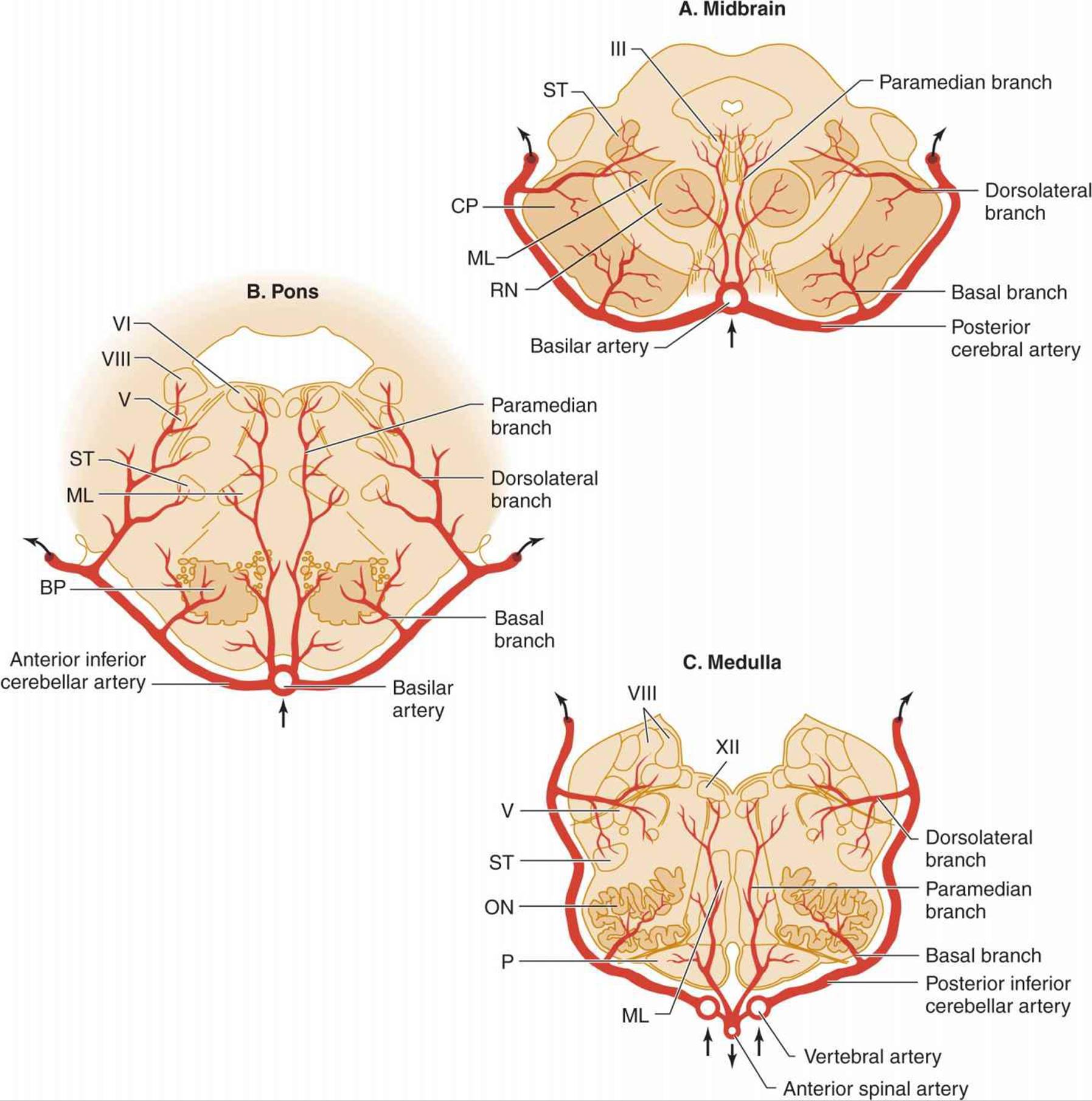
Figure 13-7. Arterial supply of the brainstem. A: Midbrain. The basilar artery gives off paramedian branches that supply the oculomotor (III) nerve nucleus and the red nucleus (RN). A larger branch, the posterior cerebral artery, courses laterally around the midbrain, giving off a basal branch that supplies the cerebral peduncle (CP) and a dorsolateral branch supplying the spinothalamic tract (ST) and medial lemniscus (ML). The posterior cerebral artery continues (upper arrows) to supply the thalamus, occipital lobe, and medial temporal lobe. B: Pons. Paramedian branches of the basilar artery supply the abducens (VI) nucleus and the medial lemniscus (ML). The anterior inferior cerebellar artery gives off a basal branch to the descending motor pathways in the basis pontis (BP) and a dorsolateral branch to the trigeminal (V) nucleus, the vestibular (VIII) nucleus, and the spinothalamic tract (ST), before passing to the cerebellum (upper arrows). C: Medulla. Paramedian branches of the vertebral arteries supply descending motor pathways in the pyramid (P), the medial lemniscus (ML), and the hypoglossal (XII) nucleus. Another vertebral branch, the posterior inferior cerebellar artery, gives off a basal branch to the olivary nuclei (ON) and a dorsolateral branch that supplies the trigeminal (V) nucleus, the vestibular (VIII) nucleus, and the spinothalamic tract (ST), on its way to the cerebellum (upper arrows). (Reproduced, with permission, from Waxman S. Clinical Neuroanatomy. 26th ed. New York, NY: McGraw-Hill; 2010.)
Major stenosis or occlusion of the subclavian artery before it has given rise to the vertebral artery can lead to the subclavian steal syndrome, in which blood passes from the vertebral artery into the distal subclavian artery with physical activity of the ipsilateral arm. The syndrome is a manifestation of generalized atherosclerosis and is not predictive of stroke in the vertebrobasilar system. Patients are usually asymptomatic, and stroke, when it occurs, is typically due to coexisting carotid lesions.
Basilar thrombosis usually affects the proximal portion of the basilar artery (Figure 13-6), which supplies the pons. Involvement of the dorsal portion (tegmentum) of the pons produces unilateral or bilateral abducens (VI) nerve palsy; horizontal eye movements are impaired, but vertical nystagmus and ocular bobbing may be present. The pupils are constricted as a result of the involvement of descending sympathetic pupillodilator fibers in the pons, but they may remain reactive. Hemiplegia or quadriplegia is usually present, and coma is common. Although the syndrome of basilar occlusion in unconscious patients may be confused with pontine hemorrhage, a CT or MRI brain scan will differentiate the two.
In some patients with basilar occlusion, the ventral portion of the pons (basis pontis) is infarcted and the tegmentum is spared. Such patients remain conscious but quadriplegic. The term locked-in syndrome is applied to this state. Locked-in patients may be able to signify that they are conscious by opening their eyes or moving their eyes vertically on command. In other cases, a conventional electroencephalogram (EEG) with stimulation may be needed to distinguish the locked-in state (in which the EEG is normal) from coma (Chapter 3).
2. Embolism—Emboli small enough to pass through the vertebral arteries into the larger basilar artery are usually arrested at the top of the basilar artery, where it bifurcates into the posterior cerebral arteries (Figure 13-6). The resulting reduction in blood flow to the ascending reticular formation of the midbrain and thalamus produces immediate loss or impairment of consciousness. Unilateral or bilateral oculomotor (III) nerve palsies are characteristic. Hemiplegia or quadriplegia with decerebrate or decorticate posturing occurs because of the involvement of the cerebral peduncles in the midbrain. Thus the top of the basilar syndrome may be confused with midbrain damage caused by transtentorial uncal herniation. Less commonly, an embolus may lodge more proximally in an atheromatous narrowed portion of the basilar artery, producing a syndrome indistinguishable from basilar thrombosis.
Smaller emboli may occlude the rostral basilar artery transiently before fragmenting and passing into one or both posterior cerebral arteries (Figure 13-6). In such cases, portions of the midbrain, thalamus, and temporal and occipital lobes can be infarcted. If conscious, these patients display a variety of visual (homonymous hemianopia, cortical blindness), visuomotor (impaired convergence, paralysis of upward or downward gaze, diplopia), and behavioral (especially confusion) abnormalities without prominent motor dysfunction. Sluggish pupillary responses are a helpful sign of midbrain involvement.
LONG CIRCUMFERENTIAL VERTEBROBASILAR BRANCHES
Anatomy
The long circumferential branches arising from the vertebral and basilar arteries are the posterior inferior cerebellar, anterior inferior cerebellar, and superior cerebellar arteries (Figure 13-2). These vessels supply the dorsolateral brainstem, including dorsolaterally situated cranial nerve nuclei (V, VII, and VIII) and pathways entering and leaving the cerebellum in the cerebellar peduncles.
Clinical Syndromes
Occlusion of one of the circumferential branches produces infarction in the dorsolateral medulla or pons.
1. Posterior inferior cerebellar artery occlusion results in the lateral medullary (Wallenberg) syndrome (Chapter 8). This syndrome varies in its presentation with the extent of infarction, but it can include ipsilateral cerebellar ataxia, Horner syndrome, and facial sensory deficit; contralateral impaired pain and temperature sensation; and nystagmus, vertigo, nausea, vomiting, dysphagia, dysarthria, and hiccup. The motor system is characteristically spared because of its ventral location in the brainstem.
2. Anterior inferior cerebellar artery occlusion leads to infarction of the lateral portion of the caudal pons and produces a syndrome with many of the same features. Horner syndrome, dysphagia, dysarthria, and hiccup do not occur, however, but ipsilateral facial weakness, gaze palsy, deafness, and tinnitus are common findings.
3. The syndrome of lateral rostral pontine infarction from superior cerebellar artery occlusion resembles that associated with anterior inferior cerebellar artery lesions, but impaired optokinetic nystagmus or skew deviation of the eyes may occur. Auditory function is unaffected, and the contralateral sensory disturbance may involve touch, vibration, and position sense as well as pain and temperature sense.
LONG PENETRATING PARAMEDIAN VERTEBROBASILAR BRANCHES
Anatomy
Long penetrating paramedian arteries supply the medial brainstem from its ventral surface to the floor of the fourth ventricle. Structures located in this region include the medial portion of the cerebral peduncle, sensory pathways, the red nucleus, the reticular formation, and the midline cranial nerve nuclei (III, IV, VI, XII).
Clinical Syndrome
Occlusion of a long penetrating artery causes paramedian infarction of the brainstem and results in contralateral hemiparesis if the cerebral peduncle is affected. Associated cranial nerve involvement depends on the level of the brainstem at which occlusion occurs. Occlusion in the mid-brain results in ipsilateral oculomotor (III) nerve palsy, which may be associated with contralateral tremor or ataxia from involvement of pathways connecting the red nucleus and cerebellum. Ipsilateral abducens (VI) and facial (VII) nerve palsies are seen in the pons, and hypoglossal (XII) nerve involvement can occur in the medulla.
If the lesion appears patchy or involves both sides of the brainstem (as manifested by coma or quadriparesis), the differential diagnosis includes occlusion of a main trunk vessel (both vertebral arteries or the basilar artery); intramedullary lesions such as hemorrhage, pontine glioma, or multiple sclerosis; and compression of the brainstem by a cerebellar mass (hemorrhage, infarct, or tumor).
SHORT BASAL VERTEBROBASILAR BRANCHES
Anatomy
Short branches arising from the long circumferential arteries (discussed previously) penetrate the ventral brainstem to supply the brainstem motor pathways.
Clinical Syndrome
The most striking finding is contralateral hemiparesis caused by corticospinal tract involvement in the cerebral peduncle or basis pontis. Cranial nerves (eg, III, VI, VII) that emerge from the ventral surface of the brainstem may be affected as well, giving rise to ipsilateral cranial nerve palsies.
LACUNAR INFARCTION
Anatomy
Small penetrating arteries located deep in the brain may become occluded as a result of changes in the vessel wall associated with chronic hypertension. The resulting lacunar infarcts are most common in deep nuclei of the brain (putamen, 37%; thalamus, 14%; caudate nucleus, 10%), the pons (16%), and the posterior limb of the internal capsule (10%) (Figure 13-8). They occur in lesser numbers in the deep cerebral white matter, the anterior limb of the internal capsule, and the cerebellum.

Figure 13-8. Arterial supply of deep cerebral structures frequently involved in lacunar infarction. The basal ganglia (caudate nucleus, putamen, and globus pallidus; light blue) and internal capsule are supplied by the anterior circulation (lenticulostriate branches of the middle cerebral artery and the anterior choroidal artery). The thalamus (dark blue) is supplied by the posterior circulation (thalamoperforate and thalamogeniculate branches of the posterior cerebral artery). Descending motor fibers to the face (F), arm (A), and leg (L) and ascending sensory fibers from face (f), arm (a), and leg (l) are shown in the posterior limb of the internal capsule. (Reproduced, with permission, from Waxman S. Clinical Neuroanatomy. 26th ed. New York, NY: McGraw-Hill; 2010.)
Clinical Syndromes
Because of their small size and their frequent location in relatively “silent” areas of the brain, many lacunar infarctions are not recognized clinically. In as many as three-fourths of autopsy-proved cases, there is no history of stroke or clear evidence of neurologic deficit on antemortem examinations.
In many cases, the isolated nature of the neurologic deficit makes the clinical picture of lacunar infarction distinctive. The onset of lacunar stroke may be gradual, developing over several hours or days. Headache is absent or minor, and the level of consciousness is unchanged.
Recognition of lacunar stroke syndromes is important because the prognosis for complete or nearly complete recovery is good. In addition, the likelihood of future lacunar strokes may be reduced by treating the hypertension that is usually associated with them. Because the arteries involved are small, angiography is normal and not indicated. The CSF is also normal. A CT scan or MRI may not disclose the lesion, but should be performed to exclude other causes of stroke. Anticoagulation is not indicated because there is no evidence that it confers any benefit in this context. Aspirin is also of uncertain benefit, but it is often given because of the low risk of serious complications. Although a wide variety of deficits can be produced, there are four classic and distinctive lacunar syndromes.
1. Pure motor hemiparesis—This consists of hemiparesis affecting the face, arm, and leg to a roughly equal extent, without an associated disturbance of sensation, vision, or language. When lacunar in origin, it is usually due to a lesion in the contralateral internal capsule or pons. Pure motor hemiparesis also may be caused by internal carotid or middle cerebral artery occlusion, subdural hematoma, or intracerebral mass lesions.
2. Pure sensory stroke—This is characterized by hemisensory loss, which may be associated with paresthesia, and results from lacunar infarction in the contralateral thalamus. It may be mimicked by occlusion of the posterior cerebral artery or by a small hemorrhage in the thalamus or midbrain.
3. Ataxic hemiparesis—In this syndrome, sometimes called ipsilateral ataxia and crural (leg) paresis, pure motor hemiparesis is combined with ataxia of the hemiparetic side and usually predominantly affects the leg. Symptoms result from a lesion in the contralateral pons, internal capsule, or subcortical white matter.
4. Dysarthria-clumsy hand syndrome—This consists of dysarthria, facial weakness, dysphagia, and mild weakness and clumsiness of the hand on the side of facial involvement. When the syndrome is caused by a lacunar infarct, the lesion is in the contralateral pons or internal capsule. Infarcts or small intracerebral hemorrhages at a variety of locations can produce a similar syndrome, however. In contrast to the lacunar syndromes described earlier, premonitory TIAs are unusual.
ETIOLOGY
Focal cerebral ischemia can result from underlying disorders that primarily affect the blood, blood vessels, or heart (Table 13-5).

Table 13-5. Conditions associated with focal cerebral ischemia.
VASCULAR DISORDERS
Atherosclerosis
Atherosclerosis of the large extracranial arteries in the neck and at the base of the brain is the underlying cause of focal cerebral ischemia in the great majority of cases. Atherosclerosis affects large and medium-sized elastic and muscular arteries. Within the cerebral circulation, the sites of predilection (Figure 13-9) are the origin of the common carotid artery, the internal carotid artery just above the common carotid bifurcation and within the cavernous sinus, the origin of the middle cerebral artery, the vertebral artery at its origin and just above where it enters the skull, and the basilar artery.

Figure 13-9. Sites of predilection (dark red areas) for atherosclerosis in the intracranial arterial circulation.
The pathogenesis of atherosclerosis is incompletely understood, but injury to and resulting dysfunction of vascular endothelial cells is thought to be an early step (Figure 13-10). Endothelial cells may be injured by circulating low-density lipoprotein (LDL), free radicals, hypertension, diabetes, homocysteine, infectious agents, or shear stress related to turbulent blood flow. As a consequence, endothelial permeability is altered, permitting entry of LDL into the arterial wall. Blood monocytes and T lymphocytes adhere to sites of endothelial injury and migrate subendothelially, where monocytes and monocyte-derived macrophages endocytose LDL particles and transform into lipid-laden foam cells. The resulting lesion is called a fatty streak.

Figure 13-10. Arterial lesion in atherosclerosis. Endothelial injury permits entry of low-density lipoprotein cholesterol and circulating mononuclear cells into the vessel wall, where they form a fatty streak. The subsequent attachment of platelets and proliferation of smooth muscle cells within this lesion leads to production of a fibrous plaque, which may encroach on the arterial lumen or rupture to occlude the vessel and provide a source of emboli.
At this stage, release of growth and chemotactic factors from endothelial cells and macrophages stimulates proliferation of intimal smooth muscle cells and migration of additional smooth muscle cells to the intima from the tunica media. These cells secrete extracellular matrix constituents, leading to the formation of a fibrous cap over the atherosclerotic plaque, in which a necrotic core develops. In some cases, fractures in the cap lead to plaque rupture, a serious complication associated with the release of procoagulant factors and subsequent thrombosis. Possible outcomes include thrombotic occlusion of the vessel lumen or embolization.
Major risk factors for atherosclerosis leading to stroke include systolic or diastolic hypertension, elevated serum LDL cholesterol, and diabetes mellitus. Current recommendations are to reduce blood pressure to < 140 mm Hg systolic and < 90 mm Hg diastolic (<130 systolic and <80 diastolic if other risk factors are present), hold LDL cholesterol below 160 mg/dL (lower with other risk factors), and maintain pre- and postprandial capillary blood glucose concentrations at 90–130 and <180 mg/dL, respectively. These goals may be achieved by lifestyle change, dietary modification, or pharmacotherapy (eg, antihypertensives, statins, oral hypoglycemic drugs, or insulin).
Other Inflammatory Disorders
1. Giant cell arteritis, also called temporal arteritis, produces inflammatory changes that affect branches of the external carotid, cervical internal carotid, posterior ciliary, extracranial vertebral, and intracranial arteries. Inflammatory changes in the arterial wall may stimulate platelet adhesion and aggregation on damaged surfaces, leading to thrombosis or distal embolism. Physical examination may show tender, nodular, or pulseless temporal arteries. Laboratory findings include an increased erythrocyte sedimentation rate and evidence of vascular stenosis or occlusion on angiography or color duplex ultrasonography. Definitive diagnosis is by temporal artery biopsy. Although it is an uncommon cause of cerebral ischemic symptoms, giant cell arteritis should be considered in patients with transient monocular blindness or transient cerebral ischemic attacks—especially elderly patients—because the disorder is responsive to corticosteroid therapy and its complications (especially permanent blindness) may thus be avoided. Treatment is discussed in more detail in Chapter 6.
2. Systemic lupus erythematosus is associated with a vasculopathy that involves small cerebral vessels and leads to multiple microinfarctions. Inflammatory changes characteristic of true vasculitis are absent. Libman-Sacks endocarditis accompanying systemic lupus may be a source of cardiogenic emboli.
3. Polyarteritis nodosa is a segmental vasculitis of small-and medium-sized arteries that affects multiple organs. Transient symptoms of cerebral ischemia, including typical spells of transient monocular blindness, can occur.
4. Primary angiitis of the central nervous system (also referred to as granulomatous angiitis) is an idiopathic inflammatory disease that affects small arteries and veins in the central nervous system and can cause transient or progressive multifocal lesions. Clinical features include headache, hemiparesis and other focal neurologic abnormalities, and cognitive disturbances. The cerebrospinal fluid (CSF) usually shows pleocytosis and elevated protein, but because the systemic vasculature is spared, the erythrocyte sedimentation rate is typically normal. The diagnosis should be suspected in any patient with multifocal central nervous system dysfunction and CSF pleocytosis. Angiography demonstrates focal and segmental narrowing of small arteries and veins, and a meningeal biopsy is diagnostic. Treatment is discussed in Chapter 4.
5. Syphilitic arteritis occurs within 5 years after a primary syphilitic infection and reflects the underlying meningeal inflammatory process. It is important to recognize and treat the disorder at this early stage to prevent the development of tertiary parenchymal neurosyphilis (general paresis or tabes dorsalis). Medium-sized penetrating vessels are typically involved (Figure 13-11), producing punctate areas of infarction in the deep white matter of the cerebral hemisphere that can be seen on CT scan or MRI.

Figure 13-11. Left carotid angiogram (AP projection) in syphilitic arteritis showing marked narrowing of the proximal middle cerebral artery (arrows at right) and anterior cerebral artery (arrow at left). (Reproduced, with permission, from Lowenstein DH, Mills C, Simon RP. Acute syphilitic transverse myelitis: unusual presentation of meningovascular syphilis. Genitourin Med. 1987;63:333-338.)
6. AIDS is associated with an increased incidence of TIAs and ischemic stroke. In some cases, ischemic neurologic complications of AIDS are related to endocarditis or opportunistic infections of the central nervous system, such as toxoplasmosis or cryptococcal meningitis.
Fibromuscular Dysplasia
This uncommon condition affects large (especially renal and carotid) arteries of children and young adults, producing segmental thinning of the media and fragmentation of the elastic lamina, alternating with rings of fibrous and muscular hyperplasia within the media. Fibromuscular dysphasia is familial in approximately 10% of cases and is more common in women than in men. Cerebral symptoms result from arterial dissection, stenosis, thromboembolism, or aneurysmal rupture. A characteristic “string-of-beads” appearance on angiography is diagnostically helpful. Treatment of symptomatic carotid artery disease may include antiplatelet drugs, anticoagulation, percutaneous or surgical dilation of stenotic arterial segments, and micro-vascular clipping or endovascular coiling of aneurysms.
Carotid or Vertebral Artery Dissection
Dissection of the carotid or vertebral artery is associated with hemorrhage into the vessel wall, which can occlude the vessel or predispose to thrombus formation and embolization. Posttraumatic carotid dissections present little difficulty in diagnosis. Certain patients, however—usually young men—suffer cerebral infarction after apparently spontaneous carotid artery dissection. Internal carotid artery dissections usually originate near the carotid bifurcation and can extend to the base of the skull. The underlying pathologic process is usually cystic medial necrosis. Prodromal transient hemispheric ischemia or monocular blindness sometimes precedes a devastating stroke. Carotid dissection may be accompanied by pain in the jaw or neck, visual abnormalities akin to those that occur in migraine, or Horner syndrome.
Dissection of the vertebral or basilar artery is less common. The clinical features of this disorder include headache, posterior neck pain, and the sudden onset of signs of brainstem dysfunction.
The treatment of carotid or vertebral artery dissection is controversial. Approaches include no treatment, removal of the intramural hematoma, administration of aspirin or anticoagulants, and occlusion of the vessel distal to the dissection. Recurrent dissection is uncommon and usually occurs within 1 month of the initial event.
Lacunar Infarction
Lacunar infarction of the brain (discussed in more detail previously under Clinical-Anatomic Correlation) results from the occlusion of small penetrating branches of the major cerebral arteries, especially those that supply the basal ganglia, thalamus, internal capsule, and pons. Lacunar infarcts are believed to be caused by either atherosclerosis or degenerative changes in arterial walls (including lipohyalinosis and fibrinoid necrosis) that are related to longstanding hypertension. Both hypertension and diabetes appear to predispose to this type of stroke.
Drug Abuse
Use of cocaine, amphetamines, or heroin is a risk factor for stroke in patients younger than 35 years old. Patients who take these agents intravenously may develop infective endocarditis (see later), leading to embolic stroke. Stroke also occurs in drug users without endocarditis, however, including those who take drugs only intranasally or by smoke or vapor inhalation, and often has its onset within hours of drug use. Mechanisms that have been proposed to explain these events include drug-induced vasospasm, vasculitis, and the rupture of preexisting aneurysms or vascular malformation.
1. Cocaine hydrochloride is most often associated with intracerebral hemorrhage but can also cause subarachnoid hemorrhage or ischemic stroke.
2. Alkaloidal (crack) cocaine use can lead to stroke that is usually ischemic in origin, but intracerebral or subarachnoid hemorrhage also occurs.
3. Amphetamines can produce vasculitis, with necrosis of the vessel wall leading to intracerebral hemorrhage; ischemic stroke and subarachnoid hemorrhage are less frequent.
4. Other sympathomimetic amines, including phenylpropanolamine and ephedrine, are also associated with an increased risk of stroke.
5. Heroin is associated primarily with embolic stroke related to endocarditis.
Multiple Progressive Intracranial Arterial Occlusions (Moyamoya)
This syndrome has two essential features: (1) bilateral narrowing or occlusion of the distal internal carotid arteries and the adjacent anterior and middle cerebral artery trunks; and (2) the presence of a fine network of collateral channels at the base of the brain. The term moyamoya derives from a Japanese word meaning smoke or haze, which characterizes the angiographic appearance of these fine collaterals (Figure 13-12). Moyamoya is most common in Japanese girls, but occurs in all ethnic groups and in patients with atherosclerosis, sickle cell anemia, or a history of basilar meningitis. Both sporadic and genetic forms of the syndrome occur, and the latter are heterogeneous. Autosomal dominant moyamoya is caused by mutations in the gene for vascular smooth muscle actin (ACTA2) in some cases. Children tend to present with ischemic strokes and adults with intracerebral, subdural, or subarachnoid hemorrhage. Transient episodes of cerebral ischemia are infrequent.

Figure 13-12. Right carotid angiogram in moyamoya. The middle cerebral artery and its branches are replaced by a diffuse capillary pattern that has the appearance of a puff of smoke. A: AP view. B: Lateral view.
Migraine
Migraine with aura has been proposed as a cause of stroke, but in many cases, other stroke risk factors (eg, oral contraceptive use) may coexist. Stroke in migraineurs may occur during an attack of classic migraine and in the same vascular territory affected by previous migraine attacks. The anterior (especially the middle cerebral artery) and posterior (especially the posterior cerebral artery) cerebral circulations are affected about equally often.
Venous or Sinus Thrombosis
Thrombotic occlusion of a cerebral vein or venous sinus (Figure 13-13) is an uncommon cause of stroke; it is typically associated with a predisposing condition, such as otitis or sinusitis, a postpartum state, dehydration, or coagulopathy. Clinical features include headache, papilledema, impaired consciousness, seizures, and focal neurologic deficits. CSF pressure is typically increased, and in cases of septic thrombosis, pleocytosis may occur. A CT scan may demonstrate hemorrhage associated with venous infarction, and in superior sagittal sinus thrombosis a CT scan with contrast sometimes shows a filling defect corresponding to the clot (delta sign). However, MRI with contrast is the diagnostic procedure of choice in most cases. The diagnosis may be confirmed by MR angiography. In patients presenting with headache and papilledema, venous or sinus thrombosis must be differentiated from intracranial mass lesions and idiopathic intracranial hypertension (pseudotumor cerebri; Chapter 6). The radiologic studies mentioned previously are useful in this regard. Septic thromboses are treated with antibiotics. Anticoagulation has been used for aseptic thrombosis, but its efficacy is uncertain, and it may precipitate intracranial hemorrhage.

Figure 13-13. Major cerebral veins and venous sinuses subject to thrombotic occlusion. (Reproduced, with permission, from Waxman S. Clinical Neuroanatomy. 26th ed. New York, NY: McGraw-Hill; 2010.)
CARDIAC DISORDERS
Mural Thrombus
Mural thrombus complicating myocardial infarction or cardiomyopathy is a recognized source of cerebral embolism. The risk of stroke in the first weeks after myocardial infarction is related to the size of the cardiac lesion. More extensive myocardial damage may increase the tendency for mural thrombi to form, exacerbate the generalized hypercoagulable state that accompanies infarction, or both. Warfarin, or the combination of warfarin and antiplatelet drugs, may be superior to antiplatelet drugs alone for preventing stroke after myocardial infarction, but is also associated with an increase in major bleeding complications.
Rheumatic Heart Disease
The incidence of focal cerebral ischemia is increased in patients with rheumatic heart disease—particularly those with mitral stenosis and atrial fibrillation—presumably as a result of embolization. Such patients should receive anticoagulants (see Treatment later). In other cases, cerebral symptoms are temporally related to exertion, suggesting hypoperfusion as the cause.
Arrhythmia
Atrial fibrillation (especially when associated with rheumatic heart disease) and the tachycardia-bradycardia syndrome (a form of sick sinus syndrome) are well-recognized causes of embolic stroke. Other cardiac arrhythmias are more likely to produce pancerebral hypoperfusion with diffuse rather than focal symptoms (eg, syncope, dimming of vision, nonspecific lightheadedness, generalized seizures) unless severe carotid artery stenosis is also present. Patients with atrial fibrillation should be treated with oral anticoagulants (see Treatment later) to reduce the risk of stroke and other embolic complications. Administration of the antiarrhythmic drug dronedarone (400 mg orally twice daily) appears to produce additional reduction of stroke risk.
Endocarditis
1. Infective (bacterial or fungal) endocarditis is a cause of transient cerebral ischemia, embolic cerebral infarction (especially within the middle cerebral artery territory), and intracerebral or subarachnoid hemorrhage (from rupture of a mycotic aneurysm). The incidence of these complications is highest during the active phase of infection and the first few months after antibiotic cure. Except where rheumatic heart disease is still common, the risk of endocarditis is associated primarily with intravenous drug use, hemodialysis, intravenous catheterization, and degenerative or prosthetic heart valves. Staphylococcus aureus and viridans streptococci are the most common causal organisms in patients with native valves and community-acquired endocarditis, whereas Staphylococcus aureus predominates in intravenous drug users, patients with hospital-acquired or other treatment-related infections, and recent recipients of prosthetic heart valves. Fungal endocarditis is rare (approximately 2% of cases), is usually caused by Candida or Aspergillus, and has a worse prognosis.
Signs of infective endocarditis include heart murmur, petechiae, subungual splinter hemorrhages, retinal Roth spots (red spots with white centers), Osler nodes (painful red or purple digital nodules), Janeway lesions(red macules on the palms or soles), and clubbing of the fingers or toes. The diagnosis is usually made by culturing the responsible organism from the blood and imaging vegetations with echocardiography. Treatment is with antibiotics and, in the setting of congestive heart failure, persistent infection, recurrent emboli, or large vegetations, valve replacement surgery. Anticoagulation should be avoided because of the risk of intracranial hemorrhage. Mortality is 10% to 30% in bacterial and approximately 50% in fungal endocarditis.
2. Nonbacterial (marantic) endocarditis is most frequent in patients with cancer and causes the majority of ischemic strokes in this population. The tumors most often associated with this type of stroke are adenocarcinomas of the lung or gastrointestinal tract. Vegetations are present on the mitral or aortic valves, but associated murmurs are rare. Vegetations may be detected by two-dimensional echocardiography, but failure to demonstrate vegetations does not exclude the diagnosis. Anticoagulation with heparin can be useful in patients with treatable tumors or with other treatable causes of marantic endocarditis, such as sepsis.
Mitral Valve Prolapse
Buckling of the mitral valve due to stretching of the mitral annulus (mitral valve prolapse) is common, occurring in 4% to 8% of young adults, and usually produces no symptoms. In some cases there appears to be an association with cerebral ischemia, but the degree to which the disorder increases the risk of stroke is apparently small, and massive strokes related to mitral valve prolapse are rare.
Paradoxical Embolus
Congenital cardiac anomalies associated with a pathologic communication between the right and left sides of the heart, such as atrial septal defect or patent foramen ovale, may permit the passage of embolic material from the systemic venous circulation to the brain. Some but not all studies suggest that patent foramen ovale, which is present in up to one-third of adults, may explain some strokes for which no other cause can be found. However, evidence that closure of a patent foramen ovale reduces stroke risk is lacking.
Atrial Myxoma
This rare disorder can lead to either embolization (producing stroke) or cardiac outflow obstruction (producing syncope). Embolic events occur in one-fourth to one-half of patients with sporadic left atrial myxoma, and hemorrhagic strokes may occur. Diagnosis is by echocardiography.
Prosthetic Heart Valves
Patients with prosthetic heart valves are at increased risk for embolic stroke, which varies with the composition and location of the valve. Mechanical valves present the highest risk and require chronic administration of warfarin, with or without adjunctive low-dose (eg, 81 mg/d) aspirin. Transcatheter valves are associated with fewer thromboembolic complications, and antiplatelet treatment with aspirin and clopidogrel for 6 months is thought to be adequate. Bioprosthetic (bovine or porcine) valves are the least thrombogenic and are usually managed with a 3-month course of warfarin, although low-dose aspirin is often used instead. Mitral valve prostheses are generally associated with a higher risk of thromboembolic complications than are aortic valve prostheses.
HEMATOLOGIC DISORDERS
Thrombocytosis
Thrombocytosis occurs in myeloproliferative disorders, other neoplastic or infectious diseases, and after splenectomy. Thrombocytosis may predispose to focal cerebral ischemia when the platelet count exceeds 1,000,000/μL. Treatment is of the underlying disorder and, especially in the elderly or those with a history of thrombotic disease, reduction of the platelet count to ≤400,000/μL with hydroxyurea or anagrelide. Addition of an antiplatelet agent is of uncertain value.
Polycythemia
Polycythemia with hematocrits above 46% is associated with reduced cerebral blood flow and increased risk of stroke. This risk rises dramatically when the hematocrit exceeds 60%. Treatment is by venesection to reduce the hematocrit to <45% (for men) or <42% (for women) and, in some cases, administration of hydroxyurea.
Sickle Cell Disease
Sickle cell (hemoglobin S) disease results from a mutation in the hemoglobin beta chain gene (HBB) that produces a single (GLU6VAL) amino acid substitution in the protein. Persons of West African descent are most frequently affected. The mutation causes sickle-shaped deformation of erythrocytes when the partial pressure of oxygen in blood is reduced and produces hemolytic anemia and vascular occlusions, which may be extremely painful (sickle cell crises). Homozygotes are more severely affected than heterozygotes.
The most frequent neurologic complication is stroke, which occurs in approximately 10% of patients by age 20 and 25% by age 45 years. Stroke related to sickle cell disease usually affects the intracranial internal carotid or proximal middle or anterior cerebral artery. Detection of increased cerebral blood flow velocity by transcranial Doppler studies may help to identify individuals at increased risk for stroke. If patients with sickle cell disease require angiography, the level of hemoglobin S should be reduced by exchange transfusion to less than 20%, because radiologic contrast media may induce sickling.
Primary prevention of stroke in patients with sickle cell disease and abnormal transcranial Doppler studies involves blood transfusion every 3 to 4 weeks to reduce hemoglobin S levels to ≤30%. Complications include iron overload, which can be reduced by chelation therapy, and a rebound increase in stroke risk if transfusions are discontinued. Administration of hydroxyurea may be an alternative approach. Patients with prior stroke are also treated with chronic transfusion to prevent stroke recurrence and can be switched to hydroxyurea after a period in which the two treatments are overlapped.
Leukocytosis
Transient cerebral ischemia has been reported in association with leukocytosis, usually in patients with leukemia and white blood cell counts in excess of 150,000/μL.
Hypercoagulable States
Hyperviscosity of the serum from paraproteinemia (especially macroglobulinemia) is an infrequent cause of focal cerebral ischemia. Additional causes of hypercoagulable states that may be associated with stroke include estrogen therapy, oral contraceptives, postpartum and postoperative states, cancer, antiphospholipid or anti-cardiolipin antibodies, and hereditary coagulopathies (eg, heparin cofactor II deficiency, protein C deficiency, defective release of plasminogen activator, or factor XII deficiency).
CLINICAL FINDINGS
HISTORY
Predisposing Factors
In patients with cerebrovascular disorders, possible risk factors (Table 13-1) such as TIAs, hypertension, cigarette smoking, diabetes, and dyslipidemia should be sought. In women, the use of oral contraceptives has been associated with cerebral arterial and venous occlusive disease, especially in the presence of hypertension and cigarette smoking. The presence of such medical conditions as ischemic or valvular heart disease or cardiac arrhythmias must also be ascertained. A variety of systemic disorders involving the blood or blood vessels (Table 13-5) also increase the risk of stroke. Antihypertensive drugs can precipitate cerebrovascular symptoms if the blood pressure is lowered excessively in patients with nearly total cerebrovascular occlusion and poor collateral circulation.
Onset & Course
The history must address whether the clinical picture is that of TIA, stroke in evolution, or completed stroke (Figure 13-1). In some cases, it also may be possible to assess the likelihood that a stroke is thrombotic or embolic in origin from the clinical history.
1. Features suggesting thrombotic stroke—Patients with thrombotic vascular occlusion often present with stepwise incremental neurologic deficits; the occlusion may be preceded by a series of TIAs with identical symptoms. TIAs precede infarction in 25% to 50% of patients with occlusive atherosclerotic disease of the extracranial internal carotid arteries. In approximately one-third of such patients, however, the onset of infarction is abrupt, suggesting that embolization from the distal extracranial artery to the intracranial artery may be the cause of stroke.
2. Features suggesting embolic stroke—Cerebral embolism typically causes neurologic deficits that occur abruptly with no warning and are maximal at onset. In many patients, a cardiac origin of emboli is suggested by multifocal cerebral infarction, cardiac valvular disease, cardiomegaly, arrhythmias, or endocarditis.
Associated Symptoms
1. Seizures accompany the onset of stroke in a small number of cases; in other instances, they follow the stroke by weeks to years. The presence of seizures does not definitively distinguish embolic from thrombotic strokes, but seizure at the onset of stroke may be more common with embolus. Excluding patients with vertebrobasilar stroke or another cause of seizures, the incidence of epilepsy after stroke is approximately 10%. The risk of epilepsy increases to approximately 25% with cortical strokes and to 50% when cortical strokes are associated with a persistent motor deficit.
2. Headache occurs in approximately 25% of patients with ischemic stroke, possibly because of the acute dilation of collateral vessels.
PHYSICAL EXAMINATION
General Physical Examination
The general physical examination of a patient with a cerebrovascular disorder should focus on searching for an underlying systemic cause, especially a treatable one.
1. The blood pressure should be measured to ascertain whether hypertension—a major risk factor for stroke—is present.
2. Comparison of blood pressure and pulse on the two sides can reveal differences related to atherosclerotic disease of the aortic arch or coarctation of the aorta.
3. Ophthalmoscopic examination of the retina can provide evidence of embolization in the anterior circulation, in the form of visible embolic material in retinal blood vessels.
4. Neck examination may reveal the absence of carotid pulses or the presence of carotid bruits. Reduced carotid artery pulsation in the neck is a poor indicator of internal carotid artery disease, however. Although carotid bruits have been associated with cerebrovascular disease, significant carotid stenosis can occur without an audible bruit; conversely, a loud bruit can occur without stenosis.
5. Cardiac examination is essential to detect arrhythmias or murmurs related to valvular disease, either of which may predispose to cardiogenic embolization and stroke.
6. Palpation of the temporal arteries is useful in the diagnosis of giant cell arteritis, in which these vessels may be tender, nodular, or pulseless.
Neurologic Examination
Patients with cerebrovascular disorders may or may not have abnormal neurologic findings on examination. A normal examination is expected, for example, after a TIA has resolved. Where deficits are found, the goal of the neurologic examination is to define the anatomic site of the lesion, which may suggest the cause or optimal management of the stroke. For example, clear evidence that the anterior circulation is involved may lead to angiographic evaluation in contemplation of possible surgical correction of an internal carotid lesion. Establishing that the symptoms are associated with vertebrobasilar circulation or to a lacunar infarction will dictate a different course of action.
1. Cognitive deficits that indicate cortical lesions in the anterior circulation should be sought. For example, if aphasia is present, the underlying disorder cannot be in the posterior circulation and is unlikely to represent lacunar infarction. The same is true for nondominant hemisphere lesions producing parietal lobe syndromes such as unilateral neglect or constructional apraxia.
2. Visual field abnormalities similarly exclude lacunar infarction. Hemianopia may occur, however, with involvement of either the middle or posterior cerebral artery, which supplies the optic radiations and primary visual cortex, respectively. Isolated hemianopia suggests posterior cerebral artery infarction, because hemianopia from middle cerebral artery stroke should produce additional deficits.
3. Ocular palsies, nystagmus, or internuclear ophthalmoplegia assign the underlying lesion to the brainstem and thus to the posterior circulation.
4. Hemiparesis can be due to lesions in cerebral cortical regions supplied by the anterior circulation, descending motor pathways in the brainstem supplied by the vertebrobasilar system, or lacunes at subcortical (corona radiata, internal capsule) or brainstem sites. However, hemiparesis affecting the face, hand, and arm more than the leg is characteristic of lesions within the distribution of the middle cerebral artery. Hemiparesis that is nonselective with respect to the face, arm, and leg is consistent with occlusion of the internal carotid artery or the stem of the middle cerebral artery, lacunar infarction in the internal capsule or basal ganglia, or brainstem disease. A crossed hemiparesis—that is, one that involves the face on one side and the rest of the body on the other—means that the abnormality must lie between the level of the facial (VII) nerve nucleus in the pons and the decussation of the pyramids in the medulla.
5. Cortical sensory deficits such as astereognosis or agraphesthesia with preserved primary sensory modalities imply a cerebral cortical deficit within the territory of the middle cerebral artery. Isolated hemisensory deficits without associated motor involvement are usually lacunar in origin. Crossed sensory deficits result from brainstem lesions in the medulla, as seen in the lateral medullary syndrome (Wallenberg syndrome, Chapter 8).
6. Hemiataxia usually points to a lesion in the ipsilateral brainstem or cerebellum but can also be produced by lacunar stroke in the internal capsule.
INVESTIGATIVE STUDIES
BLOOD TESTS
These should be obtained routinely to detect treatable causes of stroke and to exclude conditions that can mimic stroke. The recommended studies are listed below.
Complete Blood Count
A complete blood count can identify possible causes of stroke (eg, thrombocytosis, polycythemia, sickle cell disease, or leukocytosis) or concomitant infection that may complicate the clinical course. For unclear reasons, an elevated white blood cell count on admission may also be an independent predictor of worse outcome from stroke.
Inflammatory Markers
An increased erythrocyte sedimentation rate (ESR) is seen in giant cell arteritis and other vasculitides. Elevated C-reactive protein (CRP) is a marker of systemic inflammation associated with increased stroke risk.
Serologic Assay for Syphilis
A treponemal assay, such as the blood FTA-ABS or MHA-TP (or CSF VDRL), indicates whether the patient has ever had syphilis. A negative test, therefore, excludes syphilitic arteritis as the cause of stroke.
Serum Glucose
Determining the blood glucose level is extremely important because hypoglycemia or hyperglycemia can present with focal neurologic signs and masquerade as stroke. Hypoglycemia associated with focal signs, seizures, or coma demands immediate administration of glucose to avoid permanent brain injury (Chapter 3) and hyperglycemia (hyperosmolar nonketotic hyperglycemia or diabetic ketoacidosis) also requires prompt specific treatment (Chapter 4). Hyperglycemia at presentation also independently portends a worse outcome from stroke, although the benefit of treatment to lower blood glucose in this setting is uncertain.
Serum Lipids
Measurement of total and HDL cholesterol can help to assess risk factors for stroke.
ELECTROCARDIOGRAM (ECG)
An ECG should be obtained routinely to detect unrecognized myocardial infarction or cardiac arrhythmias, such as atrial fibrillation, which predispose to embolic stroke.
LUMBAR PUNCTURE
Lumbar puncture should be performed in selected cases to exclude subarachnoid hemorrhage (manifested by xanthochromia and red blood cells) or to document meningovascular syphilis (reactive VDRL) as the cause of stroke. The procedure, possible complications, and analysis of results are described in detail in Chapter 2.
CT SCAN OR MRI
A CT scan or MRI (Figure 13-14) should be obtained routinely to distinguish between infarction and hemorrhage as the cause of stroke, to exclude other lesions (eg, tumor, abscess) that can mimic stroke, and to localize the lesion. Noncontrast CT is usually preferred for initial diagnosis because it is widely available and rapid and can readily make the critical distinction between ischemia and hemorrhage. However, its sensitivity within the first 6 hours is limited. MRI may be superior to CT scan for demonstrating early ischemic infarcts, showing ischemic strokes in the brainstem or cerebellum, and detecting thrombotic occlusion of venous sinuses. The relative benefits of CT and MRI in the diagnosis of stroke are discussed in more detail in Chapter 2.


Figure 13-14. Imaging studies in ischemic stroke in the right middle cerebral artery territory. A: CT scan showing low density and effacement of cortical sulci (between arrowheads) and compression of the anterior horn of the lateral ventricle (arrow). B: T1-weighted MRI scan showing loss of sulcal markings (between arrowheads) and compression of the anterior horn of the lateral ventricle (arrow). C: T2-weighted MRI scan showing increased signal intensity (between arrowheads) and ventricular compression (arrow).
Diffusion-weighted MRI (DWI) and perfusion-weighted MRI (PWI) are additional imaging techniques that may be useful for early detection (enabling early treatment) and prognostication in stroke. Diffusion-weighted MRI measures brownian movement of water molecules in tissue. Because arterial occlusion reduces the diffusion of water within minutes to hours, DWI is superior to CT for detecting stroke during the first 12 hours after onset. DWI abnormalities also tend to predict ultimate infarct volume in anterior circulation stroke, although diffusion defects are sometimes seen with TIAs, and small strokes or brainstem strokes may escape detection. PWI measures capillary perfusion of tissue and allows cerebral blood flow to be calculated. Some studies suggest that DWI delineates the ischemic core, whereas PWI outlines the full extent of hypoperfusion. According to this formulation, the difference between DWI and PWI abnormalities (diffusion-perfusion mismatch) represents tissue that is at risk of infarction, but potentially salvageable, which equates roughly to the ischemic penumbra. Thus diffusion-perfusion mismatch may indicate the extent to which thrombolysis or other treatments might be beneficial. Both techniques are considered further in Chapter 2.
VESSEL IMAGING
A variety of imaging techniques are available to identify underlying structural causes of cerebrovascular disease (eg, vasculitis, fibromuscular dysplasia, arterial dissection, aneurysm, arteriovenous malformation) and to detect and characterize operable extracranial carotid lesions in appropriate patients. Digital subtraction angiography, in which contrast medium is injected into the femoral artery followed by x-ray imaging, is highly sensitive and specific for detecting moderate to severe stenosis of the extracranial internal carotid artery. However, this technique is invasive and carries a risk of neurovascular complications and has been largely supplanted by noninvasive methods.
Doppler ultrasonography can also detect stenosis of the internal carotid artery and is noninvasive, but may not be able to distinguish stenosis from occlusion and does not visualize the surrounding vascular anatomy. However, its safety and low false-negative rate have led to its wide use as a screening test.
Magnetic resonance angiography (MRA) with contrast enhancement is now commonly used as a noninvasive substitute for digital subtraction angiography, despite somewhat lower resolution. CT angiography is an alternative approach, although it involves radiation exposure and may be obscured by artifact caused by calcium within atherosclerotic plaques.
Certain anatomic features of atherosclerotic plaques—such as a large lipid core, attenuated or ruptured fibrous cap, or intraplaque hemorrhage—may be associated with an increased risk of stroke. For this reason, methods for carotid-plaque imaging, such as high-resolution MRI or 18fluorodeoxyglucose positron emission tomography, may be diagnostically useful.
Transcranial Doppler ultrasonography is sometimes used in the evaluation of suspected stenosis of the intracranial internal carotid artery, middle cerebral artery, or basilar artery and for detecting and following the course of cerebral vasospasm after aneurysmal subarachnoid hemorrhage.
ECHOCARDIOGRAPHY
Echocardiography may be useful for demonstrating the cardiac lesions responsible for embolic stroke in patients with, for example, mural thrombus, valvular disease, or atrial myxoma.
ELECTROENCEPHALOGRAM (EEG)
The EEG is rarely useful in evaluating stroke. It may, however, help differentiate between a seizure disorder and TIAs or between lacunar and cortical infarcts in the occasional patient in whom these possibilities cannot otherwise be distinguished.
DIFFERENTIAL DIAGNOSIS
In patients presenting with focal central nervous system dysfunction of sudden onset, ischemic stroke must be distinguished from structural and metabolic processes that can mimic it. An underlying process other than focal cerebral ischemia should be suspected when the resulting neurologic deficit does not conform to the distribution of any single cerebral artery. In addition, strokes do not typically impair consciousness in the absence of profound focal deficits, whereas other cerebral disorders may do so.
Vascular disorders sometimes mistaken for ischemic stroke include intracerebral hemorrhage, subdural or epidural hematoma, and subarachnoid hemorrhage from rupture of an aneurysm or vascular malformation. These conditions often can be distinguished by a history of trauma or of excruciating headache at onset, by a more marked depression of consciousness, or by the presence of neck stiffness on examination. They can be excluded by CT scan or MRI.
Other structural brain lesions such as tumor or abscess can also produce focal cerebral symptoms of acute onset. Brain abscess is suggested by concurrent fever, and both abscess and tumor usually can be diagnosed by CT scan or MRI. Metabolic disturbances, particularly hypoglycemia and hyperosmolar nonketotic hyperglycemia, may present in stroke-like fashion. The serum glucose level therefore should be determined in all patients with apparent stroke.
TREATMENT
Drugs used in the prevention or treatment of cerebrovascular disease are listed in Table 13-6. Treatment related to specific underlying vascular, cardiac, and hematologic causes of stroke (eg, anti-inflammatory, antibiotic or anti-arrhythmic drugs) is addressed earlier in the section on Etiology.

Table 13-6. Drugs for treatment of cerebrovascular disease.
PRIMARY PREVENTION
Risk Factors
Primary prevention of stroke should be directed first at the correction or amelioration of known modifiable risk factors, such as hypertension, cigarette smoking, dyslipidemia, diabetes, obesity, and physical inactivity (Table 13-1).
Aspirin
Current evidence also supports the administration of aspirin to reduce the incidence of cardiovascular events (including, but not specifically, stroke) in patients with increased (6%-10%) 10-year risk for such events. This would include, for example, a 65-year-old man or 71-year-old woman with untreated high-normal systolic blood pressure of 140 mm Hg and no other risk factors (diabetes, cigarette smoking, coronary vascular disease, atrial fibrillation, or left ventricular hypertrophy).
Cardioembolic Source
Primary preventive therapy is also indicated in patients with cardiac disorders that predispose to stroke, such as atrial fibrillation, acute myocardial infarction with left ventricular thrombus, or a mechanical prosthetic heart valve. Adjusted-dose warfarin treatment with a target international normalized ratio (INR) of 2.0 to 3.0 reduces stroke risk in such patients by approximately 50% to 75%. Rivaroxaban (20 mg orally daily), a factor Xa inhibitor that is given at a fixed dose and does not require monitoring coagulation factors, appears to be as effective as warfarin for stroke prophylaxis in nonvalvular atrial fibrillation, with fewer bleeding complications. Aspirin is much less effective, and cardioversion is ineffective for this purpose.
Asymptomatic Carotid Artery Stenosis
Asymptomatic carotid stenosis producing 70% to 99% reduction in vessel diameter is also associated with an increased risk of stroke. Total carotid occlusion is not a risk factor, because the absence of symptoms implies adequate compensation by collateral circulation. Whether asymptomatic stenosis should be managed medically (with antiplatelet drugs) or surgically (with carotid endarterectomy or carotid artery stenting [discussed later]) depends on several factors, including the surgical complication rate, which should be ≤3% for surgery to be considered in asymptomatic patients.
TRANSIENT ISCHEMIC ATTACK
A transient ischemic attack, or TIA, is an episode of focal cerebral ischemia that resolves fully and rapidly, usually within 1 hour. By definition, a patient with TIA has no residual neurologic deficit at the time of diagnosis. The goal of treatment is to prevent subsequent stroke, which occurs in approximately 5% to 7% of patients within 48 hours and 10% to 15% of patients within 3 months after TIA.
Investigations
Admission to the hospital is preferred so that if symptoms recur, they can be managed expeditiously. Patients should be evaluated urgently with blood tests (complete blood count, prothrombin and partial thromboplastin time, erythrocyte sedimentation rate, treponemal test for syphilis, glucose) and ECG to identify underlying causes of cerebrovascular disease, and MRI with diffusion-weighted imaging or CT scan to document TIA or stroke and exclude other disorders. Patients with symptoms or imaging findings consistent with anterior circulation ischemia who are good surgical candidates should undergo MR angiography, CT angiography, or Doppler ultrasonography to detect operable lesions in the extracranial carotid artery. Echocardiography should be performed if there is a predisposing cardiac disorder or if symptoms suggest cardiogenic embolus (eg, recurrent TIAs with symptoms related to different territories).
Medical Treatment
1. Blood pressure should usually not be lowered acutely because of the risk of decreasing perfusion of at-risk brain tissue.
2. Anticoagulation with heparin given by continuous intravenous infusion to achieve an activated partial thromboplastin time (aPTT) 1.5 to 2.5 times control followed by warfarin given orally daily to achieve an international normalized ratio (INR) of 2.0 to 3.5 or another oral anticoagulant (Table 13-6) is indicated if a cardiac embolic source appears to be responsible.
3. Antiplatelet therapy with aspirin/dipyridamole, aspirin alone, or clopidogrel (Table 13-6) is recommended for presumedly noncardiogenic TIAs, if either no surgically amenable vascular lesion is found (ie, there is no lesion, extracranial carotid stenosis of <50%-70%, complete carotid occlusion, an intracranial lesion, or a posterior circulation lesion) or the patient is not a candidate for carotid endarterectomy or stenting.
4. Statins (eg, atorvastatin 80 mg orally daily) also reduce the risk for stroke and other cardiovascular events in patients with TIA.
Interventional Treatment
In contrast to acute stroke (discussed later), TIA is not usually considered an indication for thrombolytic treatment, although there is controversy in this regard.
Surgical Treatment
1. Carotid endarterectomy (surgical removal of thrombus from a stenotic common or internal carotid artery in the neck) is indicated for patients with anterior circulation TIAs and high-grade (70%-99%) carotid stenosis—and for selected patients with moderate (50%-70%) stenosis—on the side appropriate to account for the symptoms. The net benefit of endarterectomy assumes a combined perioperative morbidity and mortality rate of less than 6%.
2. Carotid artery stenting, which involves percutaneous transluminal insertion of a nickel and titanium stent and embolic protection device, is similarly effective for extracranial carotid stenosis compared with endarterectomy, assuming a similar perioperative morbidity and mortality rate. Stenting is associated with an increased risk of periprocedural stroke, but a decreased risk of periprocedural myocardial infarction. Considering the generally greater adverse effect of stroke than of myocardial infarction on quality of life, carotid endarterectomy probably remains superior overall, although stenting may be preferable for some (eg, younger) patients.
3. Intracranial artery stenosis is not currently an indication for surgery. For patients with symptomatic, greater than 70% stenosis of the intracranial internal carotid, middle cerebral, vertebral, or basilar artery, aggressive medical therapy (two antiplatelet drugs and correction of cardiovascular risk factors) is superior to stenting for preventing stroke.
ACUTE STROKE
In contrast to TIA, a diagnosis of stroke implies a persistent focal neurologic deficit, which may be improving, stable, or worsening (stroke in evolution or progressing stroke) when the patient is seen.
Investigations
These are as described earlier for TIA.
Medical Treatment
1. Blood pressure should usually not be lowered acutely, unless it is high enough (>185 mm Hg systolic or >110 mm Hg diastolic) to make an otherwise suitable candidate ineligible for thrombolytic therapy (see later).
2. Anticoagulation with heparin followed by an oral anticoagulant is indicated, as described earlier for TIA and in Table 13-6, if the history, examination, and diagnostic investigations point to cardiogenic embolus as the likely cause of stroke. Although immediate anticoagulation may result in hemorrhage into the infarct, this rarely affects the ultimate outcome adversely unless the infarct is massive. The risk of hemorrhage is more than offset by the particularly high risk of recurrent embolization soon after an embolic stroke, so that anticoagulation should not be delayed in this setting.
3. Antiplatelet therapy with aspirin/dipyridamole, aspirin alone, or clopidogrel (Table 13-6), should be given in the absence of a cardioembolic source, if either no surgically amenable vascular lesion is found (ie, if there is no lesion, extracranial carotid stenosis of <50%-70%, complete carotid occlusion, an intracranial lesion, or a posterior circulation lesion) or the patient is not a candidate for interventional or surgical treatment.
4. Statins can be given to reduce the risk of future stroke and other cardiovascular events, as described earlier for TIA.
5. Antiedema agents such as mannitol and corticosteroids have not been shown to improve outcome from stroke.
6. Neuroprotective agents, such as barbiturates, calcium channel antagonists, excitatory amino acid receptor antagonists, and antioxidants, have not been shown to improve outcome from stroke.
Interventional Treatment
1. Intravenous thrombolysis—Intravenous administration of recombinant tissue plasminogen activator (rtPA or alteplase) within 4.5 hours of the onset of symptoms reduces disability and mortality from ischemic stroke. The drug is administered at a dose of 0.9 mg/kg, up to a maximum total dose of 90 mg; 10% of the dose is given as an intravenous bolus and the remainder as a continuous intravenous infusion over 60 minutes.
Contraindications to thrombolysis relate to the desire to avoid unnecessarily treating patients who are improving spontaneously or unlikely to benefit, or exacerbating bleeding complications (including intracerebral hemorrhage). Contraindications designed to avert unnecessary or ineffectual treatment include the presence of only minor or resolving neurologic deficits; onset of symptoms more than 6 hours prior to initiating treatment; and hypoglycemia (blood glucose <50 mg/dL), which can mimic stroke. Contraindications related to bleeding complications include recent trauma, surgery, or hemorrhage; blood pressure greater than 185 mm Hg systolic or greater than 110 mm Hg diastolic; and impaired coagulation (INR >1.7, elevated aPTT, or platelet count <100,000/μL).
Within the first 24 hours after administration of rtPA, anticoagulants and antiplatelet agents should not be given, blood pressure should be carefully monitored, and arterial puncture and placement of central venous lines, bladder catheters, and nasogastric tubes should be avoided.
2. Intraarterial thrombolysis—Intraarterial administration of rtPA is an investigational approach for the treatment of stroke that may be associated with improved recanalization rates compared with intravenous rtPA, but at the expense of slower onset of treatment. Intraarterial rtPA and combined intravenous and intraarterial rtPA are under study.
3. Clot retrieval—Mechanical endovascular therapy with clot-retrieval devices, such as the Food and Drug Administration–approved MERCI (Mechanical Embolus Removal in Cerebral Embolism) device, may prove useful for treating stroke, especially in isolated middle cerebral artery occlusion, and when combined with rtPA. However, additional studies of this approach are needed.
Surgical Treatment
The indications for surgical treatment of completed stroke are limited. When patients deteriorate as a consequence of brainstem compression after cerebellar infarction, however, posterior fossa decompression with evacuation of infarcted cerebellar tissue can be lifesaving.
Emergency carotid endarterectomy is sometimes performed in the setting of acute stroke, but has a high complication rate. Endarterectomy undertaken 2 or more weeks after stroke may be useful when less than the entire territory supplied by the affected vessel has been infarcted (partial stroke). Decompressive craniectomy is occasionally used in an effort to prevent transtentorial herniation and death in patients with extensive hemispheric stroke. Extracranial (superficial temporal artery)-to-intracranial (middle cerebral artery) (EC-IC) bypass surgery is usually reserved for special circumstances, such as moyamoya.
CHRONIC STROKE
Treatment of stroke in the chronic phase is directed at secondary prevention to avoid recurrence and optimizing outcome from the event already experienced.
Secondary Prevention
This involves measures similar to those employed in primary prevention, including correction of modifiable risk factors and administration of antiplatelet drugs and statins (or oral anticoagulants, if a cardioembolic source persists).
Rehabilitative Therapy
Most patients show spontaneous improvement in neurologic function in the 3 to 6 months after stroke, but physical, occupational, and speech therapy can provide additional benefit. Early commencement of rehabilitative therapy and high-intensity regimens are recommended, and patient motivation is an important factor. Examples of treatments for which evidence of benefit is strong include constraint-induced movement therapy and robot-assisted training for arm function, task-oriented and cardiorespiratory fitness training for walking, and occupational therapy for activities of daily living.
Noninvasive Brain Stimulation
Some studies indicate that stimulation of the affected hemisphere or inhibition of the contralateral hemisphere with transcranial magnetic or direct current stimulation can transiently improve motor or language function in patients with stroke. The clinical significance of these effects is not yet clear.
Pharmacotherapy
Recovery from stroke may be impaired by sedative and enhanced by stimulant drugs. The latter include antidepressants (tricyclics and selective serotonin reuptake inhibitors), which are often given for post-stroke depression, but which some studies suggest may have additional beneficial effects on activities of daily living, cognitive function, dexterity, gait, and mortality.
COMPLICATIONS
Medical complications are common after stroke and strongly affect outcome. They include infections (especially aspiration pneumonia and urinary tract infections), cardiac arrhythmias, dysphagia, gastrointestinal bleeding, deep vein thrombosis, pulmonary embolism, and depression. In some cases, such as arrhythmia and depression, complications may result directly from brain injury. These disorders are treated as under other circumstances.
PROGNOSIS
Outcome after stroke is influenced by a number of factors, the most important being the nature and severity of the resulting neurologic deficit. The patient’s age, the cause of stroke, and coexisting medical disorders also affect prognosis. Overall, somewhat fewer than 80% of patients with stroke survive for at least 1 month, and 10-year survival rates in the neighborhood of 35% have been cited. The latter figure is not surprising, considering the advanced age at which stroke commonly occurs. Of patients who survive the acute period, approximately one-half to two-thirds regain independent function, whereas approximately 15% require institutional care.
INTRACEREBRAL HEMORRHAGE
HYPERTENSIVE HEMORRHAGE
EPIDEMIOLOGY
Intracerebral hemorrhage causes 10% to 15% of strokes, and hypertension is the most common underlying cause of nontraumatic intracerebral hemorrhage. The incidence is lower in whites than in other groups, which relates to rates of hypertension.
PATHOPHYSIOLOGY
Cerebral Autoregulation
Autoregulation of cerebral blood flow (Figure 13-15), which is achieved by changes in the caliber of small resistance cerebral arteries, maintains constant cerebral blood flow as systemic blood pressure rises and falls. The range of autoregulated blood pressures is variable but normally wide.

Figure 13-15. Cerebrovascular autoregulation. A: Cerebral blood flow is maintained in the normal range over a wide range of blood pressures. At very low pressures, cerebral hypoperfusion occurs, producing syncope. Pressures that rise beyond the autoregulatory range can cause hypertensive encephalopathy. B: Structural changes in cerebral arteries shift the autoregulatory range to higher blood pressures. Hypoperfusion and syncope can occur at normal pressures, and pressures associated with hypertensive encephalopathy are increased.
In normotensive individuals, the lowest mean blood pressure at which autoregulation is effective is approximately 60 mm Hg. Below this level, changes in the caliber of cerebral arteries cannot compensate for decreased per-fusion pressure. Cerebral blood flow therefore declines, causing lightheadedness, confusion, and dimming of vision. If the mean blood pressure falls to less than 35 to 40 mm Hg, somnolence and then loss of consciousness follow. In contrast, at blood pressures above the upper limit of the range of autoregulation (150-200 mm Hg), cerebral blood flow increases, which can produce hypertensive encephalopathy (Chapter 4).
In chronically hypertensive individuals, the autoregulation curve is shifted, which may be due to damage to small arterial walls. The lower limit of the autoregulatory range rises, so cerebral blood flow declines when the mean arterial blood pressure falls to less than approximately 120 mm Hg. This makes marked reduction of blood pressure—especially to hypotensive levels—potentially hazardous. As noted previously in the section on treatment of focal cerebral ischemia, this is a particular hazard in patients with acute ischemic stroke.
Chronic Hypertension
Chronic hypertension appears to promote structural changes in the walls of penetrating small cerebral arteries and arterioles, consisting of fibrinoid necrosis (sometimes termed lipohyalinosis) and miliary (Charcot-Bouchard) aneurysms (Figure 13-16). Although their precise relationship to each other and to hypertensive hemorrhage is debated, fibrinoid necrosis may weaken vessel walls, predisposing them to aneurysm formation and rupture.


Figure 13-16. Distribution of Charcot-Bouchard aneurysms (stippling) underlying hypertensive intracerebral hemorrhage.
Acute Hypertension
The role of acute—as opposed to chronic—hypertension in the pathogenesis of intracerebral hemorrhage is uncertain. Most patients with intracerebral hemorrhage are hypertensive after the event, but this may be explained by baseline hypertension, the vasopressor response to increased intracranial pressure (Cushing reflex), or both. In addition, large intracerebral hemorrhages are an uncommon feature of acute hypertensive encephalopathy. Nevertheless, the fact that some patients with spontaneous intracerebral hemorrhage have no history of hypertension and lack signs of hypertensive end-organ disease suggests that more acute hypertensive episodes might be involved, as does the occurrence of intracerebral hemorrhage in young patients after amphetamine or cocaine abuse.
Hematoma Effects
Hypertensive hemorrhage causes compression rather than destruction of adjacent brain tissue, resulting in reduced perfusion and metabolism in the perihematomal region. However, the clinical significance of these phenomena is unclear. Breakdown products of extravasated blood may cause inflammation and secondary injury. Perihematomal edema is maximal approximately 3 days after nontraumatic intracerebral hemorrhage. The extent of edema correlates with hematoma size, which is a predictor of poor outcome, but edema itself is not an independent prognostic factor. Increased intracranial pressure results in tamponade of the ruptured vessel, but can also lead to brain herniation and death.
PATHOLOGY
Most hypertensive hemorrhages originate in areas of predilection corresponding to long, narrow penetrating arterial branches along which fibrinoid necrosis and Charcot-Bouchard aneurysms (see earlier) are found at autopsy. These include the caudate and putaminal branches of the middle cerebral arteries (42%), branches of the basilar artery supplying the pons (16%), thalamic branches of the posterior cerebral arteries (15%), branches of the superior cerebellar arteries supplying the dentate nuclei and the deep white matter of the cerebellum (12%), and some white matter branches of the cerebral arteries (10%), especially in the parietooccipital and temporal lobes. In the acute phase after intracerebral hemorrhage, there is edema surrounding the hematoma and often displacement of adjacent brain structures and ventricular effacement. In the chronic phase, the only abnormality may be a slitlike defect corresponding to the resorbed hematoma, surrounded by pigmented margins containing hemosiderin-laden macrophages.
CLINICAL FINDINGS
Hypertensive hemorrhage occurs without warning, most commonly while the patient is awake. Headache is present in 50% of patients and may be severe; vomiting is common. Blood pressure is elevated, so normal or low blood pressure in a patient with stroke makes the diagnosis of hypertensive hemorrhage unlikely.
After the hemorrhage, increasing edema produces clinical worsening over minutes to days. However, the duration of active bleeding is brief, and rebleeding usually does not occur. Once the deficit stabilizes, improvement is slow. Considerable return of neurologic function can occur, although mortality is approximately one-third at 6 months, and many patients remain permanently unable to care for themselves.
Massive hypertensive hemorrhages may rupture through brain tissue into the ventricles, which is associated with poor prognosis; direct rupture through the cortical mantle is unusual. Fatality is most often due to herniation caused by the mass effect.
Clinical features vary with the site of hemorrhage (Table 13-7).

Table 13-7. Clinical features of hypertensive intracerebral hemorrhage.
Deep Cerebral Hemorrhage
The two most common sites of hypertensive hemorrhage are the putamen and thalamus, which are separated by the posterior limb of the internal capsule. This segment of the internal capsule is traversed by descending motor fibers and ascending sensory fibers, including the optic radiations (Figure 13-17). Pressure from an expanding lateral (putaminal) or medial (thalamic) hematoma, therefore, produces a contralateral sensorimotor deficit.

Figure 13-17. Anatomic relationships in deep cerebral hemorrhage. Top: Plane of section. Bottom: Putaminal (1) and thalamic (2) hemorrhages can compress or transect the adjacent posterior limb of the internal capsule. Thalamic hemorrhages can also extend into the ventricles or compress the hypothalamus or midbrain upgaze center (3).
In general, putaminal hemorrhage leads to more severe motor deficit and thalamic hemorrhage more marked sensory disturbance. Homonymous hemianopia may occur as a transient phenomenon after thalamic hemorrhage and is often a persistent finding in putaminal hemorrhage. In large thalamic hemorrhages, the eyes may exhibit tonic downward deviation, as if staring at the tip of the nose, because of impingement on the midbrain center for upward gaze. Aphasia may occur if hemorrhage at either site exerts pressure on the cortical language areas. An aphasic syndrome associated with localized hemorrhage into the thalamus has been described and carries an excellent prognosis for full recovery.
Lobar Hemorrhage
Hypertensive hemorrhages also occur in subcortical white matter underlying the frontal, parietal, temporal, and occipital lobes. Symptoms and signs vary according to the location, but can include headache, vomiting, hemiparesis, hemisensory deficits, aphasia, and visual field abnormalities. Seizures are more frequent than with hemorrhages in other locations, whereas coma is less so.
Pontine Hemorrhage
Bleeding into the pons produces coma within seconds to minutes and usually leads to death within 48 hours. Ocular findings typically include pinpoint pupils. Horizontal eye movements are absent or impaired, but vertical eye movements may be preserved. In some patients, there may be ocular bobbing, a bilateral downbeating excursion of the eyes at approximately 5-second intervals. Patients are commonly quadriparetic and exhibit decerebrate posturing. Hyperthermia is sometimes present. The hemorrhage usually ruptures into the fourth ventricle, and rostral extension of the hemorrhage into the midbrain with resultant midposition fixed pupils is common. In contrast to the classic presentation of pontine hemorrhage described previously, small hemorrhages that spare the reticular activating system—and that are associated with less severe deficits and excellent recovery—also occur.
Cerebellar Hemorrhage
Distinctive symptoms of cerebellar hemorrhage—headache, dizziness, vomiting, and inability to stand or walk—begin suddenly, within minutes after onset of bleeding. Although patients may initially be alert or only mildly confused, large hemorrhages lead to coma within 12 hours in 75% of patients and within 24 hours in 90%. When coma is present at the onset, the clinical picture may be indistinguishable from that of pontine hemorrhage.
Common ocular findings include impairment of gaze to the side of the lesion or forced deviation away from the lesion caused by pressure on the pontine lateral gaze center. Skew deviation may also occur, in which case the eye ipsilateral to the lesion is depressed. The pupils are small and reactive. Ipsilateral facial weakness of lower motor neuron type occurs in approximately 50% of cases, but strength in the limbs is normal. Limb ataxia is usually slight or absent. Plantar responses are flexor early in the course but become extensor as the brainstem becomes compromised and the patient deteriorates. Impairment of voluntary or reflex upward gaze indicates upward transtentorial herniation of the cerebellar vermis and midbrain, leading to compression of the pretectum. It implies a poor prognosis.
DIFFERENTIAL DIAGNOSIS
Putaminal, thalamic, and lobar hypertensive hemorrhages may be difficult to distinguish from cerebral infarctions. To some extent, the presence of severe headache, nausea and vomiting, and impaired consciousness are useful clues that hemorrhage may have occurred. A CT scan (Figure 13-18) or MRI with gradient echo identifies the underlying disorder definitively.

Figure 13-18. CT scan in hypertensive intracerebral hemorrhage. Blood is seen as a high-density signal at the site of hemorrhage in the thalamus (left arrow) and extends into the third ventricle (top arrow) and the occipital horns of the ipsilateral (bottom arrow) and contralateral (right arrow) lateral ventricles.
The principal diagnostic pitfall in cerebellar hemorrhage is failing to consider the diagnosis because of the absence of limb ataxia. As noted previously, the most prominent cerebellar sign in these patients is typically the inability to stand or walk; for this reason, stance and gait should be examined in any patient who presents acutely with headache, dizziness, or vomiting.
Brainstem stroke or cerebellar infarction can mimic cerebellar hemorrhage, but these are readily distinguished by CT scan or MRI. Bloody CSF will confirm the diagnosis of hemorrhage, but a clear tap does not exclude the possibility of an intracerebellar hematoma, and lumbar puncture may hasten the process of herniation. Lumbar puncture is therefore not advised if a cerebellar hemorrhage is suspected. Like cerebellar hemorrhage, acute peripheral vestibulopathy also produces nausea, vomiting, and gait ataxia. Severe headache, impaired consciousness, elevated blood pressure, or later age at onset strongly favors cerebellar hemorrhage.
TREATMENT
Medical
Initial medical management includes airway support with ventilatory assistance if required. The use of antihypertensive drugs is controversial because reducing systemic blood pressure may compromise cerebral blood flow and lead to infarction surrounding the hematoma, whereas continued hypertension may increase hematoma size. One recommendation is to lower systolic blood pressure to approximately 180 mm Hg, using intravenous vasodilator drugs with short half-lives. Another approach involves the use of nitroglycerin paste (½-1 inch topically), which can be wiped off the skin to quickly terminate its effect if needed. If volume overload contributes to hypertension, judicious use of a diuretic (eg, furosemide, from 10 mg intravenously in patients new to the drug to 40 mg intravenously in patients accustomed to receiving it) can be helpful.
Corticosteroids are sometimes prescribed to reduce vasogenic edema in patients with intracerebral hemorrhage, but the evidence of their benefit is poor. Antiedema agents provide only temporary benefit. Hemostatic treatment with activated recombinant factor VII does not improve outcome.
Surgical
1. Cerebellar hemorrhage—The most important therapeutic intervention in hypertensive hemorrhage is decompressive posterior fossa surgery or endoscopic removal of cerebellar hematomas. This may reverse the neurologic deficit and, if undertaken promptly, may avert a fatal outcome or unexpected deterioration. Indications include neurologic deterioration, brainstem compression, and hydrocephalus, but results are best in conscious patients.
2. Lobar hemorrhage—Surgical evacuation can also be useful for lobar hematomas, especially those located within approximately 1 cm of the brain surface and operated on within approximately 12 hours. Patients with good neurologic function who begin to deteriorate are optimal candidates. Stereotactic or endoscopic evacuation, if available, is associated with fewer complications than open craniotomy. The prognosis is directly related to the level of consciousness before the operation, and surgery is usually fruitless in an already comatose patient.
3. Deep hemorrhage—Surgery is not indicated for pontine or deep cerebral hypertensive hemorrhages, because in most cases spontaneous decompression occurs with rupture into the ventricles—and the areas in question are accessible only at the expense of normal overlying brain.
4. Intraventricular hemorrhage and hydrocephalus—Each of these complications is a poor prognostic indicator. Ventricular drainage and ventriculoperitoneal shunting may help to remove intraventricular clots and relieve obstruction of the CSF pathways. Endoscopic removal of intraventricular blood and intraventricular administration of thrombolytic agents are under study.
OTHER CAUSES OF INTRACEREBRAL HEMORRHAGE
TRAUMA
Intracerebral hemorrhage is a frequent consequence of closed-head trauma. Such hemorrhages may occur under the skull at the site of impact or directly opposite the site of impact (contrecoup injury). The most common locations are the frontal and temporal poles. The appearance of traumatic hemorrhages on CT scans may be delayed for as much as 24 hours after injury; MRI permits earlier detection.
VASCULAR MALFORMATIONS
Vascular anomalies that can produce intracerebral hemorrhage include arteriovenous malformations (AVMs) and saccular (berry) aneurysms. AVMs consist of tortuous arteries and dilated veins and arise developmentally when the intervening capillary beds fail to form. Saccular aneurysms result from developmental weakness of arterial walls, especially at branch points around the circle of Willis.
AVMs may be asymptomatic or may cause hemorrhage, seizures, headache, or focal neurologic deficits. When the problem is seizures, anticonvulsants are the treatment of choice. In the case of hemorrhage, however, which is usually intracerebral but may extend into the subarachnoid space, surgical removal is indicated to prevent rebleeding if the malformation is surgically accessible.
Aneurysms usually present with subarachnoid hemorrhage, but at some sites (eg, middle cerebral artery) they may bleed into brain parenchyma. Aneurysms are discussed in detail in Chapter 6.
HEMORRHAGE INTO CEREBRAL INFARCTS
Some cerebral infarcts, especially when embolic in origin, are accompanied by spontaneous hemorrhage into the infarct. Hemorrhagic conversion may also occur in the setting of anticoagulant or thrombolytic therapy. Hemorrhagic conversion of an ischemic stroke does not necessarily worsen prognosis.
AMPHETAMINE OR COCAINE ABUSE
Intravenous, intranasal, and oral amphetamine or cocaine use can result in intracerebral hemorrhage, which typically occurs within minutes to hours after the drug is administered. Most such hemorrhages are located in subcortical white matter and may be related to either acute elevation of blood pressure, leading to spontaneous hemorrhage or rupture of a vascular anomaly, or drug-induced arteritis.
CEREBRAL AMYLOID ANGIOPATHY
Cerebral amyloid (congophilic) angiopathy is a rare cause of intracerebral hemorrhage. Amyloid deposits are present in the walls of small cortical blood vessels and in the meninges. The disorder is most common in elderly patients (mean age, 70 years) and typically produces lobar hemorrhage at multiple sites. Some cases result from mutations in the amyloid βA4 precursor protein (APP) or cystatin 3 (CST3) gene and are inherited in autosomal dominant fashion.
ACUTE HEMORRHAGIC LEUKOENCEPHALITIS
This is a demyelinating and hemorrhagic disorder that characteristically follows a respiratory infection and has a fulminant course resulting in death within several days. Multiple small hemorrhages are found in the brain, and red blood cells may be present in the CSF.
HEMORRHAGE INTO TUMORS
Bleeding into primary or metastatic brain tumors is an occasional cause of intracerebral hemorrhage. Tumors associated with hemorrhage include melanoma, lung carcinoma, glioma, breast carcinoma, renal cell carcinoma, and oligodendroglioma. Bleeding into a tumor should be considered when a patient with known cancer experiences acute neurologic deterioration; it may also be the presenting manifestation of cancer.
COAGULOPATHIES
Intracerebral hemorrhage is a complication of disorders of both clotting factors and platelets, such as hemophilia (factor VIII deficiency) and idiopathic thrombocytopenic purpura.
ANTICOAGULATION & THROMBOLYTIC THERAPY
Patients receiving heparin or warfarin are at increased risk for developing spontaneous or traumatic intracerebral hemorrhage.
GLOBAL CEREBRAL ISCHEMIA
ETIOLOGY
Global cerebral ischemia occurs when the blood flow is inadequate to meet the metabolic requirements of the brain, as in cardiac arrest. A spectrum of neurologic disorders can result. The greater severity of neurologic involvement in ischemia than in pure anoxia may be due to the fact that in the former condition, the delivery of glucose and removal of potentially toxic metabolites are also impaired.
PATHOLOGY
Neuropathologic changes depend on the degree and duration of cerebral ischemia.
DISTRIBUTION
Complete interruption of cerebral blood flow followed by reperfusion, such as occurs in cardiac arrest with resuscitation, produces damage that selectively affects metabolically vulnerable neurons of the cerebral cortex, basal ganglia, and cerebellum. With less profound hypotension for prolonged periods, the damage is concentrated in the anatomically vulnerable border zones (watersheds) between the territories supplied by the major arteries of cerebral cortex, cerebellum, basal ganglia, and spinal cord. It is most severe in the region between the territories supplied by the anterior, middle, and posterior cerebral arteries (Figure 13-19).


Figure 13-19. Distribution of watershed cerebral infarcts (blue areas).
MODIFYING FACTORS
Reduced cerebral energy requirements, such as occur with deep anesthesia or hypothermia, can minimize or prevent brain damage from ischemic insults. Hyperglycemia or hypermetabolic states such as status epilepticus, on the other hand, can increase ischemic damage. Superimposed occlusive atherosclerotic disease of the craniocervical arteries may lead to asymmetries in the distribution of cerebral damage from panhypoperfusion.
CLINICAL FINDINGS
BRIEF ISCHEMIA
Reversible encephalopathies are common after brief episodes of systemic circulatory arrest. In such cases, coma persists for less than 12 hours. Transient confusion or amnesia may occur on awakening, but recovery is rapid and complete. Some patients show severe anterograde and variable retrograde amnesia and a bland, unconcerned affect with or without confabulation. Recovery often occurs within 7 to 10 days but may be delayed by 1 month or longer. This syndrome may reflect reversible bilateral damage to the thalamus or hippocampus.
PROLONGED ISCHEMIA
Coma
Patients are usually comatose for at least 12 hours after cardiac arrest and may have lasting focal or multifocal motor, sensory, and cognitive deficits if they awaken. Full recovery may not occur or may require weeks to months. Some patients are eventually capable of leading an independent existence, whereas those who are more severely disabled may require institutional care. Coma is considered in detail in Chapter 3.
Focal Cerebral Dysfunction
Focal neurologic signs after cardiac arrest include partial or complete cortical blindness, weakness of both arms (bibrachial paresis), and quadriparesis. Cortical blindness is usually transient but can be permanent. It probably results from disproportionate ischemia of the occipital poles because of their location in the border zone between the middle and posterior cerebral arteries. Bibrachial paresis (man-in-a-barrel syndrome) results from bilateral infarction of the motor cortex in the border zone between the anterior and middle cerebral arteries.
Persistent Vegetative State
Some patients who are initially comatose after cardiac arrest survive and awaken but remain functionally decorticate and unaware of their surroundings. They typically regain spontaneous eye-opening, sleep-wake cycles, roving eye movements and brainstem and spinal cord reflexes. The persistent vegetative state (Chapter 3) is thus distinct from coma and appears to be associated with destruction of the neocortex. A persistent vegetative state associated with an isoelectric (flat) EEG is termed neocortical death. Persistent vegetative states must be distinguished from brain death (Chapter 3), in which both cerebral and brainstem functions are absent.
Spinal Cord Syndromes
The spinal cord seems to be more resistant to transient ischemia than the brain, so that cord damage from hypoperfusion is usually accompanied by profound cerebral involvement. Hypoperfusion does occasionally lead to isolated spinal cord infarction, however. In such cases, the anterior and central structures of the spinal cord are more involved because of their location in the critical border zones between territories supplied by the anterior and posterior spinal arteries (Chapter 9). These watersheds, especially in the upper and lower levels of the thoracic cord, are vulnerable to profound drops in perfusion pressure. In the acute period, spinal stroke from hypotension produces a flaccid paraplegia and urinary retention. The sensory level in the thoracic region is characterized by more marked impairment of pain and temperature sensation than of light touch. With time, flaccid paralysis is replaced by spastic paraplegia with brisk tendon reflexes in the legs and extensor plantar responses.
TREATMENT
Clinical management of patients in coma caused by global cerebral ischemia involves immediate restoration of adequate cerebral circulation, elimination of cardiac dysrhythmias, maintenance of effective systemic blood pressure, and correction of acid-base or electrolyte abnormalities. Ventilatory assistance may be necessary if either medullary depression or injury to the chest wall prevents adequate ventilation, and supplemental oxygen can also be administered.
Induced hypothermia to 32 to 34°C for 12 to 24 hours has been shown to improve neurologic outcome after cardiac arrest. The mechanisms involved are uncertain and likely multiple, but may include reducing cerebral metabolic demands and inhibiting inflammation and coagulation cascades.
Treatment with corticosteroids, osmotic diuretics, calcium channel antagonists, and hyperventilation do not improve prognosis.
PROGNOSIS
The goal of neurologic prognostication after cardiac arrest is to distinguish between patients who do and do not have a chance to make a meaningful recovery, defined as something other than a vegetative or completely dependent state. The best prognostic indicators remain clinical. Specifically, meaningful recovery is excluded by any of the following findings 72 hours after arrest:
1. Bilaterally absent pupillary reaction to light—This assumes no other cause, such as prior administration of cycloplegic drugs.
2. Bilaterally absent or decerebrate (extension in upper and lower limbs) motor response to pain—Thus decorticate posturing (flexion in upper and extension in lower limbs), although reflexive in nature, does not exclude recovery.
3. Bilaterally absent corneal reflex—This assumes no local mechanical explanation, such as corneal scarring or the presence of contact lenses.
4. No eye movements in response to ice-water irrigation of each ear—Drugs that can impair reflex eye movements, especially sedative-hypnotics, must not have been administered recently.
5. Myoclonic status epilepticus—In contrast, neither generalized tonic-clonic seizures (including status epilepticus) nor asynchronous multifocal myoclonus is prognostically useful.
REFERENCES
General
Andersen KK, Olsen TS, Dehlendorff C, Kammersgaard LP. Hemorrhagic and ischemic strokes compared. Stroke severity, mortality, and risk factors. Stroke. 2009;40:2068-2072.
Grinberg LT, Thal DR. Vascular pathology in the aged human brain. Acta Neuropathol. 2010;119:277-290.
Oldenbeuving AW, de Kort PL, Jansen BP, Algra A, Kappelle LJ, Roks G. Delirium in the acute phase after stroke. Incidence, risk factors, and outcome. Neurology. 2011;76:993-999.
Wu T-C, Grotta JC. Stroke treatment and prevention. Five new things. Neurology Clin Pract. 2010;75(suppl 1):S16-S21.
Risk Factors
Bradley TD, Floras JS. Obstructive sleep apnoea and its cardiovascular consequences. Lancet. 2009;373:82-93.
Chen RL, Balami JS, Esiri MM, Chen LK, Buchan AM. Ischemic stroke in the elderly: an overview of evidence. Nat Rev Neurol. 2010;6:256-265.
Feig DI, Kang DH, Johnson RJ. Uric acid and cardiovascular risk. N Engl J Med. 2008;359:1811-1821.
Galimanis A, Mono ML, Arnold M, Nedeltchev K, Mattle HP. Lifestyle and stroke risk: a review. Curr Opin Neurol. 2009;22:60-68.
Gall SL, Donnan G, Dewey HM, et al. Sex differences in presentation, severity, and management of stroke in a population based study. Neurology. 2010;74:975-981.
Goldstein LB, Bushnell CD, Adams RJ, et al. Guidelines for the primary prevention of stroke. A guideline for healthcare professionals from the American Heart Association/American Stroke Association. Stroke. 2011;42:517-584.
O’Donnell MJ, Xavier D, Liu L, et al. Risk factors for ischaemic and intracerebral haemorrhagic stroke in 22 countries (the INTERSTROKE study): a case-control study. Lancet. 2010;376:112-123.
Ridker PM, Danielson E, Fonseca FA, et al. Rosuvastatin to prevent vascular events in men and women with elevated C-reactive protein. N Engl J Med. 2008;359:2195-2207.
Rosendorff C, Black HR, Cannon CP, et al. Treatment of hypertension in the prevention and management of ischemic heart disease. A scientific statement from the American Heart Association Council for High Blood Pressure Research and the Councils on Clinical Cardiology and Epidemiology and Prevention. Circulation. 2007;115:2761-2788.
The Emerging Risk Factors Collaboration. C-reactive protein concentration and risk of coronary heart disease, stroke, and mortality: an individual participant meta-analysis. Lancet. 2010;375:132-140.
Focal Cerebral Ischemia—Pathophysiology
Brown GC. Nitric oxide and neuronal death. Nitric Oxide. 2010;23:153-165.
Carmichael ST. Cellular and molecular mechanisms of neural repair after stroke: making waves. Ann Neurol. 2006;59: 735-742.
Clarkson AN, Huang BS, Macisaac SE, Mody I, Carmichael ST. Reducing excessive GABA-mediated tonic inhibition promotes functional recovery after stroke. Nature. 2010;468:305-309.
Cramer SC. Repairing the human brain after stroke: I. Mechanisms of spontaneous recovery. Ann Neurol. 2008; 63:272-287.
Cramer SC. Repairing the human brain after stroke: II. Restorative therapies. Ann Neurol. 2008;63:549-560.
del Zoppo GJ, Sharp FR, Heiss WD, Albers GW. Heterogeneity in the penumbra. J Cereb Blood Flow Metab. 2011;31:1836-1851.
Edvinsson LIH, Povlsen GK. Vascular plasticity in cerebrovascular disorders. J Cereb Blood Flow Metab. 2011;31:1554-1571.
Hardingham GE, Bading H. Synaptic versus extrasynaptic NMDA receptor signalling: implications for neurodegenerative disorders. Nat Rev Neurosci. 2010;11:682-696.
Iadecola C, Anrather J. The immunology of stroke: from mechanisms to translation. Nat Med. 2011;17:796-808.
Jin K, Wang X, Xie L, et al. Evidence for stroke-induced neurogenesis in human brain. Proc Natl Acad Sci USA. 2006;103: 13198-13202.
Liebeskind DS. Stroke: the currency of collateral circulation in acute ischemic stroke. Nat Rev Neurol. 2009;5:645-646.
Moskowitz MA, Lo EH, Iadecola C, et al. The science of stroke: mechanisms in search of treatments. Neuron. 2010;67:181-198.
Semenza GL. Oxygen sensing, homeostasis, and disease. N Engl J Med. 2011;365:537-547.
Sims NR, Muyderman H. Mitochondria, oxidative metabolism and cell death in stroke. Biochim Biophys Acta. 2010; 1802:80-91.
Xiong ZG, Pignataro G, Li M, Chang SY, Simon RP. Acid-sensing ion channels (ASICs) as pharmacological targets for neurodegenerative diseases. Curr Opin Pharmacol. 2008;8:25-32.
Focal Cerebral Ischemia—Causes
Birnbaum J, Hellman DB. Primary angiitis of the central nervous system. Arch Neurol. 2009;66:704-709.
Calvert PA, Rana BS, Kydd AC, Shapiro LM. Patent foramen ovale: anatomy, outcomes, and closure. Nat Rev Cardiol. 2011;8:148-160.
Fagniez O, Tertian G, Dreyfus M, Ducreux D, Adams D, Denier C. Hematological disorders related cerebral infarctions are mostly multifocal. J Neurol Sci. 2011;304:87-92.
Gonzalez-Gay MA, Vazquez-Rodriguez TR, Gomez-Acebo I, et al. Strokes at time of disease diagnosis in a series of 287 patients with biopsy-proven giant cell arteritis. Medicine. 2009;88:227-235.
Hansson GK. Atherosclerosis—an immune disease. The Anitschkov Lecture 2007. Atherosclerosis. 2009;202:2-10.
Haq SA, Heitner JF, Sacchi TJ, Brener SJ. Long-term effect of chronic oral anticoagulation with warfarin after acute myocardial infarction. Am J Med. 2010;123:250-258.
Libby P, Ridker PM, Hansson GK. Progress and challenges in translating the biology of atherosclerosis. Nature. 2011;473:317-325.
Pantoni L. Cerebral small vessel disease: from pathogenesis and clinical characteristics to therapeutic challenges. Lancet Neurol. 2010;9:689-701.
Plouin P-F, Ridker PM, Hansson GK. Fibromuscular dysplasia. Orphanet J Rare Dis. 2007;2:28.
Que Y-A, Moreillon P. Infective endocarditis. Nat Rev Cardiol. 2011;8:322-336.
Saposnik G, et al. Diagnosis and management of cerebral venous thrombosis. Stroke. 2011;42:1158-1192.
Shroyer AL, Grover FL, Hattler B, et al. On-pump versus off-pump coronary-artery bypass surgery. N Engl J Med. 2009; 361:1827-1837.
Solans-Laqué R, Bosch-Gil JA, Molina-Catenario CA, et al. Stroke and multi-infarct dementia as presenting symptoms of giant cell arteritis. Report of 7 cases and review of the literature. Medicine. 2008;87:335-344.
Spector JT, Kahn SR, Jones MR, Jayakumar M, Dalal D, Nazarian S. Migraine headache and ischemic stroke risk: an updated meta-analysis. Am J Med. 2010;123:612-624.
Treadwell SD, Robinson TG. Cocaine use and stroke. Postgrad Med J. 2007;83:389-394.
Verduzco LA, Nathan DG. Sickle cell disease and stroke. Blood. 2009;114:5117-5125.
Watson T, Shantsila E, Lip GY. Mechanisms of thrombogenesis in atrial fibrillation: Virchow’s triad revisited. Lancet. 2009;373:155-66
Clinical Findings & Investigative Studies
Campbell BCV, Purushotham A, Christensen S, et al. The infarct core is well represented by the acute diffusion lesion: sustained reversal is infrequent. J Cereb Blood Flow Metab. 2011;32:50-56.
Easton JD, Saver JL, Albers GW, et al. Definition and evaluation of transient ischemic attack. Stroke. 2009;40:2276-2293.
Krishnaswamy A, Klein JP, Kapadia SR. Clinical cerebrovascular anatomy. Catheter Cardiovasc Interv. 2010;75:530-539.
Kruyt ND, Biessels GJ, Devries JH, Roos YB. Hyperglycemia in acute ischemic stroke: pathophysiology and clinical management. Nat Rev Neurol. 2010;6:145-155.
Liebeskind DS. Imaging the future of stroke: I. Ischemia. Ann Neurol. 2009;66:574-590.
Lövblad K-O, Baird A. Computed tomography in acute ischemic stroke. Neuroradiology. 2010;52:175-187.
Nardi K, Milia P, Eusebi P, Paciaroni M, Caso V, Agnelli G. Admission leukocytosis in acute cerebral ischemia: influence on early outcome. J Stroke Cerebrovasc Dis. 2011 Jun 22. [Epub ahead of print]
Schellinger PD, Bryan RN, Caplan LR, et al. Evidence-based guideline: The role of diffusion and perfusion MRI for the diagnosis of acute ischemic stroke. Neurology. 2010;75:177-185.
U-King-Im JM, Young V, Gillard JH. Carotid-artery imaging in the diagnosis and management of patients at risk of stroke. Lancet Neurol. 2009;8:569-580.
Focal Cerebral Ischemia—Medical Treatment
Adams HP, del Zoppo G, Alberts MJ, et al. Guidelines for the early management of adults with ischemic stroke. Stroke. 2007;38:1655-1711.
Adams RJ, Albers G, Alberts MJ, et al. Update to the AHA/ASA recommendations for the prevention of stroke in patients with stroke and transient ischemic attack. Stroke. 2008;39:1647-1652.
Antithrombotic Trialists’ (ATT) Collaboration. Aspirin in the primary and secondary prevention of vascular disease: collaborative meta-analysis of individual participant data from randomised trials. Lancet. 2009;373:1849-1860.
Bang OY, Saver JL, Kim SJ, et al. Collateral flow predicts response to endovascular therapy for acute ischemic stroke. Stroke. 2011;42:693-699.
Connolly SJ, Eikelboom J, Joyner C, et al. Apixaban in patients with atrial fibrillation. N Engl J Med. 2011;364:806-817.
Connolly SJ, Ezekowitz MD, Yusuf S, et al. Dabigatran versus warfarin in patients with atrial fibrillation. N Engl J Med. 2009;361:1139-1151.
Furie KL, Kasner SE, Adams RJ, et al. Guidelines for the prevention of stroke in patients with stroke or transient ischemic attack. Stroke. 2011; 42:227-276.
Johnston SC, Albers GW, Gorelick PB, et al. National Stroke Association recommendations for systems of care for transient ischemic attack. Ann Neurol. 2011;69:872-877.
Kharbanda RK, Nielsen TT, Redington AN. Translation of remote ischaemic preconditioning into clinical practice. Lancet. 2009;374:1557-1565.
Leiria TLL, Lopes RD, Williams JB, Katz JN, Kalil RA, Alexander JH. Antithrombotic therapies in patients with prosthetic heart valves: guidelines translated for the clinician. J Thromb Thrombolysis. 2011;31:514-522.
Mistri AK, Robinson TG, Potter JF. Pressor therapy in acute ischemic stroke: systematic review. Stroke. 2006;37:1565-1571
Patel MR, Mahaffey KW, Garg J, et al. Rivaroxaban versus warfarin in nonvalvular atrial fibrillation. N Engl J Med. 2011;365:883-891.
Röther J, Crijns H. Prevention of stroke in patients with atrial fibrillation: the role of new antiarrhythmic and antithrombotic drugs. Cerebrovasc Dis. 2010;30:314-322.
The ACTIVE Investigators. Effect of clopidogrel added to aspirin in patients with atrial fibrillation. N Engl J Med. 2009; 360:2066-2078
Van Gelder IC, Groenveld HF, Crijns HJ, et al. Lenient versus strict rate control in patients with atrial fibrillation. N Engl J Med. 2010;362:1363-1373.
Yusuf S, Diener HC, Sacco RL, et al. Telmisartan to prevent recurrent stroke and cardiovascular events. N Engl J Med. 2008;359: 1225-1237.
Focal Cerebral Ischemia—Interventional Treatment
Hacke W, Kaste M, Bluhmki E, et al. Thrombolysis with alteplase 3 to 4.5 hours after acute ischemic stroke. N Engl J Med. 2008;359:1317-1329.
Holmes DR, Reddy VY, Turi ZG, et al. Percutaneous closure of the left atrial appendage versus warfarin therapy for prevention of stroke in patients with atrial fibrillation: a randomised non-inferiority trial. Lancet. 2009;374:534-542.
Rouchaud A, Mazighi M, Labreuche J, et al. Outcomes of mechanical endovascular therapy for acute ischemic stroke. A clinical registry study and systematic review. Stroke. 2011;42:1289-1294.
Tomsick TA, Khatri P, Jovin T, et al. Equipoise among recanalization strategies. Neurology. 2010;74:1069-1076.
Wechsler LR. Intravenous thrombolytic therapy for acute ischemic stroke. N Engl J Med. 2011;364:2138-2146.
Focal Cerebral Ischemia—Surgical Treatment
Brott TG, Hobson RW 2nd, Howard G, et al. Stenting versus endarterectomy for treatment of carotid-artery stenosis. N Engl J Med. 2010;363:11-23.
Chimowitz MI, Lynn MJ, Derdeyn CP, et al. Stenting versus aggressive medical therapy for intracranial arterial stenosis. N Engl J Med. 2011;365:993-1003.
Economopoulos KP, Sergentanis TN, Tsivgoulis G, Mariolis AD, Stefanadis C. Carotid artery stenting versus carotid endarterectomy. A comprehensive meta-analysis of short-term and long-term outcomes. Stroke. 2011;42:687-692.
Gurm HS, Yadav JS, Fayad P, et al. Long-term results of carotid stenting versus endarterectomy in high-risk patients. N Engl J Med. 2008; 358:1572-1579.
International Carotid Stenting Investigators. Carotid artery stenting compared with endarterectomy in patients with symptomatic carotid stenosis (International Carotid Stenting Study): an interim analysis of a randomized controlled trial. Lancet. 2010;375:985-997.
Vahedi K, Hofmeijer J, Juettler E, et al. Early decompressive surgery in malignant infarction of the middle cerebral artery: a pooled analysis of three randomised controlled trials. Lancet Neurol. 2007;6:215-222.
Focal Cerebral Ischemia—Rehabilitation
Burns MM, Greenberg DA. Antidepressants in the treatment of stroke. Expert Rev Neurother. 2010;10:1237-1241.
Floel A, Cohen LG. Recovery of function in humans: cortical stimulation and pharmacological treatments after stroke. Neurobiol Dis. 2010;37:243-251.
Langhorne P, Bernhardt J, Kwakkel G. Stroke rehabilitation. Lancet. 2011;377:1693-1702.
Focal Cerebral Ischemia—Complications & Prognosis
Kumar S, Selim MH, Caplan LR. Medical complications after stroke. Lancet Neurol. 2010;9:105-118.
Shobha N, Sylaja PN, Kapral MK, Fang J, Hill MD; Investigators of the Registry of the Canadian Stroke Network. Differences in stroke outcome based on sex. Neurology. 2010;74:767-771.
Intracerebral Hemorrhage
Arima H, Anderson CS, Wang JG, et al. Lower treatment blood pressure is associated with greatest reduction in hematoma growth after acute intracerebral hemorrhage. Hypertension. 2010;56:852-858.
Arima H, Wang JG, Huang Y, et al. Significance of perihematomal edema in acute intracerebral hemorrhage. The INTERACT trial. Neurology. 2009;73:1963-1968.
Batra S, Lin D, Recinos PF, Zhang J, Rigamonti D. Cavernous malformations: natural history, diagnosis and treatment. Nat Rev Neurol. 2009;5:659-670.
Mayer SA, Brun NC, Begtrup K, et al. Efficacy and safety of recombinant activated factor VII for acute intracerebral hemorrhage. N Engl J Med. 2008;358:2127-2137.
Navi BB, Reichman JS, Berlin D, et al. Intracerebral and subarachnoid hemorrhage in patients with cancer. Neurology. 2010;74:494-501.
Qureshi AI, Mendelow AD, Hanley DF. Intracerebral haemmorhage. Lancet. 2009;373:1632-1644.
Rosenblum WI. Fibrinoid necrosis of small brain arteries and arterioles and miliary aneurysms as causes of hypertensive hemorrhage: a critical reappraisal. Acta Neuropathol. 2008; 116:361-369.
Global Cerebral Ischemia
Fugate JE, Wijdicks EF, Mandrekar J, et al. Predictors of neurologic outcome in hypothermia after cardiac arrest. Ann Neurol. 2010;68:907-914.
Holzer M. Targeted temperature management for comatose survivors of cardiac arrest. N Engl J Med. 2010;363:1256-1264.
Lay C, Badjatia N. Therapeutic hypothermia after cardiac arrest. Curr Atheroscler Rep. 2010;12:336-342.
Young GB. Neurologic prognosis after cardiac arrest. N Engl J Med. 2009;361:605-611.