
Approach to Diagnosis
History
General Physical Examination
Neurologic Examination
Laboratory Investigations
Causes of Confusional States
Drugs
Alcohol Intoxication
Alcohol Withdrawal
Sedative Drug Intoxication
Sedative Drug Withdrawal
Opiates
Anticholinergics
Sympathomimetics
Hallucinogens
Inhalants
Endocrine Disorders
Hypothyroidism
Hyperthyroidism
Hypoglycemia
Hyperglycemia
Hypoadrenalism
Hyperadrenalism
Electrolyte Disorders
Hyponatremia
Hypercalcemia
Hypocalcemia
Nutritional Disorders
Wernicke Encephalopathy
Vitamin B12 Deficiency
Organ System Failure
Hepatic Encephalopathy
Uremia
Pulmonary Encephalopathy
Organ Transplantation
Meningitis, Encephalitis, & Sepsis
Bacterial Meningitis
Tuberculous Meningitis
Syphilitic Meningitis
Lyme Disease
Viral Meningitis & Encephalitis
Herpes Simplex Virus Encephalitis
Human Immunodeficiency Virus Infection
Fungal Meningitis
Parasitic Infections
Postinfectious Encephalomyelitis
Leptomeningeal Metastases
Sepsis-Associated Encephalopathy
Vascular Disorders
Hypertensive Encephalopathy
Subarachnoid Hemorrhage
Vertebrobasilar Ischemia
Nondominant Hemispheric Infarction
Systemic Lupus Erythematosus
Vasculitis
Complications of Cardiac Surgery
Disseminated Intravascular Coagulation
Thrombotic Thrombocytopenic Purpura
Head Trauma
Concussion
Intracranial Hemorrhage
Seizures
Postictal State
Complex Partial Seizures
Nonconvulsive Status Epilepticus
Psychiatric Disorders
References
A confusional state, sometimes referred to as encephalopathy or delirium, is a state in which the level of consciousness is depressed, but to a lesser extent than in coma (unarousable unresponsiveness; see Chapter 3). In confusional states, responses to stimulation are at least semi-purposeful, whereas in coma, patients fail to respond to even painful stimulation or do so only in reflex fashion. Thus the difference between a confusional state and coma is largely one of degree, and the causes overlap extensively.
APPROACH TO DIAGNOSIS
Evaluation of a patient with altered consciousness is aimed first at characterizing the nature of the disorder (confusional state, coma, or a more chronic condition, such as dementia) and second at determining the cause. If the patient is the only source of information, little history may be available from him or her. However, old medical charts and the patient’s clothing and other personal effects can provide diagnostic clues, and the general physical examination may be similarly helpful.
A confusional state can be most readily distinguished from dementia by the time course of the impairment: confusional states are acute or subacute in onset, typically developing over hours to days, whereas dementia is a chronic disorder that evolves over months or years.
Certain causes of confusional state must be identified urgently because they may lead rapidly to severe structural brain damage or death, and prompt treatment can prevent these complications: examples include hypoglycemia, bacterial meningitis, subarachnoid hemorrhage, traumatic intracranial hemorrhage, and Wernicke encephalopathy (Table 4-1).

Table 4-1. Most urgent causes of confusional states.
HISTORY
History of Present Illness
The history should establish the time course of the disorder and provide clues to its nature and cause. Confusional states are acute to subacute in onset, whereas dementias are chronic disorders. In either case, the observations of others may be the only history available. It is, therefore, useful to have access to a relative or friend who can furnish details about the patient’s previous level of functioning, the time when dysfunction became evident, and the nature of observed changes.
Past History
Preexisting conditions that predispose to confusional states should be noted. Examples include alcoholism (intoxication or withdrawal, or Wernicke encephalopathy), other drug abuse (intoxication or infection), diabetes (hypo- or hyperglycemia), heart disease (stroke), epilepsy (seizures or postictal state), and head trauma (concussion, intracranial hemorrhage). A thorough medication history is also important, because numerous therapeutic drugs can impair consciousness as a side effect.
GENERAL PHYSICAL EXAMINATION
Findings on general physical examination of a confused patient that suggest specific causes are listed in Table 4-2.


Table 4-2. General physical examination in confusional states.
NEUROLOGIC EXAMINATION
Mental Status Examination
The mental status exam of a patient with a possible confusional state should focus on confirming whether the level of consciousness is indeed depressed by evaluating the following functions.
A. Wakefulness—In confusional states, the patient often appears sleepy; this may alternate with apparent hyper-alertness, which is, however, belied by defects in orientation and attention (discussed later).
B. Arousability—In mild confusional states, the patient may still be readily arousable when spoken to or lightly shaken. As consciousness is further impaired, the intensity of stimulation required for arousal increases, the duration of arousal declines, and the responses elicited become less purposeful.
C. Orientation—With a confusional state, the patient loses orientation to time and later to place.
D. Attention—Confused patients are inattentive, as demonstrated by the inability to immediately repeat a list of numbers or words.
E. Memory—Confusion impairs short-term memory so that the patient cannot remember a short list of items when asked to repeat them after a delay of several minutes.
One pitfall to avoid in the assessment of seemingly confused patients is to mistake receptive or fluent (Wernicke) aphasia for confusion. Although patients with this aphasia cannot comprehend written or spoken language and may speak incomprehensibly, they appear normally awake and alert, can respond appropriately to nonverbal commands (such as gestures), and usually have associated right-sided neurologic abnormalities such as hemiparesis, hemisensory deficit, and visual field deficits.
Signs of Diffuse Versus Focal Disorders Causing Confusional States
Certain findings on neurologic examination help to distinguish diffuse (including metabolic) from focal (including mass) lesions as the likely cause of a confusional state.
A. Diffuse disorders—Findings suggesting a diffuse disorder include fever or hypothermia, nystagmus, tremor, asterixis, and myoclonus.
B. Focal disorders—Findings suggesting a focal disorder include signs of head trauma, papilledema, hemiparesis, focal seizures, asymmetric hyperreflexia, and unilateral Babinski signs.
Signs of Specific Disorders Causing Confusional States
Findings on neurologic examination of a confused patient that suggest specific causes of a confusional state are listed in Table 4-3.

Table 4-3. Neurologic examination in confusional states.
LABORATORY INVESTIGATIONS
Laboratory tests that suggest specific causes of a confusional state are listed in Table 4-4. Cerebrospinal fluid (CSF) profiles in disorders associated with confusional states are described in more detail in Table 4-5.


Table 4-4. Laboratory studies in confusional states.

Table 4-5. Cerebrospinal fluid (CSF) profiles in confusional states.
CAUSES OF CONFUSIONAL STATES
DRUGS
Many drugs can cause confusional states, especially when taken in greater than customary doses, in combination with other drugs, by patients with altered drug metabolism from hepatic or renal failure, by the elderly, or in the setting of preexisting cognitive impairment. Evaluation of any patient with a confusional state should always include a thorough review of medications, including both prescribed and over-the-counter preparations. For a variety of reasons, “recreational” and psychotherapeutic drugs are the most likely to produce altered consciousness and are therefore emphasized here.
ALCOHOL INTOXICATION
Ethyl alcohol (ethanol) intoxication produces a confusional state with nystagmus, dysarthria, and limb and gait ataxia. In nonalcoholics, signs correlate roughly with blood alcohol levels, but chronic alcoholics, who have developed tolerance, may have very high levels without appearing intoxicated. Laboratory studies useful in confirming the diagnosis include blood alcohol levels and serum osmolality. In alcohol intoxication, serum osmolality determined by direct measurement exceeds the calculated osmolality (2 × serum sodium + 1/20 serum glucose + 1/3 serum urea nitrogen) by 22 mosm/L for every 100 mg/dL of alcohol present. Intoxicated patients are at high risk for head trauma. Alcohol ingestion may cause life-threatening hypoglycemia, and chronic alcoholism increases the risk of bacterial meningitis. Treatment is not required unless a withdrawal syndrome ensues, but alcoholic patients should receive thiamine to prevent malnutrition-related Wernicke encephalopathy (see next section).
ALCOHOL WITHDRAWAL
Three common withdrawal syndromes are recognized (Figure 4-1). Patients with these syndromes are also at risk for Wernicke encephalopathy and should be given thia-mine (typically 100 mg/d, by the intravenous or intramuscular route, until a normal diet is restored).

Figure 4-1. Alcohol withdrawal syndromes in relation to the time since cessation of drinking. (Data from Victor M, Adams RD. The effect of alcohol on the nervous system. Res Publ Assoc Res Nerv Ment Dis. 1952;32:526-573.)
Tremulousness & Hallucinations
This self-limited condition occurs within 2 days after cessation of drinking and is characterized by tremulousness, agitation, anorexia, nausea, insomnia, tachycardia, and hypertension. Confusion, if present, is mild. Illusions and hallucinations, usually visual, occur in approximately 25% of patients. Lorazepam 1 to 4 mg or diazepam 5 to 20 mg given intravenously every 5 to 15 minutes until calm and hourly thereafter to maintain light sedation will terminate the syndrome and prevent more serious consequences of withdrawal.
Seizures
Alcohol withdrawal seizures occur within 48 hours of abstinence and within 7 to 24 hours in approximately two-thirds of cases. Roughly 40% of patients who experience seizures have a single seizure; more than 90% have between one and six seizures. In approximately 85% of cases, the interval between the first and last seizures is 6 hours or less. Treatment is not usually required, as seizures cease spontaneously in most cases, but lorazepam 2 mg intravenously may reduce the number of seizures that occur. Unusual features such as focal seizures, prolonged duration of seizures (>6 to 12 hours), more than six seizures, status epilepticus, or a prolonged postictal state should prompt a search for other causes or complicating factors, such as head trauma or infection. The patient should be observed for 6 to 12 hours after the onset of seizures to make certain that atypical features suggesting another cause do not develop.
Delirium Tremens
This most serious ethanol withdrawal syndrome typically begins 3 to 5 days after cessation of drinking and lasts for up to 72 hours. It is characterized by confusion, agitation, fever, sweating, tachycardia, hypertension, and hallucinations. Death may result from concomitant infection, pancreatitis, cardiovascular collapse, or trauma. Treatment consists of lorazepam or diazepam as described previously for tremulousness and hallucinations and correction of fluid and electrolyte abnormalities and hypoglycemia, if present. Concomitant β-adrenergic receptor blockade with atenolol 50 to 100 mg/d may be useful for patients with persistent hypertension or tachycardia.
SEDATIVE DRUG INTOXICATION
Sedative drugs include barbiturates, benzodiazepines, gamma hydroxybutyrate (GHB), propofol (Diprivan), methaqualone, glutethimide, and chloral hydrate. The classic signs of sedative drug intoxication are confusional state or coma, respiratory depression, hypotension, hypothermia, reactive pupils, nystagmus or absence of ocular movements, ataxia, dysarthria, and hyporeflexia. Glutethimide or very high doses of barbiturates may produce large, fixed pupils, and decerebrate or decorticate posturing can occur in sedative drug-induced coma. The diagnosis of sedative drug intoxication can be confirmed by toxicologic analysis of blood, urine, or gastric aspirate, but blood levels of short-acting sedatives do not correlate with clinical severity.
Management is directed at supporting the patient’s respiratory and circulatory function while the drug is being cleared, primarily by hepatic metabolism. In addition, patients with benzodiazepine intoxication can be treated with the benzodiazepine receptor antagonist flumazenil 1 to 5 mg intravenously over 2 to 10 minutes, repeated at 20- to 30-minute intervals as needed.
Complications of sedative drug intoxication include aspiration pneumonia, hypotension, and renal failure. However, barring the development of such complications, patients who arrive at the hospital with adequate cardio-pulmonary function should survive without sequelae.
SEDATIVE DRUG WITHDRAWAL
Like alcohol, sedative drugs can produce withdrawal syndromes manifested by confusional states, seizures, or delirium tremens when intake is stopped abruptly. The likelihood and severity of withdrawal syndromes depend on the duration of drug intake and the dose and half-life of the drug and are greatest in patients taking large doses of intermediate- or short-acting drugs for at least several weeks. Withdrawal syndromes typically develop 1 to 3 days after cessation of short-acting sedatives but may not appear until 1 week or more with longer-acting drugs. Sedative drug withdrawal can be confirmed by the failure of a normally sedating or hypnotic dose to produce signs of sedative drug intoxication (sedation, nystagmus, dysarthria, or ataxia). Symptoms and signs of withdrawal are usually self-limited, but myoclonus and seizures—most common in patients taking several times a drug’s sedative dose daily—may require treatment.
OPIATES
Opiates (narcotics) include morphine, heroin, codeine, hydromorphone (Dilaudid), oxycodone (OxyContin), hydrocodone (Vicodin), meperidine (Demerol), fentanyl, and methadone. These drugs can produce analgesia, mood changes, confusional states, coma, respiratory depression, pulmonary edema, nausea and vomiting, pupillary constriction, hypotension, urinary retention, and reduced gastrointestinal motility. Their chronic use is associated with tolerance and physical dependence.
Examination of a patient with opiate intoxication may reveal needle tracks from intravenous administration or the previously mentioned signs, but the cardinal features of opiate overdose are pinpoint pupils, which usually constrict in bright light, and respiratory depression. These features can also result from pontine hemorrhage, but opiate overdose can be distinguished by the patient’s response to the opiate antagonist naloxone and the ability to induce horizontal eye movements. After administration of naloxone, pupillary dilation and full recovery of consciousness occur promptly. When large doses of opiates or multiple drug ingestions are involved, however, slight dilation of the pupils may be the only observable effect.
Treatment consists of intravenous administration of naloxone 0.4 to 0.8 mg and sometimes ventilatory support. Because the action of naloxone may be as short as 1 hour—and many opiates are longer-acting—naloxone should be readministered as the patient’s condition dictates. With appropriate treatment, patients should recover uneventfully.
ANTICHOLINERGICS
Muscarinic anticholinergic drugs are used to treat parkinsonism (eg, trihexyphenidyl, or Artane), motion sickness (eg, dimenhydrinate, or Dramamine), allergies (eg, diphenhydramine, or Benadryl), and gastrointestinal disturbances (eg, dicyclomine, or Bentyl). Antipsychotic drugs, tricyclic antidepressants, and many antihistamines also have prominent anticholinergic activity. Overdose with any of these agents can produce a confusional state with agitation, hallucinations, fixed and dilated pupils, blurred vision, dry skin and mucous membranes, flushing, fever, urinary retention, and tachycardia. In some cases, the diagnosis can be confirmed by toxicologic analysis of blood or urine. Symptoms usually resolve spontaneously, but treatment may be required, especially if life-threatening cardiac arrhythmias occur. In such cases, the cholinesterase inhibitor physostigmine can reverse the abnormality by interfering with the breakdown of acetylcholine. However, physostigmine may produce bradycardia and seizures, so it is rarely used.
SYMPATHOMIMETICS
Sympathomimetics include cocaine, amphetamine, methamphetamine, 3,4-methylenedioxymethamphetmaine (ecstasy), dextroamphetamine (Dexedrine), methylphenidate (Ritalin), phentermine, fenfluramine, ephedrine, and antidepressants. Some tricyclic antidepressants also have anticholinergic effects. Sympathomimetic intoxication can produce a confusional state with hallucinations, motor hyperactivity, stereotypic behavior, and paranoid psychosis. Physical examination typically shows tachycardia, hypertension, and dilated pupils. Hyperthermia, tremor, seizures, and cardiac arrhythmias may occur. In addition, cocaine or amphetamine use can be associated with stroke.
Agitation may be treated with benzodiazepines, psychosis with haloperidol, and hypertension with sodium nitroprusside or phentolamine.
HALLUCINOGENS
Hallucinogens include lysergide acid diethylamide (LSD), psilocybin, mescaline, phencyclidine (PCP), ketamine, ibogaine, and bufotenin. Most do not produce confusional states that come to medical attention, but PCP can be an exception. Clinical features of PCP intoxication include drowsiness, agitation, disorientation, amnesia, hallucinations, paranoia, and violent behavior. Neurologic examination may show large or small pupils, horizontal and vertical nystagmus, ataxia, increased muscle tone, analgesia, hyper-reflexia, and myoclonus. In severe cases, complications include hypertension, malignant hyperthermia, status epilepticus, coma, and death. Benzodiazepines may be useful for sedation and for treating muscle spasms, and antihypertensives, anticonvulsants, and dantrolene (for malignant hyperthermia) may be required. Symptoms and signs usually resolve within 24 hours.
INHALANTS
These include volatile solvents (eg, glue), volatile nitrites (eg, amyl nitrite), anesthetics (eg, ether, chloroform, nitrous oxide), and propellants. Their pharmacologic actions are diverse, but most can produce euphoria followed by depression, and sometimes respiratory compromise. Withdrawal may be associated with irritability, anxiety, tremor, and seizures. There is no specific treatment.
ENDOCRINE DISORDERS
HYPOTHYROIDISM
The most common cause of hypothyroidism is Hashimoto thyroiditis, an autoimmune disorder. Symptoms and signs include fatigue, depression, weight gain, constipation, bradycardia, dry skin, and hair loss (Figure 4-2). Profound hypothyroidism may produce a confusional state, coma, or dementia. Cognitive disturbances include flat affect, psychomotor retardation, agitation, and psychosis (myxedema madness). The neurologic examination may show dysarthria, deafness, or ataxia, but the most characteristic abnormality is delayed relaxation of the tendon reflexes. Untreated, the condition can progress to seizures, coma, and death.

Figure 4-2. Clinical features of hypothyroidism. The patient shows a lack of facial expression, together with pallor, dry skin, loss of hair in the lateral eyebrows, facial puffiness, broadening of the nose, and drooping eyelids. (Wolff K, Goldsmith LA, Katz, et al. Fitzpatrick’s Dermatology in General Medicine. 7th ed. New York: McGraw-Hill, 2007.)
Laboratory abnormalities include low serum thyroid hormone—triiodothyronine (T3) and tetraiodothyronine (T4)—levels and elevated thyroid-stimulating hormone (TSH). Hypothermia, hypoglycemia, hyponatremia, and respiratory acidosis may occur. CSF protein is typically elevated, and CSF pressure is occasionally increased. Treatment is of the precipitating cause and the underlying thyroid disorder. In severe myxedema madness or coma, this involves intravenous administration of levothyroxine (400 μg, then 50-100 μg daily) and hydrocortisone (100 mg, then 25-50 mg every 8 hours) for associated adrenal insufficiency.
HYPERTHYROIDISM
Hyperthyroidism is most often due to Graves disease, an autoimmune disorder, which causes anxiety, palpitations, sweating, and weight loss. Acute exacerbation of hyperthyroidism (Figure 4-3) may cause a confusional state, coma, or death. In younger patients, agitation, hallucinations, and psychosis are common (activated thyrotoxic crisis), whereas those older than age 50 tend to be apathetic and depressed (apathetic thyrotoxic crisis). Seizures may occur. Neurologic examination shows an exaggerated physiologic (action) tremor and hyperreflexia, but ankle clonus and extensor plantar responses are rare. The diagnosis is confirmed by elevated serum T4, free T4, T3 and free T3, and low serum TSH. Treatment includes correction of hyperthermia, fluid and electrolyte disorders, cardiac arrhythmias, and congestive heart failure, and administration of antithyroid drugs (propylthiouracil or methimazole) and iodide to inhibit thyroid hormone synthesis and secretion, cholestyramine to promote T4 elimination, propranolol for tachycardia, and hydrocortisone for associated adrenal insufficiency. The underlying disorder that precipitated thyrotoxic crisis should also be sought and corrected.


Figure 4-3. Clinical features of hyperthyroidism. The patient shows (A) ophthalmopathy with exophthalmos (proptosis), and (B) pretibial myxedema. (From Brunicardi CF, Andersen DK, Billiar TR, et al. Schwartz’s Principles of Surgery. 9th ed. New York: McGraw Hill, 2009.)
HYPOGLYCEMIA
Prompt treatment of hypoglycemia is essential because hypoglycemic encephalopathy may progress rapidly from a reversible to an irreversible stage, and definitive therapy can be quickly and easily administered.
The most common cause is insulin overdose in diabetic patients, but oral hypoglycemic drugs, alcoholism, malnutrition, hepatic failure, insulinoma, and non–insulin-secreting fibromas, sarcomas, or fibrosarcomas may also be responsible. Neurologic symptoms develop over minutes to hours. Although no strict correlation between blood glucose levels and the severity of neurologic dysfunction can be demonstrated, prolonged hypoglycemia at levels of 30 mg/dL or lower invariably leads to irreversible brain damage.
Clinical Findings
Early signs of hypoglycemia include tachycardia, sweating, and pupillary dilation, which may be followed by a confusional state with somnolence or agitation (Figure 4-4). Neurologic dysfunction progresses in a rostral-caudal fashion (see Chapter 3, Coma) and may mimic a mass lesion causing transtentorial herniation. Coma ensues with spasticity, extensor plantar responses, and decorticate or decerebrate posturing. Signs of brainstem dysfunction subsequently appear, including abnormal ocular movements and loss of pupillary reflexes. Respiratory depression, bradycardia, hypotonia, and hyporeflexia ultimately supervene, at which point irreversible brain damage is imminent. Hypoglycemic coma may be associated with focal neurologic signs and focal or generalized seizures.

Figure 4-4. Relationship between blood glucose concentrations and impaired consciousness. Note that once hypoglycemia becomes symptomatic, even slight further decreases in plasma glucose lead to increasingly severe neurologic complications. (Modified from Barrett KE, Barman SM, Boitano S, Brooks H. Ganong’s Review of Medical Physiology. 23rd ed. New York: McGraw-Hill, 2009.)
Treatment
The diagnosis is confirmed by measuring blood glucose levels, but intravenous glucose (50 mL of 50% dextrose) should be given immediately, without waiting for the blood glucose level to be measured. Improvement in the level of consciousness is evident within minutes after glucose administration in patients with reversible hypoglycemic encephalopathy. The consequences of inadvertently worsening what later proves to be hyperglycemic encephalopathy are never as serious as those of failure to treat hypoglycemia.
HYPERGLYCEMIA
Two hyperglycemic syndromes, diabetic ketoacidosis and hyperosmolar nonketotic hyperglycemia, can produce encephalopathy or coma. Either syndrome, distinguished by a variety of clinical and laboratory features (Table 4-6), may be the presenting manifestation of diabetes. Impaired cerebral metabolism, intravascular coagulation from hyperviscosity, and brain edema from rapid correction of hyperglycemia contribute to pathogenesis. Whereas the severity of hyperosmolarity correlates well with depression of consciousness, the degree of systemic acidosis does not.

Table 4-6. Features of hyperglycemic encephalopathies.
Clinical Findings
Symptoms include blurred vision, dry skin, anorexia, polyuria, and polydipsia. Physical examination may show hypotension and other signs of dehydration, especially in hyperosmolar nonketotic hyperglycemia. Deep, rapid (Kussmaul) respiration characterizes diabetic ketoacidosis. Impairment of consciousness varies from mild confusion to coma. Focal neurologic signs and generalized or focal seizures are common in hyperosmolar nonketotic hyperglycemia. Laboratory findings are summarized in Table 4-6.
Treatment
Treatment of diabetic ketoacidosis includes intravenous regular insulin (0.15 units/kg, then 0.1 units/kg/h), fluid (0.9% saline, 1 L/h for 1-2 hours, then 300-400 mL/h), potassium (10-30 mEq/h for 2 hours, starting after acidosis has begun to resolve), and antibiotics for concomitant infections. Treatment is adjusted as needed, based on monitoring of urine glucose and ketones, arterial pH, and blood glucose, acetone, bicarbonate, electrolytes, and urea nitrogen. Deaths are usually related to sepsis, cardiovascular or cerebrovascular complications, or renal failure. In hyperosmolar nonketotic hyperglycemia, fluid replacement is most important; 0.5 N saline is administered, except to patients with circulatory collapse, who should receive normal saline. Potassium and, if indicated, phosphate should be replaced. Less insulin is required than for diabetic ketoacidosis. When death occurs, it is usually caused by coexisting disease or delayed treatment due to misdiagnosis.
HYPOADRENALISM
Adrenocortical insufficiency (Addison disease) produces fatigue, weakness, weight loss, anorexia, hyperpigmentation of the skin, hypotension, nausea and vomiting, abdominal pain, and diarrhea or constipation. Neurologic manifestations include confusional states, seizures, or coma. Treatment is administration of hydrocortisone and correction of hypovolemia, hypoglycemia, electrolyte disturbances, and precipitating illnesses.
HYPERADRENALISM
Hyperadrenalism (Cushing syndrome) usually results from the administration of exogenous glucocorticoids. Clinical features include moon facies with facial flushing (Figure 4-5), truncal obesity, hirsutism, menstrual irregularities, hypertension, weakness, cutaneous striae, acne, and ecchymoses. Neuropsychiatric disturbances are common and include depression or euphoria, anxiety, irritability, memory impairment, psychosis, delusions, and hallucinations. The diagnosis can be confirmed by a dexamethasone suppression test, 24-hour urine free cortisol level, or late night salivary cortisol assay. Measurement of serum adrenocorticotropic hormone (ACTH) distinguishes adrenal from pituitary causes of hyperadrenalism, and magnetic resonance imaging (MRI) is used to localize pituitary or other ACTH-secreting tumors. Treatment options depend on the cause and include transphenoidal resection or stereotactic radiotherapy of pituitary adenomas and laparoscopic resection of cortisol-secreting adrenal neoplasms or ectopic ACTH-secreting tumors.

Figure 4-5. Moon (round, full, puffy) facies and facial flushing in Cushing syndrome. (From Wolff K, Johnson RA. Fitzpatrick’s Color Atlas and Synopsis of Clinical Dermatology. 6th ed. New York: McGraw-Hill; 2009.)
ELECTROLYTE DISORDERS
HYPONATREMIA
Clinical Findings
Hyponatremia, particularly when acute, produces brain swelling because of hypoosmolality of the extracellular fluid. Causes include hypothyroidism, adrenal insufficiency, drugs (eg, thiazide diuretics, nonsteriodal anti-inflammatory drugs, ecstasy), and the syndrome of inappropriate antidiuretic hormone (ADH) secretion (SIADH). Hyponatremia produces headache, lethargy, confusion, weakness, muscle cramps, nausea, and vomiting. Neurologic signs include confusional state or coma, papilledema, tremor, asterixis, rigidity, extensor plantar responses, focal or generalized seizures, and occasionally focal neurologic deficits. Neurologic complications are usually associated with serum sodium levels less than 120 mEq/L (Figure 4-6), but may be seen after a rapid fall to 130 mEq/L; conversely, chronic hyponatremia with levels as low as 110 mEq/L may be asymptomatic.

Figure 4-6. Relationship between serum sodium concentration and neurologic manifestations of hyponatremia. (Reproduced, with permission, from Arieff AI, Llach F, Massry SG. Neurologic manifestations and morbidity of hyponatremia: correlation with brain water and electrolytes. Medicine. 1976;55:121-129.)
Treatment
Treatment is most effective when the underlying cause of hyponatremia is corrected. Immediate management includes water restriction or, for severe symptoms, infusion of hypertonic saline with or without intravenous furosemide. Excessively rapid correction of hyponatremia may lead to central pontine myelinolysis (osmotic demyelination syndrome), a disorder of white matter characterized by a confusional state, paraparesis or quad-riparesis, dysarthria, dysphagia, hyper- or hyporeflexia, and extensor plantar responses. Severe cases can result in the locked-in syndrome (see Chapter 3, Coma), coma, or death. MRI may show pontine and extrapontine white matter lesions. There is no treatment for central pontine myelinolysis, so prevention is essential and may best be achieved by restricting water intake and using small amounts of hypertonic saline to raise the serum sodium concentration to between 125 and 130 mmol/L, at a rate not exceeding 8 mmol/L/d.
HYPERCALCEMIA
Hypercalcemia may result from primary hyperparathyroidism, multiple myeloma, or tumors that secrete parathyroid hormone-related protein. Symptoms include thirst, polyuria, constipation, nausea and vomiting, abdominal pain, anorexia, and flank pain from nephrolithiasis. Neurologic symptoms are always present with serum calcium levels higher than 17 mg/dL (8.5 mEq/L) and include headache, weakness, and lethargy.
Physical examination may show dehydration, abdominal distention, focal neurologic signs, myopathic weakness, and a confusional state that can progress to coma. Seizures are rare. The myopathy spares bulbar muscles, and tendon reflexes are usually normal. The diagnosis is confirmed by an elevated serum calcium level, and sometimes increased parathyroid hormone levels and a shortened QT interval on the electrocardiogram (ECG). Severe hypercalcemia in patients with normal cardiac and renal function is treated by vigorous intravenous hydration with 0.45% or 0.9% saline and usually requires central venous pressure monitoring. Bisphosphonates are added to treat hypercalcemia associated with malignancy.
HYPOCALCEMIA
Symptoms include irritability, delirium, psychosis with hallucinations, depression, nausea and vomiting, abdominal pain, and paresthesias of the circumoral region and distal extremities. The most characteristic physical signs are those of overt or latent tetany. Neural hyperexcitability is exhibited by contraction of facial muscles in response to percussion of the facial (VII) nerve anterior to the ear (Chvostek sign). Carpopedal spasm (Figure 4-7) may occur spontaneously or after tourniquet-induced limb ischemia (Trousseau sign). Cataracts and papilledema are sometimes present, and chorea is reported. Seizures or laryngospasm can be life-threatening. Serum calcium levels are below 9 mg/dL (4.5 mEq/L), but total serum calcium is also decreased in the setting of hypoalbuminemia without affecting ionized calcium, and hypocalcemia in this case is asymptomatic. The ECG may show a prolonged QT interval. Treatment is with intravenous calcium gluconate 10 to 15 mg/kg intravenously infused over 4 to 6 hours. Seizures, if present, are treated with phenytoin or phenobarbital.

Figure 4-7. Carpal spasm, a sign of tetany (neuronal hyperexcitability) in hypocalcemia. (From Gardner DG, Shoback D. Greenspan’s Basic & Clinical Endocrinology, 8th edition. New York: McGraw-Hill, 2007.)
NUTRITIONAL DISORDERS
WERNICKE ENCEPHALOPATHY
Wernicke encephalopathy is usually a complication of chronic alcoholism, but also occurs in other disorders associated with malnutrition, such as cancer, and after bariatric surgery. It is caused by deficiency of thiamine (vitamin B1). Pathologic features include neuronal loss, demyelination, and gliosis in periventricular gray matter. Proliferation of small blood vessels and petechial hemorrhages may be seen. The areas most commonly involved are the medial thalamus, mammillary bodies, periaqueductal gray matter, cerebellar vermis, and oculomotor, abducens, and vestibular nuclei.
Clinical Findings
The classic syndrome comprises the triad of ophthalmoplegia, ataxia, and confusional state. The most common ocular abnormalities are nystagmus, abducens (VI) nerve palsy, and horizontal or combined horizontal–vertical gaze palsy. Ataxia affects gait primarily; ataxia of the arms is uncommon, as is dysarthria. The mental status examination reveals global confusion with a prominent disorder of immediate recall and recent memory. The confusional state progresses to coma in a small percentage of patients. Most patients have associated neuropathy with absent ankle jerks. Hypothermia and hypotension may occur because of hypothalamic involvement. Pupillary abnormalities, including mild anisocoria, or a sluggish reaction to light, are occasionally seen. The peripheral blood smear may show macrocytic anemia, and MRI may show atrophy of the mammillary bodies (Figure 4-8).

Figure 4-8. Coronal T1-weighted MRI with contrast showing abnormal enhancement of the mammillary bodies (arrows) in a patient with Wernicke encephalopathy. (From Fauci A, Braunwald E, Kasper D, et al. Harrison’s Principles of Internal Medicine. 17th ed. New York: McGraw-Hill, 2008.)
Treatment
Treatment requires prompt administration of thiamine. An initial dose of 100 mg is given intravenously, before or with dextrose, to avoid precipitating or exacerbating the disorder. Parenteral thiamine is continued for several days. The maintenance requirement for thiamine, approximately 1 mg/d, is usually available in the diet, although enteric absorption of thiamine is impaired in alcoholics.
After treatment, ocular abnormalities usually begin to improve within 1 day and ataxia and confusion within 1 week. Ophthalmoplegia, vertical nystagmus, and acute confusion are entirely reversible, usually within 1 month. Horizontal nystagmus and ataxia, however, resolve completely in only approximately 40% of cases. The major long-term complication of Wernicke encephalopathy is Korsakoff syndrome (see Chapter 5, Dementia & Amnestic Disorders).
VITAMIN B12 DEFICIENCY
Vitamin B12 (cyanocobalamin) deficiency produces peripheral neuropathy, subacute combined degeneration of the spinal cord (combined systems disease) affecting the corticospinal tracts and dorsal columns, nutritional amblyopia (visual loss), and cognitive dysfunction that ranges from a mild confusional state to dementia or psychosis (megaloblastic madness). Neurologic abnormalities may precede the development of macrocytic anemia. The most frequent cause of vitamin B12 deficiency is pernicious anemia, a defect in the production of intrinsic factor associated with atrophic gastritis, anti-parietal cell antibodies, and achlorhydria, which is most common in those of northern European ancestry. Other causes include gastric resection and vegan diet.
Clinical Findings
The presenting symptoms are usually caused by anemia or orthostatic lightheadedness, but may also be neurologic. Distal paresthesias, gait ataxia, a bandlike sensation of tightness around the trunk or limbs, and Lhermitte sign (an electric shock–like sensation along the spine precipitated by neck flexion) may be present. Physical examination may show low-grade fever, glossitis, lemon-yellow discoloration of the skin, and cutaneous hyperpigmentation. Cerebral involvement produces confusion, depression, agitation, or psychosis with hallucinations. Spinal cord involvement is manifested by impaired vibratory and joint position sense, sensory gait ataxia, and spastic paraparesis with extensor plantar responses. Associated peripheral nerve involvement may cause loss of tendon reflexes in the legs and urinary retention.
Laboratory Findings
Hematologic abnormalities (Figure 4-9) include macro-cytic anemia, leukopenia with hypersegmented neutrophils, and thrombocytopenia with giant platelets. Because folate deficiency can produce identical changes, the diagnosis must be confirmed by the serum vitamin B12 level. When this is low (<100 pg/mL), a Schilling test determines whether defective intestinal absorption of vitamin B12 (as in pernicious anemia) is the cause. In pernicious anemia, the urinary excretion of orally administered vitamin B12 is abnormally low, and this abnormality can be corrected by coadministration of intrinsic factor. Diagnosis may be difficult when cerebral symptoms occur without anemia or spinal cord disease, requiring that the serum vitamin B12 level be determined routinely in patients with cognitive disorders, myelopathy, or peripheral neuropathy, regardless of whether anemia is present. T1-weighted MRI may show gadolinium enhancement of the posterior cord in B12 myelopathy (Figure 10–9) and deep T2-signal abnormalities in B12 encephalopathy, which resolve with treatment.


Figure 4-9. Peripheral blood smear from a patient with vitamin B12 deficiency showing oval macrocytes (A) and hypersegmented neutrophil (B). (From Kaushansky K, Lichtman M, Beutler E, Kipps T. Williams Hematology. 8th ed. New York: McGraw-Hill, 2010.)
Treatment
Treatment of neurologic manifestations is by prompt intramuscular administration of cyanocobalamin, as soon as blood is drawn to determine the serum vitamin B12 level. Daily injections are continued for 1 week, and a Schilling test is performed to determine the cause of deficiency. If, as in pernicious anemia, deficiency is not correctable by dietary supplementation or treatment of an underlying cause (eg, intestinal malabsorption), intramuscular vitamin B12(typically 100 μg) is given at weekly intervals for several months and monthly thereafter. Alternatively, oral treatment with 100 to 250 μg/d may be possible, because even with impaired absorption, this can provide the required 5 μg/d. The reversibility of neurologic complications depends on their duration, and abnormalities present for more than 1 year are less likely to resolve with treatment. Encephalopathy may begin to clear within 24 hours after the first vitamin B12 dose, but full neurologic recovery, when it occurs, may take several months.
ORGAN SYSTEM FAILURE
HEPATIC ENCEPHALOPATHY
Hepatic encephalopathy occurs as a complication of cirrhosis, portosystemic shunting, chronic active hepatitis, or fulminant hepatic necrosis after viral hepatitis. Alcoholism is the most common underlying disorder. The syndrome may be chronic and progressive or acute in onset; in the latter case, gastrointestinal hemorrhage, systemic infection, dehydration, and sedative drugs are frequent precipitating factors. Liver disease produces cerebral symptoms by impairing hepatocellular detoxifying mechanisms or by the portosystemic shunting of venous blood. As a result, ammonia and perhaps other nitrogenous toxins accumulate in the blood and diffuse into the brain, where they appear to cause astrocyte swelling and brain edema.
Clinical Findings
Symptoms of encephalopathy may precede systemic symptoms such as nausea, anorexia, and weight loss. Recent gastrointestinal bleeding, consumption of high-protein foods, use of sedatives or diuretics, or systemic infection may cause clinical decompensation.
Physical examination may reveal systemic signs of liver disease. Cognitive disturbances include somnolence, agitation, and coma. Ocular reflexes are usually brisk. Nystagmus, tonic downward ocular deviation, and disconjugate eye movements may be seen. The most helpful neurologic sign of a metabolic disturbance, although not restricted to liver disease, is asterixis (Figure 4-10)—a flapping tremor of the outstretched, dorsiflexed hands or feet that results from impaired postural control. Other motor abnormalities include tremor, myoclonus, paratonic rigidity, spasticity, decorticate or decerebrate posturing, and extensor plantar responses. Focal neurologic signs and focal or generalized seizures may occur.
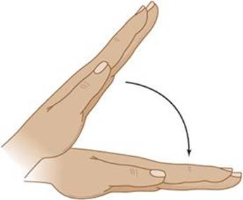
Figure 4-10. Asterixis, a flapping tremor of the outstretched hands or feet, is often associated with hepatic encephalopathy, but can be seen in a variety of metabolic disorders.
Laboratory Findings
Laboratory studies may show elevated serum bilirubin, transaminases, ammonia, prothrombin time (PT) and partial thromboplastin time (PTT), and respiratory alkalosis. The most specific CSF abnormality is elevated glutamine(Figure 4-11). The electroencephalogram (EEG) may be diffusely slow with triphasic waves.

Figure 4-11. Range of CSF glutamine concentrations in hepatic encephalopathy. (Reproduced, with permission, from Plum F. The CSF in hepatic encephalopathy. Exp Biol Med. 1971;4:34-41.)
Treatment
Underlying factors that may have precipitated acute decompensation (discussed previously) should be corrected, and, when indicated, coagulopathy should be reversed with fresh-frozen plasma or vitamin K. Encephalopathy is treated with rifaximin, a poorly absorbed antibiotic that reduces ammonia-forming bacteria in the colon (200 mg orally three times daily), and lactulose, a nonabsorbable disaccharide that decreases colonic pH and ammonia absorption (20 to 30 g orally or per rectum, three to four times daily). Rifaximin can also reduce time to recurrence in patients in remission from hepatic encephalopathy. Severe restriction of dietary protein is not recommended. Liver transplantation is required in some cases. Prognosis in hepatic encephalopathy correlates best with the severity of hepatocellular rather than neurologic dysfunction.
UREMIA
Renal failure, particularly when acute in onset or rapidly progressive, may produce encephalopathy or coma with hyperventilation and prominent motor manifestations. These include tremor, asterixis, myoclonus, and tetany. Focal or generalized seizures and focal neurologic signs are common, and decorticate or decerebrate posturing may occur. Laboratory abnormalities include elevated serum urea nitrogen, creatinine, and potassium, and metabolic acidosis, but their severity correlates poorly with symptoms. The EEG is diffusely slow and may show triphasic waves or paroxysmal spikes or sharp waves.
Acute management includes hydration, protein and salt restriction, and treatment of complications such as seizures. Long-term management requires reversing the cause (eg, urinary tract obstruction), dialysis, or kidney transplantation. Although dialysis reverses the encephalopathy, clinical improvement often lags behind normalization of serum urea nitrogen and creatinine. Dialysis itself can produce an encephalopathy, termed dialysis disequilibrium syndrome, that is thought to result from hypo-osmolality. This is most common with a patient’s first hemodialysis and can be prevented by correcting uremia more gradually or using briefer periods of dialysis at reduced rates of blood flow.
PULMONARY ENCEPHALOPATHY
Patients with lung disease or brainstem or neurologic disorders that affect respiratory function may develop encephalopathy related to hypoventilation. Symptoms include headache, confusion, and somnolence. Examination shows papilledema, asterixis or myoclonus, and a confusional state or coma. Tendon reflexes are often decreased, but pyramidal signs may be present, and seizures occur occasionally. Arterial blood gases show respiratory acidosis. Treatment involves ventilatory support to decrease hyper-capnia and maintain adequate oxygenation.
ORGAN TRANSPLANTATION
Treatment of organ failure by transplantation can lead to acute confusional states as a consequence of surgical complications, immunosuppressive drug treatment, stroke, opportunistic infection, lymphoproliferative disorders, or transplant rejection. The problems encountered depend on the time in relation to transplantation and on the organ transplanted.
Surgical complications that may produce encephalopathy include hypotension, hypoxia, thromboembolism, and air embolism and are most common with heart and liver transplants.
Immunosuppressive drugs used to prevent transplant rejection can cause acute confusional states by direct effects on the nervous system or as a consequence of immunologic impairment. Cyclosporine and tacrolimus produce encephalopathy that may be associated with seizures, tremor, visual disturbances, weakness, sensory symptoms, or ataxia. MRI shows abnormalities in the subcortical white matter. Symptoms are often associated with excessively high drug levels in the blood and may improve with reduction of drug dosage. Corticosteroids can produce psychosis, which may respond to dexamethasone. Corticosteroid withdrawal is sometimes associated with lethargy, headache, myalgia, and arthralgia. OKT3 causes encephalopathy, aseptic meningitis, and seizures. Gabapentin is often used to treat seizures in transplant recipients because of its relative lack of pharmacokinetic interaction with other drugs typically given to these patients.
Infections causing confusional states are most prominent after bone marrow transplantation, but are also common after transplantation of other organs. They are comparatively rare in the first month after transplantation and, when they occur, usually reflect preexisting infection in the recipient or in the donor organ, or a perioperative complication. Within this period, the most frequent organisms are gram-negative bacteria, herpes simplex virus, and fungi. Opportunistic infections are more common between 1 and 6 months after transplant and include acute Listeria meningitis or encephalitis, chronic meningitis from Cryptococcus or Mycobacterium tuberculosis, and brain abscesses related to infection with Aspergillus, Nocardia, or Toxoplasma. Past 6 months, cytomegalovirus, Toxoplasma, Cryptococcus, Listeria, or Nocardia infection may be seen.
Posttransplant lymphoproliferative disorder is related to immunosuppression and may be associated with primary central nervous system (CNS) lymphoma.
Transplant rejection may also produce encephalopathy, especially in recipients of kidney transplants.
MENINGITIS, ENCEPHALITIS, & SEPSIS
BACTERIAL MENINGITIS
Bacterial meningitis is a leading cause of acute confusional states and one in which early diagnosis is crucial to a good outcome. Predisposing conditions include systemic (especially respiratory) or parameningeal infection, head trauma, anatomic meningeal defects, prior neurosurgery, cancer, alcoholism, and other immunodeficiency states. The etiologic organism varies with age and with the presence of predisposing conditions (Table 4-7).


Table 4-7. Etiologic agents and empirical antibiotic treatment in bacterial meningitis.
Pathogenesis & Pathology
Bacteria typically gain access to the CNS by colonizing the mucous membranes of the nasopharynx, leading to local tissue invasion, bacteremia, and hematogenous seeding of the subarachnoid space. Listeriais an exception in that it is ingested. Bacteria can also spread to the meninges directly, through anatomic defects in the skull or from parameningeal sites such as the paranasal sinuses or middle ear. Polysaccharide bacterial capsules, lipopolysaccharides, and outer membrane proteins may contribute to bacterial invasion and virulence. The low levels of antibody and complement present in the cerebrospinal fluid are inadequate to contain the infection. The resulting inflammatory response is associated with the release of inflammatory cytokines that promote blood–brain barrier permeability, vasogenic cerebral edema, changes in cerebral blood flow, and perhaps direct neuronal toxicity.
Pathologically, bacterial meningitis is characterized by leptomeningeal and perivascular infiltration with polymorphonuclear leukocytes and an inflammatory exudate. These changes tend to be most prominent over the cerebral convexities in Streptococcus pneumoniae and Haemophilus infection and over the base of the brain with Neisseria meningitidis. Brain edema, hydrocephalus, and cerebral infarction may occur, although bacterial invasion of the brain parenchyma is rare.
Clinical Findings
At presentation, most patients have had symptoms of meningitis for 1 to 7 days. These include fever, confusion, vomiting, headache, and neck stiffness, but the full syndrome is not usually present (Table 4-8).

Table 4-8. Clinical findings in patients with bacterial meningitis.
Physical examination may show fever and signs of systemic or parameningeal infection, such as skin abscess or otitis. A petechial rash is seen in 50% to 60% of patients with N. meningitidis meningitis. Signs of meningeal irritation (meningismus) are seen in approximately 80% of cases, but are often absent in the very young and very old, or with profoundly impaired consciousness. These signs include neck stiffness on passive flexion, thigh flexion on flexion of the neck (Brudzinski sign), and resistance to passive extension of the knee with the hip flexed (Kernig sign) (Figure 1-5). The level of consciousness, when altered, ranges from mild confusion to coma. Focal neurologic signs, seizures, and cranial nerve palsies may occur. Papilledema is rare.
Laboratory Findings
Peripheral blood may reveal polymorphonuclear leukocytosis from systemic infection or leukopenia caused by immunosuppression. The causative organism can be cultured from the blood in approximately two-thirds of cases. Images of the chest, sinuses, or mastoid bones may indicate a primary site of infection. A brain CT or MRI scan may show contrast enhancement of the cerebral convexities, the base of the brain, or the ventricular ependyma. The EEG is usually diffusely slow, and focal abnormalities suggest the possibility of focal cerebritis, abscess formation, or scarring.
Although these studies may be helpful, the essential test in all cases of suspected meningitis is prompt lumbar puncture and CSF examination. CSF pressure is elevated in approximately 90% of cases, and the appearance of the fluid ranges from slightly turbid to grossly purulent. CSF white cell counts of 1,000 to 10,000/μL are usually seen, consisting chiefly of polymorphonuclear leukocytes, although mononuclear cells may predominate in Listeria monocytogenes meningitis. Protein concentrations of 100 to 500 mg/dL are most common. The CSF glucose level is lower than 40 mg/dL in approximately 80% of cases and may be too low to measure. Gram-stained smears of CSF identify the causative organism in 70% to 80% of cases. CSF culture, which is positive in approximately 80% of cases, provides a definitive diagnosis and allows determination of antibiotic sensitivity. The polymerase chain reaction has also been used with CSF specimens to identify the etiologic agent in bacterial meningitis.
Differential Diagnosis
Signs of meningeal irritation may also be seen with nonbacterial meningitis and subarachnoid hemorrhage. However, the combination of an acute to subacute course (days rather than weeks), polymorphonuclear pleocytosis, and low CSF glucose point to a bacterial cause. Early viral meningitis can produce polymorphonuclear pleocytosis and symptoms identical to those of bacterial meningitis, but a repeat lumbar puncture after 6 to 12 hours should demonstrate a shift to lymphocytic predominance in viral meningitis, and the CSF glucose level is normal. Subarachnoid hemorrhage is distinctive in that lumbar puncture yields bloody CSF, which does not clear as increasing amounts of CSF are removed.
Prevention
Vaccines are available for three bacteria that can cause meningitis: H. influenzae type b, N. meningitidis, and S. pneumoniae. Children ages 2 to 15 months should be routinely immunized against H. influenzaeand S. pneumoniae, children and adolescents ages 1 to 18 years against N. meningitidis, and adults 65 years of age and older against S. pneumoniae. Additional indications for vaccination are listed in Table 4-9. The risk of contracting H. influenzae or N. meningitidis meningitis can be reduced in household and other close contacts of affected patients by the prophylactic administration of rifampin 20 mg/kg/d orally given as a single daily dose for 4 days (H. influenzae) or as two divided doses for 2 days (N. meningitidis).

Table 4-9. Vaccinations against bacterial meningitis.
Treatment
Unless the physical examination shows focal neurologic abnormalities or papilledema, suggesting a mass lesion, lumbar puncture should be performed immediately; if the CSF is not clear and colorless, antibiotic treatment (see next paragraph) is started without delay. When focal signs or papilledema are present, blood and urine should be taken for culture, antibiotics begun, and a brain CT scan obtained. If the scan shows no focal lesion that would contraindicate lumbar puncture, the puncture is then performed.
The initial choice of antibiotics is empirical, based on the patient’s age and predisposing factors (see Table 4-7). Therapy is adjusted as indicated when the Gram stain or culture and sensitivity results become available (Table 4-10). Lumbar puncture can be repeated to assess the response to therapy. CSF should be sterile after 24 hours, and a decrease in pleocytosis and in the proportion of polymorphonuclear leukocytes should occur within 3 days.


Table 4-10. Treatment of bacterial meningitis of known cause.
Dexamethasone, given immediately before the onset of antibiotic treatment and continued for 4 days, may improve outcome and decrease mortality in immunocompetent patients with confirmed bacterial meningitis.
Prognosis
Complications of bacterial meningitis include headache, seizures, hydrocephalus, syndrome of inappropriate secretion of antidiuretic hormone (SIADH), residual neurologic deficits (including cognitive disturbances and cranial—especially VIII—nerve abnormalities), and death. A CT or MRI scan will confirm suspected hydrocephalus, and fluid and electrolyte status should be carefully monitored to detect SIADH. N. meningitidis infections may be complicated by adrenal hemorrhage related to meningococcemia (Waterhouse-Friderichsen syndrome), resulting in hypotension and often death.
Morbidity and mortality from bacterial meningitis are high. Fatalities occur in approximately 20% of affected adults, and more often with some pathogens (eg, S. pneumoniae, gram-negative bacilli) than others (eg, H. influenzae, N. meningitidis). Factors that worsen prognosis include extremes of age, delay in diagnosis and treatment, complicating illness, stupor or coma, seizures, and focal neurologic signs.
TUBERCULOUS MENINGITIS
Tuberculous meningitis must be considered in patients who present with a confusional state, especially if there is a history of pulmonary tuberculosis, alcoholism, corticosteroid treatment, HIV infection, or other conditions associated with impaired immune responses. It should also be considered in patients from areas (eg, Asia, Africa) or groups (eg, the homeless and inner-city drug users) with a high incidence of tuberculosis.
Pathogenesis & Pathology
Tuberculous meningitis usually results from reactivation of latent infection with Mycobacterium tuberculosis. Primary infection, typically acquired by inhaling bacillus-containing droplets, may be associated with metastatic dissemination of blood-borne bacilli from the lungs to the meninges and the surface of the brain. Here the organisms remain in a dormant state in tubercles that can rupture into the subarachnoid space at a later time, resulting in tuberculous meningitis.
The main pathologic finding is a basal meningeal exudate containing primarily mononuclear cells. Tubercles may be seen on the meninges and surface of the brain. The ventricles may be enlarged as a result of hydrocephalus, and their surfaces may show ependymal exudate or granular ependymitis. Arteritis can result in cerebral infarction, and basal inflammation and fibrosis can compress cranial nerves.
Clinical Findings
Symptoms usually have been present for less than 4 weeks at the time of presentation and include headache, fever, neck stiffness, vomiting, and lethargy or confusion. Weight loss, visual impairment, diplopia, focal weakness, and seizures may also occur. A history of contact with known cases of tuberculosis is usually absent.
Fever, signs of meningeal irritation, and a confusional state are the most common findings on physical examination, but all may be absent. Papilledema, ocular palsies, and hemiparesis or paraparesis are sometimes seen. Complications include hyponatremia, hydrocephalus, brain edema, visual loss, cranial nerve (especially VI) palsies, spinal subarachnoid block, and stroke, which usually affect the internal capsule, basal ganglia, or thalamus.
Only one-half to two-thirds of patients show a positive skin test for tuberculosis or evidence of active or healed tubercular infection on chest x-ray; chest CT is more sensitive. The diagnosis is established by CSF analysis. CSF pressure is usually increased, and the fluid is typically clear and colorless. Lymphocytic and mononuclear cell pleocytosis of 50 to 500 cells/mL is most often seen, but polymorpho-nuclear pleocytosis can occur early and may give an erroneous impression of bacterial meningitis. CSF protein is usually more than 100 mg/dL and may exceed 500 mg/dL, particularly in patients with spinal subarachnoid block. The glucose level is usually decreased and may be less than 20 mg/dL. Acid-fast bacillus (AFB) smears of CSF (Figure 4-12) should be performed in all cases of suspected tuberculous meningitis, but they are positive in only a minority of cases. Definitive diagnosis is most often made by culturing M. tuberculosis from the CSF, a process that usually takes several weeks and requires large quantities of spinal fluid for maximum yield. In some cases, polymerase chain reaction can be used for diagnosis. A CT or MRI scan may show enhancement of the basal cisterns and cortical meninges or hydrocephalus.

Figure 4-12. Acid-fast bacillus (AFB) stain showing Mycobacterium tuberculosis bacilli (red rods).
Differential Diagnosis
Many other conditions can cause a subacute to chronic confusional state with mononuclear cell pleocytosis (Table 4-11). These can usually be distinguished based on the history, associated physical findings, and appropriate laboratory studies.
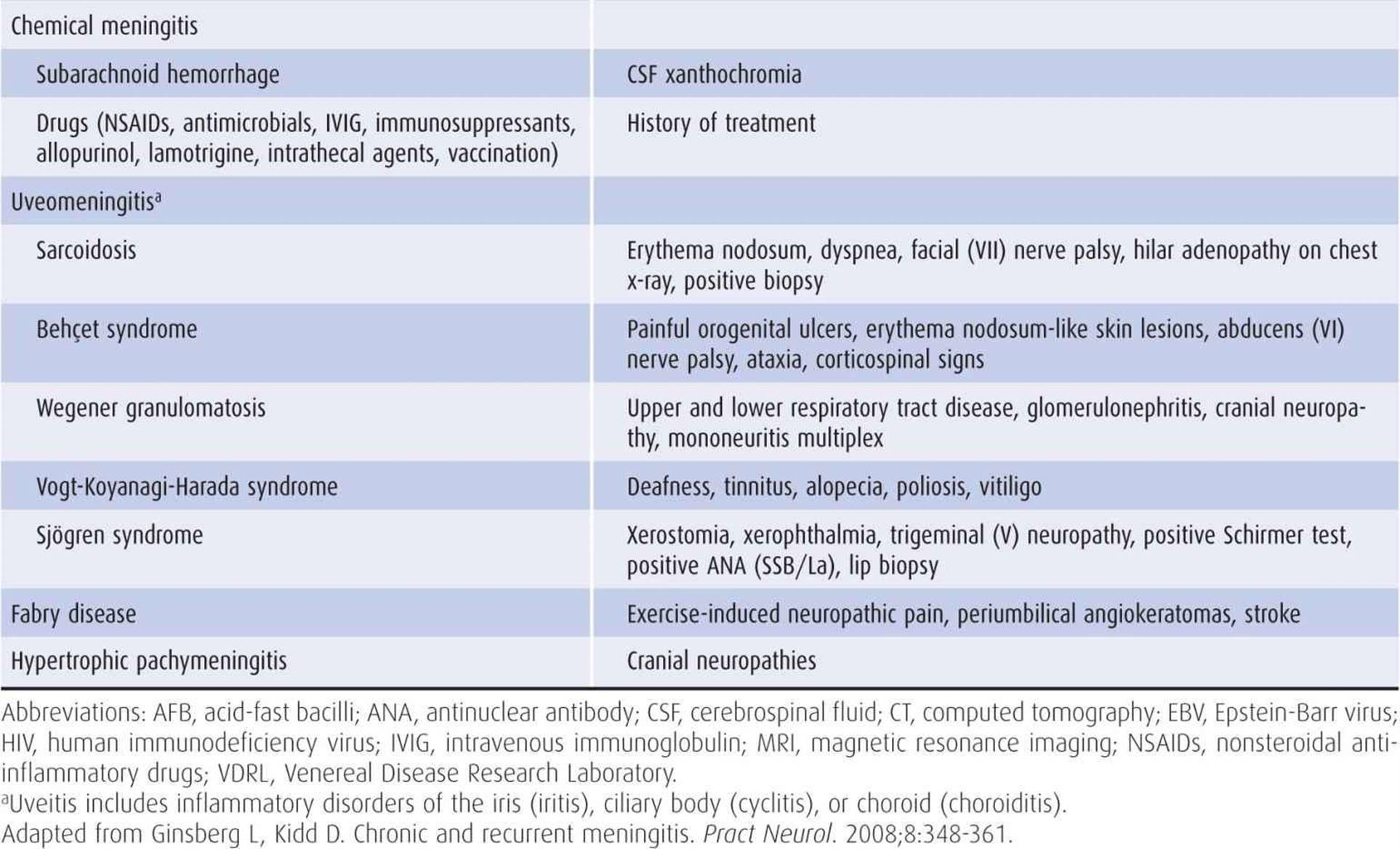
Table 4-11. Causes of chronic meningitis.
Treatment
Treatment should be started as early as possible; it should not be withheld while awaiting culture results. The decision to treat is based on the CSF findings described previously; lymphocytic pleocytosis and decreased glucose are particularly suggestive, even if AFB smears are negative.
Four drugs are used for the 2-month initiation phase of therapy: isoniazid 300 mg, rifampin 600 mg, pyrazinamide 1,600 mg, and streptomycin 1,000 mg, each given orally once daily. During the subsequent, 7- to 12-month continuation phase, only isoniazid and rifampin are used, at the same doses. For multidrug-resistant tuberculous meningitis, the initiation phase is extended to 4 months and employs five drugs: amikacin or kanamycin (1,000 mg daily by the intravenous or intramuscular route) and ethionamide (1,000 mg), pyrazinamide (1,600 mg), ofloxacin (800 mg), and either ethambutol (1,200 mg) or cycloserine (1,000 mg), each given orally daily. The continuation phase involves treatment for an additional 12 to 18 months with ethionamide, ofloxacin, and ethambutol or cycloserine, at the same doses. Pyridoxine 50 mg/d can be used to decrease the likelihood of isoniazid- or cycloserine-induced polyneuropathy or seizures.
Complications of therapy include hepatic dysfunction (isoniazid, rifampin, pyrazinamide, ethionamide), polyneuropathy (isoniazid, cycloserine), optic neuritis (ethambutol), seizures (isoniazid, cycloserine), and ototoxicity (streptomycin, amikacin, kanamycin).
Corticosteroids (eg, prednisone, 60 mg/d orally in adults or 1-3 mg/kg/d orally in children, tapered gradually over 3-4 weeks) are indicated as adjunctive therapy in HIV-negative patients. Their use is associated with reductions in mortality, neurologic sequelae, and antituberculous drug toxicity. Corticosteroids are especially warranted for patients with spinal subarachnoid block and for seriously ill patients with focal neurologic signs or increased intracranial pressure. Because corticosteroids may exacerbate fungal meningitis, however, antifungal therapy (see later) should be added along with corticosteroids unless fungal meningitis has been excluded.
Ventriculoperitoneal shunting or endoscopic third ventriculostomy can be useful for relieving hydrocephalus.
Prognosis
Even with appropriate treatment, approximately one-third of patients with tuberculous meningitis succumb. Adverse prognostic factors include age <5 or >50 years, coma, seizures, and concomitant HIV infection. Neurologic sequelae include cognitive disturbances, visual loss, motor deficits, and cranial nerve palsies.
SYPHILITIC MENINGITIS
Acute or subacute syphilitic meningitis usually occurs within 2 years after primary syphilitic infection (Figure 4-13). It is most common in young adults, affects men more often than women, and requires prompt treatment to prevent the irreversible manifestations of tertiary neurosyphilis.

Figure 4-13. Interval between primary syphilitic infection and symptomatic neurosyphilis by presentation. (GPI, general paresis of the insane)
In approximately one-fourth of patients with Treponema pallidum infection, treponemes gain access to the CNS, where they produce a meningitis that is usually asymptomatic (asymptomatic neurosyphilis). Asymptomatic invasion of the CNS is associated with CSF pleocytosis, elevated protein, and positive serologic tests for syphilis.
Clinical Findings
In a few patients, syphilitic meningitis is a clinically apparent acute or subacute disorder. At the time of presentation, symptoms such as headache, nausea and vomiting, stiff neck, mental disturbances, focal weakness, seizures, deafness, and visual impairment usually have been present for up to 2 months.
Physical examination may show signs of meningeal irritation, confusion or delirium, papilledema, hemiparesis, and aphasia. The cranial nerves most frequently affected are (in order) the facial (VII), acoustic (VIII), oculomotor (III), trigeminal (V), abducens (VI), and optic (II) nerves, but other nerves may be involved as well. Fever is typically absent.
The diagnosis is established by CSF findings. Opening pressure is normal or slightly elevated. Pleocytosis is lymphocytic or mononuclear in character, with white blood cell counts usually in the range of 100 to 1,000/mL. Protein may be mildly or moderately elevated (<200 mg/dL) and glucose mildly decreased. CSF Venereal Disease Research Laboratory (VDRL) and serum fluorescent treponemal antibody (FTA) or microhemagglutination-Treponema pallidum (MHA-TP) tests are usually positive. Protein electrophoretograms of CSF may show discrete γ-globulin bands (oligoclonal bands) not visible in normal CSF.
Treatment
Acute syphilitic meningitis is usually a self-limited disorder with no or minimal sequelae. More advanced manifestations of neurosyphilis, including vascular and parenchymatous disease (tabes dorsalis, general paresis, optic neuritis, myelitis), can be prevented by adequate treatment of the early syphilitic infection.
Syphilitic meningitis is treated with aqueous penicillin G 2 to 4 × 10⁶ units intravenously every 4 hours for 10 days. For penicillin-allergic patients, tetracycline or erythromycin 500 mg orally every 6 hours for 20 days can be substituted. The CSF should be examined every 6 months until all findings are normal. Another course of therapy must be given if the CSF cell count or protein remains elevated.
LYME DISEASE
Clinical Findings
Lyme disease is a tick-borne disorder that results from systemic infection with the spirochete Borrelia burgdorferi. Most cases occur during the summer months. Primary infection may be manifested by an expanding erythematous annular skin lesion (erythema migrans) (Figure 4-14) that usually appears over the thigh, groin, or axilla. Less distinctive symptoms include fatigue, headache, fever, neck stiffness, joint or muscle pain, anorexia, sore throat, and nausea. Neurologic involvement may be delayed for up to 10 weeks and is characterized by meningitis or meningoencephalitis and disorders of the cranial or peripheral nerves or nerve roots; bilateral facial weakness from involvement of cranial nerve VII is particularly common. Cardiac abnormalities (conduction defects, myocarditis, pericarditis, cardiomegaly, or heart failure) can also occur at this stage. Lyme meningitis usually produces prominent headache that may be accompanied by signs of meningeal irritation, photophobia, pain when moving the eyes, nausea, and vomiting. When encephalitis is present, it is usually mild and characterized by insomnia, emotional lability, or impaired concentration and memory. European Lyme disease differs clinically from that seen in the United States in that the infective organism is Borrelia garinii or Borrelia afzelii, erythema migrans is not a feature, and painful radiculopathy (Bannwarth syndrome) is common.

Figure 4-14. Erythema migrans due to Borrelia burgdorferi (Lyme disease). Courtesy of James Gathany, Public Health Image Library, US Centers for Disease Control and Prevention.
Laboratory Findings
The CSF usually shows a lymphocytic pleocytosis with 100 to 200 cells/mL, slightly elevated protein, and normal glucose. Oligoclonal immunoglobulin G (IgG) bands may be detected. Definitive diagnosis is usually made by serologic testing for B. burgdorferi, preferably by enzyme-linked immunosorbent assay (ELISA) followed by Western blot, but the polymerase chain reaction, which can amplify spirochetal DNA in synovial fluid, blood, or CSF, has also been used.
Treatment
Preventive measures include avoiding tick-infested areas and using insect repellents and protective clothing when avoidance is impossible. A Lyme disease vaccine is also available, but its use is controversial.
For patients with Lyme disease and cranial or peripheral nerve involvement, treatment is with doxycycline (100 mg twice daily) or amoxicillin (500 mg three times daily), each given orally for 2 to 3 weeks. With meningitis or other CNS involvement, intravenous treatment is indicated with ceftriaxone (2 g intravenously daily) for 2 to 4 weeks.
Symptoms typically resolve within 10 days in treated cases. Untreated or inadequately treated infections may lead to recurrent oligoarthritis, and chronic neurologic disorders including memory, language, and other cognitive disturbances; focal weakness; and ataxia. In such cases, a CT scan or MRI may show hydrocephalus, lesions in white matter resembling those seen in multiple sclerosis, or abnormalities suggestive of cerebral infarction. Subtle chronic cognitive or behavioral symptoms should not be attributed to Lyme encephalitis in the absence of serologic evidence of B. burgdorferi exposure, CSF abnormalities, or focal neurologic signs. The peripheral neurologic manifestations of Lyme disease are discussed in Chapter 10.
VIRAL MENINGITIS & ENCEPHALITIS
Viral infections of the meninges (meningitis) or brain parenchyma (encephalitis) often present as acute confusional states. The most common causative agents are listed in Table 4-12. Clues in the history that may suggest a specific virus or virus family include the time of year, recent travel, and contact with insects or other animals, sexual contacts, and immunosuppression. Some viruses (eg, herpesviruses) can cause either meningitis or encephalitis, but others preferentially affect the meninges (eg, enteroviruses) or brain parenchyma (eg, arthropod-borne—or arbo—viruses).
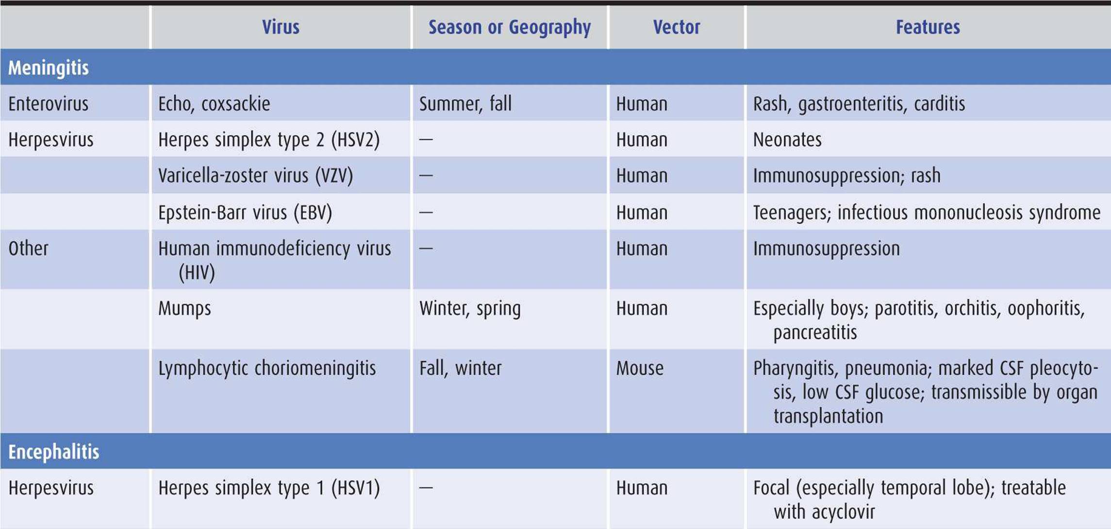

Table 4-12. Etiologic agents in viral meningitis and encephalitis.
Pathology
Viral infections can affect the CNS in three ways—hematogenous dissemination of a systemic viral infection (eg, arthropod-borne viruses), neuronal spread of the virus by axonal transport (eg, herpes simplex, rabies), and autoimmune postinfectious demyelination (eg, varicella, influenza). Pathologic changes in viral meningitis consist of an inflammatory meningeal reaction mediated by lymphocytes. Encephalitis is characterized by perivascular cuffing, lymphocytic infiltration, and microglial proliferation mainly involving subcortical gray matter regions. Intranuclear or intracytoplasmic inclusions are often seen.
Clinical Findings
Clinical manifestations of viral meningitis include fever, headache, neck stiffness, photophobia, pain with eye movement, and mild impairment of consciousness. Patients usually do not appear as ill as those with bacterial meningitis. Systemic viral infection may cause skin rash, pharyngitis, lymphadenopathy, pleuritis, carditis, jaundice, organomegaly, diarrhea, or orchitis, and these findings may suggest a particular etiologic agent. Because viral encephalitis involves the brain directly, more marked alterations of consciousness, seizures, and focal neurologic signs can occur. When signs of meningeal irritation and brain dysfunction coexist, the condition is termed meningoencephalitis.
Laboratory Findings
CSF analysis is the most important laboratory test. CSF pressure is normal or increased, and a lymphocytic or monocytic pleocytosis is present, with cell counts usually less than 1,000/mL. Higher counts can be seen in lymphocytic choriomeningitis or herpes simplex encephalitis. A polymorphonuclear pleocytosis can occur early in viral meningitis, whereas red blood cells may be seen with herpes simplex encephalitis. Protein is normal or slightly increased (usually 80-200 mg/dL). Glucose is usually normal, but may be decreased in mumps, herpes zoster, or herpes simplex encephalitis. Gram stains and bacterial, fungal, and AFB cultures are negative. Oligoclonal bands and CSF protein electrophoresis abnormalities may be present. An etiologic diagnosis often can be made from CSF by virus isolation, polymerase chain reaction, or detection of antiviral antibodies.
Blood counts may show a normal white cell count, leukopenia, or mild leukocytosis. Atypical lymphocytes in blood smears and a positive heterophil (Monospot) test suggest infectious mononucleosis. Serum amylase is frequently elevated in mumps; abnormal liver function tests are associated with both hepatitis viruses and infectious mononucleosis. The EEG is diffusely slow, especially if there is direct cerebral involvement.
Differential Diagnosis
The differential diagnosis of meningitis with mononuclear cell pleocytosis includes partially treated bacterial meningitis; syphilitic, tuberculous, fungal, parasitic, and neoplastic meningitis; and postinfectious encephalomyelitis after infections or vaccinations (see later). Evidence of systemic viral infection and CSF wet mounts, stained smears, cultures, and cytology can distinguish among these possibilities. When presumed early viral meningitis is associated with a polymorphonuclear pleocytosis of less than 1,000 white blood cells/mL and normal CSF glucose, one of two strategies can be used. The patient can be treated for bacterial meningitis until the results of CSF cultures are known, or treatment can be withheld and lumbar puncture repeated in 6 to 12 hours. If the meningitis is viral in origin, the second sample should show a mononuclear cell pleocytosis.
Prevention & Treatment
Vaccines are available against varicella-zoster virus and Japanese encephalitis, and postexposure prophylaxis against rabies can be achieved through active immunization by vaccine combined with passive immunization using human rabies-immune globulin. Except for herpes simplex virus (HSV) and varicella-zoster virus encephalitis, however, which are sensitive to acyclovir (discussed in next section), no satisfactory treatment for viral meningitis or encephalitis is available. Cytomegalovirus (CMV) encephalitis may respond to induction therapy for 3 weeks with a combination of ganciclovir (5 mg/kg intravenously twice daily) and foscarnet (60 mg/kg intravenously every 8 hours or 90 mg/kg every 12 hours), followed by maintenance therapy for 3 to 6 weeks with ganciclovir (5 mg/kg intravenously daily) or foscarnet (60-120 mg/kg intravenously daily). Corticosteroids are of no proven benefit except in immune-mediated postinfectious syndromes. Headache and fever can be treated with acetaminophen or nonsteroidal anti-inflammatory drugs. Seizures usually respond to phenytoin or phenobarbital. Supportive measures in comatose patients include mechanical ventilation and intravenous or nasogastric feeding.
Prognosis
Symptoms of viral meningitis usually resolve spontaneously within 2 weeks regardless of the causative agent, although residual deficits may be seen. The outcome of viral encephalitis varies with the specific virus—for example, eastern equine and HSV infections are associated with severe morbidity and high mortality rates. Mortality rates as high as 20% have also been reported in immune-mediated encephalomyelitis after measles infections.
HERPES SIMPLEX VIRUS (HSV) ENCEPHALITIS
HSV type 1 (oral herpes) is the most common cause of sporadic fatal encephalitis in the United States. Approximately one-half of cases involve patients older than 50 years of age. The virus migrates along nerve axons to sensory ganglia, where it persists in a latent form and may be subsequently reactivated. HSV type 1 encephalitis can result from either primary infection or reactivation of latent infection. Neonatal HSV encephalitis usually results from acquisition of HSV type 2 (genital herpes) during passage through the birth canal of a mother with active genital lesions. CNS involvement by HSV type 2 in adults usually causes meningitis, rather than encephalitis.
Pathology
HSV type 1 encephalitis is an acute, necrotizing, asymmetric hemorrhagic process with lymphocytic and plasma cell reaction and usually involves the medial temporal and inferior frontal lobes. Intranuclear inclusions may be seen in neurons and glia. Patients who recover may show cystic necrosis of the involved regions.
Clinical Findings
The clinical syndrome may include headache, stiff neck, vomiting, behavioral disorders, memory loss, anosmia, aphasia, hemiparesis, and focal or generalized seizures. Active herpes labialis is seen occasionally, but does not reliably implicate HSV as the cause of encephalitis. HSV encephalitis is usually rapidly progressive over several days and may result in coma or death. The most common sequelae in patients who survive are memory and behavior disturbances, reflecting the predilection of HSV for limbic structures.
Laboratory Findings
The CSF in HSV type 1 encephalitis most often shows increased pressure, lymphocytic or mixed lymphocytic and polymorphonuclear pleocytosis (50-100 white blood cells/mL), mild protein elevation, and normal glucose. Red blood cells, xanthochromia, and decreased glucose are seen in some cases. The virus generally cannot be isolated from the CSF, but can be detected by the polymerase chain reaction and serologic testing. The EEG may show periodic slow-wave complexes arising from one or both temporal lobes. MRI is more sensitive than CT for early detection of edema and mass effect in one or both temporal lobes and cingulate gyri (Figure 4-15). However, imaging studies may also be normal.

Figure 4-15. MRI in herpes simplex encephalitis. FLAIR I (A) and T2 (B) sequences show mild mass effect, loss of gray-white differentiation caused by edema, and characteristic involvement of the temporal lobe (arrow). T2 image shows involvement on the other side as well. (Courtesy of J. Handwerke.)
Differential Diagnosis
The symptoms and signs are not specific for herpes virus infection. The greatest diagnostic difficulty is distinguishing between HSV encephalitis and brain abscess, and the two disorders often cannot be differentiated on clinical grounds alone. Other CNS infections and vasculitis can also mimic HSV encephalitis. Definitive diagnosis can be made by biopsy of affected brain areas, with the choice of biopsy site guided by the EEG, CT scan, or MRI findings. However, because treatment is most effective when begun early and is comparatively safe, the most common approach is to treat patients with possible HSV encephalitis as described next and to reserve biopsy for those who fail to improve.
Treatment
The most effective drug is acyclovir, given intravenously at a dosage of 10 to 15 mg/kg every 8 hours, with each dose given over 1 hour. Treatment is continued for 14 to 21 days. Complications include erythema at the infusion site, gastrointestinal disturbances, headache, skin rash, tremor, seizures, and encephalopathy or coma. Treatment is started as early as possible, because outcome is greatly influenced by the severity of dysfunction at the time treatment is initiated.
Prognosis
Patients younger than age 30 and those who are only lethargic at the onset of treatment are more likely to survive than are older or comatose patients. The mortality rate is approximately 70% without treatment, which is reduced to approximately 25% at 18 months in patients given acyclovir.
HUMAN IMMUNODEFICIENCY VIRUS INFECTION
Acquired immune deficiency syndrome (AIDS) is caused by infection with human immunodeficiency virus type 1 (HIV-1) and is characterized by opportunistic infections, malignant neoplasms (typically non-Hodgkin lymphoma or Kaposi sarcoma), and a variety of neurologic disturbances. Transmission occurs through sexual activity or by transfer of virus-contaminated blood or blood products. Individuals at particular risk of infection include those who engage in unprotected sexual intercourse, intravenous drug users who share needles, hemophiliacs who have received factor VIII transfusions, and their sexual partners.
Neurologic complications of HIV infection include dementia (see Chapter 5), myelopathy (see Chapter 10), neuropathy (see Chapter 10), myopathy (see Chapter 9), and stroke (see Chapter 13). HIV may enter the CNS through penetration of the blood–brain barrier by HIV-infected circulating monocytes or direct infection of the choroid plexus or capillary endothelial cells. In addition to monocytes, choroid plexus, and capillary endothelium, the virus ultimately infects brain microglia and astrocytes, but not neurons or oligodendrocytes. Patients with systemic HIV infection are at increased risk of neurologic involvement not only from direct HIV infection of the nervous system, but also from opportunistic infections and tumors.
A. HIV-1 Meningitis
Patients infected with HIV-1 can develop a syndrome characterized by headache, fever, signs of meningeal irritation, cranial nerve (especially VII) palsies, other focal neurologic abnormalities, or seizures. This usually occurs at about the time of HIV-1 seroconversion. An acute confusional state is occasionally present. HIV-1 meningitis is associated with mononuclear CSF pleocytosis of up to approximately 200 cells/μL with normal or slightly elevated protein and normal glucose. HIV may be detectable in the CSF by polymerase chain reaction. Symptoms usually resolve spontaneously within about 1 month. Other causes of pleocytosis associated with HIV infection, including cryptococcal meningitis and cerebral toxoplasmosis, must be excluded.
B. Cryptococcal Meningitis or Meningoencephalitis
Cryptococcus neoformans meningitis or meningoencephalitis occurs in patients with HIV infection and CD4 cell counts of <100/μL. Clinical features include headache, confusion, stiff neck, fever, nausea and vomiting, seizures, and cranial nerve palsies. CSF cell counts, protein, and glucose may be normal, so CSF India ink staining or cryptococcal antigen titers should always be obtained when cryptococcal meningitis is suspected in HIV-infected patients. CT or MRI scans may be normal or may show meningeal enhancement, intraventricular or intraparenchymal cryptococcomas, gelatinous pseudocysts, abscesses, or hydrocephalus. Treatment is with amphotericin B and flucytosine, followed by fluconazole (see Fungal Meningitis section).
C. Cerebral Toxoplasmosis
Cerebral toxoplasmosis produces intracerebral mass lesions in patients with HIV infection and CD4 cell counts of <200/μL. Presenting symptoms usually include fever, headache, altered mental status, and seizures. Focal neurologic abnormalities such as cranial nerve palsies or hemiparesis may also occur. MRI is more sensitive than CT scanning and typically reveals one or more supratentorial lesions with a predilection for gray-white matter junctions or the basal ganglia; with contrast, the lesions may show rim enhancement. Lumbar puncture may be inadvisable in the presence of mass lesions. Intracerebral mass lesions in HIV-infected patients are typically due to either toxoplasmosis or primary central nervous system lymphoma (see later), but the two disorders may not be clinically distinguishable. Therefore, because toxoplasmosis is readily treatable, patients with HIV infection and intracerebral mass lesions that are not obviously caused by stroke should be treated for presumed toxoplasmosis. Treatment is with pyrimethamine and sulfadiazine (see Parasitic Meningitis section). Folinic acid (10-25 mg orally daily) should also be given to prevent pyrimethamine-induced hematotoxicity. Up to 90% of patients respond favorably to therapy within the first few weeks, and the majority survive longer than 6 months. Absence of a response to treatment for toxoplasmosis should prompt brain biopsy for diagnosis of possible lymphoma.
D. Cytomegalovirus Encephalitis
Cytomegalovirus produces encephalitis in patients with HIV infection and CD4 cell counts <50/μL. The virus can infect neurons, astrocytes, oligodendrocytes, endothelial cells, and ependymal cells. Clinical features include fever, confusion or coma, seizures, and focal neurologic signs. Myelopathy and radiculopathy may also occur. CSF cell count, protein, and glucose are variable, but the diagnosis can be made from CSF by polymerase chain reaction. Treatment is with ganciclovir 5 mg/kg intravenously twice daily and is continued until the CD4 cell count exceeds 100/μL.
E. Progressive Multifocal Leukoencephalopathy
This demyelinating disorder is caused by infection with JC virus and occurs in HIV-infected patients with CD4 counts <100/μL. Altered mental status is accompanied by focal neurologic signs including hemianopsia, ataxia, or hemiparesis, and seizures. Headache and fever are usually absent. CT or MRI scan shows one or more white matter lesions. The CSF typically shows mild lymphocytic pleocytosis, elevated protein, and normal glucose, and polymerase chain reaction may provide evidence for JC virus infection. There is no proven effective treatment.
F. Primary CNS Lymphoma
Primary CNS lymphoma is the most common brain tumor associated with HIV infection. Clinical features include confusional state, hemiparesis, aphasia, seizures, cranial nerve palsies, and headache; signs of meningeal irritation are uncommon. CSF commonly shows elevated protein and mild mononuclear pleocytosis, and glucose may be low; cytology is rarely positive. MRI is more sensitive than CT scanning and shows single or multiple contrast-enhancing lesions, which may not be distinguishable from those seen in toxoplasmosis. Patients with HIV infection and one or more intracerebral mass lesions that fail to respond to treatment for toxoplasmosis within 3 weeks should undergo brain biopsy for diagnosis of lymphoma. Treatment of primary CNS lymphoma involves corticosteroids, whole-brain radiation, and combination chemotherapy with methotrexate and additional agents and is associated with significant cognitive neurotoxicity.
G. Other Disorders
Pneumocystis carinii pneumonia in patients with HIV infection may lead to hypoxia and a resulting confusional state. Patients with HIV infection, especially those with CNS involvement, may be especially sensitive to drugs (eg, antidepressants) and metabolic disorders, and the antiretroviral drug zidovudine can produce a confusional state. Stroke can occur in patients with HIV infection, especially when it is complicated by cryptococcal meningitis, and may produce an acute confusional state. Seizures are common in HIV infection, especially in HIV-associated dementia, cerebral toxoplasmosis, or cryptococcal meningitis, and both complex partial seizures and the postictal state that follows generalized tonic-clonic seizures are associated with confusional states.
FUNGAL MENINGITIS
In a small fraction of patients with systemic fungal infections (mycoses), fungi invade the CNS to produce meningitis or focal intraparenchymal lesions (Table 4-13). Several fungi are opportunistic organisms that cause infection in patients with cancer, those receiving corticosteroids or other immunosuppressive drugs, and other debilitated hosts. Intravenous drug abuse is a potential route for infection with Candidaand Aspergillus. Diabetic acidosis is strongly correlated with rhinocerebral mucormycosis. In contrast, meningeal infections with Coccidioides, Blastomyces, and Actinomyces usually occur in previously healthy individuals. Cryptococcus (the most common cause of fungal meningitis in the United States) and Histoplasma infection can occur in either healthy or immunosup-pressed patients. Cryptococcal meningitis is the most common fungal infection of the nervous system in patients with HIV infection, but Coccidioides and Histoplasma infections can also occur in this setting. Geographic factors are also important in the epidemiology of certain mycoses: Blastomyces is seen especially in the Mississippi River Valley, Coccidioides in the southwestern United States, and Histoplasma in the eastern and midwestern United States.


Table 4-13. Etiologic agents in fungal meningitis.
Pathogenesis & Pathology
Fungi reach the CNS by hematogenous spread from the lungs, heart, gastrointestinal or genitourinary tract, or skin, or by direct extension from parameningeal sites such as the orbits or paranasal sinuses. Invasion of the meninges from a contiguous focus of infection is particularly common in mucormycosis but may also occur in aspergillosis and actinomycosis.
Pathologic findings in fungal infections of the nervous system include a primarily mononuclear meningeal exudative reaction, focal abscesses or granulomas in the brain or spinal epidural space, cerebral infarction related to vasculitis, and ventricular enlargement caused by communicating hydrocephalus.
Clinical Findings
Fungal meningitis is usually a subacute illness and clinically resembles tuberculous meningitis. Common symptoms include headache and lethargy or confusion. Nausea and vomiting, visual loss, seizures, or focal weakness may be noted, whereas fever may be absent. In a diabetic patient with acidosis, complaints of facial or eye pain, nasal discharge, proptosis, or visual loss should urgently alert the physician to the likelihood of Mucor infection.
Careful examination of the skin, orbits, sinuses, and chest may reveal evidence of systemic fungal infection. Neurologic examination may show signs of meningeal irritation, a confusional state, papilledema, visual loss, ptosis, exophthalmos, ocular or other cranial nerve palsies, and focal neurologic abnormalities such as hemiparesis. Because some fungi (eg, Cryptococcus) can cause spinal cord compression, there may be evidence of spine tenderness, paraparesis, pyramidal signs in the legs, and loss of sensation over the legs and trunk.
Laboratory Findings
Blood cultures should be obtained. Serum glucose and arterial blood gas levels should be determined in diabetic patients. The urine should be examined for Candida. Chest x-ray may show hilar lymphadenopathy, patchy or miliary infiltrates, cavitation, or pleural effusion. The CT scan or MRI may demonstrate intracerebral mass lesions associated with Cryptococcus (Figure 4-16) or other organisms, a contiguous infectious source in the orbit or paranasal sinuses, or hydrocephalus.

Figure 4-16. T2-weighted MRI in cryptococcal meningitis. Note the bilateral increase in signal in the basal ganglia (arrows) with relative sparing of the thalami (T). This is caused by gelatinous fungal pseudocysts in the territory of the lenticulostriate arteries. (Courtesy of A. Gean.)
CSF pressure may be normal or elevated, and the fluid is usually clear, but may be viscous in the presence of numerous cryptococci. Lymphocytic pleocytosis of up to 1,000 cells/μL is common, but a normal cell count or polymorphonuclear pleocytosis can be seen in early fungal meningitis, and normal cell counts are common in immunosuppressed patients. Aspergillus infection typically produces a polymorphonuclear pleocytosis. CSF protein, which may be normal initially, subsequently rises, usually to levels not exceeding 200 mg/dL. Higher levels (up to 1 g/dL) suggest possible subarachnoid block. Glucose is normal or decreased but rarely below 10 mg/dL. Microscopic examination of Gram-stained and acid-fast smears and India ink preparations may reveal the infecting organism. Fungal cultures of CSF and other body fluids and tissues should be obtained, but are often negative. In suspected mucormycosis, biopsy of the affected tissue (usually nasal mucosa) is essential. Useful CSF serologic studies include cryptococcal antigen and Coccidioides complement-fixing antibody. Cryptococcal antigen is more sensitive than India ink for detecting Cryptococcus and should always be looked for in both CSF and serum when that organism is suspected, as in patients with HIV infection.
Differential Diagnosis
Fungal meningitis may mimic brain abscess and other subacute or chronic meningitides, such as those caused by tuberculosis or syphilis. CSF findings and CT or MRI scans are useful in differential diagnosis.
Treatment & Prognosis
Amphotericin B is the most common treatment for fungal meningitis, but nephrotoxicity is common and may be accompanied by fever, shaking chills, hypokalemia, hypomagnesemia, and anemia. Newer, lipid-based formulations are less nephrotoxic. In patients with Coccidioides meningitis or those not responding to intravenous therapy, intrathecal amphotericin B (usually administered via an Ommaya reservoir) is sometimes used. Flucytosine is often given together with amphotericin B to treat cryptococcal meningitis; it may cause bone marrow suppression, which is usually reversible, and its dose must be reduced in patients with renal failure. Azoles, including fluconazole, itracinazole, and voriconazole, are first-line antifungal agents for meningitis due to several mycoses. Most can cause skin rash, nausea, and elevated serum transaminases, and voriconazole can also produce transient visual disturbances, consisting of perceptual alterations, photophobia, blurring, or changes in color vision. Mortality in fungal meningitis is high, complications of treatment are common, and neurologic sequelae are frequent.
PARASITIC INFECTIONS
Protozoal and helminthic infections are important causes of CNS disease, particularly in immunosuppressed patients (including those with HIV infection), and in certain regions of the world (Table 4-14). Rickettsia, the parasitic bacteria that cause Rocky Mountain spotted fever, rarely affect the nervous system.


Table 4-14. Parasitic infections of the central nervous system.
A. Malaria
Malaria, the most common parasitic infection of humans worldwide, is caused by the protozoan Plasmodium falciparum or other Plasmodium species and is transferred to humans by the female Anophelesmosquito. Clinical features include fever, chills, myalgia, nausea and vomiting, anemia, renal failure, hypoglycemia, and pulmonary edema. Cerebral involvement is rare, but occurs when plasmodia reach the CNS in infected red blood cells and cause occlusion of cerebral capillaries. Neurologic involvement becomes apparent weeks after infection. In addition to acute confusional states, cerebral malaria can produce seizures and, rarely, focal neurologic abnormalities. The diagnosis is made by finding plasmodia in red blood cells on peripheral blood smears (Figure 4-17). The CSF may show increased pressure, xanthochromia, mononuclear pleocytosis, or mildly elevated protein. Treatment is described in Table 4-14. Artemisinins are alternative first-line drugs for cerebral malaria, but are not readily available in the United States. Intravenous administration of quinidine should be accompanied by ECG monitoring to detect QTc segment prolongation. Hypoglycemia may occur in treated patients and requires IV administration of dextrose. Cerebral edema is not a consistent finding. Mannitol and corticosteroids are not helpful and may be deleterious. The mortality rate in cerebral malaria is about 20%.

Figure 4-17. Peripheral blood smear from a patient with Plasmodium falciparum malaria, showing parasites (dark spots) within red blood cells. (From Kaushansky K, Lichtman M, Beutler E, Kipps T. Williams Hematology. 8th ed. New York: McGraw-Hill, 2010.)
B. Toxoplasmosis
Toxoplasmosis results from ingestion of Toxoplasma gondii cysts in raw meat or cat excrement and is usually asymptomatic. Symptomatic infection is associated with underlying malignancy, immunosuppressive therapy, or HIV infection. Systemic manifestations include skin rash, lymphadenopathy, myalgias, arthralgias, carditis, pneumonitis, and splenomegaly. CNS involvement leads to mass lesions or encephalitis, and symptoms and signs include headache, altered mental status, seizures and focal deficits. The CSF may show mild mononuclear cell pleocytosis or slight protein elevation, and the organism may be seen on wet mounts of centrifuged CSF. MRI is superior to CT scanning for demonstrating the characteristic ring-enhancing lesions (Figure 4-18). Diagnosis can be made by serologic methods. Treatment is given in Table 4-14. Folinic acid 10 mg orally daily is added to prevent pyrimethamine-induced leukopenia and thrombocytopenia.

Figure 4-18. T1-weighted, gadolinium-enhanced MRI in cerebral toxoplasmosis complicating HIV infection. Note the multiple calcifications (arrow, right) and ring-enhanced lesions (arrow, left) in the basal ganglia and cerebral cortex.
C. Primary Amebic Meningoencephalitis
The free-living ameba Naegleria fowleri causes primary amebic meningoencephalitis in previously healthy young persons exposed to warm, polluted fresh water. It is reported most often in the southeastern United States. Amebas gain entry to the CNS through the cribriform plate, producing a diffuse meningoencephalitis that affects the base of the frontal lobes and posterior fossa. It is characterized by headache, fever, nausea and vomiting, signs of meningeal irritation, and disordered mental status. The CSF shows a polymorphonuclear pleocytosis with elevated protein and low glucose; highly motile, refractile trophozoites can sometimes be seen on CSF wet mounts. Treatment options are listed in Table 4-14, but the disease is usually fatal within 1 week.
D. Granulomatous Amebic Encephalitis
Granulomatous amebic encephalitis results from infection with Acanthamoeba/Hartmanella species and commonly occurs with chronic illness or immunosuppression. The disorder typically lasts 1 week to 3 months and is characterized by subacute or chronic meningitis and granulomatous encephalitis. The cerebellum, brainstem, basal ganglia, and cerebral hemispheres are affected. An acute confusional state is the most common clinical finding. Fever, headache, and meningeal signs each occur in approximately one-half of patients. Seizures, hemiparesis cranial nerve palsies, cerebellar ataxia, and aphasia may also be seen. CSF pleocytosis can be lymphocytic or polymorphonuclear, protein is elevated, and glucose is low or normal. Sluggishly motile trophozoites may be seen on CSF wet mounts. Successful treatment has been reported occasionally (Table 4-14).
E. Cysticercosis
Cysticercosis is the most common helminthic infection of the CNS and is observed most often in Mexico, Central and South America, Africa, and Asia. Infection follows ingestion of larvae of the pork tapeworm Taenia solium. Larvae form single or multiple cysts in the brain, ventricles, and subarachnoid space, and neurologic manifestations result from mass effect, obstruction of CSF flow, or inflammation. Seizures are common, but other complications include headache, focal neurologic deficits, hydrocephalus, myelopathy, and subacute meningitis. The ophthalmoscopic examination may show ocular cysts, and there may be peripheral blood eosinophilia, soft tissue calcifications, or parasites in the stool. The CSF shows a lymphocytic pleocytosis, with eosinophils usually present (Table 4-15). Opening pressure is often increased, but if it is decreased, myelography should be performed to detect possible spinal subarachnoid block. CSF protein is 50 to 100 mg/dL and glucose is 20 to 50 mg/dL in most cases. The CT scan or MRI is the most useful diagnostic test. It may show contrast-enhanced mass lesions (sometimes containing live parasites) with surrounding edema, intracerebral calcifications, or ventricular enlargement (Figure 4-19).

Table 4-15. Causes of CSF eosinophilia.

Figure 4-19. Neurocysticercosis. Noncontrast head CT showing new (cystic, black) and old (calcified, white) lesions of neurocysticercosis. (Photo contributors: Seth W. Wright, MD, and Universidad Peruana Cayetano Heredia, Lima, Peru.)
The indications for treatment of cerebral cysticercosis are controversial, partly because the parasites usually die spontaneously and because antihelminthic drugs can worsen inflammation. Patients with seizures and calcified cysts should b e treated with anticonvulsants. Cysts containing viable parasites or persistent or multiple enhancing lesions are usually treated with anticonvulsants, antihelminthic drugs, and corticosteroids (Table 4-14). Intraventricular, ocular, and spinal cysts may be amenable to surgical removal, and hydrocephalus is treated by ventriculoperitoneal shunting. Patients with ocular cysts should not be given antihelminthics.
F. Angiostrongylus cantonensis Meningitis
Angiostrongylus cantonensis is endemic to Southeast Asia, Hawaii, and other Pacific islands. Infection is transmitted by ingestion of infected raw mollusks and produces meningitis with peripheral blood and CSF eosinophilia (Table 4-15). Most patients complain of headache, and approximately half report stiff neck, vomiting, fever, and paresthesias. Lymphocytic CSF pleocytosis, mild elevation of protein, and normal glucose are typical. The acute illness usually resolves spontaneously in 1 to 2 weeks, although corticosteroids, analgesics, reduction of CSF pressure by repeated lumbar punctures, and antihelminthic drugs may be helpful (Table 4-14).
G. Rocky Mountain Spotted Fever
Rocky Mountain spotted fever is caused by Rickettsia rickettsii, an intracellular parasite transmitted to humans by tick bites. R. rickettsii damages endothelial cells, leading to vasculitis, microinfarcts, and petechial hemorrhage. Initial symptoms include fever, headache, and a characteristic rash that involves the palms and soles and spreads centrally. Neurologic involvement, which is uncommon, produces a confusional state and, less often, coma or focal neurologic abnormalities. The CSF is normal or shows a mild mononuclear pleocytosis. Treatment is listed in Table 4-14.
POSTINFECTIOUS ENCEPHALOMYELITIS
Postinfectious encephalitis is an immune-mediated demyelinating disorder that typically occurs within 1 month after a bacterial or viral (usually upper respiratory) infection or vaccination. Clinical features include confusion or coma and focal neurologic deficits. Three forms of postinfectious encephalomyelitis are recognized. In the most common of these, acute disseminated encephalomyelitis, MRI shows multifocal demyelinating lesions affecting primarily the supratentorial white matter, although gray matter and spinal cord can also be involved. The CSF may show lymphocytic or, less commonly, polymorphonuclear pleocytosis. Bickerstaff brainstem encephalitis is similar except that it affects the brainstem and cerebellum predominantly. In acute hemorrhagic leukoencephalitis, MRI shows bihemispheric demyelinating lesions associated with hemorrhage and edema, and the CSF may contain red blood cells. Treatment of postinfectious encephalomyelitis is with methylprednisolone up to 1 g/d intravenously for 3 to 5 days, followed by prednisone 1 mg/kg/d orally tapered over 4 to 6 weeks. Outcome in acute disseminated encephalomyelitis and Bickerstaff brainstem encephalitis is usually good, whereas acute hemorrhagic leukoencephalitis is associated with high mortality.
LEPTOMENINGEAL METASTASES
Diffuse metastatic seeding of the leptomeninges may complicate systemic cancer (especially carcinoma of the breast, carcinoma of the lung, lymphoma, leukemia, carcinoma of the gastrointestinal tract, and melanoma), producing neurologic syndromes with prominent cognitive dysfunction. Primary brain tumors may be associated with meningeal gliomatosis, and medulloblastomas and pineal tumors have a propensity for meningeal dissemination. Neoplastic meningitis usually occurs 3 months to 5 years after the diagnosis of cancer, but may precede it. Abnormal neurologic signs are often more striking than the symptoms and usually suggest involvement at multiple levels of the neuraxis; diagnosis is by MRI (Figure 4-20), CSF cytology, or both (Table 4-16). Untreated, leptomeningeal metastases are typically associated with death within about 2 months, and treatment in most cases is palliative. However, radiation and intrathecal chemotherapy are beneficial in some cases and can increase survival by several months, depending on the tumor type.

Figure 4-20. Gadolinium-enhanced T1 coronal MRI showing meningeal spread of breast cancer. There are contrast-enhancing (white) focal lesions in the meninges on the left, diffuse meningeal enhancement, and mass effect from a hemispheric lesion on the left.


Table 4-16. Presenting features of leptomeningeal metastases.
SEPSIS-ASSOCIATED ENCEPHALOPATHY
Systemic sepsis can produce an encephalopathy that may be related to impaired cerebral blood flow, disruption of the blood–brain barrier, or cerebral edema. Gram-negative infections are the most common cause. Bacteremia, liver failure, or kidney failure may be present. Neurologic manifestations include confusional states or coma, seizures, focal neurologic deficits, rigidity, myoclonus, and asterixis. CSF examination is essential to exclude meningitis. The EEG is often abnormal. Therapy involves supportive measures, such as assisted ventilation, and treatment of the underlying infection. Mortality is high, but can be reduced by prompt diagnosis and treatment.
VASCULAR DISORDERS
Vascular causes of acute confusional states can be classified as disorders of the blood vessels, heart, or blood (Table 4-17).

Table 4-17. Vascular causes of acute confusional states.
HYPERTENSIVE ENCEPHALOPATHY
A sudden increase in blood pressure, with or without preexisting chronic hypertension, may result in encephalopathy and headache, which develop over a period of hours to days. Patients at risk include those with acute glomerulonephritis or eclampsia. Impaired autoregulation of cerebral blood flow, vasospasm, and intravascular coagulation have all been proposed as causes. Vomiting, visual disturbances, focal neurologic deficits, and focal or generalized seizures can occur. Blood pressure in excess of 250/150 mm Hg is usually required to precipitate the syndrome in patients with chronic hypertension, but previously normotensive patients may be affected at lower pressures. Retinal arteriolar spasm is almost invariable, and papilledema, retinal hemorrhages, and exudates are usually present. Lumbar puncture may show normal or elevated CSF pressure and protein. Areas of edema, located especially in parieto-occipital white matter, are seen on CT scan and MRI (Figure 4-21) and are reversible with treatment. Stroke and subarachnoid hemorrhage also produce encephalopathy with acutely elevated blood pressure; when focal neurologic abnormalities are also present, stroke is most likely. A similar syndrome (posterior reversible encephalopathy) can occur in normotensive individuals and may be related to autoimmune-induced endothelial dysfunction.

Figure 4-21. Axial FLAIR MRI in hypertensive encephalopathy showing increased signal (white) in the subcortical occipital white matter and occipital cortex bilaterally. These findings may represent reversible vasogenic edema.
The diagnosis is established when lowering the blood pressure results in rapid resolution of symptoms. This is accomplished with sodium nitroprusside, given by continuous intravenous infusion at an initial rate of 0.25 μg/kg/min and increased to as much as 10 μg/kg/min as required. The patient must be carefully monitored and the infusion rate adjusted to maintain a therapeutic effect without producing hypotension. Mean arterial blood pressure should be reduced by no more than 25% in the first 2 hours of treatment, and a target of 160/100 mm Hg should be aimed for in the following 4 hours. Treatment should be terminated immediately if neurologic function worsens. Untreated hypertensive encephalopathy can result in renal failure, stroke, coma, or death, but prompt treatment usually produces full clinical recovery.
SUBARACHNOID HEMORRHAGE
Subarachnoid hemorrhage, usually due to rupture of a cerebral aneurysm, must receive early consideration in the differential diagnosis of an acute confusional state. Subarachnoid hemorrhage may produce an acute confusional state, coma, meningeal signs, and focal neurologic deficits, but the most prominent symptom is usually headache. For this reason, the disorder is discussed in Chapter 6, Headache & Facial Pain.
VERTEBROBASILAR ISCHEMIA
An embolus to the top of the basilar artery that subsequently breaks up and sends fragments distally can produce ischemia affecting both posterior cerebral arteries. This condition (top of the basilar syndrome) may produce an acute confusional state accompanied by pupillary (sluggish responses to light and accommodation), visual (homonymous hemianopia, cortical blindness), visuomotor (impaired convergence, paralysis of upward or downward gaze, diplopia), and behavioral (hypersomnolence, peduncular hallucinosis) abnormalities. Vertebrobasilar ischemia is discussed in more detail in Chapter 13, Stroke.
NONDOMINANT HEMISPHERIC INFARCTION
Agitated confusion of sudden onset can result from infarction (usually embolic) in the territory of the inferior division of the nondominant (usually right) middle cerebral artery. If the superior division is spared, there is no associated hemiparesis. Agitation may be so pronounced as to suggest drug intoxication or withdrawal, but autonomic hyperactivity is absent. The diagnosis is confirmed by brain CT scan or MRI. Rarely, isolated anterior cerebral artery infarcts or posterior cerebral artery infarcts cause acute confusion.
SYSTEMIC LUPUS ERYTHEMATOSUS
Systemic lupus erythematosus (SLE) is an autoimmune disorder that affects primarily young women and causes skin rash, arthritis, serositis, nephritis, anemia, leukopenia, and thrombocytopenia. SLE produces neurologic involvement in 37% to 75% of patients and is the most common autoimmune cause of encephalopathy. Clinically active systemic disease need not be present for neurologic symptoms to occur. Neuropathologic findings include fibrinoid degeneration of arterioles and capillaries, microinfarcts, and intracerebral hemorrhages, but true vasculitis of cerebral blood vessels is rare. Clinical features include headache, seizures, visual defects, hemiparesis, acute confusional states, schizophreniform psychosis, depression, and mania. Seizures are usually generalized but may be focal. Laboratory abnormalities include antinuclear antibodies, anti-native DNA antibodies, anti-Sm antibodies, and false-positive serologic test for syphilis. CSF shows mild elevation of protein or a modest, usually mononuclear, pleocytosis in some cases, and MRI may show white matter lesions.
Even in patients with known SLE, encephalopathy can be caused by a variety of factors, including coagulopathy, infection, uremia, emboli from endocarditis, and corticosteroid therapy. Cerebral lupus is treated with corticosteroids, beginning at 60 mg/d of prednisone or the equivalent. In patients already receiving steroids, the dose should be increased by the equivalent of 5 to 10 mg/d of prednisone. After symptoms resolve, steroids should be tapered to a maintenance dose of 5 to 10 mg/d. Seizures are treated with anticonvulsants. Neurologic symptoms of SLE improve in more than 80% of patients treated with corticosteroids, but may also resolve without treatment. Cerebral involvement in SLE has not been shown to adversely affect the overall prognosis.
VASCULITIS
Acute confusional states can occur in primary central nervous system vasculitis, primary systemic vasculitis, and vasculitis secondary to systemic infection or neoplasm.
Primary angiitis of the central nervous system, sometimes referred to as granulomatous angiitis, is a rare disorder. Headache and encephalopathy are the most common clinical features, but stroke and seizures may also occur. There is no involvement of other organs, and laboratory studies reveal no evidence of systemic vasculitis. The CSF usually shows mild lymphocytic pleocytosis and elevated protein. MRI may demonstrate bilateral, multifocal infarcts or diffuse changes consistent with ischemic demyelination. Angiography shows beading of small to medium-sized arteries due to multifocal narrowing. This finding also occurs in reversible cerebral vasoconstriction syndrome which, however, is not due to vasculitis; it is characterized by thunderclap headache, does not produce a confusional state, and resolves spontaneously. Amphetamines and other drugs can also produce a nonvasculitic syndrome that mimics primary angiitis of the nervous system. Definitive diagnosis is by brain biopsy. Treatment is with methylprednisolone 1 g/d intravenously for 3 days, followed by prednisone 1/mg/kg/d orally for 1 month and then tapered over 1 year and cyclophosphamide 2 mg/kg/d orally for 3 to 6 months, then switched to azathioprine, which is continued for 2 to 3 years.
Large vessel systemic vasculitis (eg, giant cell or Takayasu arteritis) produces ischemic optic neuropathy and stroke, rather than confusional states. Medium-size vessel systemic vasculitis due to polyarteritis nodosa can cause encephalopathy, focal neurologic deficits, and seizures, but these occur late in the course, when the diagnosis is likely already known. Small vessel systemic vasculitis due to cryoglobulinemia, Henoch-Schönlein purpura, or Wegener granulomatosis can also produce encephalopathy. These diseases are diagnosed based on the pattern of systemic involvement and by laboratory tests.
COMPLICATIONS OF CARDIAC SURGERY
Cardiac surgery, including coronary artery bypass grafting and valve repair or replacement, is associated with neurologic complications, especially stroke and encephalopathy. Several factors—embolization, hypoperfusion, arrhythmia, metabolic disturbances, and pharmacologic agents—may contribute. Evaluation should include a review of medications, search for metabolic derangements, and CT scan or MRI to detect perioperative stroke. Sedatives and other psychoactive medications should be avoided. Postoperative encephalopathy is typically transient, but some patients show more persistent cognitive dysfunction, which affects memory disproportionately and lasts for weeks to months. Cognitive decline that continues for years after cardiac surgery is likely due to another cause.
DISSEMINATED INTRAVASCULAR COAGULATION
Disseminated intravascular coagulation (DIC) results from pathologic activation of the coagulation and fibrinolytic systems in the setting of an underlying disorder such as sepsis, malignancy, or trauma. The principal manifestation is hemorrhage. Common findings in the brain include small multifocal infarctions and petechial hemorrhages involving gray and white matter. Subdural hematoma, subarachnoid hemorrhage, and hemorrhagic infarction in the distribution of large vessels may also occur.
Neurologic manifestations are common and include confusional states, coma, focal signs, and seizures. They may precede hematologic abnormalities, which include hypofibrinogenemia, thrombocytopenia, fibrin degradation products, and prolonged prothrombin time. Microangiopathic hemolytic anemia may also occur. The differential diagnosis includes thrombotic thrombocytopenic purpura (see later), which is distinguished by its tendency to occur in previously healthy individuals and its association with normal plasma fibrinogen and normal or only slightly elevated fibrin degradation products. Treatment is directed at the underlying disease and correction of anemia, thrombocytopenia, and coagulopathy. Prognosis is related to the severity of the underlying disease.
THROMBOTIC THROMBOCYTOPENIC PURPURA
TTP (Moschcowitz disease) is a rare multisystem disorder defined by the pentad of thrombocytopenic purpura, microangiopathic hemolytic anemia, neurologic dysfunction, fever, and renal disease. In most cases, the cause is an autoimmune reaction against the metalloprotease ADAMTS13, which allows multimers of von Willebrand factor to accumulate in the plasma, where they stimulate platelet aggregation. The result is platelet-fibrin thrombus formation with occlusion of small blood vessels, especially at arteriolar-capillary junctions. Pathologic findings in the brain include disseminated microinfarcts and, less frequently, petechial hemorrhages that are present mainly in gray matter.
Patients usually present with altered consciousness, headache, focal neurologic signs, or seizures, or with cutaneous purpura, ecchymoses, or petechiae. Neurologic symptoms may be fleeting and recurrent. Hematologic studies show Coombs-negative hemolytic anemia, thrombocytopenia, and normal or only slightly abnormal PT, PTT, fibrinogen, and fibrin degradation products. Compared with DIC (see preceding section), TTP is suggested by a platelet count of <20,000/μL and PT within 5 seconds of the upper limit of the normal range. There may be hematuria, proteinuria, or azotemia. CSF is usually normal. The diagnosis can be made by gingival biopsy or splenectomy.
Treatment is by plasma or cryosupernatant (cryoprecipitate-poor plasma) infusion, which provides ADAMTS13. Plasma exchange can help by removing anti-ADAMTS13 antibodies. Corticosteroids and rituximab, a B-cell–depleting monoclonal antibody, may also be useful. With treatment, mortality is 10% to 20%.
HEAD TRAUMA
Blunt head trauma can produce a confusional state or coma. Acceleration or deceleration forces and physical deformation of the skull can cause shearing of white matter, contusion from contact between the inner surface of the skull and the polar regions of the cerebral hemispheres, torn blood vessels, vasomotor changes, brain edema, and increased intracranial pressure.
CONCUSSION
Concussion is characterized by transient loss of consciousness for seconds to minutes, followed by a confusional state that usually lasts from minutes to hours and is characterized by prominent retrograde and anterograde amnesia (see Chapter 5, Dementia & Amnestic Disorders). If examined during the period of unconsciousness, the patient shows normal pupillary and ocular reflexes, flaccidity, and extensor plantar responses. A CT scan should be obtained to detect possible posttraumatic intracranial hemorrhage in patients <16 or >65 years of age, patients taking anticoagulants, and those with prolonged (>1 minute) unconsciousness, severe persistent headache, vomiting, intoxication, focal neurologic signs, seizure, skull fracture, or soft tissue signs of head injury. Patients usually recover uneventfully, but headache, dizziness, or mild cognitive impairment (postconcussion syndrome) may persist for weeks. Some evidence suggests that a concussion increases the risk of subsequent concussion. Athletes who sustain a concussion while playing sports should probably delay their return to play until postconcussive symptoms have resolved and then gradually resume normal activity over approximately 1 week.
INTRACRANIAL HEMORRHAGE
Traumatic intracranial hemorrhage can be epidural, subdural, or intracerebral. Epidural hematoma (Figure 4-22) most often results from a lateral skull fracture that lacerates the middle meningeal artery or vein. Patients may or may not lose consciousness initially, but in either event a lucid interval lasting several hours to 1 to 2 days is followed by the rapid evolution, over hours, of headache, progressive obtundation, hemiparesis, and finally ipsilateral pupillary dilatation from uncal herniation. Death may follow if treatment is delayed.

Figure 4-22. Epidural (left) and subdural (right) hematomas. (From Waxman SG. Clinical Neuroanatomy. 26th ed. New York: McGraw-Hill, 2009.)
Subdural hematoma (Figure 4-22) after head injury can be acute, subacute, or chronic. In each case, headache and altered consciousness are the principal manifestations. Delay in diagnosis and treatment may lead to a fatal outcome. In contrast to epidural hematoma, the time between trauma and the onset of symptoms is typically longer, the hemorrhage tends to be located over the cerebral convexities, and associated skull fractures are uncommon. Subdural hematoma in the posterior fossa is uncommon.
Intracerebral contusion (bruising) or intracerebral hemorrhage related to head injury is usually located at the frontal or temporal poles. Blood typically enters the CSF, resulting in signs of meningeal irritation and sometimes hydrocephalus. Focal neurologic signs are usually absent or subtle.
The diagnosis of posttraumatic intracranial hemorrhage is made by CT scan or MRI. Epidural hematoma tends to appear as a biconvex, lens-shaped, extra-axial mass that may cross the midline or the tentorium but not the cranial sutures. Subdural hematoma is typically crescent-shaped and may cross the cranial sutures but not the midline or tentorium. Midline structures may be displaced contralaterally.
Epidural and subdural hematomas are treated by surgical evacuation. The decision to operate for intracerebral hematoma depends on the clinical course and location. Evacuation, decompression, or shunting for hydrocephalus may be indicated.
SEIZURES
POSTICTAL STATE
Generalized tonic-clonic (grand mal) seizures are typically followed by a transient confusional state (postictal state) that resolves within 1 to 2 hours. Sleepiness and confusion are usually prominent, but coma, agitation, amnesia, aphasia, or psychosis may occur. When postictal confusion does not clear rapidly, an explanation for the prolonged postictal state must be sought. This occurs in three settings: status epilepticus, an underlying structural brain abnormality (eg, stroke, intracranial hemorrhage), or an underlying diffuse cerebral disorder (eg, dementia, meningitis or encephalitis, metabolic encephalopathy). Patients with an unexplained prolonged postictal state should be evaluated with blood chemistry studies, lumbar puncture, EEG, and CT scan or MRI.
COMPLEX PARTIAL SEIZURES
Complex partial (formerly called temporal lobe or psycho-motor) seizures produce alterations in consciousness characterized by confusion or by other cognitive, affective, psychomotor, or psychosensory symptoms. Such symptoms include withdrawal, agitation, and automatisms such as staring, repetitive chewing, swallowing, lip-smacking, or picking at clothing. Complex partial seizures rarely cause diagnostic problems in the patient presenting with an acute confusional state, because spells are typically brief and stereotypical, psychomotor manifestations are often obvious to the observer, and patients themselves may describe classic cognitive, affective, or psychosensory symptoms (see Chapter 12, Seizures & Syncope). The diagnosis is made or confirmed by EEG.
NONCONVULSIVE STATUS EPILEPTICUS
Nonconvulsive (complex partial, other focal, absence, or tonic) status epilepticus can produce confusion or coma, personality change, aphasia, subtle motor activity, or nystagmus. The diagnosis is established by a favorable clinical or EEG response to the administration of anticonvulsants (eg, lorazepam 4 mg or diazepam 10 mg given intravenously).
PSYCHIATRIC DISORDERS
Symptoms similar to those associated with acute confusional states—including incoherence, agitation, distractibility, hypervigilance, delusions, and hallucinations—can also be seen in a variety of psychiatric disorders. These include psychotic disorders (schizophrenia, schizophreni-form disorder, schizoaffective disorder, delusional disorder, and brief psychotic disorder), mood (depressive and bipolar) disorders, anxiety disorders (posttraumatic stress disorder), and factitious disorders. Such diagnoses may be mistakenly assigned to patients with acute confusional states; conversely, patients with psychiatric disturbances may be thought, incorrectly, to have “organic” disease.
Unlike acute confusional states, psychiatric disorders are rarely acute in onset but typically develop over a period of at least several weeks. The history may reveal previous psychiatric disease or hospitalization or a precipitating psychologic stress. Physical examination may show abnormalities related to autonomic overactivity, including tachycardia, tachypnea, and hyperreflexia, but no definitive signs of neurologic dysfunction. Routine laboratory studies are normal in the psychiatric disorders listed previously, but are useful for excluding organic disorders.
Although the mental status examination in acute confusional states is often characterized by disorientation and fluctuating consciousness, patients with psychiatric disorders tend to maintain a consistent degree of cognitive impairment, appear awake and alert, have intact memory, and are oriented to person, place, and time. Disturbances in the content and form of thought (eg, persecutory delusions, delusions of reference, loosening of associations), perceptual abnormalities (eg, auditory hallucinations), and flat or inappropriate affect are common, however. Psychiatric consultation should be sought regarding diagnosis and management.
REFERENCES
General
McPhee SJ, Papadakis M, Rabow MW. Current Medical Diagnosis and Treatment. 51st ed. New York, NY: McGraw-Hill; 2011.
Skrobik Y. Delirium prevention and treatment. Crit Care Clin. 2009;25:585-591.
Drugs
Devlin JW. The pharmacology of oversedation in mechanically ventilated adults. Curr Opin Crit Care. 2008;14:403-407.
Greene SL, Kerr F, Braitberg G. Amphetamines and related drugs of abuse. Emerg Med Australas. 2008;20:391-402.
Hughes JR. Alcohol withdrawal seizures. Epilepsy Behav. 2009; 15:92-97.
Lubman DI, Yücel M, Lawrence AJ. Inhalant abuse among adolescents: neurobiological considerations. Br J Pharmacol. 2008;154:316-326.
Tetrault JM, O’Connor PG. Substance abuse and withdrawal in the critical care setting. Crit Care Clin. 2008;24:767-788.
Virmani A, Ali SF, Binienda ZK. Neuroprotective strategies in drug abuse-evoked encephalopathy. Ann N Y Acad Sci. 2010;1199:52-68.
Endocrine Disorders
Anglin RE, Rosebush PI, Mazurek MF. The neuropsychiatric profile of Addison’s disease: revisiting a forgotten phenomenon. J Neuropsychiatry Clin Neurosci. 2006;18:450-459.
Boscaro M, Arnaldi G. Approach to the patient with possible Cushing’s syndrome. J Clin Endocrinol Metab. 2009;94: 3121-3131.
Kearney T, Dang C. Diabetic and endocrine emergencies. Postgrad Med J. 2007;83:79-86.
Schiess N, Pardo CA. Hashimoto’s encephalopathy. Ann N Y Acad Sci. 2008;1142:254-265.
Tamagno G, Celik Y, Simó R, et al. Encephalopathy associated with autoimmune thyroid disease in patients with Graves’ disease: clinical manifestations, follow-up, and outcomes. BMC Neurology. 2010;10:27.
Electrolyte Disorders
Kacprowicz RF, Lloyd JD. Electrolyte complications of malignancy. Emerg Med Clin North Am. 2009;27:257-269.
King JD, Rosner MH. Osmotic demyelination syndrome. Am J Med Sci. 2010;339:561-567.
Moe SM. Disorders involving calcium, phosphorus, and magnesium. Prim Care. 2008;35:215-237.
Zenenberg RD, Carluccio AL, Merlin MA. Hyponatremia: evaluation and management. Hosp Pract. 2010;38:89-96.
Nutritional Disorders
Graber JJ, McGraw CA, Kimbrough D, Dhib-Jalbut S. Vitamin B12-responsive severe leukoencephalopathy and autonomic dysfunction in a patient with “normal” serum B12 levels. J Neurol Neurosurg Psychiatry. 2010;81:1369-1371.
Santos Andrade C, Tavares Lucato L, da Graça Morais Martin M, et al. Non-alcoholic Wernicke’s encephalopathy: broadening the clinicoradiological spectrum. Br J Radiol. 2010;83:437-446.
Zuccoli G, Pipitone N. Neuroimaging findings in acute Wernicke’s encephalopathy: review of the literature. AJR. 2009;192:501-508.
Organ System Failure
Bass NM, Mullen KD, Sanyal A, et al. Rifaximin treatment in hepatic encephalopathy. N Engl J Med. 2010;362:1071-1081.
Bernal W, Auzinger G, Dhawan A, Wendon J. Acute liver failure. Lancet. 2010;376:190-201.
Cash WJ, McConville P, McDermott E, et al. Current concepts in the assessment and treatment of hepatic encephalopathy. Q J Med. 2010;103:9-16.
Dodd JW, Getov SV, Jones PW. Cognitive function in COPD. Eur Respir J. 2010;35:913-922.
Krishnan AV, Kiernan MC. Neurological complications of chronic kidney disease. Nat Rev Neurol. 2009;5:542-551.
Lacerda G, Krummel T, Hirsch E. Neurologic presentations of renal diseases. Neurol Clin. 2010;28:45-59.
Mathew RM, Rosenfeld MR. Neurologic complications of bone marrow and stem-cell transplantation in patients with cancer. Curr Treat Options Neurol. 2007;9:308-314.
Muñoz P, Valerio M, Palomo J, et al. Infectious and non-infectious neurologic complications in heart transplant recipients. Medicine. 2010;89:166-175.
Saiz A, Graus F. Neurologic complications of hematopoietic cell transplantation. Semin Neurol. 2010;30:287-295.
Senzolo M, Ferronato C, Burra P. Neurologic complications after solid organ transplantation. Transpl Int. 2009;22:269-278.
Bacterial Meningitis
Aguilar J, Urday-Cornejo V, Donabedian S, et al. Staphylococcus aureus meningitis: case series and literature review. Medicine. 2010;89:117-125.
Brigham KS, Sandora TJ. Neisseria meningitidis: epidemiology, treatment and prevention in adolescents. Curr Opin Pediatr. 2009;21:437-443.
Greenberg BM. Central nervous system infections in the intensive care unit. Semin Neurol. 2008;28:682-689.
Hsu HE, Shutt KA, Moore MR, et al. Effect of pneumococcal conjugate vaccine on pneumococcal meningitis. N Engl J Med. 2009;360:244-256.
Kasanmoentalib ES, Brouwer MC, van der Ende A, et al. Hydrocephalus in adults with community-acquired bacterial meningitis. Neurology. 2010;75:918-923.
Lin AL, Safdieh JE. The evaluation and management of bacterial meningitis. Neurologist. 2010;16:143-151.
Miranda J, Tunkel AR. Strategies and new developments in the management of bacterial meningitis. Infect Dis Clin North Am. 2010;23:925-943.
van de Beek D, Farrar JJ, de Gans J, et al. Adjunctive dexamethasone in bacterial meningitis: a meta-analysis of individual patient data. Lancet Neurol. 2010;9:254-263.
van de Beek D, Drake JM, Tunkel AR. Nosocomial bacterial meningitis. N Engl J Med. 2010;362:146-154.
Viral Meningitis & Encephalitis
Gerstner ER, Batchelor TT. Primary central nervous system lymphoma. Arch Neurol. 2010;67:291-297.
Gyure KA. West Nile virus infection. J Neuropathol Exp Neurol. 2009;68:1053-1060.
James SH, Kimberlin DW, Whitley RJ. Antiviral therapy for herpesvirus central nervous system infections: neonatal herpes simplex virus infection, herpes simplex encephalitis, and congenital cytomegalovirus infection. Antiviral Res. 2009;83:207-213.
Singer EJ, Valdes-Sueiras M, Commins D, Levine A. Neurologic presentations of AIDS. Neurol Clin. 2010;28:253-275.
Steiner I, Budka H, Chaudhuri A, et al. Viral meningoencephalitis: a review of diagnostic methods and guidelines for management. Eur J Neurol. 2010;17:999-1009.
Volberding PA, Deeks SG. Antiretroviral therapy and management of HIV infection. Lancet. 2010;376:49-62.
Other Infectious Meningitis & Encephalitis
Drake KW, Adam RD. Coccidioidal meningitis and brain abscess. Analysis of 71 cases at a referral center. Neurology. 2009;73:1780-1786.
Garg RK. Tuberculous meningitis. Acta Neurol Scand. 2010;122:75-90.
Ginsberg L, Kidd D. Chronic and recurrent meningitis. Pract Neurol. 2008;8:348-361.
Helbok R, Broessner G, Pfausler B, Schmutzhard E. Chronic meningitis. J Neurol. 2009;256:168-175.
Laupland KB. Vascular and parameningeal infections of the head and neck. Infect Dis Clin N Am. 2007;21:577-590.
Mishra SK, Wiese L. Advances in the management of cerebral malaria in adults. Curr Opin Neurol. 2009;22:302-307.
Murray TS, Shapiro ED. Lyme disease. Clin Lab Med. 2010;30: 311-328.
Murthy JM. Fungal infections of the central nervous system: the clinical syndromes. Neurol India. 2007;55:221-225.
Mygland A, Ljøstad U, Fingerle V, Rupprecht T, Schmutzhard E, Steiner I. EFNS guidelines on the diagnosis and management of European Lyme neuroborreliosis. Eur J Neurosci. 2010; 17:8-16.
Redmond A, Dancer C, Woods ML. Fungal infections of the central nervous system: a review of fungal pathogens and treatment. Neurol India. 2007;55:251-259.
Sinha S, Sharma BS. Neurocysticercosis: a review of current status and management. J Clin Neurosci. 2009;16: 867-876.
Sullivan DC, Chapman SW. Bacteria that masquerade as fungi: Actonomycosis/Nocardia. Proc Am Thorac Soc. 2010;7:216-221.
Visvesvara GS. Amebic meningoencephalitides and keratitis: challenges in diagnosis and treatment. Curr Opin Infect Dis. 2010;23:590-594.
Noninfectious Meningitis & Encephalitis
Chamberlain MC. Leptomeningeal metastasis. Semin Neurol. 2010;30:236-244.
Clarke JL, Perez HR, Jacks LM, Panageas KS, Deangelis LM. Leptomeningeal metastases in the MRI era. Neurology. 2010;74:1449-1454.
Fugate JE, Claassen DO, Cloft HJ, Kallmes DF, Kozak OS, Rabinstein AA. Posterior reversible encephalopathy syndrome: associated clinical and radiologic findings. Mayo Clin Proc. 2010;85:427-432.
Groves MD. New strategies in the management of leptomeningeal metastases. Arch Neurol. 2010;67:305-312.
Sonneville R, Klein I, de Broucker T, Wolff M. Post-infectious encephalitis in adults: diagnosis and management. J Infect. 2009;58:321-328.
Terushkin V, Stern BJ, Judson MA, et al. Neurosarcoidosis: presentations and management. Neurologist. 2010;16:2-15.
Sepsis
Iacabone E, Bailly-Salin J, Polito A, Friedman D, Stevens RD, Sharshar T. Sepsis-associated encephalopathy and its differential diagnosis. Crit Care Med. 2009;37:S331-336.
Vascular Disorders
Birnbaum J, Hellman DB. Primary angiitis of the central nervous system. Arch Neurol. 2009;66:704-709.
Gottesman RF, McKhann GM, Hogue CW. Neurological complications of cardiac surgery. Semin Neurol. 2008;28: 703-715.
Hajj-Ali RA, Calabrese LH. Central nervous system vasculitis. Curr Opin Rheumatol. 2009;21:10-18.
Joseph FG, Scolding NJ. Neurolupus. Pract Neurol. 2010; 10:4-5.
Park YA, Waldrum MR, Marques MB. Platelet count and prothrombin time help distinguishthrombotic thrombocytopenic purpura–hemolytic uremicsyndrome from disseminated intravascular coagulationin adults. Am J Clin Pathol. 2010;133:460-465.
Rossi CM, Di Comite G. The clinical spectrum of the neurological involvement in vasculitides. J Neurol Sci. 2009;285:13-21.
Shroyer AL, Grover FL, Hattler B et al. On-pump versus off-pump coronary-artery bypass surgery. N Engl J Med. 2009; 361:1827-1837.
Head Trauma
Le TH, Gean AD. Neuroimaging of traumatic brain injury. Mt Sinai J Med. 2009;76:145-162.
McCrory P, Meeuwisse W, Johnston K, et al. Consensus statement on concussion in sport—the 3rd International Conference on concussion in sport, held in Zurich, November 2008. J Clin Neurosci. 2009;16:755-763.
Ropper AH, Gorson KC. Concussion. N Engl J Med. 2007;356: 166-172.
Su I-C, Wang KC, Huang SH, et al. Differential CT features of acute lentiform subdural hematoma and epidural hematoma. Clin Neurol Neurosurg. 2010;112:552-556.
Seizures
Beyenburg S, Elger CE, Reuber M. Acute confusion or altered mental status: consider nonconvulsive status epilepticus. Gerontology. 2007;53:388-396.
Jetter GM, Cavazos JE. Epilepsy in the elderly. Semin Neurol. 2008;28:336-341.
Krauss G, Theodore WH. Treatment strategies in the postictal state. Epilepsy Behav. 2010;19:188-190.
Theodore WH. The postictal state: effects of age and underlying brain dysfunction. Epilepsy Behav. 2010;19:118-120.
Psychiatric Disorders
Young JL, Rund D. Psychiatric considerations in patients with decreased levels of consciousness. Emerg Med Clin North Am. 2010;28:595-609.