
Approach to Diagnosis
Pathophysiology of Headache & Facial Pain
Pain-Sensitive Structures
Radiation or Projection of Pain
History
Classification & Approach to the Differential Diagnosis
Precipitating Factors
Prodromal Symptoms (Auras)
Characteristics of Pain
Location of Pain
Associated Symptoms
Other Features of Headache
Physical Examination
Vital Signs
General Physical Examination
Neurologic Examination
Acute Headache
Subarachnoid Hemorrhage
Pathology
Pathophysiology
Clinical Findings
Differential Diagnosis
Complications & Sequelae
Treatment
Prognosis
Other Cerebrovascular Disorders
Intracerebral Hemorrhage
Cerebral Ischemia
Meningitis or Encephalitis
Other Causes of Acute Headache
Seizures
Lumbar Puncture
Hypertensive Encephalopathy
Coitus
Ophthalmic Disorders
Subacute Headache
Giant Cell Arteritis
Intracranial Mass
Idiopathic Intracranial Hypertension
Trigeminal Neuralgia
Glossopharyngeal Neuralgia
Postherpetic Neuralgia
Hypertension
Persistent Idiopathic Facial Pain
Chronic Headache
Migraine
Genetics
Pathogenesis
Clinical Findings
Precipitating Factors
Treatment
Medication Overuse Headache
Cluster Headache
Clinical Findings
Treatment
Tension Headache
Icepick-Like Pain
Cervical Spine Disease
Sinusitis
Dental Disease
References
Headache occurs in all age groups and accounts for 1% to 2% of emergency department evaluations and up to 4% of medical office visits; the causes are myriad (Table 6-1). Although most often a benign condition (especially when chronic and recurrent), headache of new onset may be the earliest or the principal manifestation of serious systemic or intracranial disease and therefore requires thorough and systematic evaluation.

Table 6-1. Causes of headache and facial pain.
An etiologic diagnosis of headache is based on understanding the pathophysiology of head pain; obtaining a history, with characterization of the pain as acute, sub-acute, or chronic; performing a careful physical examination; and formulating a differential diagnosis.
APPROACH TO DIAGNOSIS
PATHOPHYSIOLOGY OF HEADACHE & FACIAL PAIN
PAIN-SENSITIVE STRUCTURES
Headache is caused by traction, displacement, inflammation, vascular spasm, or distention of the pain-sensitive structures in the head or neck. Isolated involvement of the bony skull, most of the dura, or most regions of brain parenchyma does not produce pain.
A. Pain-Sensitive Structures Within the Cranial Vault
These include the venous sinuses (eg, sagittal sinus); anterior and middle meningeal arteries; dura at the base of the skull; trigeminal (V), glossopharyngeal (IX), and vagus (X) nerves; proximal portions of the internal carotid artery and its branches near the circle of Willis; brainstem periaqueductal gray matter; and sensory nuclei of the thalamus.
B. Extracranial Pain-Sensitive Structures
These include periosteum of the skull; skin; subcutaneous tissues, muscles, and arteries; neck muscles; second (C2) and third (C3) cervical nerves; eyes, ears, teeth, sinuses, and oropharynx; and mucous membranes of the nasal cavity.
RADIATION OR PROJECTION OF PAIN
A. The trigeminal (V) nerve carries sensation from intracranial structures in the anterior and middle fossae of the skull (above the cerebellar tentorium). Discrete intracranial lesions in these locations can produce pain that radiates in the trigeminal nerve distribution (Figure 6-1).

Figure 6-1. Innervation of pain-sensitive intracranial compartments (A) and corresponding extracranial sites of pain radiation (B). The trigeminal (V) nerve, especially its ophthalmic (V1) division, innervates the anterior and middle cranial fossae; lesions in these areas can produce frontal headache. The upper cervical nerve roots (especially C2) innervate the posterior fossa; lesions here can cause occipital headache.
B. The glossopharyngeal (IX) and vagus (X) nerves convey sensation from part of the posterior fossa; pain originating in this area may also be referred to the ear or throat, as in glossopharyngeal neuralgia.
C. The upper cervical (C2-C3) nerves transmit stimuli from infratentorial and cervical structures; therefore, pain from posterior fossa lesions often projects to the second and third cervical dermatomes (Figure 6-1).
HISTORY
CLASSIFICATION & APPROACH TO THE DIFFERENTIAL DIAGNOSIS
A. Acute Headache & Facial Pain
Headaches that are new in onset or clearly different from any the patient has experienced previously are commonly a symptom of serious illness and demand prompt evaluation. The sudden onset of “the worst headache I’ve ever had in my life” (classically due to subarachnoid hemorrhage), diffuse headache with neck stiffness and fever (meningitis), and head pain centered about one eye (acute glaucoma) are striking examples. Acute headaches may also accompany more benign processes such as systemic viral infections or other febrile illnesses.
B. Subacute Headache & Facial Pain
Subacute headaches are those that persist or recur over weeks to months. Such headaches may also signify serious medical disorders, especially when the pain is progressive or when it occurs in elderly patients. Patients with subacute headache should be asked about recent head trauma (subdural hematoma or postconcussive syndrome); malaise, fever, or neck stiffness (subacute meningitis); focal neurologic abnormalities or weight loss (primary or metastatic brain tumor); visual changes (giant cell arteritis, benign intracranial hypertension); or medications predisposing to headache (nitrates).
C. Chronic Headache & Facial Pain
Headaches that have occurred for years (eg, migraine or tension headaches) usually have a benign cause, although each acute attack may be profoundly disabling. When treating these patients, it is important to determine whether the present headache is similar to those suffered previously or is new—and thus represents a different process.
PRECIPITATING FACTORS
Precipitating factors can provide a guide to the cause of headache. Such factors include recent eye or dental surgery; acute exacerbation of chronic sinusitis or hay fever; systemic viral infection; psychological tension, emotional stress, or fatigue; menses; hunger; consumption of ice cream or foods containing nitrite (hot dogs, salami, ham, and most sausage), phenylethylamine (chocolate), or tyramine (cheddar cheese); and exposure to bright lights. Precipitation of headache by alcohol is especially typical of cluster headache. Chewing and eating commonly trigger pain associated with glossopharyngeal neuralgia, tic douloureux, jaw claudication associated with giant cell arteritis, or temporomandibular joint dysfunction. Oral contraceptive agents and nitrates may precipitate or exacerbate migraine. Headache may also be provoked by coughing, especially in patients with structural lesions in the posterior fossa.
PRODROMAL SYMPTOMS (AURAS)
Prodromal symptoms or auras, such as scintillating scotomas or other visual changes, often occur with migraine; they may also occur in patients with seizure disorders and postictal headaches.
CHARACTERISTICS OF PAIN
Headache or facial pain may be described in a variety of ways. Pulsating or throbbing pain is frequently ascribed to migraine, but it is equally common in patients with tension headache. A steady sensation of tightness or pressureis also commonly seen with tension headache. The pain produced by intracranial mass lesions is typically dull and steady. Sharp, lancinating (stabbing) pain suggests a neuritic cause such as trigeminal neuralgia. Icepick-like pain may be described by patients with migraine, cluster headache, or giant cell arteritis.
Headache of virtually any description can occur in patients with migraine or brain tumors; therefore, the character of the pain alone does not provide a reliable etiologic guide.
LOCATION OF PAIN
A. Unilateral headache is an invariable feature of cluster headache and occurs in the majority of migraine attacks; most patients with tension headache report bilateral pain.
B. Ocular or retroorbital pain suggests a primary ophthalmic disorder such as acute iritis or glaucoma, optic (II) nerve disease (eg, optic neuritis), or retroorbital inflammation (eg, Tolosa-Hunt syndrome). It is also common in migraine or cluster headache.
C. Paranasal pain localized to one or several sinuses, often associated with tenderness of the overlying periosteum and skin, occurs with acute sinus infection or outlet obstruction.
D. Focal headache may result from intracranial mass lesions, but even in such cases it is replaced by bioccipital and bifrontal pain when the intracranial pressure becomes elevated.
E. Bandlike or occipital discomfort is commonly associated with tension headache. Occipital localization can also occur with meningeal irritation from infection or hemorrhage and with disorders of the joints, muscles, or ligaments of the upper cervical spine.
F. Pain within the first (V1) division of the trigeminal nerve, characteristically described as burning in quality, is a common feature of postherpetic neuralgia.
G. Lancinating pain localized to the second (V2) or third (V3) division of the trigeminal (V) nerve suggests trigeminal neuralgia (tic douloureux).
H. The pharynx and external auditory meatus are the most frequent sites of pain caused by glossopharyngeal neuralgia.
ASSOCIATED SYMPTOMS
Manifestations of underlying systemic disease can aid in the etiologic diagnosis of headache and should always be sought.
A. Recent weight loss may accompany cancer, giant cell arteritis, or depression.
B. Fever or chills may indicate systemic infection or meningitis.
C. Dyspnea or other symptoms of heart disease raise the possibility of subacute infective endocarditis and resultant brain abscess.
D. Visual disturbances suggest an ocular disorder (eg, glaucoma), migraine, or an intracranial process involving the optic nerve or tract or central visual pathways.
E. Nausea and vomiting are common in migraine and post-traumatic headache and can also be seen with intracranial mass lesions. Some patients with migraine report that diarrhea or other gastrointestinal symptoms accompany attacks.
F. Photophobia may be prominent in migraine, acute meningitis, or subarachnoid hemorrhage.
G. Myalgias often accompany tension headache, systemic viral infections, and giant cell arteritis.
H. Ipsilateral rhinorrhea and lacrimation during attacks typify cluster headache.
I. Transient loss of consciousness may be seen in both migraine (basilar migraine) and glossopharyngeal neuralgia (due to cardiac syncope; Chapter 12).
OTHER FEATURES OF HEADACHE
A. Temporal Pattern of Headache
Headaches from mass lesions are commonly maximal on awakening, as are sinus headaches. Headaches from mass lesions, however, increase in severity over time. Cluster headaches frequently awaken patients from sleep; they often recur at the same time each day or night. Tension headaches can develop whenever stressful situations occur and are often maximal at the end of a workday. Migraine headaches are episodic and may be worse during menses (Figure 6-2).

Figure 6-2. Temporal patterns of headache. Migraine headache is episodic and may occur at varying intervals. Tension headache may be present every day. Cluster headache occurs in bouts separated by symptom-free periods. Headache caused by brain tumor often increases in severity with time.
B. Conditions Relieving Headache
Migraine headaches are frequently relieved by darkness, sleep, vomiting, or pressing on the ipsilateral temporal artery, and their frequency may diminish during pregnancy. Post–lumbar-puncture and low-pressure headaches are typically relieved by recumbency, whereas headaches caused by intracranial mass lesions may be less severe with the patient standing.
C. Conditions Exacerbating Headache
Discomfort exacerbated by rapid changes in head position or by events that transiently raise intracranial pressure, such as coughing and sneezing, is often associated with an intracranial mass but can also occur in migraine. Anger, excitement, or irritation can precipitate or worsen both migraine and tension headaches. Stooping, bending forward, sneezing, or blowing the nose characteristically worsens the pain of sinusitis. Postural headache (maximal when upright, nearly absent when lying down) occurs with low cerebro-spinal fluid (CSF) pressure caused by lumbar puncture, head injury, or spontaneous spinal fluid leak. Fluctuations in intensity and duration of the headache with no obvious cause, especially when associated with similar fluctuations in mental status, are seen with subdural hematoma.
D. History of Headache
The characteristics of the present headache should be compared with those of previous occurrences, because headache with features different from those previously experienced calls for especially careful investigation.
PHYSICAL EXAMINATION
A general physical examination is mandatory, because headache is a nonspecific accompaniment of many systemic disorders. If possible, the patient should be observed during an episode of headache or facial pain.
VITAL SIGNS
A. Temperature
Although fever suggests a viral syndrome, meningitis, encephalitis, or brain abscess, headache from these causes can also occur without fever. Moreover, headache can accompany many systemic infections.
B. Pulse
Tachycardia can occur in a tense, anxious patient with a tension headache or accompany any severe pain. Paroxysmal headache associated with tachycardia and perspiration is characteristic of pheochromocytoma.
C. Blood Pressure
Hypertension per se rarely causes headache unless the blood pressure elevation is acute, as with pheochromocytoma, or very high, as with early hypertensive encephalopathy. Chronic hypertension, however, is the major risk factor for stroke, which can be associated with acute headache. Subarachnoid hemorrhage is commonly followed by marked acute blood pressure elevation.
D. Respiration
Hypercapnia from respiratory insufficiency from any cause can elevate intracranial pressure and produce headache.
GENERAL PHYSICAL EXAMINATION
A. Weight Loss
Weight loss or cachexia in a patient with headache suggests cancer or chronic infection. Polymyalgia rheumatica and giant cell arteritis can also be accompanied by weight loss.
B. Skin
Focal inflammation (cellulitis) of the face or overlying the skull indicates local infection, which may be the source of intracranial abscess or cause venous sinus thrombosis. Cutaneous abnormalities elsewhere may suggest vasculitis (including that from meningococcemia), endocarditis, or cancer. The neurofibromas or café-au-lait spots of von Recklinghausen disease (neurofibromatosis) may be associated with benign or malignant intracranial tumors that can produce headache. Cutaneous angiomas sometimes accompany arteriovenous malformations (AVMs) of the central nervous system, which may be associated with chronic headache—or acute headache if they bleed. Herpes zoster that affects the face and head most often involves the eye and the skin around the periorbital tissue, causing facial pain.
C. Scalp, Face, & Head
Scalp tenderness is characteristic of migraine headache, subdural hematoma, giant cell arteritis, and postherpetic neuralgia. Nodularity, erythema, or tenderness over the temporal artery suggests giant cell arteritis. Localized tenderness of the superficial temporal artery also accompanies acute migraine. Recent head trauma or a mass lesion can cause a localized area of tenderness. Trauma causes characteristic ecchymosis (Figure 1-4).
Paget disease, myeloma, or metastatic cancer of the skull may produce head pain that is boring in quality and associated with skull tenderness. In Paget disease, arteriovenous shunting within bone may make the scalp feel warm.
Disorders of the eyes, ears, or teeth may cause headache. Tooth percussion may reveal periodontal abscess. Sinus tenderness may indicate sinusitis. A bruit over the orbit or skull suggests an intracranial AVM, carotid artery–cavernous sinus fistula, aneurysm, or meningioma. Lacerations of the tongue raise the possibility of postictal headache. Ipsilateral conjunctival injection, lacrimation, Horner syndrome (Chapter 7), and rhinorrhea occur with cluster headache. Temporomandibular joint disease is accompanied by local tenderness and crepitus over the joint.
D. Neck
Cervical muscle spasm occurs with tension and migraine headaches, cervical spine injuries, cervical arthritis, or meningitis. Carotid bruits may be associated with cerebrovascular disease.
Meningeal signs must be carefully sought, especially if headache is of recent onset. Meningeal irritation causes nuchal (neck) rigidity mainly in the anteroposterior direction, whereas cervical spine disorders restrict movement in all directions. Discomfort or hip and knee flexion during neck flexion (Brudzinski sign) indicates meningeal irritation (Figure 1-5). Meningeal signs may be absent or difficult to demonstrate in the early stages of subacute (eg, tuberculous) meningitis, in the first few hours after subarachnoid hemorrhage, and in comatose patients.
E. Heart & Lungs
Brain abscess may be associated with congenital heart disease, which is evidenced by heart murmur or cyanosis. Lung abscess may also be a source of brain abscess.
NEUROLOGIC EXAMINATION
A. Mental Status Examination
During the mental status examination, patients with acute headache may be confused, as is commonly seen with subarachnoid hemorrhage and meningitis. Headache with dementia may be indicative of intracranial tumor, particularly one in the frontal lobe or infiltrating across the corpus callosum.
B. Cranial Nerve Examination
Cranial nerve abnormalities can suggest and localize an intracranial tumor or other mass lesion. Papilledema, the hallmark of increased intracranial pressure, may be seen with space-occupying intracranial lesions, carotid artery–cavernous sinus fistula, pseudotumor cerebri, or hypertensive encephalopathy. Superficial retinal hemorrhages (subhyaloid hemorrhages) are characteristic of subarachnoid hemorrhage in adults (Figure 6-3). Ischemic retinopathy may be found in patients with vasculitis.

Figure 6-3. Peripapillary intraretinal (streak-like) and preretinal or subhyaloid (globular) hemorrhages associated with a sudden increase in intracranial pressure caused by aneurysmal rupture. (Courtesy of W.F. Hoyt.)
Progressive oculomotor (III) nerve palsy, especially when it causes pupillary dilatation, may be the presenting sign of an expanding posterior communicating artery aneurysm; alternatively, it may reflect increasing intracranial pressure and incipient brain herniation. Decreased pupillary reactivity to light occurs in optic neuritis. Extraocular muscle palsies occur in Tolosa-Hunt syndrome (Chapter 7). Proptosis suggests an orbital mass lesion or carotid artery–cavernous sinus fistula.
Decreased sensation over the site of facial pain—most commonly the first (V1) division of the trigeminal (V) nerve—is found in postherpetic neuralgia. Trigger areas on the face and pharynx suggest trigeminal and glossopharyngeal neuralgia, respectively.
C. Motor Examination
Asymmetric motor function or gait ataxia in a patient with a history of subacute headache demands complete evaluation to exclude intracranial mass lesions.
D. Sensory Examination
Focal or segmental sensory impairment or diminished corneal sensation (corneal reflex) is strong evidence against a benign cause of pain.
ACUTE HEADACHE
Sudden onset of a new headache may be a symptom of serious intracranial or systemic disease; it must be investigated promptly and thoroughly.
SUBARACHNOID HEMORRHAGE
Spontaneous (nontraumatic) subarachnoid hemorrhage (bleeding into the subarachnoid space) is usually the result of a ruptured cerebral arterial aneurysm or an AVM.
Rupture of a saccular (berry) aneurysm accounts for approximately 75% of cases of subarachnoid hemorrhage, with an annual incidence of 6 per 100,000. Most arise sporadically, but because some are familial, families with two or more affected persons should have all members screened. Both autosomal and recessive patterns of inheritance occur.
Rupture occurs most often during the fifth and sixth decades, with an approximately equal sex distribution. The risk of rupture of an intracranial aneurysm varies with the patient’s age and the site and size of the aneurysm; approximately 5% of autopsied individuals have cerebral aneurysms, and most have never experienced symptoms. Hypertension has not been conclusively demonstrated to predispose to the formation of aneurysms, but acute elevation of blood pressure (eg, at orgasm) may be responsible for rupture.
Fusiform aneurysms result from circumferential dilation of a cerebral arterial trunk. In contrast to saccular aneurysms, they are thought to be caused by atherosclerosis or dissection, affect the vertebrobasilar system preferentially, and can present with symptoms from ischemia or mass effect, in addition to rupture.
Intracranial AVMs, a less frequent cause of subarachnoid hemorrhage (10%), occur twice as often in men as in women and usually bleed in the second to fourth decades, although a significant incidence extends into the sixties. Blood in the subarachnoid space can also result from intracerebral hemorrhage, embolic stroke, and trauma.
PATHOLOGY
Cerebral arterial aneurysms are usually congenital and result from developmental weakness of the vessel wall, especially at sites of branching. They typically arise from intracranial arteries about the circle of Willis at the base of the brain (Figure 6-4) and are multiple in approximately 20% of cases. Other congenital abnormalities, such as polycystic kidney disease or coarctation of the aorta, may be associated with berry aneurysms.

Figure 6-4. Frequency distribution of intracranial aneurysms.
Occasionally, systemic infections such as infective endocarditis disseminate to a cerebral artery and cause aneurysm formation; such mycotic aneurysms account for 2% to 3% of aneurysmal ruptures. Mycotic aneurysms are usually more distal along the course of cerebral arteries than are berry aneurysms.
AVMs consist of abnormal vascular communications that permit arterial blood to enter the venous system without passing through a capillary bed. They are most common in the middle cerebral artery distribution.
PATHOPHYSIOLOGY
Rupture of an intracranial artery elevates intracranial pressure and distorts pain-sensitive structures, producing headache. Intracranial pressure may reach systemic perfusion pressure and acutely decrease cerebral blood flow; together with the concussive effect of the rupture, this is thought to cause the loss of consciousness that occurs at the onset in approximately 50% of patients. Rapid elevation of intracranial pressure can also produce subhyaloid retinal hemorrhages (Figure 6-3).
CLINICAL FINDINGS
A. Symptoms & Signs
The classic (but not invariable) presentation of subarachnoid hemorrhage is with the sudden onset of an unusually severe generalized headache, classically described as the worst headache the patient has ever had. The absence of headache essentially precludes the diagnosis, and one-third of patients present with headache alone. In the remainder, loss of consciousness is frequent at onset, as are vomiting and neck stiffness. Symptoms may begin at any time of day and during either rest or exertion.
The most significant feature of the headache is that it is new. Milder but otherwise similar headache (sentinel headache) may have occurred in the weeks prior to the acute event and probably result from small prodromal hemorrhages or aneurysmal stretch.
The headache is not always severe, especially if hemorrhage is from a ruptured AVM rather than an aneurysm. Although the duration of the hemorrhage is brief, the intensity of the headache may remain unchanged for several days and may subside only slowly over approximately 2 weeks. Recurrence of the headache usually signifies rebleeding.
Blood pressure frequently rises precipitously as a result of the hemorrhage. Meningeal irritation may induce temperature elevations up to 39°C (102.2°F) during the first 2 weeks. There is frequently confusion, stupor, or coma. Nuchal rigidity and other evidence of meningeal irritation (Figure 1-5) are common, but may not occur for several hours after the onset of headache. Preretinal globular subhyaloid hemorrhages (found in 20% of cases; Figure 6-3) are most suggestive of the diagnosis.
With aneurysmal rupture, bleeding occurs mainly in the subarachnoid space rather than brain parenchyma. Therefore, prominent focal neurologic signs are uncommon, and even when present, they may bear no relationship to the site of the aneurysm. An exception is oculomotor (III) nerve palsy from compression of the nerve ipsilateral to a posterior communicating artery aneurysm. Bilateral extensor plantar responses and abducens (VI) nerve palsy are frequent, nonlocalizing signs that result from increased intracranial pressure.
Ruptured AVMs tend to occur within brain tissue and may produce focal neurologic signs, such as hemiparesis, aphasia, or visual field defects.
B. Laboratory Findings
Patients presenting with subarachnoid hemorrhage are investigated first by computed tomography (CT) scan (Figure 6-5), which confirms the hemorrhage in >90% of patients with aneurysmal rupture and can help identify a focal source. The test is most sensitive on the day bleeding occurs and in patients with altered consciousness. Complications, including intracerebral or intraventricular extension of blood, hydrocephalus, and infarction, can also be identified. Aneurysms themselves may not be evident on CT, but most AVMs can be seen after administration of contrast material. Magnetic resonance imaging (MRI) is especially useful for detecting small AVMs in the brainstem, which are poorly seen on CT scan.

Figure 6-5. A: Nonenhanced brain CT scan from a patient with an acute aneurysmal subarachnoid hemorrhage. Areas of high density (arrows) represent blood in the subarachnoid space at the base of the brain (most aneurysms occur in this region about the circle of Willis; see Figure 6-4). B: A normal nonenhanced brain CT scan of the same region. Interpeduncular cistern, large arrow; suprasellar cistern, small arrow. (Courtesy of C. Jungreis.)
If a CT scan fails to confirm the diagnosis of subarachnoid hemorrhage, lumbar puncture (Chapter 2) is performed. The CSF in subarachnoid hemorrhage usually shows markedly elevated opening pressure and is grossly bloody, with 100,000 to 1 million or more red blood cells/μL. As heme from these cells is degraded, first (by heme oxygenase) to the green pigment, biliverdin, and then (by biliverdin reductase) to the yellow pigment, bilirubin, the supernatant of the centrifuged CSF becomes yellow-tinged (xanthochromic). This becomes apparent within 12 hours after hemorrhage. Because traumatic lumbar puncture can also introduce blood into the CSF, it is important to exclude this possibility by comparing red blood cell counts in the first and last tubes of CSF obtained. Blood clears after a traumatic tap, but not after subarachnoid hemorrhage. In addition, the CSF supernatant is not xanthochromic after a traumatic tap, because the conversion of heme to bilirubin takes time.
White blood cells are initially present in the spinal fluid in the same proportion to red cells as in the peripheral blood. However, chemical meningitis caused by blood in the subarachnoid space may produce a pleocytosis of several thousand white blood cells during the first 48 hours and a reduction in CSF glucose 4 to 8 days after hemorrhage. In the absence of pleocytosis, CSF glucose after subarachnoid hemorrhage is normal. The peripheral white blood cell count is often modestly elevated but rarely exceeds 15,000/μL.
The electrocardiogram (ECG) may reveal a host of abnormalities, including peaked or deeply inverted T waves, short PR interval, or tall U waves.
Once the diagnosis of subarachnoid hemorrhage is confirmed by CT or lumbar puncture, four-vessel cerebral arteriography is undertaken. Both the carotid and vertebral arteries should be studied to visualize the entire cerebral vascular anatomy, as multiple aneurysms occur in 20% of patients and AVMs are frequently supplied from multiple vessels. Angiography can be performed at the earliest time convenient for radiology department personnel; emergency studies in the middle of the night are rarely indicated. Angiography is a prerequisite to the rational planning of surgical treatment and is therefore not necessary for patients who are not surgical candidates, such as those who are deeply comatose.
DIFFERENTIAL DIAGNOSIS
The sudden onset of severe headache, with confusion or obtundation, nuchal rigidity, absence of focal neurologic deficits, and bloody spinal fluid, is highly specific for subarachnoid hemorrhage.
Other disorders can also produce obtundation and bloody spinal fluid, but are distinguished by additional findings, such as prominent focal neurologic deficits (hypertensive intracerebral hemorrhage) or signs of endocarditis (ruptured mycotic aneurysm).
Traumatic lumbar puncture can be excluded as the cause of bloody CSF by examination of the centrifuged CSF specimen. Blood clears as fluid is removed after a traumatic tap, but not after subarachnoid hemorrhage. In addition, because blood introduced by traumatic lumbar puncture does not have time to undergo enzymatic breakdown to bilirubin, centrifugation of the specimen reveals a colorless supernatant.
Bacterial meningitis is excluded by blood in the CSF or on CT scan.
COMPLICATIONS & SEQUELAE
A. Recurrence of Hemorrhage
Recurrence of aneurysmal hemorrhage occurs in approximately 20% of patients over 10 to 14 days. It is the major acute complication and roughly doubles the mortality rate. Acute recurrence of hemorrhage from AVM is less common.
B. Intraparenchymal Extension of Hemorrhage
Although hemorrhages from an AVM commonly involve the brain parenchyma, this is far less common with aneurysmal hemorrhage. Nevertheless, rupture of an aneurysm of the anterior cerebral or middle cerebral artery may direct a jet of blood into the brain, producing hemiparesis, aphasia, and sometimes transtentorial herniation.
C. Arterial Vasospasm
Delayed arterial narrowing (vasospasm) occurs in vessels surrounded by subarachnoid blood and can lead to cerebral ischemia in more than one-third of cases. Clinical ischemia typically does not appear before day 4 after the hemorrhage, peaks at day 10 to 14, and then spontaneously resolves. The diagnosis can be confirmed by transcranial Doppler or cerebral angiography (Chapter 2). The severity of spasm is related to the amount of subarachnoid blood and is less common when less blood is present, such as after traumatic subarachnoid hemorrhage or rupture of an AVM.
D. Acute or Subacute Hydrocephalus
Acute or subacute hydrocephalus may develop during the first day—or after several weeks—as a result of impaired CSF absorption in the subarachnoid space. Progressive somnolence, nonfocal findings, and impaired upgaze due to downward pressure on the midbrain should suggest the diagnosis.
E. Seizures
Seizures occur in fewer than 10% of cases and only after damage to the cerebral cortex. Decorticate or decerebrate posturing is common acutely and may be mistaken for seizures.
F. Other Complications
Although inappropriate secretion of antidiuretic hormone and resultant diabetes insipidus can occur, they are uncommon.
TREATMENT
A. Medical Treatment
Medical treatment is directed toward preventing elevation of arterial or intracranial pressure that might re-rupture the aneurysm or AVM. Typical measures include absolute bed rest with the head of the bed elevated 15 to 20 degrees, mild sedation, and analgesics for headache (antiplatelet drugs should be avoided). Because patients who are hypertensive on admission have an increased mortality risk, reducing the blood pressure (to approximately 160/100 mm Hg) is prudent, but bed rest and mild sedation are often adequate in this regard.
Hypotension should be prevented to ensure adequate cerebral perfusion, but intravenous fluids should be isosmotic (normal saline) and should be administered with care, as overhydration can exacerbate cerebral swelling. Hyponatremia is frequently seen and should be managed by oral administration of NaCl, or intravenous 3% normal saline, rather than by fluid restriction.
The calcium channel antagonist nimodipine, 60 mg orally (or by nasogastric tube) every 4 hours for 21 days, may reduce the ischemic sequelae of cerebral vasospasm in patients with a ruptured aneurysm. Vasospasm is treated by inducing hypertension with phenylephrine or dopamine, ideally after definitive surgical treatment of the aneurysm. Magnesium sulfate and endothelin ETA receptor antagonists are also under investigation as potential treatments for vasospasm.
Although seizures are uncommon after aneurysmal rupture, the hypertension accompanying a seizure increases the risk of re-rupture, so routine prophylactic administration of an anticonvulsant (eg, phenytoin, 300 mg/d) is recommended.
B. Surgical Treatment
1. Aneurysm—Definitive surgical therapy of a ruptured aneurysm consists of clipping the neck of the aneurysm or endovascular placement of a coil to induce clotting.
The neurologic examination is used to grade the patient’s surgical candidacy (Table 6-2). In patients who are fully alert (Hunt and Hess grades I and II) or only mildly confused (grade III), surgery has been shown to improve the clinical outcome. In contrast, stuporous (grade IV) or comatose (grade V) patients do not appear to benefit.

Table 6-2. Clinical grading of patients with aneurysmal subarachnoid hemorrhage.
Although there is some controversy about the optimal timing of surgery, current evidence supports early intervention, within approximately 2 days after the hemorrhage. This approach reduces the period at risk for rebleeding and permits aggressive treatment of vasospasm with volume expansion and pharmacologic elevation of blood pressure.
Treatment of unruptured aneurysm is individualized. Surgery is favored by young age, previous rupture, family history of aneurysmal rupture, observed aneurysm growth, and low operative risk. Decreased life expectancy and asymptomatic small (<7-mm diameter) aneurysms favor conservative management.
2. AVM—Surgically accessible AVMs may be removed by en-bloc resection or obliterated by ligation of feeding vessels or by embolization with a local intra-arterial catheter. Because the risk of an early second hemorrhage is much less with AVMs than with aneurysms, surgical treatment can be undertaken electively at a convenient time after the bleeding episode.
PROGNOSIS
The mortality rate from aneurysmal subarachnoid hemorrhage is high. Approximately 20% of patients die before reaching a hospital, 25% die subsequently from the initial hemorrhage or its complications, and 20% die from rebleeding if the aneurysm is not surgically corrected. Most deaths occur in the first few days after the hemorrhage.
The probability of survival after aneurysmal rupture is related to the patient’s state of consciousness and the time elapsed since rupture. On day 1, the probability of survival is 60% for symptom-free and 30% for somnolent patients; at 1 month, these groups have survival probabilities of 90% and 60%, respectively. Among survivors of aneurysmal subarachnoid hemorrhage, approximately one-half have permanent brain injury.
Nearly 90% of patients recover after subarachnoid hemorrhage from ruptured AVM. Although recurrent hemorrhage remains a danger, conservative management compares favorably with surgery.
OTHER CEREBROVASCULAR DISORDERS
INTRACEREBRAL HEMORRHAGE
Intracerebral hemorrhage commonly presents with headache, vomiting, altered consciousness, and focal neurologic deficits. Headache in this setting results from compression of pain-sensitive structures by the hematoma. The most common cause of nontraumatic intracerebral hemorrhage is hypertension, but AVMs and bleeding into tumors can also present in this way. The CT scan shows a hematoma, which is usually located in the basal ganglia, thalamus, cerebellum, pons, or subcortical white matter. Intracerebral hemorrhage is discussed in more detail in Chapter 13.
CEREBRAL ISCHEMIA
Headache may be associated with or, rarely, be the presenting symptom of stroke. The frequency of headache is greatest with arterial thrombosis, lower with embolism, and lowest with lacunar infarction.
Headaches associated with ischemic stroke are typically mild to moderate in intensity, ipsilateral to the involved hemisphere, and nonthrobbing in character. Their location is determined by the pain projection sites of the involved arteries: Carotid lesions usually produce frontal (trigeminal distribution) pain, whereas posterior fossa strokes usually present with occipital headache. Transient ischemic attacks are also associated with headache in as many as 50% of cases; in perhaps one-third of these, headache is the first symptom.
Headache accompanying retinal artery embolism or posterior cerebral artery spasm or occlusion may be erroneously diagnosed as migraine because of the associated visual impairment.
Headache also occurs as part of the cerebral hyperper-fusion syndrome after carotid endarterectomy and may be associated with hypertension, focal sensory or motor signs, seizures, and altered consciousness. This syndrome occurs on the second or third postoperative day and typically produces intense throbbing anterior headache on the operated side, often associated with nausea. The cause is thought to be impaired autoregulation of cerebral blood flow.
Headache associated with cerebral infarction may require analgesics for symptomatic relief.
Cerebral ischemia is discussed in more detail in Chapter 13.
MENINGITIS OR ENCEPHALITIS
Headache is a prominent feature of inflammation of the brain (encephalitis) or its meningeal coverings (meningitis) caused by bacterial, viral, or other infections, as well as granulomatous processes, neoplasms, or chemical irritants. The pain is caused by inflammation of intracranial pain-sensitive structures, including blood vessels at the base of the brain.
Headache is commonly throbbing in character, bilateral, and occipital or nuchal in location. Its severity is increased by sitting upright, moving the head, compressing the jugular vein, or performing other maneuvers (eg, sneezing, coughing) that transiently increase intracranial pressure. Photophobia may be prominent. The headache rarely presents suddenly, developing instead over hours to days.
Neck stiffness and other signs of meningeal irritation (Figure 1-5) must be sought with care, as they may not be obvious early in the course or in encephalitis. Fever and lethargy or confusion are often, but not always, prominent features.
The diagnosis is suggested by a CSF examination that shows an increased white blood cell count (pleocytosis) and low glucose concentration (hypoglycorrhachia). Bacterial, syphilitic, tuberculous, viral, fungal, and parasitic infections may be distinguished by CSF glucose, Gram stain, acid-fast stain, India ink preparation, cryptococcal antibody assays, Venereal Disease Research Laboratory (VDRL), and cultures (Table 4-5). Treatment of meningitis and encephalitis is discussed in detail in Chapter 4.
OTHER CAUSES OF ACUTE HEADACHE
SEIZURES
Postictal headache sometimes follows generalized tonic-clonic seizures and may be accompanied by resolving lethargy, diffuse muscle soreness, or tongue laceration. Although this headache requires no specific treatment, it is important to differentiate it from subarachnoid hemorrhage and meningitis. If doubt exists about the cause, lumbar puncture should be undertaken. Seizures may produce mild CSF pleocytosis (up to approximately 10 cells/μL after single seizures or up to approximately 100 cells/μL after status epilepticus), but CSF glucose is normal.
LUMBAR PUNCTURE
Post–lumbar-puncture headache is diagnosed by a history of lumbar puncture and is characteristically a postural headache, with marked increase in pain in the upright position and relief with recumbency. The pain is typically occipital, comes on within 72 hours after the procedure, and lasts 1 to 2 days. Nausea and vomiting may occur. Headache is caused by persistent leak of CSF from the spinal subarachnoid space, with resultant traction on pain-sensitive structures at the base of the brain.
The risk of this complication can be reduced by using a small-gauge needle (22 gauge or smaller) for the puncture. Lying flat afterward, for any length of time, does not lessen the risk.
Low-pressure headache syndromes are usually self-limited. When this is not the case, they may respond to the administration of caffeine sodium benzoate, 500 mg intravenously, which can be repeated after 45 minutes if headache persists or recurs upon standing. In persistent cases, the subarachnoid rent can be sealed by injection of autologous blood into the epidural space at the site of the puncture; this requires an experienced anesthesiologist.
Spontaneous intracranial hypotension can produce headache similar in character to that caused by lumbar puncture. T1-weighted, gadolinium-enhanced MRI may show smooth enhancement of the pachymeninges and a “sagging brain” (Figure 6-6); the enhancement may be confused with that associated with meningitis. Low CSF pressure can produce the same MRI picture in the absence of headache.

Figure 6-6. Spontaneous intracranial hypotension in a 27-year-old woman presenting with severe postural headaches. Sagittal and axial T1-weighted images obtained after gadolinium injections show features of “sagging brain”: downward displacement of the cerebellar tonsils into the foramen magnum (arrows, left image), effacement of the brainstem cisterns (left image; compare with normal sagittal MRI in Figure 2-3), and diffuse dural enhancement (arrows, right image). MRI abnormalities and symptoms reversed following an epidural blood patch. (Courtesy of H.A. Rowley.)
HYPERTENSIVE ENCEPHALOPATHY
Headache may be due to a sudden elevation in blood pressure caused by pheochromocytoma, sexual intercourse, the combination of monoamine oxidase inhibitors and tyramine-containing foods such as cheddar cheese, or—the most important cause—malignant hypertension. Blood pressures of 250/150 mm Hg or higher—characteristic of malignant hypertension—produce cerebral edema and displace pain-sensitive structures. The pain is described as severe and throbbing. Other signs of diffuse or focal central nervous system dysfunction are also present, such as lethargy, hemiparesis, or focal seizures; on CT or MRI, posterior white matter changes may be seen (Figure 4-21). Treatment is with antihypertensive drugs (Chapter 4), but care must be taken to avoid hypotension, which can produce cerebral ischemia and cause stroke.
COITUS
Headache during sexual intercourse has been described as the presenting sign of subarachnoid hemorrhage from aneurysmal rupture, but coital headache is usually benign. Men are more often affected than women. The pain may be either a dull, bilateral pain occurring during sexual excitement or a severe, sudden headache occurring at the time of orgasm, presumably caused by a marked increase in systemic blood pressure. Persistent headache after orgasm—worse in the upright posture—has also been described. In the latter case, the symptoms are reminiscent of post–lumbar-puncture headache, and patients have low opening pressures at lumbar puncture. Each of these headaches—except that associated with aneurysmal rupture—is benign and subsides over minutes to days. Patients reporting severe headache in association with orgasm should be evaluated for possible subarachnoid hemorrhage (see earlier discussion). If no hemorrhage is found, prophylactic treatment with indomethacin, 50 mg orally prior to intercourse, may be effective.
OPHTHALMIC DISORDERS
Pain about the eye may occur in migraine and cluster headache and is also the presenting feature of iritis and glaucoma. Acute iritis produces extreme eye pain with photophobia. The diagnosis is confirmed by slit lamp examination; acute management involves pharmacologic dilatation of the pupil. Angle-closure glaucoma produces pain within the globe that radiates to the forehead. When it occurs after middle age, such a pain syndrome should prompt diagnostic tonometry.
SUBACUTE HEADACHE
GIANT CELL ARTERITIS
This disorder, also known as temporal arteritis, is a systemic vasculitis that affects medium-sized and large arteries, especially branches of the external carotid artery. It is characterized pathologically by subacute granulomatous inflammation (consisting of lymphocytes, neutrophils, and giant cells). Inflammation of the pain-sensitive arterial wall produces headache, and arterial stenosis leads to ischemia.
Giant cell arteritis affects women twice as frequently as men and is uncommon before age 50 years. It is frequently associated with malaise, myalgia, weight loss, arthralgia, and fever (polymyalgia rheumatica complex) due to periarticular inflammation. The headache can be unilateral or bilateral and is often fairly severe and boring in quality. It is characteristically localized to the scalp, especially over the temporal arteries. Scalp tenderness may be especially apparent when lying with the head on a pillow or brushing the hair. Pain or stiffness in the jaw during chewing (jaw claudication) is highly suggestive of giant cell arteritis and is due to arterial ischemia in the muscles of mastication.
Involvement of the ophthalmic artery leads to permanent blindness in 50% of untreated patients; in half of these, blindness is bilateral. The visual loss is most often sudden in onset. Although episodes of transient prodromal blindness have been reported, blindness is unusual as an initial symptom. However, it often occurs within 1 month after the onset of symptoms.
The erythrocyte sedimentation rate (ESR) is almost invariably elevated. The mean Westergren ESR is approximately 100 mm/h in giant cell arteritis (range, 29-144 mm/h) and polymyalgia rheumatica (range, 58-160 mm/h); the normal upper limit of the Westergren ESR in elderly patients is reported to be as high as 40 mm/h. Elevated ESR, C-reactive protein level >2.45 mg/dL, and thrombocytosis (>400,000 platelets/μL) each make the diagnosis more likely.
The diagnosis is made by biopsy of an affected temporal artery, which is characteristically thickened, nonpulsa-tile, dilated, and tender. The arteries may be affected in a patchy manner, and serial sections may be necessary to demonstrate histologic vasculitis.
Possible giant cell arteritis requires prompt evaluation to avoid visual loss, but therapy should not be withheld pending biopsy diagnosis and should be continued despite negative biopsy findings if the diagnosis can be made with confidence on clinical grounds. Initial therapy is with prednisone 40 to 60 mg/d orally. The dose is decreased, usually after 1 to 2 months, depending on the clinical response. Alternatively, treatment can be started with intravenous methylprednisolone (500-1,000 mg every 12 hours for 2 days), with prednisone substituted thereafter. The sedimentation rate returns rapidly toward normal with prednisone therapy and must be maintained within normal limits as the drug dose is tapered over 1 to 2 years. Although dramatic improvement in headache may occur within 2 to 3 days after institution of therapy, associated blindness is usually irreversible.
INTRACRANIAL MASS
The new onset of headache in middle or late life should always raise concern about a mass lesion, such as a brain tumor (Table 6-3), subdural hematoma (Chapter 3), or brain abscess (Chapter 3), although these may or may not produce headache, depending on whether they compress or distort pain-sensitive intracranial structures. Only about one-half of patients with brain tumor complain of headache (Table 6-4), although symptoms vary to some extent with tumor type.

Table 6-3. Relative incidence of primary central nervous system tumors.

Table 6-4. Symptoms of brain tumors.
Headaches associated with brain tumors are most often nonspecific in character, mild to moderate in severity, dull and steady in nature, and intermittent. The pain is characteristically bifrontal, worse ipsilaterally, and aggravated by a change in position or by maneuvers that increase intracranial pressure, such as coughing, sneezing, and straining at stool. The headache is classically maximal on awakening in the morning and is associated with nausea and vomiting. Subdural hematoma frequently presents with conspicuous headache, because its large size increases the likelihood of impinging upon pain-sensitive areas.
An uncommon type of headache that suggests brain tumor is characterized by a sudden onset of severe pain that reaches maximal intensity within seconds, persists for minutes to hours, and subsides rapidly. This may be associated with altered consciousness or “drop attacks”. Although classically associated with third ventricular colloid cysts, these paroxysmal headaches can be produced by tumors at many different intracranial sites.
Suspicion of an intracranial mass lesion demands prompt evaluation by CT scan or MRI. Brain tumor is excluded by a normal contrast-enhanced brain MRI scan. Treatment depends on the tumor type (benign or malignant, primary or metastatic, pathologic grade), but may include surgical excision, radiation, and chemotherapy (eg, temozolomide for glioma). Prognosis also varies with tumor characteristics, as well as age, biomarkers, and gene polymorphisms.
IDIOPATHIC INTRACRANIAL HYPERTENSION
Idiopathic intracranial hypertension (pseudotumor cerebri) is a diffuse increase in intracranial pressure that can cause headache, papilledema, pulsatile tinnitus, visual loss, and diplopia (from abducens [VI] nerve palsy). Impaired CSF absorption may be involved in pathogenesis. Intracranial hypertension can also be symptomatic of a variety of disorders (Table 6-5), but this is less common than the idiopathic form.

Table 6-5. Disorders associated with intracranial hypertension.
Women are affected much more commonly than men, with a peak incidence in the third decade. Most patients are obese. Diffuse headache is almost always a presenting symptom. Transient (seconds-long) visual obscurations and visual blurring also occur in most cases. Visual acuity is normal in most patients at presentation, but moderate to severe papilledema is seen in almost all. Visual loss can develop from increased intracranial pressure, which leads to optic (II) nerve atrophy. Like visual loss from glaucoma, that due to idiopathic intracranial hypertension is characterized by gradually constricting visual fields with late loss of central acuity.
Symptoms of idiopathic intracranial hypertension are generally self-limited over several months and papilledema may disappear, but CSF pressure remains elevated for years, and recurrent symptomatic episodes occur in 10%. The condition must be differentiated from intracerebral mass lesions and from the disorders listed in Table 6-5 by MRI or CT brain scanning and lumbar puncture. Imaging studies may show small (“slitlike”) ventricles and demonstrate an empty sella turcica in 70% of instances. The optic nerve sheath is characteristically dilated, and the back of the globe is flattened. Elevated intracranial pressure (CSF opening pressure >200 mm H2O) is documented by lumbar puncture, and removal of 20 to 40 mL of CSF may transiently relieve headache. However, it should be noted that obese individuals without idiopathic intracranial hypertension may have opening pressures as high as 250 mm H2O. Cells, glucose, and protein content of the CSF are normal.
If a cause (Table 6-5) is identified, specific treatment should be given. Treatment of idiopathic cases is with acetazolamide (1-2 g/d); furosemide or topiramate may also be effective. Corticosteroids are effective, but have untoward side effects, and papilledema tends to rebound when they are discontinued. Obese patients should be encouraged to lose weight. In cases refractory to medical treatment, optic nerve sheath fenestration or lumboperito-neal or ventriculoperitoneal shunting may be needed to preserve vision and decrease headache.
TRIGEMINAL NEURALGIA
Trigeminal neuralgia (tic douloureux) is a facial pain syndrome that develops in middle to late life and is more common in women than men. In many instances, the trigeminal (V) nerve roots are close to a vascular structure, and microvascular compression followed by demyelination of the nerve is believed to cause the disorder.
Pain is unilateral and typically confined to the area supplied by the second (V2) and third (V3) divisions of the trigeminal (V) nerve (Figure 6-7). Involvement of the first division or bilateral disease occurs in less than 5% of cases. Pain occurs as lightning-like momentary (>1 second to approximately 2 minutes) jabs of excruciating pain that spontaneously abate, and attacks are stereotypic in a given patient. Occurrence during sleep is rare. Pain-free intervals may last for minutes to weeks, but long-term spontaneous remission is rare. Stimulation of trigger zones about the cheek, nose, or mouth by touch, cold, wind, talking, or chewing can precipitate the pain. Physical examination discloses no abnormalities; trigeminal sensory deficits or abnormal trigeminal (eg, corneal or jaw jerk) reflexes exclude the diagnosis. Rarely, similar pain may occur in multiple sclerosis or brainstem tumors, which should be considered in young patients and in all patients who show neurologic abnormalities on examination or experience bilateral symptoms.

Figure 6-7. Distribution of symptoms in trigeminal neuralgia.
CT scan, conventional MRI, and arteriography are normal, although high-resolution MRI techniques may show microvascular nerve compression.
Treatment with carbamazepine 400 to 1200 mg/d orally in three divided doses produces remission of pain within 24 hours in a high percentage of cases. Rarely, blood dyscrasia occurs as an adverse reaction to carbamazepine. Oxcarbazepine (600-1,800 mg/d in two divided doses) appears equally effective, without risk of blood dyscrasia. Intravenous administration of phenytoin 250 mg will abort an acute attack, and phenytoin 200 to 400 mg/d orally may be effective alone or in combination with carbamazepine if a second drug is necessary. Lamotrigine (400 mg/d) or baclofen (10 mg three times daily to 20 mg four times daily) can be used in refractory cases. Posterior fossa microvascular decompression surgery is used in patients who fail or cease to respond to drugs.
GLOSSOPHARYNGEAL NEURALGIA
Glossopharyngeal neuralgia is a rare syndrome characterized by unilateral pain localized to the oropharynx, tonsil-lar pillars, base of the tongue, or auditory meatus. Pain may be paroxysmal, resembling that described in the preceding section for trigeminal neuralgia, or more continuous and burning or aching in quality. Rarely, cardiac syncope due to bradyarrhythmia (Chapter 12) accompanies the pain. Trigger areas are usually around the tonsillar pillars, so symptoms are initiated by swallowing or talking. Paroxysms of pain can occur many times daily.
Men and women are affected about equally often, and symptoms begin at a somewhat younger age than in trigeminal neuralgia. The diagnosis is established by the history and by reproducing pain through stimulation of peritonsillar trigger zones. There are no abnormal neurologic signs. Bilateral symptoms, abnormal signs, or other atypical features should prompt a search for disorders that can mimic glossopharyngeal neuralgia, such as multiple sclerosis, cerebellopontine angle tumor, and nasopharyngeal carcinoma.
As in trigeminal neuralgia, conventional imaging techniques show no abnormalities, but high-resolution MRI techniques may reveal microvascular compression of the glossopharyngeal (IX) nerve.
Application of local anesthetics (eg, 10% lidocaine) to the trigger area may block the pain response. Carbamazepine or phenytoin (as described earlier for trigeminal neuralgia) usually produces dramatic relief; microvascular decompression has also been used in drug-resistant cases.
POSTHERPETIC NEURALGIA
Reactivation of varicella-zoster virus in the trigeminal, or Gasserian, ganglion causes herpes zoster (shingles), a vesicular skin eruption in the distribution of the trigeminal (V) nerve accompanied by pain and tenderness. In most patients, symptoms resolve within 1 to 2 months, but in some the pain persists for months to years as postherpeticneuralgia. The syndrome does not occur before age 50 years and becomes increasingly common with advancing age. It is also observed in immunocompromised patients and those with leukemia or lymphoma.
Postherpetic neuralgia is characterized by constant, severe, stabbing or burning, dysesthetic pain. The first division (V1) of the trigeminal (V) nerve is most commonly affected, so pain is usually localized to the forehead on one side (Figure 6-8). Scarring may be present. Careful testing of the painful area reveals decreased cutaneous sensitivity to pinprick. The other major complication of herpes zoster in the trigeminal distribution is decreased corneal sensation with impaired blink reflex, which can lead to corneal abrasion, scarring, and ultimately loss of vision.

Figure 6-8. Distribution of symptoms and signs in postherpetic neuralgia.
The intensity and duration of the cutaneous eruption and the acute pain of herpes zoster can be reduced by treatment for 7 to 10 days with acyclovir (800 mg five times daily), famciclovir, or valacyclovir, but this treatment has not been shown to lessen the likelihood of postherpetic neuralgia. Corticosteroids (60 mg/d prednisone, orally for 2 weeks, with rapid tapering) taken during the acute herpetic eruption also reduce the incidence of acute herpetic pain, but have an uncertain effect on postherpetic pain.
Once the postherpetic pain syndrome is established, the most useful treatment has been tricyclic antidepressants such as amitriptyline, 25 to 150 mg/d orally, which are thought to act directly on central nervous system pain-integration pathways, rather than via an antidepressant effect; nortriptyline or desipramine may be better tolerated. Tricyclic antidepressant drugs may be more effective when combined with a phenothiazine. Gabapentin (1,800-3,600 mg/d) has shown some effect. Carbamazepine, phenytoin, and pregabalin in anticonvulsant doses also have been used.
Lidocaine-prilocaine (2.5% cream) or lidocaine (5% gel or cutaneous patch) is an effective topical therapy. Topical capsaicin (0.075% cream or 8% patch) can also be helpful, but may cause local skin irritation. Capsaicin acts as a transient receptor potential cation channel (TRPV1) antagonist to deplete pain-mediating peptides (substance P, calcitonin gene-related peptide) from peripheral sensory neurons. In otherwise intractable cases, weekly intrathecal administration of methylprednisolone may reduce pain.
HYPERTENSION
Chronic hypertension is often invoked as a cause of headache, but evidence to support such a connection is sparse. In contrast, headache is a well-established complication of paroxysmal hypertension such as seen in patients with pheochromocytoma or those ingesting tyramine-rich foods while being treated with monoamine oxidase inhibitors. In pheochromocytoma, headache attacks are brief. They last less than 15 minutes in one-half of patients and are characteristically associated with sweating and with palpitations from tachycardia. The headache is usually bilateral and may be precipitated by urination if the bladder is involved. Diagnosis is by measurement of fractionated plasma meta-nephrines or 24-hour total urinary catecholamines and metanephrines, followed by CT scan or MRI for localization if the diagnosis is confirmed biochemically.
PERSISTENT IDIOPATHIC FACIAL PAIN
Constant, boring, mainly unilateral, lower facial pain for which no cause can be found is referred to as persistent idiopathic or atypical facial pain. Unlike trigeminal neuralgia, it is not confined to the trigeminal nerve distribution and is not paroxysmal. Neurologic examination in normal. This idiopathic disorder must be distinguished from similar pain syndromes related to nasopharyngeal carcinoma, intracranial extension of squamous cell carcinoma of the face, or infection at the site of a tooth extraction. Treatment is with amitriptyline 20 to 250 mg/d orally, alone or in combination with phenelzine 30 to 75 mg/d orally. Phenytoin can be an effective alternative, especially if a tricyclic antidepressant is poorly tolerated.
CHRONIC HEADACHE
MIGRAINE
Migraine is a common syndrome manifested by recurrent headache, which is usually unilateral and frequently pulsatile in quality and often associated with nausea, vomiting, photophobia, phonophobia, and lassitude. Prodromal symptoms affecting mood, appetite, or cognition occur in many patients. The migraine attack begins with visual or other neurologic (usually sensory) symptoms in approximately 15% to 20% of patients (migraine with aura, or classic migraine), followed by the headache phase. In most cases, however, no aura occurs (migraine without aura, or common migraine). Two-thirds to three-fourths of cases of migraine occur in women. Onset is early in life—approximately 25% of cases begin during the first decade, 55% by 20 years of age, and more than 90% before age 40.
GENETICS
Susceptibility to migraine is strongly influenced by genetics. A family history of migraine in at least one first-degree relative is present in most cases, and twin studies demonstrate the involvement of both genetic and environmental factors. Autosomal dominant inheritance of migraine occurs in several well-recognized syndromes, including familial hemiplegic migraine and cerebral autosomal dominant arteriopathy with subcortical infarcts (CADASIL) (Table 6-6).

Table 6-6. Autosomal dominant disorders associated with migraine.
PATHOGENESIS
Intracranial vasoconstriction and extracranial vasodilation were long held to be the respective causes of the aura and headache phases of migraine. This theory received support from the efficacy of vasoconstrictive ergot alkaloids (eg, ergotamine) in aborting the acute migraine attack and of vasodilators such as amyl nitrite in abolishing the migraine aura. Recent studies show a more complicated picture and suggest that a primary disturbance of central neuronal activity may be responsible for both the aura and headache phases.
In many patients, migraine attacks are heralded by prodromal fatigue, or cognitive, affective, or gastrointestinal symptoms, which can last for up to 1 day. The basis for this premonitory phase is poorly understood, but it may reflect altered hypothalamic or brainstem functions.
At the onset of the aura phase, a decrease in cerebral blood flow is observed in the occipital cortex and spreads anteriorly across the cortex according to cytoarchitectural rather than vascular boundaries (Figure 6-9). In this respect, and in its rate of propagation (2-5 mm/min), it resembles the phenomenon of spreading depression, in which a slow wave of neuronal and glial depolarization decreases blood flow and inhibits neuronal activity in its wake. However, the areas of decreased blood flow do not correspond to the cortical regions responsible for a particular aura, the extent of decrease is insufficient to cause ischemic symptoms, and blood flow may remain depressed after aura symptoms have resolved and headache has begun (Figure 6-10). In addition, inhibiting spreading depression can prevent the migraine aura (but not the subsequent headache). These findings suggest that changes in neuronal activity, rather than ischemia, produce the aura. However, what initiates spreading depression and its relationship, if any, to the premonitory phase are poorly understood.

Figure 6-9. Central and peripheral nervous system sites proposed to be involved in migraine pathogenesis. During the aura phase, a reduction in cortical blood flow spreads anteriorly from the occipital cortex (large arrow), which is thought to be due to spreading depression. During the headache phase, sterile inflammation in the meninges may activate trigeminal (V) nerve sensory fibers that project to the nucleus caudalis, periaqueductal gray, sensory thalamic nuclei, and primary somatosensory cortex (small arrows). Alternatively, this central sensory pathway may convey normal afferent signals that are interpreted as noxious.

Figure 6-10. Time course of changes in cerebral blood flow in migraine with aura. (Adapted from Olesen J, Friberg L, Olsen TS, et al. Timing and topography of cerebral blood flow, aura, and headache during migraine attacks. Ann Neurol. 1990;28:791-798.)
Two principal mechanisms have been proposed to explain the headache phase. According to one theory, pain is triggered peripherally in primary sensory trigeminal neurons innervating the meninges and blood vessels, perhaps as a result of sterile inflammation. These neurons project to the nucleus caudalis in the brainstem, and from there to the periaqueductal gray, sensory thalamic nuclei, and somatosensory cortex. Another theory holds that there is a primary disturbance of central pain pathways, so that normally innocuous sensory input is misinterpreted as signaling pain, a phenomenon called allodynia.
CLINICAL FINDINGS
A. Migraine With Aura (Classic Migraine)
In migraine with aura, headache is preceded by transient neurologic symptoms. The most common auras are visual alterations, particularly hemianopic field defects and scotomas (blind spots) and scintillations (flickerings) that enlarge and spread peripherally (Figure 6-11). A throbbing unilateral headache ensues (Figure 6-12). The frequency of headache varies, but more than 50% of patients experience no more than one attack per week. The duration of episodes is greater than 2 hours and less than 1 day in most patients. Remissions are common during the second and third trimesters of pregnancy and after menopause.

Figure 6-11. Successive maps of scintillating scotoma (blind spot) to show the evolution of fortification figures (jagged edges) and associated scotoma in a patient with classic migraine. As the fortification moves laterally, a region of transient blindness remains.

Figure 6-12. Distribution of pain in migraine. Hemicranial pain (the pattern shown) is most common, but the pain can also be holocephalic, bifrontal, or unilateral frontal in distribution or, less commonly, localized to the occiput or the vertex of the skull.
Although hemicranial pain is a hallmark of classic migraine, headaches can also be bilateral. Bilateral headache, therefore, does not exclude the diagnosis of migraine, nor does an occipital location—a characteristic commonly attributed to tension headaches.
During the headache, prominent associated symptoms include nausea, vomiting, photophobia, phonophobia, irritability, osmophobia, and lassitude. Vasomotor and autonomic symptoms are also common. Light-headedness, vertigo, ataxia, or altered consciousness may occur in basilar migraine, which can be distinguished from stroke by both the gradual onset (“migrainous march”) and spontaneous resolution of symptoms. Migraine occasionally produces neurologic deficits that persist into or beyond the pain phase (eg, hemiplegic migraine) and may rarely cause stroke.
Especially after age 50 years, the aura may occur without headache (migraine equivalents). Symptoms include visual disturbance, hemiparesis, hemisensory loss, dysarthria, or aphasia and usually last for 15 to 60 minutes.
B. Migraine Without Aura (Common Migraine)
Migraine without aura is much more common than migraine with aura and produces headache that is most often bilateral and periorbital in location. The pain may be described as throbbing, particularly when severe. Nausea, vomiting, and photophobia are common. As the pain persists, associated cervical muscle contraction can compound the symptoms. Scalp tenderness is often present during the episode. If untreated, headache usually persists for 4 to 72 hours and is occasionally terminated by vomiting. In both common and classic migraine, compressing the ipsilateral carotid or superficial temporal artery during headache may reduce its severity.
C. Chronic (Transformed) Migraine
Episodic migraine can change its clinical features over months to years, evolving into a chronic headache syndrome with nearly daily pain. Obesity, prior headache frequency, and caffeine use are associated risk factors. The patient’s description of the headache can include migrainous or tension headache–like features. A common subgroup of chronic migraine is that of headache from medication overuse (see later discussion).
PRECIPITATING FACTORS
Migraine attacks can be precipitated by certain foods (tyramine-containing cheeses; meat, such as hot dogs or bacon, with nitrite preservatives; chocolate containing phenylethylamine but not chocolate alone) and by food additives such as monosodium glutamate, a commonly used flavor enhancer. Fasting, emotion, menses, drugs (especially oral contraceptive agents and vasodilators such as nitroglycerin), and bright lights may also trigger attacks.
TREATMENT
Table 6-7 summarizes treatment options in migraine, which can be divided into measures used to abort attacks in progress and to prevent future episodes.


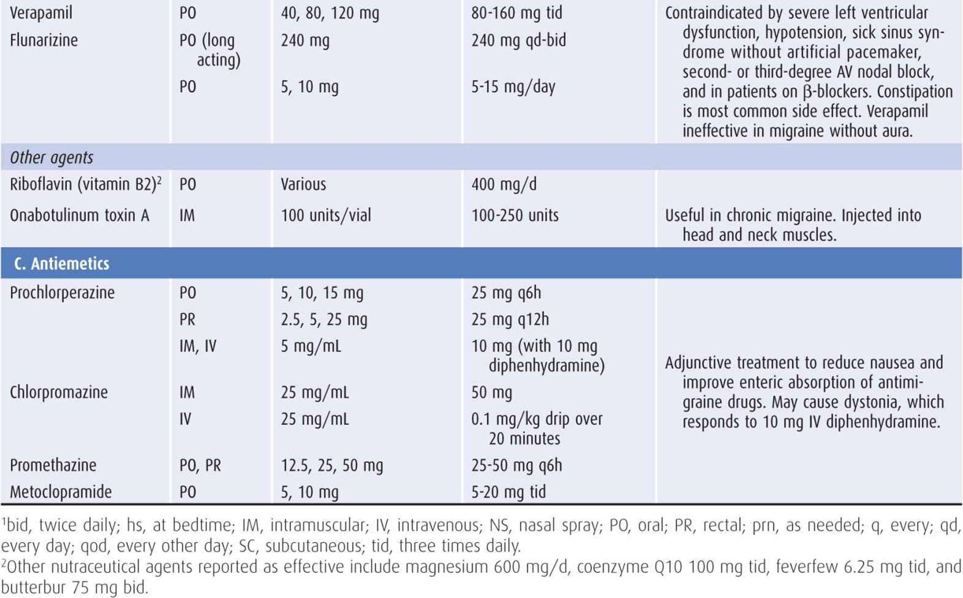
Table 6-7. Treatment of migraine headache.
Acute migraine attacks often respond to simple analgesics or, if these are ineffective, serotonin 5-HT1B/D receptor agonists (triptans) or ergot alkaloids (eg, dihydroergotamine). A new class of antimigraine drugs—calcitonin gene-related peptide receptor antagonists (eg, telcagepant, olcegepant)—may also prove to be useful for this purpose. Drugs for acute migraine headache (Table 6-7) must be taken immediately at the onset of symptoms to be maximally effective. Rapidly absorbed parenteral formulations are superior to oral preparations. Nausea, a prominent feature of migraine, is also a common side effect of some antimigraine drugs, so that administration by other than the oral route or concomitant administration of an antiemetic may be necessary. Ergot alkaloids and triptans are contraindicated in patients with hypertension or other cardiovascular disease.
Prophylactic treatment should be used in patients with two or more headaches per week or those in whom abortive treatment is inadequate. Tricyclic antidepressants, β-blockers, anticonvulsants, and calcium channel blockers can be tried in that order, unless concurrent illnesses or medications dictate differently (Table 6-7).
Migraine during pregnancy should be treated only with narcotic analgesics, although sumatriptan is safe in the first trimester. Other antimigraine drugs may be teratogenic or cause complications in pregnancy.
MEDICATION OVERUSE HEADACHE
Overuse of medications used to treat migraine or other forms of headache can lead to a chronic daily headache syndrome. Most patients have migraine as their underlying problem and have overused simple analgesics or triptans, but patients with other types of headache and using other classes of drugs are also susceptible. Most patients are women. Headache is characteristically present at least 15 days per month for at least 3 months. Treatment involves withdrawal of the offending medications, which is done abruptly except for narcotic analgesics or sedative drugs. Corticosteroids may be useful for treating withdrawal headache.
CLUSTER HEADACHE
CLINICAL FINDINGS
Cluster headache presents as clusters of brief, very severe, unilateral, constant, nonthrobbing headaches that last from 15 minutes to 3 hours. Unlike migraine headaches, cluster headaches are always unilateral and usually recur on the same side in any given patient. They commonly occur at night, awakening the patient from sleep, and recur daily, often at nearly the same time of day (circadian periodicity), for a cluster period of weeks to months. Patients may pace restlessly during attacks. Between clusters, the patient may be free from headaches for months or years. The cause is unknown, but functional MRI during attacks has shown hypothalamic activation.
Cluster headache occurs much more frequently in men than in women and typically begin at a somewhat later age than migraine (mean onset at 25 years). There is rarely a family history.
The headache may begin as a burning sensation over the lateral aspect of the nose or as pressure behind the eye (Figure 6-13). Ipsilateral conjunctival injection, lacrimation, nasal stuffiness, and Horner syndrome (Figure 6-14) are commonly associated with the attack. Episodes may be precipitated by the use of alcohol or vasodilating drugs, especially if used during a cluster siege.
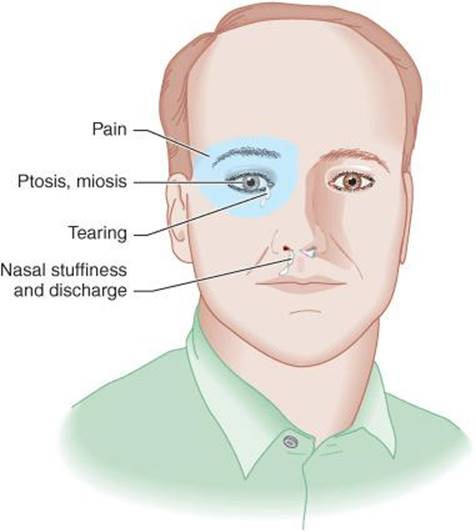
Figure 6-13. Distribution of symptoms and signs in cluster headache.

Figure 6-14. Right ptosis during acute cluster headache. Other components of Horner syndrome (pupillary miosis and regional anhidrosis) may also occur.
TREATMENT
At the onset of a headache cluster, treatment involves measures both to abort the acute attack and to prevent subsequent ones.
A. Acute Treatment
Prompt relief of pain is best achieved by inhalation of 100% oxygen (7-12 L/min for 15-20 minutes) or subcutaneous administration of sumatriptan (6 mg, repeated once per attack if necessary) (Table 6-7). Alternative abortive treatments include intranasal sumatriptan, zolmitriptan, or lidocaine; subcutaneous octreotide; and intravenous, intramuscular, subcutaneous, or intranasal dihydroergotamine.
B. Maintenance Prophylaxis
Several drugs used in the treatment of migraine (Table 6-7), including calcium channel antagonists, triptans, and ergot alkaloids, are also useful for preventing recurrent symptoms during an active bout of cluster headache (maintenance prophylaxis). The most widely used drug is verapamil (360 mg/d or sustained-release 240-480 mg/d). Lithium carbonate or lithium citrate syrup, 300 mg orally one to three times daily, is also effective in many cases. Serum lithium levels should be measured at weekly intervals for the first several weeks and should be maintained below 1.2 mEq/L to reduce the likelihood of adverse effects (nausea, diarrhea, polyuria, renal failure, hypothyroidism, tremor, dysarthria, ataxia, myoclonus, and seizures). Anticonvulsants and melatonin (10 mg/d) can also be helpful. Drugs used for maintenance prophylaxis are discontinued at the end of the cluster.
C. Transitional Prophylaxis
Administration of prednisone at the beginning of a cluster cycle can be abortive, when given as 40 to 80 mg/d orally for 1 week, and then discontinued by tapering the dose over the following week. Intravenous methylprednisolone or dihydroergotamine may be useful as well.
D. Cluster Headache Variants
Chronic rather than episodic cluster headaches may respond dramatically to indomethacin 25 mg three times daily. A bilateral variant, hypnic headache, which lacks the autonomic components of cluster headache and occurs in the elderly, responds to lithium.
E. Invasive Treatment
Invasive treatments are reserved for patients who fail to respond to medications and include greater occipital nerve or sphenopalatine ganglion block or stimulation, hypothalamic stimulation, and radiofrequency trigeminal ganglion ablation or trigeminal rhizotomy.
TENSION HEADACHE
Tension headache is the term used to describe chronic, recurrent headache of inapparent cause that lacks features of migraine or other headache syndromes. The underlying pathophysiologic mechanism is unknown, and “tension” is unlikely to be primarily responsible. Contraction of neck and scalp muscles, which has also been proposed as the cause, is probably a secondary phenomenon.
In its classic form (Figure 6-15), tension headache is a chronic disorder that begins after age 20 years. It is characterized by attacks of nonthrobbing, bilateral occipital head pain that is not associated with nausea, vomiting, or prodromal visual disturbance. Headache duration is from hours to days. The pain is sometimes likened to a tight band around the head.

Figure 6-15. Distribution of symptoms and signs in tension headache.
Women are more commonly affected than men. Although tension headache and migraine have been traditionally considered distinct disorders, many patients have headaches that exhibit features of both. Thus some patients who are classified as having tension headaches experience throbbing headaches, unilateral head pain, or nausea with attacks. In consequence, it may be more accurate to view tension headache and migraine as representing opposite poles of a single clinical spectrum.
Drugs used in the treatment of tension headache include many of the same agents used for migraine (Table 6-7). Acute attacks may respond to aspirin, other nonsteroidal anti-inflammatory drugs, acetaminophen, ergotamine, or dihydroergotamine. For prophylactic treatment, amitriptyline or imipramine is often effective, and propranolol is useful in some cases. Selective serotonin reuptake inhibitors are not useful. Although many patients respond to benzodiazepines such as diazepam 5 to 30 mg/d orally or chlordiazepoxide 10 to 75 mg/d orally, these drugs should be used sparingly because of their addictive potential. Psychotherapy, physical therapy, and relaxation techniques can provide additional benefit in selected cases.
ICEPICK-LIKE PAIN
Very brief, sharp, severe pain located in the scalp outside of the trigeminal distribution is called “icepick-like pain.” Paroxysms of pain are single or repetitive or occur in clusters, either at a single point or scattered over the scalp. Pain is experienced as an electric-like jab that reaches maximal intensity in less than 1 second, resolves rapidly, and is severe enough to cause involuntary flinching. Icepick-like pain is more common in those with migraine or cluster headache, but may occur in individuals who are otherwise headache-free. Patients frequently seek medical attention because of the intensity of the pain. If the bouts of pain are recurrent, treatment may be indicated. The syndrome responds to indomethacin (25-50 mg three times daily); gabapentin (400 mg twice daily) and melatonin (3-12 mg at bedtime) have also been reported as effective.
CERVICAL SPINE DISEASE
Injury or degenerative disease processes involving the upper neck can produce pain in the occiput or orbital regions. The most important source of discomfort is irritation of the second cervical (C2) nerve root. Disk disease or abnormalities of the articular processes affecting the lower cervical spine refer pain to the ipsilateral arm or shoulder, not to the head, although cervical muscle spasm may occur. Acute pain of cervical origin is treated with immobilization of the neck (eg, with a soft collar) and analgesic or anti-inflammatory drugs.
SINUSITIS
Acute sinusitis can produce pain and tenderness localized to the affected frontal or maxillary sinuses. Inflammation in the ethmoidal or sphenoidal sinuses produces a deep midline pain behind the nose. Sinusitis pain is increased by bending forward and by coughing or sneezing. Percussion over the frontal or maxillary area produces tenderness and accentuation of pain.
Sinusitis is treated with vasoconstrictor nose drops (eg, phenylephrine, 0.25%, instilled every 2-3 hours), antihista-mines, and antibiotics. In refractory cases, surgical sinus drainage may be necessary.
Patients who complain of chronic “sinus” headache rarely have recurrent inflammation of the sinuses; they are much more likely to have migraine or tension headaches.
DENTAL DISEASE
Temporomandibular joint dysfunction is a poorly defined syndrome characterized by preauricular facial pain, limitation of jaw movement, tenderness of the muscles of mastication, and “clicking” of the jaw with movement. Symptoms are often associated with malocclusion, bruxism, or clenching of the teeth and may result from spasm of the masticatory muscles. Some patients benefit from local application of heat, jaw exercises, nocturnal use of a bite guard, or nonsteroidal anti-inflammatory drugs.
Infected tooth extraction sites can also give rise to pain, which is characteristically constant, unilateral, and aching or burning in character. Although radiologic studies may be normal, injection of a local anesthetic at the extraction site relieves the symptoms. Treatment is with jawbone curettage and antibiotics.
REFERENCES
General
Bo SH, Davidsen EM, Gulbrandsen P, Dietrichs E. Acute headache: a prospective diagnostic work-up of patients admitted to a general hospital. Eur J Neurol. 2008;15:1293-1299.
Dodick DW. Clinical practice. Chronic daily headache. N Engl J Med. 2006;354:158-165.
Evans RW. Case studies of uncommon headaches. Neurol Clin. 2006;24:347-362.
Schievink WI. Spontaneous spinal cerebrospinal fluid leaks and intracranial hypotension. JAMA. 2006;295:2286-2296.
Walker RA, Wadman MC. Headache in the elderly. Clin Geriatr Med. 2007;23:291-305.
Subarachnoid Hemorrhage
Brown SC, Brew S, Madigan J. Investigating suspected subarachnoid haemorrhage in adults. BMJ. 2011;342:d2644.
Park S-H, Yim MB, Lee CY, Kim E, Son EI. Intracranial fusiform aneurysms: its pathogenesis, clinical characteristics and managements. J Korean Neurosurg Soc. 2008;44:116-123.
Provenzale JM. Imaging evaluation of the patient with worst headache of life—its not all subarachnoid hemorrhage. Emerg Radiol. 2010;17:403-412.
Siebert B, Tummala RP, Chow R, Faridar A, Mousavi SA, Divani AA. Intracranial aneurysms: review of current treatment options and outcomes. Front Neurol. 2011;2:45.
Suarez JI, Tarr RW, Selman WR. Aneurysmal subarachnoid hemorrhage. N Engl J Med. 2006;354:387-396.
Meningitis & Encephalitis
Bamberger DM. Diagnosis, initial management, and prevention of meningitis. Am Fam Physician. 2010;82:1491-1498.
Brouwer MC, Tunkel AR, van de Beek D. Epidemiology, diagnosis, and antimicrobial treatment of acute bacterial meningitis. Clin Microbiol Rev. 2010;23:467-492.
Gladstone J, Bigal ME. Headaches attributable to infectious diseases. Curr Pain Headache Rep. 2010;14:299-308.
Seizures
Ekstein D, Schachter SC. Postictal headache. Epilepsy Behav. 2010;19:151-155.
Lumbar Puncture
Ahmed SV, Jayawarna C, Jude E. Post lumbar puncture headache: diagnosis and management. Postgrad Med J. 2006;82:713-716.
Bezov D, Lipton RB, Ashina S. Post-dural puncture headache: Part I. Diagnosis, epidemiology, etiology, and pathophysiology. Headache. 2010;50:1144-1152.
Bezov D, Ashina S, Lipton R. Post-dural puncture headache: Part II. Prevention, management, and prognosis. Headache. 2010; 50:1482-1498.
Lavi R, Rowe JM, Avivi I. Lumbar puncture: it is time to change the needle. Eur Neurol. 2010;64:108-113.
Hypertensive Encephalopathy
Rodriguez MA, Kumar SK, De Caro M. Hypertensive crisis. Cardiol Rev. 2010: 18:102-107.
Giant Cell Arteritis
Ezeonyeji AN, Borg FA, Dasgupta B. Delays in recognition and management of giant cell arteritis: results from a retrospective audit. Clin Rheumatol. 2011;30:259-262.
Gonzalez-Gay MA, Barros S, Lopez-Diaz MJ, Garcia-Porrua C, Sanchez-Andrade A, Llorca J. Giant cell arteritis: disease patterns of clinical presentation in a series of 240 patients. Medicine (Baltimore). 2005;84:269-276.
Intracranial Mass
Buckner JC, Brown PD, O’Neill BP, Meyer FB, Wetmore CJ, Uhm JH. Central nervous system tumors. Mayo Clin Proc. 2007;82:1271-1286.
Clarke J, Butowski N, Chang S. Recent advances in therapy for glioblastoma. Arch Neurol. 2010;67:279-283.
Preussner M, de Ribaupierre S, Wöhrer A, et al. Current concepts and management of glioblastoma. Ann Neurol. 2011;70:9-21.
Idiopathic Intracranial Hypertension
Ball AK, Clarke CE. Idiopathic intracranial hypertension. Lancet Neurol. 2006;5:433-442.
Dhungana S, Sharrack B, Woodroofe N. Idiopathic intracranial hypertension. Acta Neurol Scand. 2010;121:71-82.
Trigeminal Neuralgia
Bennetto L, Patel NK, Fuller G. Trigeminal neuralgia and its management. BMJ. 2007;334:201-205.
Cruccu G, Gronseth G, Alksne J, et al. AAN-EFNS guidelines on trigeminal neuralgia management. Eur J Neurol. 2008;15: 1013-1028.
Glossopharyngeal Neuralgia
De Simone R, Ranieri A, Bilo L, Fiorillo C, Bonavita V. Cranial neuralgias: from physiopathology to pharmacological treatment. Neurol Sci. 2008;29:S69-S78.
Postherpetic Neuralgia
Baron R, Wasner G. Prevention and treatment of postherpetic neuralgia. Lancet. 2006;367:186-188.
Delaney A, Colvin LA, Fallon MT, Dalziel RG, Mitchell R, Fleetwood-Walker SM. Postherpetic neuralgia: from preclinical models to the clinic. Neurotherapeutics. 2009;6:630-637.
Hypertension
Adler JT, Meyer-Rochow GY, Chen H, et al. Pheochromocytoma: current approaches and future directions. Oncologist. 2008;13:779-793.
Persistent Idiopathic Facial Pain
Cornelissen P, van Kleef M, Mekhail N, Day M, van Zundert J. Evidence-based interventional pain medicine according to clinical diagnoses. 3. Persistent idiopathic facial pain. Pain Pract. 2009;9:443-448.
Migraine
De Vries B, Frants RR, Ferrari MD, van den Maagdenberg AM. Molecular genetics of migraine. Hum Genet. 2009;126:115-132.
Goadsby PJ, Sprenger T. Current practice and future directions in the prevention and acute management of migraine. Lancet Neurol. 2010;9:285-298.
Kirchmann M, Thomsen LL, Olesen J. Basilar-type migraine: clinical, epidemiologic, and genetic features. Neurology. 2006;66:880-886.
Kruit MC, van Buchem MA, Launer LJ, Terwindt GM, Ferrari MD. Migraine is associated with an increased risk of deep white matter lesions, subclinical posterior circulation infarcts and brain iron accumulation: the population-based MRI CAMERA study. Cephalalgia. 2010;30(2):129-36.
Levy D. Migraine pain and nociceptor activation—where do we stand? Headache. 2010;50:909-916.
Loder E. Triptan therapy in migraine. N Engl J Med. 2010;363:63-70.
Olesen J, Ashina M. Emerging migraine treatments and drug targets. Trends Pharmacol Sci. 2011;32:352-359.
van de Ven RC, Kaja S, Plomp JJ, Frants RR, van den Maagdenberg AM, Ferrari MD. Genetic models of migraine. Arch Neurol. 2007;64:643-646.
Medication Overuse Headache
Evers S, Marziniak M. Clinical features, pathophysiology, and treatment of medication-overuse headache. Lancet Neurol. 2010;9:391-401.
Cluster Headache
Ashkenazi A, Schwedt T. Cluster headache—acute and prophylactic therapy. Headache. 2011;51:272-286.
Francis GJ, Becker WJ, Pringsheim TM. Acute and preventive pharmacologic treatment of cluster headache. Neurology. 2010;75:463-473.
Tension Headache
Fumal A, Schoenen J. Tension-type headache: current research and clinical management. Lancet Neurol. 2008;7:70-83.
Loder E, Rizzoli P. Tension-type headache. BMJ. 2008; 336:88-92.
Cervical Spine Disease
Bogduk N, Govind J. Cervicogenic headache: an assessment of the evidence on clinical diagnosis, invasive tests, and treatment. Lancet Neurol. 2009;8:959-968.
Sinusitis
Cady RK, Dodick DW, Levine HL, et al. Sinus headache: a neurology, otolaryngology, allergy, and primary care consensus on diagnosis and treatment. Mayo Clin Proc. 2005;80:908-916.
Dental Disease
Scrivani SJ, Keith DA, Kaban LB. Temporomandibular disorders. N Engl J Med. 2008;359:2693-2705.