
Approach to Diagnosis
History
History of Present Illness
Past Medical History
Developmental History
Family History
Neurologic Examination
Motor System
Coordination
Tendon Reflexes
Superficial Reflexes
Gait
Clinical Localization of the Lesion
Upper Motor Neuron Lesions
Lower Motor Neuron Lesions
Cerebellar Lesions
Neuromuscular Transmission Disorders
Myopathic Disorders
Investigative Studies
Imaging
Electrodiagnostic Studies
Serum Enzymes
Muscle Biopsy
Spinal Cord Disorders (Myelopathy)
Traumatic Myelopathy
Clinical Findings
Imaging
Treatment
Prognosis
Demyelinating Myelopathies
Multiple Sclerosis
Neuromyelitis Optica
Acute Disseminated Encephalomyelitis
Other Infective or Inflammatory Myelopathies
Spinal Epidural Abscess
Acute Transverse Myelitis
Syphilis
Tuberculosis
AIDS
Other Viral Infections
Tetanus
Chronic Adhesive Arachnoiditis
Vascular Myelopathies
Spinal Cord Infarction
Hematomyelia
Spinal Epidural or Subdural Hemorrhage
Arteriovenous Malformation (AVM) or Fistula
Nutritional Myelopathies
Cervical Spondylosis
Pathogenesis
Clinical Findings
Investigative Studies
Differential Diagnosis
Treatment
Congenital Spinal Anomalies
Spinal Cord Tumors
Etiology
Pathogenesis of Myelopathy
Clinical Findings
Investigative Studies
Treatment
Prognosis
Anterior Horn Cell Disorders
Idiopathic Disorders
Motor Neuron Disease in Children
Motor Neuron Disease in Adults
Other Noninfective Anterior Horn Cell Disorders
Infective Anterior Horn Cell Disorders
Polio Virus Infection
Postpolio Syndrome
West Nile Virus Infection
Nerve Root and Plexus Lesions
Acute Intervertebral Disk prolapse
Lumbar Disk Prolapse
Cervical Disk Prolapse
Cervical Spondylosis
Traumatic Avulsion of Nerve roots
Erb-Duchenne Paralysis
Klumpke Paralysis
Brachial plexopathy
Neuralgic Amyotrophy (Idiopathic Brachial Plexopathy)
Cervical Rib Syndrome
Other Causes Of Brachial Plexopathy
Lumbosacral plexopathy
Disorders of Peripheral Nerve
Polyneuropathy
Acute Inflammatory Polyradiculoneuropathy (Guillain-Barré Syndrome)
Critical Illness Polyneuropathy
Diphtheritic POlyneuritis
Paralytic Shellfish Poisoning
Porphyria
Acute Arsenic or Thallium Poisoning
Organophosphate Polyneuropathy
Mononeuropathy Multiplex
Lead Toxicity
Multifocal Motor Neuropathy
Mononeuropathy Simplex
Bell Palsy
Disorders of Neuromuscular Transmission
Myasthenia Gravis
Pathogenesis
Clinical Findings
Diagnosis
Investigative Studies
Treatment
Prognosis
Myasthenic Syndrome (Lambert-Eaton Syndrome)
Pathogenesis
Clinical Findings
Diagnosis
Treatment
Botulism
Pathogenesis
Clinical Findings
Investigative Studies
Treatment
Aminoglycoside Antibiotics
Myopathic Disorders
Muscular Dystrophies
Duchenne Dystrophy
Becker Dystrophy
Limb-Girdle Dystrophy
Facioscapulohumeral Dystrophy
Emery-Dreifuss Muscular Dystrophy
Distal Myopathy
Ocular Dystrophy
Oculopharyngeal Dystrophy
Paraspinal DystropHy
Congenital Myopathies
Mitochondrial Myopathies
Myotonic Disorders
Myotonic Dystrophies
Nondystrophic Myotonias
Inflammatory Myopathies
Trichinosis, Toxoplasmosis, & Sarcoidosis
Polymyositis & Dermatomyositis
Inclusion-Body Myositis
AIDS
Polymyalgia Rheumatica
Eosinophilia-Myalgia Syndrome
Metabolic Myopathies
Hypokalemia
Periodic Paralyses
Osteomalacia
Endocrine Myopathies
Alcoholic Myopathies
Acute Necrotizing Alcoholic Myopathy
Chronic Alcoholic Myopathy
Drug-Induced Myopathies
Critical Illness Myopathy
Myoglobinuria
Motor-Unit Hyperactivity States
Central Nervous System Disorders
Stiff-Person Syndrome
Tetanus
Startle Syndromes
Peripheral Nerve Disorders
Cramps
Neuromyotonia
Tetany
Hemifacial Spasm
Muscle Disorders
Myotonia
Malignant Hyperthermia
References
APPROACH TO DIAGNOSIS
Normal motor function depends on the transmission of signals from the brain to the brainstem or spinal cord by upper motor neurons, and from there to skeletal muscle by lower motor neurons (Figure 9-1). Accordingly, motor function can be impaired by a lesion that involves this pathway anywhere along its length. Anatomic structures involved in the regulation or execution of motor activity include the pyramidal and extrapyramidal systems, the cerebellum, and the lower motor neurons in the cranial nerve nuclei of the brainstem and the anterior horns of the spinal cord.

Figure 9-1. Anatomic basis of the upper motor neuron and lower motor neuron concepts.
The pyramidal system (Figure 9-2) consists of upper motor neuron fibers that descend from the cerebral cortex through the internal capsule, traverse the medullary pyramid, and then mostly decussate, to descend in the lateral corticospinal tract on the side opposite that of their origin, where they synapse on interneurons and lower motor neurons in the spinal cord.

Figure 9-2. Upper motor neuron pathways. Tracts at bottom left are shown outside the cord for clarity only. (From McPhee SJ, Hammer GD: Pathophysiology of Disease: An Introduction to Clinical Medicine. 6e. New York:2009, McGraw Hill.)
All other descending influences on lower motor neurons belong to the extrapyramidal system and originate primarily in the basal ganglia and cerebellum. Disorders of the basal ganglia (Chapter 11) and cerebellum (Chapter 8) are considered separately.
The motor fibers that make up the cranial and peripheral nerves have their origin in the lower motor neurons (Figure 9-3). A disturbance of function at any point in the peripheral nervous system (anterior horn cell, nerve root, limb plexus, peripheral nerve, or neuromuscular junction) can disturb motor function, as can disease that primarily affects the muscles themselves.

Figure 9-3. Anatomic components of the motor unit.
HISTORY
Patients with motor deficits generally complain of weakness, heaviness, stiffness, clumsiness, impaired muscular control, or difficulty in executing movements. The term weakness is sometimes used in a nonspecific way to denote fatigue or loss of energy, drive, or enthusiasm, and care must be taken to clarify what the patient means. The word is properly used to mean loss of muscle power, and it is in this sense that it is employed here.
HISTORY OF PRESENT ILLNESS
Several aspects of the present complaint must be documented.
Mode of Onset
An abrupt onset suggests a vascular disturbance, such as a stroke, or certain toxic or metabolic disturbances, whereas subacute onset over days to weeks is commonly associated with a neoplastic, infective, or inflammatory process (Table 9-1). Weakness that evolves slowly over several months or years often has a hereditary, degenerative, endocrinologic, or neoplastic basis.

Table 9-1. Some causes of weakness of acute or subacute onset.
Course
A progressive increase in the motor deficit from its onset suggests continuing activity of the underlying process. Episodic progression suggests a vascular or inflammatory origin; a steadily progressive course is more suggestive of neoplastic disorder or such degenerative conditions as motor neuron disease. Rapid fluctuation of symptoms over short periods (eg, activity leads to fatigue and an exacerbation of weakness; rest is followed by recovery of strength) is characteristic of myasthenia gravis.
Distribution of Symptoms
The distribution of weakness and the presence of associated symptoms may indicate the approximate site of the lesion. For example, weakness in the right arm and leg may result from a lesion of the contralateral motor cortex or the corticospinal pathway at any point above the fifth cervical segment of the spinal cord. Associated right facial weakness indicates that the lesion must be above the level of the facial (VII) nerve nucleus in the brainstem, and an accompanying aphasia (Chapter 1) or visual field defect (Chapter 7) localizes it to the cerebral hemisphere.
Associated Symptoms
The presence and distribution of any sensory abnormalities are also helpful in localizing the lesion in patients with weakness. Sensory abnormalities lateralized to the same side as weakness suggest a hemispheric lesion; a cortical lesion is implied by sensory neglect or inattention, agraphesthesia (inability to identify by touch a number written on the skin), astereognosis (inability to identify by touch an object placed in the hand), abarognosis (inability to judge the weight of an object placed in the hand), or impaired two-point discrimination, when peripheral sensory function is intact. Sensory loss below a particular segmental level on the trunk suggests a spinal cord lesion, whereas distal sensory changes in the limbs favor a peripheral nerve lesion. Diseases of the anterior horn cells, neuromuscular junctions, or muscles are not accompanied by alterations in sensation.
The character of the associated symptoms may suggest the nature of the lesion at any given site in the nervous system. Thus progressive leg weakness caused by myelopathy is often preceded or accompanied by pain in the back or legs when the myelopathy is due to a compressive lesion—but not when it has a metabolic or hereditary basis.
Severity of Symptoms
An attempt must be made to evaluate the functional severity of a motor deficit by determining whether there has been any restriction of daily activities, difficulty in performing previously easy tasks, or reduction in exercise tolerance.
The nature of the functional disturbance depends on the muscles involved. Weakness of proximal muscles in the legs leads to difficulty in climbing or descending stairs or in getting up from a squatting position, whereas weakness in the arms leads to difficulty with such tasks as combing the hair. Distal weakness in the arms may lead to clumsiness, difficulty with such fine motor tasks as doing up buttons or tying shoelaces, and eventually the inability to pick up or grasp objects with the hands, so that even eating becomes difficult or impossible.
Involvement of the muscles supplied by the cranial nerves may lead to diplopia (oculomotor [III], trochlear [IV], or abducens [VI] nerve); difficulty in chewing (trigeminal [V] nerve) or sucking, blowing, or grimacing (facial [VII] nerve); or difficulty in swallowing, with nasal regurgitation and dysarthria (glossopharyngeal [IX], vagus [X], and hypoglossal [XII] nerves).
Weakness of the respiratory muscles leads to tachypnea, the use of accessory muscles of respiration, and anxiety at a stage when arterial blood gases are usually still normal. A vital capacity of less than 1 L in an adult generally calls for ventilatory support, especially if weakness is increasing.
PAST MEDICAL HISTORY
The importance of the history depends on the patient’s present complaint and the nature of any previous illnesses. For example, in a patient with known carcinoma of the lung, limb weakness may be due to metastasis or to a remote (nonmetastatic) complication of cancer. Leg weakness in a diabetic may reflect peripheral nerve, plexus, or multiple root involvement, and hand weakness in a myxedematous patient may be associated with carpal tunnel syndrome.
The history should include careful note of all drugs taken by the patient. Drugs can cause peripheral neuropathy, impair neuromuscular transmission, or lead to myopathy (Table 9-2).

Table 9-2. Motor disorders associated with drugs.
DEVELOPMENTAL HISTORY
When symptoms develop during infancy, childhood, or early adult life, it is particularly important to obtain a full developmental history, including details of the delivery, the birth weight, the patient’s condition in the neonatal period, and the dates at which motor milestones were attained. Congenital or perinatal cerebral disease accounts for most causes of infantile diplegia (weakness of all four limbs, with the legs more severely affected than the arms).
FAMILY HISTORY
Hereditary factors may be important, and the patient’s family background therefore must be explored. Some types of myopathy, motor neuron disease, and peripheral neuropathy have a genetic basis, as do some spinocerebellar degenerations, hereditary spastic paraparesis, and certain other neurologic disorders. In some instances, it may be necessary to examine other family members to determine whether the patient’s disorder has a hereditary basis.
NEUROLOGIC EXAMINATION
MOTOR SYSTEM
In examining the motor system, a systematic approach will help to avoid overlooking important abnormalities. A sequential routine for the examination should be developed.
Muscle Appearance
1. Wasting, or muscle atrophy, suggests that weakness is due to a lesion of the lower motor neurons or the muscle itself. The distribution of wasting may help to localize the underlying disorder. Upper motor neuron disorders are not usually accompanied by muscle wasting, though muscle atrophy may occasionally occur with prolonged disuse.
2. Pseudohypertrophy of muscles occurs in certain forms of myopathy, but the apparently enlarged muscles are actually weak and flabby.
3. Fasciculations—visible irregular flickerings over the surface of the affected muscle caused by spontaneous contractions of individual motor units—suggest that weakness is due to a lower motor neuron lesion. Fasciculations are most apt to be seen in anterior horn cell disorders, but also occur in normal individuals.
4. Flexor or extensor spasms of the limbs are sometimes seen in upper motor neuron disorders as a result of impaired supraspinal control of reflex activity.
Muscle Tone
For clinical purposes, tone can be defined as the resistance of muscle to passive movement of a joint. Tone depends on the degree of muscle contraction and on the mechanical properties of muscle and connective tissue. The degree of muscle contraction depends, in turn, on the activity of anterior horn cells, which is governed by spinal and supraspinal mechanisms.
Tone is assessed by observing the position of the extremities at rest, by palpating the muscle belly, and particularly by determining the resistance to passive stretch and movement. To assess resistance to passive movement, the patient is asked to relax while each limb is examined in turn by passively taking the major joints through their full range of movement at different speeds and estimating whether the force required is more or less than normal.
Postural abnormalities may result from the increased activity of certain muscle groups caused by disturbances of reflex function, as exemplified by the typical hemiplegic posture—flexion of the upper limb and extension of the ipsilateral lower limb—of many patients who have had a stroke.
1. Hypertonia—Two types of increased tone can be distinguished.
a. Spasticity—consists of an increase in tone that affects different muscle groups to different extents. In the arms, tone is increased to a greater extent in the flexor and adductor muscles than in the extensors and abductors; in the legs, tone is increased to a greater extent in the extensor muscles than in the flexors. Moreover, the resistance of an affected muscle is not the same throughout the range of movement, but tends to be most marked when passive movement is initiated and then diminishes as the movement continues (the clasp-knife phenomenon). The increase in tone is velocity-dependent, so that passive movement at a high velocity—but not at lower velocities—may be met with increased resistance. Spasticity is caused by an upper motor neuron lesion, such as a stroke that involves the supplementary motor cortex or corticospinal tract. Spasticity may not become apparent for several days after the onset of an acute lesion, however.
b. Rigidity—consists of increased resistance to passive movement that is independent of the direction of the movement; that is, it affects agonist and antagonist muscle groups equally. The term lead-pipe rigidity is sometimes used for descriptive purposes, whereas cogwheel rigidity is used when there are superimposed ratchet-like interruptions in the passive movement, which probably relate to underlying tremor. In general, rigidity indicates extrapyramidal dysfunction and is due to a lesion of the basal ganglia (eg, Parkinson disease).
2. Hypotonia (flaccidity)—This is characterized by excessive floppiness—a reduced resistance to passive movement—so that the distal portion of the limb is easily waved to and fro when the extremity is passively shaken. In hypotonic limbs it is often possible to hyperextend the joints, and the muscle belly may look flattened and feel less firm than usual. Although hypotonia usually relates to pathologic involvement of the lower motor neuron supply to the affected muscles, it can also occur with primary muscle disorders, disruption of the sensory (afferent) limb of the reflex arc, cerebellar disease, and certain extrapyramidal disorders such as Huntington disease, as well as in the acute stage of a pyramidal lesion.
3. Paratonia—Some patients give the impression of being unable to relax and will move the limb being examined as the physician moves it, despite instructions to the contrary. In more advanced cases, there seems to be rigidity when the examiner moves the limb rapidly but normal tone when the limb is moved slowly. This phenomenon—paratonia—is particularly apt to occur in patients with frontal lobe or diffuse cerebral disease.
Muscle Power
When muscle power is to be tested, the patient is asked to resist pressure exerted by the examiner. On the basis of the history and other findings, muscles that are particularly likely to be affected are selected for initial evaluation, and other muscles are subsequently examined to determine the distribution of the weakness more fully and to shorten the list of diagnostic possibilities. For instance, if an upper motor neuron (pyramidal) lesion is suspected, the extensors and abductors of the upper extremity and the flexors of the lower extremity are tested in the most detail, because these muscles will be the most affected. Strength on the two sides is compared so that minor degrees of weakness can be recognized.
1. Upper vs. lower motor neuron lesions—Weakness can result from a disturbance in function of the upper or the lower motor neurons; the distribution of weakness is of paramount importance in distinguishing between these two possibilities. Upper motor neuron lesions (eg, stroke) lead to weakness that characteristically involves the extensors and abductors more than the flexors and adductors of the arms—and the flexors more than the extensors of the legs. Lower motor neuron lesions produce weakness of the muscles supplied by the affected neurons; the particular distribution of the weakness may point to lower motor neuron involvement at the spinal cord, nerve root, plexus, or peripheral nerve level.
2. Myopathic vs. neuropathic disorders—Weakness may also result from a primary muscle disorder (myopathy) or from a disorder of neuromuscular transmission. In patients with a motor deficit in all limbs that is not due to an upper motor neuron lesion, proximal distribution of weakness suggests a myopathic disorder, whereas predominantly distal involvement suggests a lower motor neuron disturbance.
3. Neuromuscular junction disorders—Marked variability in the severity and distribution of weakness over short periods of time (eg, over the course of a day) suggests myasthenia gravis, a disorder of neuromuscular transmission.
4. Psychogenic disorders—Apparent weakness that is not organic in nature also shows a characteristic variability; it is often more severe on formal testing than is consistent with the patient’s daily activities. Moreover, palpation of antagonist muscles commonly reveals that they contract each time the patient is asked to activate the agonist.
5. Muscle power grading—For practical and comparative purposes, power is best graded in the manner shown in Table 9-3.

Table 9-3. Grading of muscle power according to the system suggested by the Medical Research Council.
6. Terminology for describing distribution of weakness—Monoplegia denotes paralysis or severe weakness of the muscles in one limb, and monoparesis denotes less severe weakness in one limb, although the two words are often used interchangeably. Hemiplegia or hemiparesis is weakness in both limbs (and sometimes the face) on one side of the body; paraplegia or paraparesis is weakness of both legs; and quadriplegia or quad-riparesis (also tetraplegia, tetraparesis) is weakness of all four limbs.
COORDINATION
The coordination of motor activity can be impaired by weakness, sensory disturbances, or cerebellar disease and requires careful evaluation.
Voluntary activity is observed with regard to its accuracy, velocity, range, and regularity, and the manner in which individual actions are integrated to produce a smooth complex movement.
In the finger-nose test, the patient moves the index finger to touch the tip of his or her nose and then the tip of the examiner’s index finger; the examiner can move his or her own finger about during the test to change the location of the target and should position it so that the patient’s arm must extend fully to reach it.
In the heel-knee-shin test, the recumbent patient lifts one leg off the bed, flexes it at the knee, places the heel on the other knee, and runs the heel down the shin as smoothly as possible.
Rapid alternating movement is tested by asking the patient to tap repetitively with one hand on the back of the other, to tap alternately with the palm and back of one hand on the back of the other hand or on the knee, to screw an imaginary light bulb into the ceiling with each arm in turn, and to rub the fingers of one hand in a circular polishing movement on the back of the other hand. Other tests of rapid alternating movement include tapping on the ball of the thumb with the tip of the index finger or tapping the floor as rapidly as possible with the sole while keeping the heel of the foot in place. During all these tests, the examiner looks for irregularities of rate, amplitude, and rhythm and for precision of movements. With pyramidal lesions, fine voluntary movements are performed slowly. With cerebellar lesions, the rate, rhythm, and amplitude of such movements are irregular.
If loss of sensation may be responsible for impaired coordination, the maneuver should be repeated both with eyes closed and with visual attention directed to the limb; with visual feedback the apparent weakness or incoordination will improve. In patients with cerebellar disease, the main complaint and physical finding are often of incoordination, and examination may reveal little else. Further discussion of the ataxia of cerebellar disease and the various terms used to describe aspects of it can be found in Chapter 8.
TENDON REFLEXES
Changes in the tendon (muscle stretch) reflexes may accompany disturbances in motor (or sensory) function and provide a guide to the cause of the motor deficit. The tendon is tapped with a reflex hammer to produce a sudden brisk stretch of the muscle and its contained spindles. The clinically important stretch reflexes and the nerves, roots, and spinal segments subserving them are indicated in Table 9-4. When the reflexes are tested, the limbs on each side should be placed in identical positions and the reflexes elicited in the same manner.

Table 9-4. Tendon (muscle stretch) reflexes.1
1. Areflexia—Apparent loss of the tendon reflexes in a patient may merely reflect a lack of clinical expertise on the part of the examiner. Performance of the Jendrassik maneuver (an attempt by the patient to pull apart the fingers of the two hands when they are hooked together) or some similar distracting action (such as making a fist with the hand that is not being tested) may elicit the reflex response when it is otherwise unobtainable. A reflex may be lost or depressed by any lesion that interrupts the structural or functional continuity of its reflex arc, as in a root lesion or peripheral neuropathy. In addition, reflexes are often depressed during the acute stage of an upper motor neuron lesion, in patients who are deeply comatose, and in patients with cerebellar disease.
2. Hyperreflexia—Increased reflexes occur with upper motor neuron lesions, but they may also occur with symmetric distribution in certain healthy subjects and in patients under emotional tension. The presence of reflex asymmetry is therefore of particular clinical significance. Clonus consists of a series of rhythmic reflex contractions of a muscle that is suddenly subjected to sustained stretch, with each beat caused by renewed stretch of the muscle during relaxation from its previous contracted state. Sustained clonus—more than three or four beats in response to sudden sustained stretch—is always pathologic and is associated with an abnormally brisk reflex. In hyperreflexic states, there may be spread of the region from which a particular reflex response can be elicited. For example, elicitation of the biceps reflex may be accompanied by reflex finger flexion, or eliciting the finger flexion reflex may cause flexion of the thumb (Hoffmann sign).
3. Reflex asymmetry—Although the intensity of reflex responses varies considerably among subjects, reflexes should be symmetric in any individual. Several general points can be made regarding reflex asymmetries.
a. Lateralized asymmetries of response—reflexes that are brisker on one side of the body than on the other—usually indicate an upper motor neuron disturbance, but sometimes reflect a lower motor neuron lesion on the side with the depressed reflexes.
b. Focal reflex deficits often relate to root, plexus, or peripheral nerve lesions. For example, unilateral depression of the ankle jerk commonly reflects an S1 radiculopathy resulting from a lumbosacral disk lesion.
c. Loss of distal tendon reflexes (especially ankle jerks), with preservation of more proximal ones, is common in polyneuropathies.
SUPERFICIAL REFLEXES
1. The polysynaptic superficial abdominal reflexes, which depend on the integrity of the T8-12 spinal cord segments, are elicited by gently stroking each quadrant of the abdominal wall with a blunt object such as a wooden stick. A normal response consists of contraction of the muscle in the quadrant stimulated, with a brief movement of the umbilicus toward the stimulus. Asymmetric loss of the response may be of diagnostic significance. The response may be depressed or lost on one side in patients with an upper motor neuron disturbance affecting that side. Segmental loss of the response may relate to local disease of the abdominal wall or its innervation, as in a radiculopathy. Bilaterally absent responses are usually of no significance, occurring in the elderly, the obese, multiparous women, and patients who have had abdominal surgery.
2. The cremasteric reflex, mediated through the L1 and L2 reflex arcs, consists of retraction of the ipsilateral testis when the inner aspect of the thigh is lightly stroked; it is lost in patients with a lesion involving these nerve roots. It is also lost in patients with contralateral upper motor neuron disturbances.
3. Stimulation of the lateral border of the foot in a normal adult leads to plantar flexion of the toes and dorsiflexion of the ankle. The Babinski response consists of dorsiflexion of the big toe and fanning of the other toes in response to stroking the lateral border of the foot, which is part of the S1 dermatome; flexion at the hip and knee may also occur. Such an extensor plantar response indicates an upper motor neuron lesion involving the contralateral motor cortex or the corticospinal tract. It can also be found bilaterally in anesthetized or comatose subjects, in patients who have just had a seizure, and in normal infants.
An extensor plantar response can also be elicited, though less reliably, by such maneuvers as pricking the dorsal surface of the big toe with a pin (Bing sign), firmly stroking down the anterior border of the tibia from knee to ankle (Oppenheim maneuver), squeezing the calf muscle (Gordon maneuver) or Achilles tendon (Schafer maneuver), flicking the little toe (Gonda maneuver), or stroking the back of the foot just below the lateral malleolus (Chaddock maneuver). In interpreting responses to these maneuvers, attention must be focused only on the direction in which the big toe first moves.
GAIT
In evaluating gait, the examiner first observes the patient walking at a comfortable pace. Attention is directed at the stance and posture; the facility with which the patient starts and stops walking and turns to either side; the length of the stride; the rhythm of walking; the presence of normally associated movements, such as swinging of the arms; and any involuntary movements (Figure 1-25).
Subtle gait disorders become apparent only when the patient is asked to run, walk on the balls of the feet or the heels, hop on either foot, or walk heel-to-toe along a straight line. Gait disorders occur in many neurologic disturbances and in other contexts that are beyond the scope of this chapter. A motor or sensory disturbance may lead to an abnormal gait whose nature depends on the site of pathologic involvement. Accordingly, the causes and clinical types of gait disturbance are best considered together.
1. Apraxic gait—Apraxic gait occurs in some patients with disturbances, usually bilateral, of frontal lobe function, such as may occur in hydrocephalus or progressive dementing disorders. There is no weakness or incoordination of the limbs, but the patient is unable to stand unsupported or to walk properly—the feet seem glued to the ground. If walking is possible at all, the gait is unsteady, uncertain, and short-stepped, with marked hesitation (“freezing”), and the legs are moved in a direction inappropriate to the center of gravity.
2. Corticospinal lesions—A corticospinal lesion, irrespective of its cause, can lead to a gait disturbance that varies in character depending on whether there is unilateral or bilateral involvement.
a. In patients with hemiparesis, the selective weakness and spasticity lead to a gait in which the affected leg must be circumducted to be advanced. The patient tilts at the waist toward the normal side and swings the affected leg outward as well as forward, thus compensating for any tendency to drag or catch the foot on the ground because of weakness in the hip and knee flexors or the ankle dorsiflexors. The arm on the affected side is usually held flexed and adducted. In mild cases, there may be no more than a tendency to drag the affected leg, so that the sole of that shoe tends to be excessively worn.
b. With severe bilateral spasticity, the legs are brought stiffly forward and adducted, often with compensatory movements of the trunk. Such a gait is commonly described as scissors-like. This gait is seen in its most extreme form in children with spastic diplegia from perinatally acquired static encephalopathy. In patients with mild spastic paraparesis, the gait is shuffling, slow, stiff, and awkward, with the feet tending to drag.
3. Frontal disorders—Some patients with frontal lobe or white matter lesions have a gait characterized by short, shuffling steps; hesitation in starting (“ignition failure”) or turning; unsteadiness; and a wide or narrow base. Sometimes referred to as marche à petit pas, this abnormality may be mistaken for a parkinsonian gait, but the wide base, preserved arm swing, absence of other signs of parkinsonism, and accompanying findings of cognitive impairment, frontal release signs, pseudobulbar palsy, pyramidal deficits, and sphincter disturbances are helpful in indicating the correct diagnosis. In patients with frontotemporal dementia, however, a parkinsonian gait and other extrapyramidal findings may be present.
4. Extrapyramidal disorders—Extrapyramidal disorders can produce characteristic gait disturbances.
a. In advanced parkinsonism, the patient is often stooped and has difficulty in beginning to walk. Indeed, the patient may need to lean farther and farther forward while walking in place in order to advance; once in motion, there may be unsteadiness in turning and difficulty in stopping. The gait itself is characterized by small strides, often taken at an increasing rate until the patient is almost running (festination), and by loss of the arm swinging that normally accompanies locomotion. At times, as when walking through a doorway, the patient may be unable to advance (“freezing”). Turning may require several small steps. In mild parkinsonism, a mildly slowed or unsteady gait, flexed posture, or reduced arm swinging may be the only abnormality found.
b. Abnormal posturing of the limbs or trunk is a feature of dystonia; it can interfere with locomotion or lead to a distorted and bizarre gait.
c. Chorea can cause an irregular, unpredictable, and unsteady gait, as the patient dips or lurches from side to side. Choreiform movements of the face and extremities are usually well in evidence.
d. Tremor that occurs primarily on standing (orthostatic tremor) may lead to an unsteady, uncertain gait, with hesitancy in commencing to walk.
5. Cerebellar disorders—In cerebellar disorders (Chapter 8), the gait may be disturbed in several ways.
a. Truncal ataxia results from involvement of midline cerebellar structures, especially the vermis. The gait is irregular, clumsy, unsteady, uncertain, and broad-based, with the patient walking with the feet wide apart for additional support. Turning and heel-to-toe walking are especially difficult. There are often few accompanying signs of a cerebellar disturbance in the limbs. Causes include midline cerebellar tumors and the cerebellar degeneration that can occur with alcoholism or hypothyroidism, as a nonmetastatic complication of cancer, and with certain hereditary disorders.
b. In extreme cases, with gross involvement of midline cerebellar structures (especially the vermis), the patient cannot stand without falling.
c. A lesion of one cerebellar hemisphere leads to an unsteady gait in which the patient consistently falls or lurches toward the affected side.
6. Vestibular disorders—With unilateral vestibular dysfunction, the patient is unsteady, veering to the affected side. If both sides are affected, the gait becomes especially unsteady in the dark, when visual input is reduced.
7. Impaired sensation—Impaired sensation, especially disturbed proprioception, also leads to an unsteady gait that is aggravated by walking in the dark or with the eyes closed, as visual input cannot then compensate for the sensory loss. Because of their defective position sense, many patients lift their feet higher than necessary when walking, producing a steppage gait. Causes include tabes dorsalis, sensory neuropathies, vitamin B12 deficiency, and certain hereditary disorders (Chapter 10).
8. Anterior horn cell, peripheral motor nerve, or skeletal muscle disorders—These disorders lead to gait disturbances if the muscles involved in locomotion are affected. Weakness of the anterior tibial muscles leads to foot drop; to avoid catching or scuffing the foot on the ground, the patient must lift the affected leg higher than the other, in a steppage gait resembling that due to sensory disorders. Weakness of the calf muscles leads to an inability to walk on the balls of the feet. Weakness of the trunk and girdle muscles, such as occurs in muscular dystrophy, other myopathic disorders, and Kugelberg-Welander syndrome, leads to a waddling gait because the pelvis tends to slump toward the non–weight-bearing side.
9. Unsteady or cautious gait in the elderly—Many elderly persons complain of unsteadiness when walking and of a fear of falling, but neurologic examination reveals no abnormality. Their gait is cautious, unsteady, and sometimes difficult to initiate. Frontal lobe dysfunction may be responsible, as may reduced sensory input from several different afferent systems and impaired central processing of sensory input; an impairment of vestibular function may also be important.
10. Psychogenic gait disorder—This is suggested by fluctuations of stance and gait, commonly in response to suggestion, frequently accompanied by a sudden buckling at the knees, often without falls. Apart from the bizarre gait, neurologic examination is often otherwise normal. Patients with apparent inability to stand or walk (astasia-abasia) may actually be able to walk on a narrow base, but sway wildly in all directions, often with waving of the arms as if about to fall.
CLINICAL LOCALIZATION OF THE LESION
The findings on examination should indicate whether the weakness or other motor deficit is due to an upper or lower motor neuron disturbance, a disorder of neuromuscular transmission, or a primary muscle disorder. In the case of an upper or lower motor neuron disturbance, the clinical findings may also help to localize the lesion more precisely to a single level of the nervous system. Such localization helps to reduce the number of diagnostic possibilities.
UPPER MOTOR NEURON LESIONS
Signs
The following classic signs of an upper motor neuron lesion occur with involvement of the upper motor neuron at any point; further clinical findings depend on the actual site of the lesion. Note that it may not be possible to localize a lesion by its motor signs alone.
1. Weakness or paralysis
2. Spasticity
3. Increased tendon reflexes
4. Extensor plantar (Babinski) response
5. Loss of superficial abdominal reflexes
6. Little, if any, muscle atrophy
Localization of Underlying Lesion
1. A parasagittal intracranial lesion produces an upper motor neuron deficit that characteristically affects both legs and may later involve the arms.
2. A discrete lesion of the cerebral cortex or its projections may produce a focal motor deficit involving, for example, the contralateral hand. Weakness may be restricted to the contralateral leg in patients with anterior cerebral artery occlusion or to the contralateral face and arm if the middle cerebral artery is involved. A more extensive cortical or subcortical lesion will produce weakness or paralysis of the contralateral face, arm, and leg and may be accompanied by aphasia, a visual field defect, or a sensory disturbance of cortical type.
3. A lesion at the level of the internal capsule, where the descending fibers from the cerebral cortex are closely packed, commonly results in a severe hemiparesis that involves the contralateral limbs and face.
4. A brainstem lesion commonly—but not invariably—leads to bilateral motor deficits, often with accompanying sensory and cranial nerve disturbances, and disequilibrium. A more limited lesion involving the brainstem characteristically leads to a cranial nerve disturbance on the ipsilateral side and a contralateral hemiparesis; the cranial nerves affected depend on the level at which the brainstem is involved.
5. A unilateral spinal cord lesion above the fifth cervical segment (C5) causes an ipsilateral hemiparesis that spares the face and cranial nerves. Lesions between C5 and the first thoracic segment (T1) affect the ipsilateral arm to a variable extent as well as the ipsilateral leg; a lesion below T1 will affect only the ipsilateral leg. If both sides of the cord are involved, quadriparesis or paraparesis usually results. Increased muscle tone (spasticity) may be more prominent than weakness. If there is an extensive but unilateral cord lesion, the motor deficit is accompanied by ipsilateral impairment of vibration and position sense and by contralateral loss of pain and temperature appreciation (Brown-Séquard syndrome).
6. With compressive and other focal lesions that involve the anterior horn cells in addition to the fiber tracts traversing the cord, the muscles innervated by the affected cord segment weaken and atrophy. Therefore, a focal lower motor neuron deficit exists at the level of the lesion, and an upper motor neuron deficit exists below it—in addition to any associated sensory disturbance.
LOWER MOTOR NEURON LESIONS
Signs
Lower motor neuron lesions produce the following characteristic signs at the affected levels.
1. Weakness or paralysis
2. Wasting and fasciculations of involved muscles
3. Hypotonia (flaccidity)
4. Loss of tendon reflexes when neurons subserving them are affected
5. Normal abdominal and plantar reflexes—unless the neurons subserving them are directly involved, in which case reflex responses are lost
Localization of the Underlying Lesion
In distinguishing weakness from a segmental spinal cord, anterior horn cell, radicular (nerve root), plexus, or peripheral nerve lesion, the distribution of the motor deficit is of particular importance. Only those muscles supplied wholly or partly by the involved structure are weak (Tables 9-5 and Table 9-6). The distribution of any accompanying sensory deficit similarly reflects the location of the underlying lesion (Chapter 10).

Table 9-5. Innervation of selected muscles of the upper limbs.

Table 9-6. Innervation of selected muscles of the lower limbs.
Weakness due to lesions that affect nerve roots may be difficult to distinguish from weakness caused by spinal cord lesions that involve the anterior horn cells. In the latter situation, however, there is more often a bilateral motor deficit at the level of the lesion, a corticospinal or sensory deficit below it, or a disturbance of bladder, bowel, or sexual function.
Disorders that affect the anterior horn cells of the spinal cord can often be distinguished from disorders of motor nerves (motor neuropathy) by the extensive lower motor neuron deficit without sensory changes that occurs in the former.
CEREBELLAR LESIONS
Signs
Cerebellar lesions produce characteristic signs, which are discussed in this section and in Chapter 8.
1. Hypotonia.
2. Depressed or pendular tendon reflexes.
3. Ataxia—this is a complex movement disorder caused, at least in part, by impaired coordination. It occurs in the limbs on the same side as a lesion affecting the cerebellar hemisphere. With midline lesions, incoordination may not be evident in the limbs at all, but there is marked truncal ataxia that becomes evident on walking. The term dysmetria is used when movements are not adjusted accurately for range, so that, for example, a moving finger overshoots a target at which it is aimed. Dysdiadochokinesia denotes rapid alternating movements that are clumsy and irregular in terms of rhythm and amplitude. Asynergia or dyssynergia denotes the breakdown of complex actions into the individual movements composing them; when asked to touch the tip of the nose with a finger, for example, the patient may first flex the elbow and then bring the hand up to the nose instead of combining the maneuvers into one action. Intention tremor occurs during activity and is often most marked as the target is neared. The rebound phenomenon is the overshooting of the limb when resistance to a movement or posture is suddenly withdrawn.
4. Gait disorder—The gait becomes unsteady in patients with disturbances of either the cerebellar hemispheres or midline structures.
5. Imbalance of station.
6. Disturbances of eye movement—Jerk nystagmus, which is commonly seen in patients with a unilateral lesion of the cerebellar hemisphere, is slowest and of greatest amplitude when the eyes are turned to the side of the lesion. Nystagmus is not present in patients with lesions of the anterior cerebellar vermis.
7. Dysarthria—Speech becomes dysarthric and takes on an irregular and explosive quality in patients with lesions that involve the cerebellar hemispheres. Speech is usually unremarkable when only the midline structures are involved.
Localization of the Underlying Lesion
The relationship of symptoms and signs to lesions of different parts of the cerebellum is considered in Chapter 8.
NEUROMUSCULAR TRANSMISSION DISORDERS
Signs
1. Normal or reduced muscle tone.
2. Normal or depressed tendon and superficial reflexes.
3. No sensory changes.
4. Weakness, often patchy in distribution, not conforming to the distribution of any single anatomic structure; frequently involves the cranial muscles and may fluctuate in severity over short periods, particularly in relation to activity.
Localization of the Underlying Lesion
Pathologic involvement of either the presynaptic (eg, botulism) or postsynaptic (eg, myasthenia gravis) portion of the neuromuscular junction may impair neuromuscular transmission. Disorders affecting neuromuscular transmission are discussed later in this chapter.
MYOPATHIC DISORDERS
Signs
1. Weakness, usually most marked proximally rather than distally.
2. No muscle wasting or depression of tendon reflexes, at least until an advanced stage of the disorder.
3. Normal abdominal and plantar reflexes.
4. No sensory loss or sphincter disturbances.
Localization of the Underlying Lesion
In distinguishing the various myopathic disorders, it is important to determine whether the weakness is congenital or acquired, whether there is a family history of a similar disorder, and whether there is any clinical evidence that a systemic disease may be responsible. The distribution of affected muscles is often especially important in distinguishing the various hereditary myopathies (see Myopathic Disorders, later).
INVESTIGATIVE STUDIES
Investigative studies of patients with weakness from focal cerebral deficits are considered in Chapter 2. The investigations discussed here may be helpful in evaluating patients with weakness from other causes (Table 9-7).

Table 9-7. Investigation of patients with weakness that is not of cerebral origin.
IMAGING
Plain X-Rays of the Spine
Congenital abnormalities and degenerative, inflammatory, neoplastic, or traumatic changes may be revealed by plain x-rays of the spine, but computed tomography (CT) scan or magnetic resonance imaging (MRI) is preferred in patients with suspected cord or root lesions because it provides more detail and visualization of soft tissue structures.
CT Scan or MRI
CT scan of the spine, especially when performed after instilling water-soluble contrast material into the subarachnoid space (CT myelogram), may also reveal disease involving the spinal cord or nerve roots. MRI is superior to CT scanning in this regard (see Chapter 2).
ELECTRODIAGNOSTIC STUDIES
The function of the normal motor unit, which consists of a lower motor neuron and all of the muscle fibers it innervates, may be disturbed at any of several sites in patients with weakness. A lesion may, for example, affect the anterior horn cell or its axon, interfere with neuromuscular transmission, or involve the muscle fibers directly so that they cannot respond normally to neural activation. In each circumstance, characteristic changes in the electrical activity can be recorded from affected muscle by a needle electrode inserted into it and connected to an oscilloscope (electromyography, or EMG). Depending on the site of pathology, nerve conduction studies or the muscle responses to repetitive nerve stimulation may also be abnormal (see Table 9-7 and Chapter 2 for further details).
SERUM ENZYMES
Damage to muscle fibers may lead to the release of certain enzymes [creatine kinase (CK), aldolase, lactic acid dehydrogenase (LDH), and alanine and aspartate transaminases (ALT and AST)] that can then be detected in increased amounts in serum. Serum CK shows the greatest increase and is the most useful for following the course of muscle disease. It is also present in high concentrations in the heart and brain, however, and damage to these structures can lead to increased serum CK levels. Fractionation of serum CK into isoenzyme forms is useful for determining the tissue of origin. In patients with weakness, elevated serum CK levels are generally indicative of a primary myopathy, especially one that is evolving rapidly. A moderately elevated serum CK may also occur in motor neuron disease, however, and more marked elevations can follow trauma, surgery, intramuscular injections, EMG, or vigorous activity.
MUSCLE BIOPSY
Histopathologic examination of a specimen of weak muscle can be important in determining whether the underlying weakness is neurogenic or myopathic in origin. With neurogenic disorders, muscle biopsy specimens show atrophied fibers occurring in groups, with adjacent groups of larger, uninvolved fibers. In myopathies, atrophy occurs in a random pattern; nuclei of muscle cells may be centrally situated, in contrast to their normal peripheral location; and fibrosis or fatty infiltration may be seen. In addition, examination of a muscle biopsy specimen may permit recognition of certain inflammatory muscle diseases (eg, polymyositis) for which specific treatment is available—helping to differentiate them from muscle disorders that have no specific treatment.
SPINAL CORD DISORDERS (MYELOPATHY)
Cord lesions can lead to motor, sensory, or sphincter disturbances or to some combination of these deficits. Depending on whether it is unilateral or bilateral, a lesion above C5 may cause either an ipsilateral hemiparesis or quadriparesis. With lesions located lower in the cervical cord, involvement of the upper limbs is partial, and a lesion below T1 affects only the lower limbs on one or both sides. Disturbances of sensation are considered in detail in Chapter 10, but it should be noted here that unilateral involvement of the posterior columns of the cord leads to ipsilateral loss of position and vibration sense. In addition, any disturbance in function of the spinothalamic tracts in the anterolateral columns impairs contralateral pain and temperature appreciation below the level of the lesion.
Spasticity is a common accompaniment of upper motor neuron lesions and may be especially troublesome below the level of the lesion in patients with myelopathies. When the legs are weak, the increased tone of spasticity may help to support the patient in the upright position. Marked spasticity, however, may lead to deformity, interfere with toilet functions, and cause painful flexor or extensor spasms. Pharmacologic management includes treatment with diazepam, baclofen, dantrolene, or tizanidine, as discussed later under Traumatic Myelopathy, but reduction in tone may also lead to increased disability from underlying leg weakness.
TRAUMATIC MYELOPATHY
Although cord damage may result from whiplash (recoil) injury, severe injury to the cord usually relates to fracture-dislocation in the cervical, lower thoracic, or upper lumbar region, which is commonly associated with local pain. Concomitant cerebral and systemic injuries may complicate evaluation. The most common site for traumatic spinal cord injury is in the cervical region.
CLINICAL FINDINGS
Total Cord Transection
Total transection results in immediate permanent paralysis and loss of sensation below the level of the lesion. Although reflex activity is lost for a variable period after the injury, a persistent increase in reflex function follows.
1. In the acute stage, there is flaccid paralysis with loss of tendon and other reflexes, accompanied by sensory loss and by urinary and fecal retention. This is the stage of spinal shock.
2. Over the following weeks, as reflex function returns, the clinical picture of a spastic paraplegia or quadriplegia emerges, with brisk tendon reflexes and extensor plantar responses; however, a flaccid, atrophic (lower motor neuron) paralysis may affect muscles innervated by spinal cord segments at the level of the lesion, where anterior horn cells are damaged. Sensation is reduced at the level of the lesion and lost below it. The bladder and bowel now regain some reflex function, so that urine and feces are expelled at intervals.
3. Flexor or extensor spasms of the legs may become increasingly troublesome and are ultimately elicited by even the slightest cutaneous stimulus, especially in the presence of bedsores or a urinary tract infection. Eventually, the patient assumes a posture with the legs in flexion or extension, the former being especially likely with cervical or complete cord lesions.
Less Severe Injury
With lesser degrees of injury, the neurologic deficit is less severe and less complete, but patients may be left with a mild paraparesis or quadriparesis or a distal sensory disturbance. Sphincter function may also be impaired—urinary urgency and urgency incontinence are especially common. Hyperextension injuries of the neck can lead to focal cord ischemia that causes bibrachial paresis (weakness of both arms) with sparing of the legs and variable sensory signs.
IMAGING
In all patients with suspected traumatic injury of the spinal cord, imaging is important. Plain radiographs will reveal misalignments, fractures, and soft tissue swelling, but CT scanning is more sensitive for detecting spinal fractures, especially in the cervical region, and also allows evaluation of the spinal cord. It is therefore preferred in the acute setting. Spinal MRI provides complementary information about the extent and nature of any spinal cord injury and the presence of an epidural hematoma, which is important for treatment and prognosis.
TREATMENT
Immobilization, Decompression, & Stabilization
Initial treatment consists of immobilization until the nature and extent of the injury are determined. Injury of the spinal cord should be assumed in patients with head injuries until excluded by imaging studies. If there is cord compression, urgent decompressive surgery will be necessary. An unstable spine may require surgical fixation, and vertebral dislocation may necessitate spinal traction.
General Measures
A clear airway must be ensured and the circulation, blood pressure, and ventilation maintained. Tracheostomy may be required. Respiratory complications such as pneumonia, atelectasis, and pulmonary embolism must be treated vigorously. Prophylaxis for deep venous thrombosis with low-molecular-weight heparin is important.
Corticosteroids
Corticosteroids (eg, methylprednisolone 30 mg/kg by intravenous bolus followed by intravenous infusion at 5.4 mg/kg/h for 24 hours) may improve motor and sensory function at 6 months when treatment is begun within 8 hours of traumatic spinal cord injury, although recent analysis of the published evidence casts doubt on any significant beneficial effects. The mechanism of the action is unknown, but it may involve the inhibition of lipid peroxidation and the improvement of blood flow to the injured spinal cord.
Treatment of Painful Spasms
Painful flexor or extensor spasms can be treated with drugs that enhance spinal inhibitory mechanisms (baclofen, diazepam) or uncouple muscle excitation from contraction (dantrolene). Baclofen should be given 5 mg orally twice daily, increasing up to 30 mg four times daily; diazepam, 2 mg orally twice daily up to as high as 20 mg three times daily; and dantrolene, 25 mg/d orally to 100 mg four times daily. Tizanidine, a central α2-adrenergic receptor agonist, may also be helpful, but its precise mechanism of action is unclear. The daily dose is built up gradually, usually to 8 mg three times daily. Side effects include dryness of the mouth, somnolence, and hypotension, but the drug is usually well tolerated. Patients who fail to benefit from or who cannot tolerate sufficient doses of oral medications may respond to intrathecal infusion of baclofen.
All these drugs may increase functional disability by reducing tone. Dantrolene may also increase weakness and should be avoided in patients with severely compromised respiratory function.
Skin Care
Particular attention must be given to skin care, avoiding continued pressure on any single area.
Bladder & Bowel Disorders
Depending on the severity of the injury, catheterization may be necessary initially. Subsequently, the urgency and frequency of the spastic bladder may respond to a parasympatholytic drug such as oxybutynin, 5 mg three times daily. Suppositories and enemas will help maintain regular bowel movements and may prevent or control fecal incontinence.
Experimental Therapeutics
Experimental work has focused recently on enhancing axonal regeneration in the damaged section of the spinal cord through such approaches as neutralization of neurite regrowth inhibitors, use of neurotrophic or growth factors, implantation of synthetic axonal guidance channels, and cellular therapies. Translational application to patients with spinal cord injuries is possible in the near future.
PROGNOSIS
There is a significant mortality after spinal cord injury, and this is highest in those with cervical injuries, associated head injury, cardiovascular or respiratory inadequacy, and coexisting disorders. The greatest degrees of improvement are seen in those with incomplete injury, with most recovery occurring in the first few months
DEMYELINATING MYELOPATHIES
MULTIPLE SCLEROSIS
Epidemiology
Multiple sclerosis is one of the most common neurologic disorders, affecting approximately 300,000 patients in the United States, and its highest incidence is in young adults. It is defined clinically by the involvement of different parts of the central nervous system at different times—provided that other disorders causing multifocal central dysfunction have been excluded. Initial symptoms generally commence before the age of 55 years, with a peak incidence between ages 20 and 40 years; women are affected nearly twice as often as men.
Epidemiologic studies show that the prevalence of the disease rises with increasing distance from the equator, and no population with a high risk for the disease exists between latitudes 40°N and 40°S. A genetic predisposition is suggested by twin studies, the occasional familial occurrence, and the strong association between the disease and specific HLA alleles (HLA-DRB*15:01 and HLA-DRB1* 15:03). Alleles of IL2RA (interleukin-2 receptor α gene) and IL7RA (interleukin-7 receptor α gene) have also been proposed as heritable risk factors for multiple sclerosis. Present evidence supports the belief that the disease has an autoimmune basis.
Pathology
The disorder is characterized pathologically by the development of focal—often perivenular—scattered areas of demyelination, together with reactive gliosis, axonal damage, and neuronal degeneration. These lesions occur in both white and gray matter of the brain and spinal cord and in the optic (II) nerve.
Pathophysiology
The cause of multiple sclerosis is unknown, but tissue damage and neurologic symptoms are thought to be triggered by an immune mechanism directed against myelin antigens. Viral infection or other inciting factors may promote the entry of T cells and antibodies into the central nervous system by disrupting the blood–brain barrier. This leads to increased expression of cell-adhesion molecules, matrix metalloproteinases, and proinflammatory cytokines. These molecules work in concert to attract additional immune cells, break down the extracellular matrix to aid their migration, and activate autoimmune responses against several antigens (eg, myelin basic protein, myelin-associated glycoprotein, myelin oligodendrocyte glycoprotein, proteolipid protein, α B-crystallin, phosphodiesterases, and S-100). Binding of these target antigens by antigen-presenting cells triggers an autoimmune response that may involve cytokines, macrophages, and complement. Immune attack on myelin denudes axons, which slows nerve conduction. Together with loss of axons and nerve cell bodies, this leads to progressive neurologic symptoms.
Clinical Findings
1. Initial or presenting symptoms—Patients can present with any of a variety of symptoms (Table 9-8). Common initial complaints are focal weakness, numbness, tingling, or unsteadiness in a limb; sudden loss or blurring of vision in one eye (optic neuritis); diplopia; disequilibrium; or a bladder-function disturbance (urinary urgency or hesitancy). Such symptoms are often transient, disappearing after a few days or weeks, even though some residual deficit may be found on careful neurologic examination. Other patients present with an acute or gradually progressive spastic paraparesis and sensory deficit; this should raise concern about the possibility of an underlying structural lesion unless there is evidence on clinical examination of more widespread disease.


Table 9-8. Symptoms and signs of multiple sclerosis.
2. Subsequent course—There may be an interval of months or years after the initial episode before further neurologic symptoms appear. New symptoms may then develop, or the original ones may recur and progress. Relapses may be triggered by infection and, in women, are more likely in the 3 months or so after childbirth (but are reduced during the pregnancy itself). A rise in body temperature can cause transient deterioration in patients with a fixed and stable deficit (Uhthoff phenomenon). With time—and after a number of relapses and usually incomplete remissions—the patient may become increasingly disabled by weakness, stiffness, sensory disturbances, unsteadiness of the limbs, impaired vision, and urinary incontinence.
Based on its course, the disease is divided into a relapsingremitting form (85% of cases), in which progression does not occur between attacks; a secondary progressive form (80% of cases after 25 years), which is characterized by a gradually progressive course after an initial relapsing-remitting pattern; and a primary progressive form (10% of cases), in which there is gradual progression of disability from clinical onset. A progressive-relapsing form occurs rarely, with acute relapses being superimposed on a primary progressive course.
Examination in advanced cases commonly reveals optic atrophy, nystagmus, dysarthria, and upper motor neuron, sensory, or cerebellar deficits in some or all of the limbs (Table 9-8). Note that the diagnosis cannot be based on any single symptom or sign but only on a total clinical picture that indicates involvement of different parts of the central nervous system at different times.
Investigative Studies
These may help to support the clinical diagnosis and exclude other disorders but do not themselves justify a definitive diagnosis of multiple sclerosis.
The cerebrospinal fluid (CSF) is commonly abnormal, with mild lymphocytosis or a slightly increased protein concentration, especially if examined soon after an acute relapse. CSF protein electrophoresis shows the presence of discrete bands in the immunoglobulin G (IgG) region (oligoclonal bands) in 90% of patients. The antigens responsible for these antibodies are not known.
If clinical evidence of a lesion exists at only one site in the central nervous system, a diagnosis of multiple sclerosis cannot properly be made unless other regions have been affected subclinically. Such subclinical involvement may be detected by the electrocerebral responses evoked by monocular visual stimulation with a checkerboard pattern (visual evoked potentials), monaural stimulation with repetitive clicks (brainstem auditory evoked potentials), or electrical stimulation of a peripheral nerve (somatosensory evoked potentials).
MRI may also detect subclinical lesions and has become nearly indispensable in confirming the diagnosis (Figure 9-4). T1-weighted images may reveal hypointense “black holes” that probably represent areas of permanent axonal damage; hyperintense lesions are also found. Gadolinium-enhanced T1-weighted images may highlight areas of inflammation with breakdown of the blood–brain barrier. T2-weighed images provide information about disease burdenor lesion load (ie, total number of lesions), which typically appear as areas of high signal intensity. Other MRI techniques, including measures of cerebral atrophy, magnetization transfer imaging, magnetic resonance spectroscopy, and diffusion tensor imaging, will likely provide yet more relevant information. The MRI of healthy subjects sometimes shows “unidentified bright objects” that resemble the lesions of multiple sclerosis but are without clinical correlates or significance; the imaging findings must therefore be interpreted in the clinical context in which they were obtained.

Figure 9-4. A. A mid-sagittal T2-weighted MRI of the cervical spinal cord in a young woman with multiple sclerosis. An abnormal region of high signal intensity (arrow) is seen. (Courtesy of RA Heyman.) B. Axial T2-weighted MR brain image of a patient with multiple sclerosis showing multiple, primarily punctate, white matter plaques (arrows); note the typical location in the periventricular region (arrowheads). (Courtesy of RA Heyman.)
In patients presenting with the spinal form of the disorder and no evidence of disseminated disease, spinal MRI or CT myelography may be necessary to exclude the possibility of a single congenital or acquired surgically treatable lesion. The region of the foramen magnum must be visualized to exclude the possibility of a lesion such as Arnold-Chiari malformation, in which part of the cerebellum and the lower brainstem are displaced into the cervical canal, producing mixed pyramidal and cerebellar deficits in the limbs.
Diagnosis
The diagnosis of multiple sclerosis requires evidence that at least two different regions of the central white matter have been affected at different times.
Clinically definite multiple sclerosis can be diagnosed in patients with a relapsing-remitting course and signs of at least two lesions involving different regions of the central white matter. Probable multiple sclerosis is diagnosed when patients have evidence of multifocal white matter disease but have had only one clinical attack or have a history of at least two clinical episodes but signs of only a single lesion.
Imaging can be used to show dissemination in time if there is a gadolinium-enhancing lesion at least 3 months after the onset of the initial clinical event, at a site other than that corresponding to the initial event, or if a new T2 lesion is found at any time when compared with a baseline scan obtained at least 30 days after the initial clinical event.
Dissemination in space requires three of the following: (1) at least one gadolinium-enhancing lesion or nine T2 hyperintense lesions if there is no enhancing lesion; (2) one or more infratentorial (or spinal cord) lesions; (3) one or more juxtacortical lesions; and (4) at least three periventricular lesions.
The diagnosis of primary progressive disease requires, in addition to 1 year of disease progression, two of the following: (1) positive MRI (nine T2 lesions or at least four T2 lesions with an abnormal visual evoked potential); (2) two or more focal T2 lesions on spinal MRI; and (3) positive CSF oligoclonal bands, increased IgG index, or both.
When patients have had a single clinical event and thus do not satisfy criteria for multiple sclerosis, a diagnosis of a clinically isolated syndrome (CIS) is made. These patients are at increased risk for developing multiple sclerosis and are sometimes offered treatment as if they had the disease in the hope of delaying progression to clinically definite disease. Follow-up MRI should be considered 6 to 12 months later to determine whether any new lesions have occurred.
Treatment
The treatment approach is summarized in Table 9-9.

Table 9-9. Treatment of multiple sclerosis or clinically isolated demyelinating events suggestive of multiple sclerosis.
1. Relapsing-remitting disease—Treatment on an indefinite basis with interferon β-1a or interferon β-1b, in a dosing regimen that depends on the formulation, reduces the relapse rate. Glatiramer acetate(formerly copolymer 1, a mixture of random polymers simulating the amino acid composition of myelin basic protein) given by subcutaneous injection (20 mg daily) appears to be equally effective. In addition to their effect on relapses, interferon β-1a and glatiramer acetate may also delay the onset of significant disability in patients with relapsing disease. Natalizumab, an α4 integrin antibody, reduces the relapse rate, but in rare instances is associated with progressive multifocal leukoencephalopathy. It is limited to patients with relapsing-remitting disease poorly responsive to other therapies or with an aggressive initial course. However, if JC virus antibody testing is negative, the risk is low. Intravenous immunoglobulin (IVIg) infusions may also reduce the relapse rate, but treatment recommendations are premature. Plasmapheresis is sometimes helpful when patients have severe relapses that are unresponsive to steroids.
The most common side effects of interferons are a flu-like syndrome and (in the case of interferon β-1b) injection site reactions. Glatiramer acetate is generally tolerated well, but it may produce erythema at the sites of injection, and approximately 15% of patients experience transient episodes of flushing, dyspnea, chest tightness, palpitations, and anxiety after injections. All three of these agents are approved for use in relapsingremitting multiple sclerosis and are available by prescription. They are expensive, but their cost must be balanced against the reduced need for medical care and reduced time lost from work that follows their use.
These disease-modifying agents are given parenter-ally, but oral fingolimod (0.5 mg daily) has recently been approved in the United States to reduce relapses and disease progression; it also reduces MRI lesion activity and loss of brain volume in relapsing-remitting disease. Its mechanism of action is probably through the prevention of lymphocytes from migrating into the central nervous system. It is generally safe and well tolerated. Adverse effects include headache, fatigue, back pain, diarrhea, respiratory tract infections, elevation of liver enzymes, blood pressure effects, macular edema, and—on initiation of therapy—transient bradycardia and slowed atrioventricular conduction. Therefore, heart rate should be monitored for 6 hours after the first dose, or if fingolimod is restarted, after its use has been interrupted for 2 weeks or more. Skin and certain other cancers have also been reported. At least until further experience has accumulated, use of fingolimod is probably best restricted to patients with active relapsingremitting disease who are intolerant of β interferons and glatiramer acetate; it is also prescribed for newly diagnosed patients with active relapsing disease who prefer treatment with oral rather than parenteral medications, provided they understand the associated risks. Another oral agent, cladribine, has also appeared effective in reducing relapse rate in recent phase 3 trials but has not yet been approved for use in the United States.
Corticosteroids may hasten recovery from acute relapses, but the extent of the recovery itself is unchanged. Treatment is therefore generally reserved for attacks that lead to acute change in functional ability, such as by causing visual or gait dysfunction. Long-term steroid administration does not prevent relapses and should not be used because of unacceptable side effects. There is no standard schedule of treatment with corticosteroids, but the regimen most commonly used is intravenous methylprednisolone (1 g daily) for 5 days, followed by an oral prednisone taper (1 mg/kg/d for 1 week, with rapid reduction over the ensuing 1-2 weeks).
2. Primary or secondary progressive multiple sclerosis—Optimal treatment in these forms of the disease is less clear. There is no established treatment for primary progressive multiple sclerosis. Recent studies suggest that interferon β-1b (and probably interferon β-1a) are effective in reducing the progression rate as determined clinically and by MRI in secondary progressive disease, but there is only limited experience with glatiramer acetate in this setting. Mitoxantrone probably reduces the clinical attack rate and may help to reduce disease progression in patients whose clinical condition is worsening. Treatment with cyclophosphamide, azathioprine, methotrexate, or cladribine may help to arrest the course of secondary progressive disease, but studies are inconclusive. Pulse therapy with high-dose intravenous methylprednisolone (1 g/d once a month) is also sometimes effective and may carry a lower risk of long-term complications than the cytotoxic drugs. The possible role of statins (which have immunomodulatory effects) in the treatment of multiple sclerosis is under study.
3. General health and symptomatic treatment—These should not be neglected in the comprehensive management of multiple sclerosis. Exercise and physical therapy are important, but excessive exertion must be avoided, particularly during periods of acute relapse. Fatigue is a serious problem for many patients and sometimes responds to amantadine or one of the selective serotonin reuptake inhibitor antidepressants. Treatment for spasticity (discussed earlier) is often needed, as is aggressive bladder and bowel management. Treatment for other aspects of advanced multiple sclerosis such as cognitive deficits, pain, tremor, and ataxia is generally less successful.
Prognosis
At least partial recovery from an acute episode can be anticipated, but it is impossible to predict when the next relapse will occur. Features that tend to imply a more favorable prognosis include female sex, onset before 40 years of age, and presentation with visual or somatosensory, rather than pyramidal or cerebellar, dysfunction. Although some degree of disability is likely to result eventually, approximately one-half of patients are only mildly or moderately disabled 10 years after the onset of symptoms.
NEUROMYELITIS OPTICA
This relapsing disorder (formerly known as Devic disease and once considered a variant of multiple sclerosis) is associated with a specific antibody marker, NMO-IgG, that targets the water channel aquaporin-4. The fully established disorder is characterized by optic neuritis and acute myelitis associated with MRI changes that extend over at least three segments of the spinal cord. Some patients with isolated myelitis or optic neuritis are also antibody positive. Seropositivity suggests a poor visual outcome. In contrast to multiple sclerosis, the MRI does not show widespread white matter involvement in typical cases, although such changes do not exclude the diagnosis.
Acute attacks are treated with intravenous methylprednisolone (1 g daily) for 5 days, followed by an oral prednisone taper (1 mg/kg/d for 1 week, with rapid reduction over the ensuing 1-2 weeks). If the response is poor, plasma-pheresis is undertaken. Treatment with intravenous immunoglobulins is generally unhelpful. Treatment is otherwise with long-term immunosuppressive therapy. Treatment with mycophenolate mofetil (unlabeled use; 1 g twice daily by mouth) may help to reduce the frequency of attacks and stabilize the disorder. If not, rituximab (unlabeled use) may also help in reducing the relapse rate and is undergoing further clinical trials; a common regimen is 1,000 mg for each of two intravenous infusions 2 weeks apart, but there is no agreement about re-treatment regimens.
ACUTE DISSEMINATED ENCEPHALOMYELITIS
Pathogenesis
This uncommon disorder occurs as a single episode of neurologic symptoms and signs that develop over a few days in association with a nonspecific viral infection, including measles or chickenpox, or after immunization. Pathologically, perivascular areas of demyelination are scattered throughout the brain and spinal cord, with an associated inflammatory reaction. A similar disorder may also occur independently, with no apparent infection; it may then represent the initial manifestation of multiple sclerosis. This is particularly problematic as occasional multiphasic cases have been described. Diagnostic criteria are lacking.
Clinical Findings
The disorder has its highest incidence in childhood. Initial symptoms often consist of headache, fever, and confusion; examination reveals signs of meningeal irritation. Multifocal neurologic deficits are common. The patient is typically encephalopathic, with disturbances of consciousness that range from somnolence to coma; seizures may occur. Flaccid weakness and sensory disturbance of the legs, extensor plantar responses, and urinary retention are common manifestations of spinal cord involvement. Other neurologic signs may indicate involvement of the optic nerves or cranial nerves, cerebral hemispheres, brainstem, or cerebellum; cerebellar ataxia is often conspicuous but optic neuritis, hemiparesis and other long-tract signs, aphasia, and even movement disorders may also occur.
The neurologic deficit resolves, at least in part, over a few weeks. Most patients make a virtually complete recovery, but some are left with severe residual deficits.
Investigative studies
The CSF is occasionally normal but in many cases shows an increased mononuclear cell count; protein concentration may be increased, but glucose concentration is normal. Oligoclonal bands, a nonspecific finding, are sometimes present. CT scans are often normal but MRI is helpful: T2-weighted images show asymmetric high-signal lesions particularly in the hemispheric white matter, optic nerves, basal ganglia, thalamus, cerebellum, brainstem, or spinal cord. On T1-weighted images, low-signal lesions are found in the white matter and—depending on their age—may enhance with gadolinium (Figure 9-5). Gray matter abnormalities may also be present.

Figure 9-5. Gadolinium-enhanced T1 axial MRI in a patient with acute disseminated encephalomyelitis, showing evolution of the lesions. The MRI in B was obtained 7 days after that in A. (Courtesy of A DiBernardo.)
Differential Diagnosis
The diagnosis is based on the clinical and neuroimaging features. Infective meningitis, encephalitis, and other inflammatory disorders (eg, multiple sclerosis) must be excluded. Long-term follow-up helps to confirm the diagnosis; relapses suggest alternative possibilities, such as multiple sclerosis.
Treatment
Broad-spectrum antibiotics and acyclovir are often administered until bacterial infections and herpes simplex virus encephalitis are excluded by diagnostic studies. High-dose intravenous methylprednisolone (30 mg/kg per day, up to a maximum dose of 1 g daily, for 5 days) is then usually given. Treatment with intravenous immunoglobulins or plasmapheresis is sometimes helpful in patients with an inadequate response to methylprednisolone.
OTHER INFECTIVE OR INFLAMMATORY MYELOPATHIES
SPINAL EPIDURAL ABSCESS
Pathogenesis
Epidural abscess may occur as a sequel to skin infection, septicemia, vertebral osteomyelitis, intravenous drug abuse, spinal trauma or surgery, epidural anesthesia, or lumbar puncture. Predisposing factors include acquired immunodeficiency syndrome (AIDS) and iatrogenic immunosuppression.
The most common causative organisms are Staphylococcus aureus, streptococci, gram-negative bacilli, and anaerobes. Neurologic complications result from compression of the spinal cord or its blood supply, obstructed venous drainage, inflammatory reactions, and vasculitis.
Clinical Findings
Fever, backache and tenderness, pain in the distribution of a spinal nerve root, headache, and malaise are early symptoms, followed by rapidly progressive paraparesis, sensory disturbances in the legs, and urinary and fecal retention. Spinal epidural abscess is a neurologic emergency that requires prompt diagnosis and treatment.
Investigations
MRI with gadolinium enhancement is the imaging study of choice and should be sufficient to determine the extent of the abscess. A block may be found at CT myelography. Laboratory investigations reveal a peripheral leukocytosis and increased erythrocyte sedimentation rate. A spinal tap should not be performed at the site of a suspected abscess, as it may disseminate the infection from the epidural to subarachnoid space. The CSF typically shows a mild pleocytosis with increased protein but normal glucose concentration. Blood cultures and cultures of the excised abscess or its aspirate help to identify the causal organism.
Treatment
Treatment involves surgery and antibiotics. With the advent of MRI, spinal epidural abscess is being diagnosed with increasing frequency before it has compressed the spinal cord and at a time when treatment with intravenous antibiotics alone has been successful. Nafcillin or vancomycin is administered to cover staphylococcal or streptococcal infection, and a third- or fourth-generation cephalosporin such as ceftazidime or cefepime, respectively, to cover gram-negative infections; other agents are added or substituted based on the clinical context and results of Gram stain of excised material. The results of culture of the necrotic material that makes up the abscess may subsequently alter the antibiotic regimen. The antibiotic dosages are those used to treat bacterial meningitis, as given in Chapter 4. Intravenous antibiotics are usually continued for 3 to 4 weeks, but longer treatment is required in the presence of vertebral osteomyelitis.
Prognosis
Delayed diagnosis or treatment and suboptimal management may lead to irreversible paraparesis or paraplegia, which occurs in up to 20% cases, depending on the series. The most important prognostic indicator is the patient’s clinical status before decompressive surgery; the more severe the preoperative deficit, the less recovery is to be expected.
ACUTE TRANSVERSE MYELITIS
This syndrome results from a variety of infectious and noninfectious inflammatory disorders that produce anatomic and functional disruption of the spinal cord. Children and young adults are affected most often. Transverse myelitis may be an isolated occurrence, or a feature of a more chronic disease, such as multiple sclerosis or neuromyelitis optica. Clinical findings include bilateral sensory, motor, and autonomic deficits in the limbs and trunk; a discrete sensory level corresponding to the site of inflammation in the spinal cord; a course of hours to days; an inflammatory CSF profile; and an MRI showing an intrinsic spinal cord lesion that usually enhances with gadolinium administration. It is critically important to exclude compressive lesions of the spinal cord, such as spinal epidural abscess, that require specific treatment.
Treatment is with corticosteroids, typically methylprednisolone (1 g intravenously daily for 3-5 days), although their benefit has not been rigorously established. Plasma exchange or cyclophosphamide may be useful in steroid-unresponsive patients. Patients tend to improve over several months, but may have residual deficits, and mortality rates in excess of 30% have been reported.
SYPHILIS
Syphilis can produce meningovasculitis resulting in spinal cord infarction. Vascular myelopathies are discussed later in this chapter.
TUBERCULOSIS
Tuberculosis may lead to vertebral disease (Pott disease) with secondary compression of the spinal cord, meningitis with secondary arteritis and cord infarction, or cord compression by a tuberculoma. Such complications assume great importance in certain parts of the world, especially Asia and Africa, and among such groups as the homeless and intravenous drug users, who are at increased risk for contracting tuberculosis. Tuberculous meningitis is considered in more detail in Chapter 4.
AIDS
A disorder of the spinal cord, vacuolar myelopathy, is found at autopsy in up to 50% of patients with AIDS, most of whom had no symptoms of spinal cord involvement during life. This disorder is characterized by vacuo-lation of white matter in the spinal cord, which is most pronounced in the lateral and posterior columns of the thoracic cord. Direct involvement of the spinal cord by human immunodeficiency virus-1 (HIV-1), the etiologic agent in AIDS, is thought to be the cause, but the correlation between presence and extent of HIV-1 infection and spinal pathology is poor. A metabolic basis therefore has been suggested. Myelopathy in patients with AIDS also may be caused by lymphoma, cryptococcal infection, or herpesviruses.
Most patients with vacuolar myelopathy have coexisting HIV-associated dementia (see Chapter 5). Symptoms progress over weeks to months and include leg weakness, ataxia, incontinence, erectile dysfunction, and paresthesias. Examination shows paraparesis, lower extremity monoparesis, or quadriparesis; spasticity; increased or decreased tendon reflexes; Babinski signs; and diminished vibration and position sense. Sensation over the trunk is usually normal, and a sensory level is difficult to define. MRI of the spinal cord is typically normal. Therapy is with highly active antiretroviral drug regimens, but whether this helps to arrest the myelopathy is not clear.
OTHER VIRAL INFECTIONS
Tropical Spastic Paraparesis
A retrovirus, human T-lymphotropic virus type I (HTLV-I), appears to be the cause of tropical spastic paraparesis, a disorder found especially in the Caribbean, off the Pacific coast of Colombia, and in the Seychelles. Transmission of the virus occurs in breast milk, during sexual intercourse, and by exposure to contaminated blood products. Clinical features of the disorder include spastic paraparesis, impaired vibration and joint position sense, and bowel and bladder dysfunction. Recent reports indicate that a clinically similar myelopathy may also follow infection with human T-lymphotropic virus type II (HTLV-II). The precise pathogenesis is uncertain, specific therapy is lacking, and treatment is symptomatic (primarily for spasticity and a spastic bladder). Preventive therapy is also important; patients should avoid sharing needles or syringes and breast-feeding, use condoms to prevent sexual transmission, and not donate blood, sperm, or other tissues.
Herpesvirus
Herpesviruses can also produce myelopathy, which commonly affects spinal nerve roots as well as the cord (radiculomyelopathy), especially in immunocompromised patients, such as those with AIDS. Cytomegalovirus causes a myelopathy characterized by demyelination of the posterior columns of the spinal cord and by cytomegalic cells that contain Cowdry type A inclusion bodies. MRI may show increased T2 signal with enhancement, and the CSF usually contains a lymphocytosis and increased protein concentration, although it is sometimes normal. Viral identification may be possible by polymerase chain reaction in CSF and by antibody studies. The value of treatment with antiviral drugs such as ganciclovir and foscarnet is still uncertain. Herpes zoster and herpes simplex types 1 and 2 may respond to treatment with acyclovir (see Chapter 4).
TETANUS
Pathogenesis
Tetanus is a disorder of neurotransmission associated with infection by Clostridium tetani. The organism typically becomes established in a wound, where it elaborates a toxin that is transported retrogradely along motor nerves into the spinal cord or, with wounds to the face or head, the brainstem. The toxin is also disseminated through the bloodstream to skeletal muscle, where it gains access to additional motor nerves. In the spinal cord and brainstem, tetanus toxin interferes with release of the inhibitory neurotransmitters, glycine and GABA, resulting in motor nerve hyper-activity. Autonomic nerves are also disinhibited.
Clinical Findings
After an incubation period of up to 3 weeks, tetanus usually presents with trismus (lockjaw), difficulty in swallowing, or spasm of the facial muscles that resembles a contorted smile (risus sardonicus). Painful muscle spasms and rigidity progress to involve both axial and limb musculature and may give rise to hyperextended posturing (opisthotonos). Laryngospasm and autonomic instability are potential life-threatening complications.
Investigations
Although the diagnosis is usually made on clinical grounds, the presence of continuous motor unit activity or absence of the normal silent period in the masseter muscle after elicitation of the jaw-jerk reflex is a helpful electromyographic finding. The serum CK may be elevated, and myoglobinuria may occur. The organisms can be cultured from a wound in only a minority of cases.
Prevention
1. Immunization—Tetanus is preventable through immunization with tetanus toxoid. Tetanus toxoid is usually administered routinely to infants and children in the United States, in combination with pertussis vaccine and diphtheria toxoids. In children under age 7 years, three doses of tetanus toxoid are administered at intervals of at least 1 month, followed by a booster dose 1 year later. For older children and adults, the third dose is delayed for at least 6 months after the second, and no fourth dose is required. Immunization lasts for 5 to 10 years.
2. Wound care—Debridement of wounds is an important preventive measure. Patients with open wounds should receive an additional dose of tetanus toxoid if they have not received a booster dose within 10 years—or if the last booster dose was more than 5 years ago and the risk of infection with C. tetani is moderate or high. A moderate likelihood of infection is associated with wounds that penetrate muscle, those sustained on wood or pavement, human bites, and nonabdominal bullet wounds. High-risk wounds include those acquired in barnyards or near sewers or other sources of waste material, and abdominal bullet wounds. Patients with moderate- or high-risk wounds should also be given tetanus immune globulin (3,000 to 6,000 units intramuscularly).
Treatment
The treatment of tetanus includes hospitalization in an intensive care unit to monitor respiratory and circulatory function, tetanus immune globulin to neutralize the toxin, and penicillin or metronidazole for 7 to 10 days for the infection itself. Intrathecal administration of tetanus immune globulin has been associated with better clinical progression than intramuscular administration. Diazepam, 10 to 30 mg intravenously or intramuscularly every 4 to 6 hours, is useful for treating painful spasms and rigidity, as also is intravenous infusion of propofol. Intrathecal baclofen has also been used. Chlorpromazine, 25 to 50 mg intravenously or intramuscularly every 8 hours, is now rarely used for this purpose. Neuromuscular blockade with vecuronium or pancuronium may be required when these measures fail; if so, mechanical ventilation must be used.
Autonomic hyperactivity can be treated with magnesium sulfate, which also blocks neurotransmitter release at the neuromuscular junction and helps to control muscle spasms. Intravenous administration of the mixed α- and β-adrenergic receptor antagonist labetalol (up to 1 mg/min) or morphine sulfate (0.5 to 1 mg/kg/hour) is also helpful for treating autonomic dysfunction.
Prognosis
Fatality rates of 10% to 60% are reported. Lower fatality rates are most likely to be achieved by early diagnosis, prompt institution of appropriate treatment before the onset of spasms, and perhaps by the use of intrathecal—in addition to intramuscular—tetanus immune globulin. Among patients who recover, approximately 95% do so without long-term sequelae.
CHRONIC ADHESIVE ARACHNOIDITIS
This inflammatory disorder is usually idiopathic but can follow subarachnoid hemorrhage; meningitis; intrathecal administration of penicillin, radiologic contrast materials, and certain forms of spinal anesthetic; trauma; and surgery.
The usual initial complaint is of constant radicular pain, but in other cases there is lower motor neuron weakness because of the involvement of anterior nerve roots. Eventually, a spastic ataxic paraparesis with sphincter involvement develops.
CSF protein is elevated, and the cell count may be increased. Myelography shows a characteristic fragmentation of the contrast material into pockets; MRI may disclose inflammation.
Treatment with steroids or nonsteroidal anti-inflammatory analgesics may be helpful. Surgery may be indicated in cases with localized spinal cord involvement.
VASCULAR MYELOPATHIES
SPINAL CORD INFARCTION
Pathogenesis
This rare event occurs most commonly in the territory of the anterior spinal artery (Figure 9-6). This artery, which supplies the anterior two-thirds of the cord, is itself supplied by only a limited number of feeding vessels, whereas the paired posterior spinal arteries are supplied by numerous feeders at many different levels. Thus anterior spinal artery syndrome usually results from interrupted flow in one of its feeders. Other patterns of involvement include central and posterior spinal artery syndromes and a transverse syndrome. Causes include trauma, dissecting aortic aneurysm, aortography, polyarteritis nodosa, and hypotensive crisis. Because the anterior spinal artery is particularly well supplied in the cervical region, infarcts almost always occur more caudally.

Figure 9-6. Blood supply to the cervical spinal cord (shown in transverse section). Left: Major territories supplied by the anterior spinal artery (dark shading) and the posterior spinal artery (light shading). Right: Pattern of supply by intramedullary arteries. From the pial vessels (around the circumference of the cord), radially oriented branches supply much of the white matter and the posterior horns of the gray matter. The remaining gray matter and the innermost portion of the white matter are supplied by the central artery (located in the anterior median fissure), which arises from the anterior spinal artery. The descending corticospinal tract is supplied by both the anterior and posterior spinal arteries.
Clinical Findings
The typical clinical presentation is with the acute onset of a flaccid, areflexic paraparesis that, as spinal shock wears off after a few days or weeks, evolves into a spastic paraparesis with brisk tendon reflexes and extensor plantar responses. In addition, there is dissociated sensory impairment—pain and temperature appreciation are lost, but there is sparing of vibration and position sense because the posterior columns are supplied by the posterior spinal arteries. Treatment is symptomatic. Mortality relates to the underlying cause. Survivors may show some improvement; most remain chair-bound, however, and only a minority regain the ability to walk unaided.
Differential Diagnosis
A subacute, asymmetric myelopathy sometimes develops as a consequence of a vasculitic process; the cerebrospinal fluid shows a pleocytosis, and clinical benefit may follow steroid therapy. An even more insidious, asymmetric ischemic myelopathy may relate to compression of the anterior spinal artery or its major feeder, as by degenerative disease of the spine. The resulting disorder may simulate amyotrophic lateral sclerosis when there is a combined upper and lower motor neuron deficit, without sensory changes.
HEMATOMYELIA
Hemorrhage into the spinal cord is rare; it is caused by trauma, a vascular anomaly, a bleeding disorder, or anticoagulant therapy. A severe cord syndrome develops acutely and is usually associated with blood in the CSF. The prognosis depends on the extent of the hemorrhage and the rapidity with which it occurs.
SPINAL EPIDURAL OR SUBDURAL HEMORRHAGE
Spinal epidural or subdural hemorrhage can occur in relation to trauma or tumor and as a complication of anticoagulation, aspirin therapy, thrombocytopenia, coagulopathy, epidural catheters, or lumbar puncture. It occasionally occurs spontaneously. The likelihood of hemorrhage after lumbar puncture—usually epidural in location—is increased when a disorder of coagulation is present. Therefore, the platelet count, prothrombin time, and partial thromboplastin time should be determined before lumbar puncture is performed, and if anticoagulant therapy is to be instituted, it should be delayed for at least 1 hour after the procedure. Patients with fewer than 20,000 platelets/μL or those with rapidly falling counts ≤ 50,000 should undergo platelet transfusion before lumbar puncture. Spinal epidural hemorrhage usually presents with back pain that may radiate in the distribution of one or more spinal nerve roots; it is occasionally painless. Paraparesis or quadriparesis, sensory disturbances in the lower limbs, and bowel and bladder dysfunction may develop rapidly, necessitating urgent CT scan or MRI and surgical evacuation of the hematoma.
ARTERIOVENOUS MALFORMATION (AVM) OR FISTULA
This may present with spinal subarachnoid hemorrhage or with myelopathy. Most of these lesions involve the lower part of the cord. Symptoms include motor and sensory disturbances in the legs and disorders of sphincter function. Pain in the legs or back is often conspicuous.
On examination, there may be an upper motor neuron, lower motor neuron, or mixed deficit in the legs, and sensory deficits are usually extensive but occasionally radicular; the signs indicate an extensive lesion in the longitudinal axis of the cord. In patients with cervical lesions, symptoms and signs may also be present in the arms. A bruit is sometimes audible over the spine, and there may be a cutaneous angioma.
The diagnosis is suggested by the MRI appearance of multiple flow voids (Figure 9-7) or the myelographic finding of serpiginous filling defects caused by enlarged vessels, and it is confirmed by selective spinal arteriography. Spinal MRI is sometimes normal despite the presence of an AVM and therefore cannot be relied on to exclude this diagnosis.

Figure 9-7. Spinal MRI, sagittal view, T2-weighted image, demonstrating multiple flow voids (within circle) in the posterior subarachnoid space in a patient with an arteriovenous fistula. (Courtesy of A. DiBernardo.)
Most lesions are extramedullary and posterior to the cord; they can be treated by embolization or by ligation of feeding vessels and excision of the anomalous arteriovenous nidus of the malformation, which is usually dural in location. Left untreated, the patient is likely to become increasingly disabled until chair-bound or bed-bound.
NUTRITIONAL MYELOPATHIES
Subacute combined degeneration of the cord as a result of vitamin B12 deficiency is characterized by an upper motor neuron deficit in the limbs that is usually preceded by sensory symptoms and signs caused by posterior column involvement (see Chapter 10). In addition to the myelopathy, there may be optic atrophy, mental changes, or peripheral neuropathy. Nitrous oxide toxicity can produce a similar syndrome, as can copper deficiency.
CERVICAL SPONDYLOSIS
Cervical spondylosis is characterized by any or all of the following:
1. Pain and stiffness in the neck
2. Pain in the arms, with or without a segmental motor or sensory deficit
3. Upper motor neuron deficit in the legs
PATHOGENESIS
Cervical spondylosis results from chronic cervical disk degeneration, with herniation of disk material, secondary calcification, and associated osteophytic outgrowths. It can lead to impingement on one or more nerve roots on either or both sides and to myelopathy related to compression, vascular insufficiency, or recurrent minor trauma to the cord.
CLINICAL FINDINGS
Patients often present with neck pain and limitation of head movement or with occipital headache. In some cases, radicular pain and other sensory disturbances occur in the arms, and there may be weakness of the arms or legs. Examination commonly reveals restricted lateral flexion and rotation of the neck. There may be a segmental pattern of weakness or dermatomal sensory loss in one or both arms, along with depression of those tendon reflexes mediated by the affected root(s). Cervical spondylosis tends to affect particularly the C5 and C6 nerve roots (Table 9-5), so there is commonly weakness of muscles (eg, deltoid, supra- and infraspinatus, biceps, brachioradialis) supplied from these segments, pain or sensory loss about the shoulder and outer border of the arm and forearm, and depressed biceps and brachioradialis reflexes. If there is an associated myelopathy, upper motor neuron weakness develops in one or both legs (ie, affecting flexors more than extensors), with concomitant changes in tone and reflexes. There may also be posterior column or spinothalamic sensory deficits.
INVESTIGATIVE STUDIES
Plain x-rays show osteophyte formation, narrowing of disk spaces, and encroachment on the intervertebral foramina. Spinal MRI, CT scanning, or CT myelography may be necessary to confirm the diagnosis and exclude other structural causes of myelopathy. The CSF obtained at the time of myelography is usually normal, but the protein concentration may be increased, especially if there is a block in the subarachnoid space. Electrophysiologic studies, especially needle electromyography, are helpful in identifying a radiculopathy and in determining whether degenerative anatomic abnormalities of the cervical spine are clinically significant.
DIFFERENTIAL DIAGNOSIS
Spondylotic myelopathy may resemble myelopathy caused by such disorders as multiple sclerosis, motor neuron disease, subacute combined degeneration, cord tumor, syringomyelia, or hereditary spastic paraplegia. Moreover, degenerative changes in the spine are common in the middle-aged and elderly and may coincide with one of these other disorders.
TREATMENT
Treatment with a cervical collar to restrict neck movements may relieve severe pain. Pain may also respond to simple analgesics, nonsteroidal anti-inflammatory drugs, muscle relaxants, tricyclic antidepressants (taken at night), or anticonvulsants. Activities that provoke the pain must be avoided. Physical therapy is sometimes helpful, especially once pain is less severe and increasing mobilization is desirable. Patients with cervical radiculopathy and severe pain are sometimes helped by epidural steroid injections, but complications include spinal cord or cerebral infarction.
Operative treatment may prevent further progression if there is a significant neurologic deficit; it may also be required if the root pain is severe, persistent, and unresponsive to conservative measures and root compression is present on imaging studies. In patients with progressive clinical and imaging signs of spinal cord compression, operative treatment should be suggested and this should be done early if there is any sphincter disturbance.
CONGENITAL SPINAL ANOMALIES
A combination of corticospinal and cerebellar signs may be found in the limbs of patients with congenital skeletal abnormalities such as platybasia (flattening of the base of the skull) or basilar invagination(an upward bulging of the margins of the foramen magnum). Syringomyelia (cavitation of the cord), which can be congenital or acquired, may lead to a lower motor neuron deficit, a dissociated sensory loss in the arms, and upper motor neuron signs in the legs. Because the sensory findings are so characteristic, this disorder, which is frequently associated with Arnold-Chiari malformation, is discussed in detail in Chapter 10.
SPINAL CORD TUMORS
ETIOLOGY
Tumors can be divided into two groups: intramedullary (10%) and extramedullary (90%). Ependymomas are the most common type of intramedullary tumor, and the various types of gliomas make up the remainder. Extramedullary tumors can be either extradural or intradural in location. Among the primary extramedullary tumors, neurofibromas and meningiomas are relatively common and are benign; they can be intra- or extradural. Carcinomatous metastases (especially from bronchus, breast, or prostate), lymphomatous or leukemic deposits, and myeloma are usually extradural.
PATHOGENESIS OF MYELOPATHY
Myelopathy may occur in patients with malignant neoplasms because of spinal cord compression or direct involvement by the primary tumor or by metastases, ischemic or hemorrhagic complications of the neoplasm or its treatment, complications of radiation or chemotherapy, secondary infection (especially in patients who are immunocompromised as a result of the tumor or chemotherapy), or a paraneoplastic disorder.
Opportunistic Infection
Immunocompromised patients are at particular risk of infection, often with unusual agents that may cause a myelopathy, such as varicella-zoster virus, cytomegalovirus, Epstein-Barr virus, or herpes simplex virus. Treatment of infective myelopathies is discussed earlier in this chapter.
Paraneoplastic Disorders
In paraneoplastic necrotizing myelopathy, the most common antibody is anti-Hu; other antibodies may also be found, but in some cases, no antibody can be identified. The underlying tumor is commonly a cancer of the lung or breast, lymphoma, or leukemia. Patients present with a rapidly ascending flaccid paraplegia. The myelopathy is often accompanied by an encephalopathy and neuropathy (paraneoplastic encephalomyelitis). The MRI findings are usually nonspecific or normal, but may show swelling of the spinal cord. The CSF may contain inflammatory cells. Treatment is of the underlying malignancy, but improvement of the myelopathy is uncommon. Immunosuppressive therapies are often prescribed, with limited benefit.
Spinal Cord Compression
Common causes of cord compression are disk protrusion, trauma, and tumors; in certain parts of the world, tuberculous disease of the spine is also a frequent cause. Rare but important causes of cord compression include epidural abscess and hematoma. The present section will be restricted to a consideration of tumors, as other causes are considered elsewhere in this chapter.
CLINICAL FINDINGS
Irrespective of its nature, a tumor can lead to cord dysfunction and a neurologic deficit by direct compression, ischemia secondary to arterial or venous obstruction, or, in the case of intramedullary lesions, by invasive infiltration.
Symptoms
Symptoms may develop insidiously and progress gradually or—as is often the case with spinal cord compression from metastatic carcinoma—exhibit a rapid course.
Pain is a conspicuous feature—and usually the initial abnormality—in many patients with extradural lesions; it can be radicular, localized to the back, or experienced diffusely in an extremity and is characteristically aggravated by coughing or straining (Table 9-10).

Table 9-10. Clinical features of spinal cord compression by extradural metastasis.
Motor symptoms (heaviness, weakness, stiffness, or focal wasting of one or more limbs) may develop, or there may be paresthesias or numbness, especially in the legs. When sphincter disturbances occur, they usually are particularly disabling.
Signs
Examination sometimes reveals localized spinal tenderness to percussion. Involvement of anterior roots leads to an appropriate lower motor neuron deficit, and involvement of posterior roots leads to dermatomal sensory changes at the level of the lesion. Involvement of pathways traversing the cord may cause an upper motor neuron deficit below the level of the lesion and a sensory deficit with an upper level on the trunk. The distribution of signs varies with the level of the lesion and may take the form of Brown-Séquard or central cord syndrome (see Figures 10-5 and 10-7).
INVESTIGATIVE STUDIES
The CSF is often xanthochromic, due to a greatly increased protein concentration rather than hemorrhage, with normal or elevated white blood cell count and normal or depressed glucose concentration. A plain x-ray of the spine may or may not be abnormal, and CT myelography or MRI is necessary to delineate the lesion and localize it accurately.
TREATMENT
Treatment depends on the nature of the lesion. Extradural metastases must be treated urgently. Depending on the nature of the primary neoplasm, they are best managed by analgesics, corticosteroids, radiotherapy, and hormonal treatment; decompressive laminectomy is often unnecessary. Intradural (but extramedullary) lesions are best removed if possible. Intramedullary tumors are treated by decompression and surgical excision when feasible and by radiotherapy.
PROGNOSIS
The prognosis depends on the cause and severity of the cord compression before it is relieved. Cord compression by extradural metastasis is usually manifested first by pain alone and may progress rapidly to cause permanent impairment of motor, sensory, and sphincter function. Therefore, the diagnosis must be suspected early in any patient with cancer and spinal or radicular pain, who must be investigated immediately. Reliance on motor, sensory, or sphincter disturbances to make the diagnosis will unnecessarily delay treatment and worsen the outcome.
ANTERIOR HORN CELL DISORDERS
Disorders that predominantly affect the anterior horn cells are characterized clinically by wasting and weakness of the affected muscles without accompanying sensory changes. Electromyography shows changes that are characteristic of chronic partial denervation, with abnormal spontaneous activity in resting muscle and a reduction in the number of motor units under voluntary control; signs of reinnervation may also be present. Motor conduction velocity is usually normal but may be slightly reduced, and sensory conduction studies are normal. Muscle biopsy shows the histologic changes of denervation. Serum CK may be mildly elevated, but it never reaches the extremely high values seen in some muscular dystrophies.
IDIOPATHIC DISORDERS
The clinical features and outlook depend in part on the patient’s age at onset. The cause of these disorders is unknown, but the genetic basis for some of them is being clarified.
MOTOR NEURON DISEASE IN CHILDREN
Three forms of spinal muscular atrophy (SMA-I, II, and III) have been described in infants and children, and mutations in the survival of motor neuron 1 (SMN1) gene have been identified in 95% of patients. A nearby gene, coding for neuronal apoptosis inhibitory protein (NAIP), is also affected in 45% of patients with SMA-I and 18% of those with SMA-II and SMA-III. NAIP may modify disease severity.
Infantile Spinal Muscular Atrophy (Werdnig-Hoffmann Disease or SMA-I)
This autosomal recessive disorder usually manifests itself within the first 3 months of life. The infant is floppy and may have difficulty with sucking, swallowing, or ventilation. Examination reveals impaired swallowing or sucking, atrophy and fasciculation of the tongue, and muscle wasting in the limbs that is sometimes obscured by subcutaneous fat. The tendon reflexes are normal or depressed, and the plantar responses may be absent. There is no sensory deficit. The disorder is rapidly progressive, generally leading to death from respiratory complications by approximately 3 years of age.
Intermediate Spinal Muscular Atrophy (Chronic Werdnig-Hoffmann Disease or SMA-II)
This also has an autosomal recessive mode of inheritance but usually begins in the latter half of the first year of life. Its main clinical features are wasting and weakness of the extremities; bulbar weakness occurs less commonly. The disorder progresses slowly, ultimately leading to severe disability with kyphoscoliosis and contractures, but its course is more benign than the infantile variety described in the preceding paragraph, and many patients survive into adulthood. Treatment is supportive and directed particularly at the prevention of scoliosis and other deformities.
Juvenile Spinal Muscular Atrophy (Kugelberg-Welander Disease or SMA-III)
This disorder develops in childhood or early adolescence, on either a hereditary or sporadic basis. The usual mode of inheritance is autosomal recessive. It particularly tends to affect the proximal limb muscles, with generally little involvement of the bulbar musculature. It follows a gradually progressive course, leading to disability in early adult life. The proximal weakness may lead to a mistaken diagnosis of muscular dystrophy, but serum CK determination, electromyography, and muscle biopsy will differentiate the disorders. There is no effective treatment, but noninvasive ventilatory support has extended survival.
MOTOR NEURON DISEASE IN ADULTS
These disorders are characterized by degeneration of anterior horn cells in the spinal cord, motor nuclei of the lower cranial nerves in the brainstem, and corticospinal and corticobulbar pathways.
Epidemiology
Motor neuron disease in adults generally begins between the ages of 30 and 60 years and has an annual incidence of approximately 2 per 100,000, with a male predominance, except in familial cases. Occasional familial cases have a juvenile onset. The disorder usually occurs sporadically but may be familial in 5% to 10% of cases.
Pathogenesis
1. Genetics—Approximately 90% to 95% of cases are sporadic and of unknown cause; no robust environmental risk factors have emerged.
Approximately 20% of familial cases show autosomal dominant inheritance of motor neuron disease (with upper and lower motor neuron signs) related to mutations in the copper/zinc superoxide dismutase (SOD1) gene. Other autosomal dominant forms are associated with mutations in senataxin (SETX), fused in sarcoma (FUS), vesicle-associated membrane protein-associated protein B (VAPB), angiogenin (ANG), TAR DNA-binding protein (TARDP), homolog of S. cerevisiae FIG4 (FIG4), ataxin 2 (ATXN2), or valosin-containing protein (VCP). Autosomal recessive motor neuron disease is linked in some cases to alsin (ALSN), and both dominant and recessive inheritance have been seen with mutations in optineurin (OPTN). X-linked forms may also occur.
Vascular endothelial growth factor (VEGF) gene polymorphisms may increase the risk of motor neuron disease. Other susceptibility genes include heavy neurofilament subunit (NEFH), peripherin (PRPH), and dynactin (DCTN1).
2. Mechanisms—The pathophysiologic basis of motor neuron disease is uncertain, but several mechanisms have been proposed, based largely on studies in animal models with SOD1 mutations. Because these are gain-of-function mutations, the mechanisms inferred generally involve a toxic effect of the mutated protein.
A. CELLULAR MECHANISMS—Although motor neurons are the primary cellular target, studies in which mutant SOD1 is expressed selectively in different cell types indicate that non-neuronal cells also contribute to the pathogenesis of motor neuron disease. Involvement of neurons appears to determine the age at onset and early course of disease, whereas microglia and astrocytes influence the subsequent rate of progression.
B. MOLECULAR MECHANISMS—A variety of explanations have been offered for the toxicity of mutant SOD1—and, perhaps, of factors involved in sporadic motor neuron disease. These are not mutually exclusive, as multiple mechanisms might operate in concert, or different mechanisms might underlie different forms of the disease. It is generally thought that, in the case of SOD1 mutations, an early pathogenetic step is abnormal folding and aggregation of the mutant protein, similar to that proposed for other neurodegenerative proteinopathies (see Chapter 5). How this leads to disease is disputed.
(1) Excitotoxicity— The principal excitatory neurotransmitter, glutamate, is toxic to neurons when present in excessive amounts. In motor neuron disease, spinal cord astrocytes express reduced levels of the EAAT2 excitatory amino acid (glutamate) transporter, which is the major site through which extracellular glutamate is cleared. This could expose motor neurons to toxic concentrations of glutamate.
(2) Endoplasmic reticulum stress— Mutant SOD1 accumulates in the endoplasmic reticulum, where it may interfere with the degradation of misfolded proteins or the synthesis of normal proteins.
(3) Proteasome inhibition— The proteasome is a large protein complex involved in proteolytic degradation and elimination of abnormal cellular proteins. The production of large amounts of mutant SOD1may overwhelm the ability of the proteasome to perform its normal function.
(4) Mitochondrial damage— Mutant SOD1 associates with the outer mitochondrial membrane, which might inhibit the production of ATP or the ability of mitochondria to regulate intracellular calcium levels.
(5) Secretion of mutant SOD1— SOD1 released into the extracellular space may activate microglia, resulting in immune-mediated injury of motor neurons.
(6) Increased production of superoxide— Mutant SOD1 may stimulate increased production of toxic superoxide radicals by glial cell NADPH oxidase.
(7) Impaired axonal transport— Anterograde and retrograde axonal transport may be disrupted by the accumulation of misfolded SOD1 or other proteins.
(8) Microvascular dysfunction— Loss of tight junctions between capillary endothelial cells could cause microhemorrhages that allow toxins like iron to escape into the extravascular compartment and damage motor neurons.
Classification
Five varieties of adult-onset motor neuron disease can be distinguished by their predominant distribution (limb or bulbar musculature) and the nature of their clinical deficits (upper or lower motor neuron) (Figure 9-8).

Figure 9-8. Adult-onset motor neuron disease syndromes. Upper motor neurons are shown with filled circles and lower motor neurons with open circles for cell bodies. Cerebral cortex is indicated with dark, brainstem white, and spinal cord light shading.
1. Progressive bulbar palsy—Bulbar (brainstem) involvement predominates and is due to lesions affecting the motor nuclei of cranial nerves (ie, lower motor neurons) in the brainstem.
2. Pseudobulbar palsy—This term is used when bulbar involvement predominates and is due primarily to upper motor neuron disease (ie, to bilateral involvement of corticobulbar pathways). A pseudobulbar palsy can occur in any disorder that causes bilateral corticobulbar disease (eg, vascular dementia [Chapter 5] or progressive supranuclear palsy [Chapter 11]), however, and the use of the term must not be taken to imply that the underlying cause is necessarily motor neuron disease.
3. Progressive spinal muscular atrophy—There is primarily a lower motor neuron deficit in the limbs, caused by anterior horn cell degeneration in the spinal cord. Familial forms have been recognized.
4. Primary lateral sclerosis—In this rare disorder, a purely upper motor neuron (corticospinal) deficit is found in the limbs.
5. Amyotrophic lateral sclerosis—A mixed upper and lower motor neuron deficit is found in the limbs. There may also be bulbar involvement of the upper or lower motor neuron type. Both primary lateral sclerosis and progressive spinal muscular atrophy are considered to be variants of amyotrophic lateral sclerosis because, at autopsy, abnormalities of both upper and lower motor neurons are likely. Cognitive and behavioral changes also occur in some patients.
Clinical Findings
1. Bulbar muscles—In approximately 20% of patients with amyotrophic lateral sclerosis, the initial symptoms are related to weakness of bulbar muscles. Bulbar involvement is somewhat more common in familial cases and is generally characterized by difficulty in swallowing, chewing, coughing, breathing, and speaking (dysarthria). In progressive bulbar palsy, examination may reveal drooping of the palate, a depressed gag reflex, a pool of saliva in the pharynx, a weak cough, and a wasted and fasciculating tongue. The tongue is contracted and spastic in pseudobulbar palsy and cannot be moved rapidly from side to side. There is generally no involvement of extraocular muscles.
2. Limb muscles—Weakness of the upper extremity muscles is the presenting complaint in approximately 40% of patients; lower extremity muscles are first affected in a similar proportion of patients. Limb involvement is characterized by easy fatigability, weakness, stiffness, twitching, wasting, and muscle cramps, and there may be vague sensory complaints and weight loss.
3. Other systems—Amyotrophic lateral sclerosis and frontotemporal dementia (Chapter 5) overlap clinically, pathologically, and genetically. Cognitive alterations may occur in patients with amyotrophic lateral sclerosis, including personality change, irritability, lack of insight, and deficits in executive function. In other instances, parkinsonian or dysautonomic features may be present. Sensory and sphincteric functions are characteristically spared. The CSF is normal.
Diagnosis
Diagnostic criteria for amyotrophic lateral sclerosis have been established by the World Federation of Neurology. Criteria vary depending on the level of certainty of the diagnosis, as shown in Table 9-11.Definitive diagnosis requires the presence of upper and lower motor neuron signs in the bulbar region and at least two other spinal regions (cervical, thoracic, or lumbosacral), or in three spinal regions.

Table 9-11. Clinical diagnosis of amyotrophic lateral sclerosis: El Escorial criteria of the World Federation of Neurology.
Differential Diagnosis
Other noninfective disorders of anterior horn cells (discussed later) must be excluded: They have different prognostic and therapeutic implications. Multifocal motor neuropathy is also an important consideration; its clinical features and treatment are discussed later in this chapter. Cervical spondylosis can mimic amyotrophic lateral sclerosis when it produces lower motor neuron signs in the arms and upper motor neuron signs in the legs. However, cervical spondylosis can be distinguished by the absence of clinical and electromyographic evidence of lower motor neuron involvement in the legs.
Treatment
1. Riluzole (50 mg orally twice daily) may reduce the mortality rate and slow progression of amyotrophic lateral sclerosis, possibly because it blocks NMDA receptor-mediated glutamatergic transmission in the central nervous system. However, it probably prolongs survival by only approximately 2 or 3 months. Those most likely to benefit are patients with definite or probable amyotrophic lateral sclerosis who have been symptomatic for less than 5 years, have a vital capacity exceeding 60% of the predicted value, and do not require a tracheostomy. Adverse effects of riluzole include fatigue, dizziness, gastrointestinal disorders, reduced pulmonary function, and an increase in liver enzymes.
2. Symptomatic measures may include muscarinic anti-cholinergic drugs (eg, glycopyrrolate, trihexyphenidyl, amitriptyline, transdermal hyoscine, or atropine) if drooling of saliva is troublesome. Braces or a walker may improve mobility, and physical therapy may prevent contractures.
3. Diet—A semiliquid diet or feeding via nasogastric tube may be required for severe dysphagia. Percutaneous endoscopic gastroscopy (PEG) is indicated for dysphagia with accelerated weight loss due to insufficient caloric intake, dehydration, or choking on food. For optimal safety, it should be offered when the patient’s vital capacity is more than 50% of predicted.
4. Ventilation—Noninvasive or invasive ventilation may be necessary as hypoventilation develops. In these circumstances, however, palliative care to relieve distress without prolonging life becomes an important consideration and requires detailed discussion with the patient and family. Such discussions are best initiated early in the course of the disease, with continuing discussion as the disease advances.
5. Other—Treatment with celecoxib, coenzyme Q-10, creatine, gabapentin, insulin growth factor-1, lamotrigine, lithium, minocycline, topiramate, valproic acid, verapamil, and vitamin E has been studied experimentally in the hope of slowing disease progression, but no benefit was found. Stem cell–based treatments are under consideration.
Prognosis
Motor neuron disease is progressive and usually has a fatal outcome within 3 to 5 years, most commonly from pulmonary infection. Some familial cases progress more slowly. In general, patients with bulbar involvement have a poorer prognosis than those in whom dysfunction is limited to the extremities. Patients with primarily upper motor neuron involvement (primary lateral sclerosis) often survive longer despite severe quadriparesis and spasticity.
OTHER NONINFECTIVE ANTERIOR HORN CELL DISORDERS
Bulbospinal neuronopathy (Kennedy disease) is a sex-linked recessive disorder associated with an expanded CAG trinucleotide repeat sequence in the androgen receptor (AR) gene. It has a more benign prognosis than the other motor neuron diseases. Its clinical characteristics include tremor (resembling essential tremor), cramps, fasciculations, proximal weakness, and twitching movements of the chin that are precipitated by pursing of the lips. Severity of weakness and earlier disease onset correlate with CAG repeat.
Juvenile spinal muscular atrophy can occur in patients with hexosaminidase deficiency. Rectal biopsy may be abnormal, and reduced hexosaminidase A is found in serum and leukocytes.
Patients with monoclonal gammopathy may present with pure motor syndromes. Plasmapheresis and immunosuppressive drug treatment (with dexamethasone and cyclophosphamide) may be beneficial in such cases.
Anterior horn cell disease may occur as a rare paraneo-plastic complication of lymphoma. Both men and women are affected, and the symptoms typically have their onset after the diagnosis of lymphoma has been established. The principal manifestation is weakness, which primarily affects the legs, may be patchy in its distribution, and spares bulbar and respiratory muscles. The reflexes are depressed, and sensory abnormalities are minor or absent. Neurologic deficits usually progress over months, followed by spontaneous improvement and, in some cases, resolution.
INFECTIVE ANTERIOR HORN CELL DISORDERS
POLIO VIRUS INFECTION
Poliomyelitis due to infection with polio virus—still common in certain parts of the world—became rare in developed countries with the introduction of immunization programs. It is caused by an RNA virus of the picornavirus group. The usual route of infection is fecal-oral, and the incubation period varies between 5 and 35 days.
Neurologic involvement follows a prodromal phase of fever, myalgia, malaise, and upper respiratory or gastrointestinal symptoms in a small number of cases. This involvement may consist merely of aseptic meningitis but in some instances leads to weakness or paralysis due to involvement of lower motor neurons in the spinal cord and brainstem. Weakness develops over the course of one or a few days, sometimes in association with recrudescence of fever, and is accompanied by myalgia and signs of meningeal irritation. The weakness is asymmetric in distribution and can be focal or unilateral; the bulbar and respiratory muscles may be affected either alone or in association with limb muscles. Tone is reduced in the affected muscles, and tendon reflexes may be lost. There is no sensory deficit.
CSF pressure is often mildly increased, and spinal fluid analysis characteristically shows a polymorphonuclear or lymphocytic pleocytosis, slightly elevated protein concentration, and normal glucose level. Diagnosis may be confirmed by virus isolation from the stool or nasopharyngeal secretions—and less commonly from the CSF. A rise in viral antibody titer in convalescent-phase serum, compared with serum obtained during the acute phase of the illness, is also diagnostically helpful. A clinically similar disorder is produced by coxsackievirus infection.
There is no specific treatment, and management is purely supportive, with attention directed particularly to the maintenance of respiratory function. With time, there is often useful recovery of strength even in severely weakened muscles.
POSTPOLIO SYNDROME
The postpolio syndrome is characterized by the occurrence some years after the original illness of increasing weakness in previously involved or seemingly uninvolved muscles. Muscle pain and ease of fatigue are common. Slow progression occurs and may lead to increasing restriction of daily activities. The postpolio syndrome probably relates to loss of anterior horn cells with aging from a pool that was depleted by the original infection. There is no specific treatment.
WEST NILE VIRUS INFECTION
West Nile virus infection is acquired from infected mosquitos. Its most common manifestation is meningoencephalitis. Acute paralytic poliomyelitis is another manifestation and is characterized by acute, focal or generalized, asymmetric weakness or by a rapidly ascending quadriplegia that may be mistaken for the Guillain-Barré syndrome. Electrodiagnostic studies may be helpful in showing the nature and extent of involvement, distinguishing the disorder from a neuropathy, and guiding prognosis. Examination of the CSF is also helpful; there is a pleocytosis, often with a predominance of neutrophils, and viral-specific IgM antibodies are also found. Treatment is supportive, as in paralytic polio virus infection.
NERVE ROOT AND PLEXUS LESIONS
ACUTE INTERVERTEBRAL DISK PROLAPSE
LUMBAR DISK PROLAPSE
Acute prolapse of a lumbar disk (Figure 9-9) generally leads to pain in the back and in a radicular distribution (L5 or S1) in the leg, where it is often accompanied by numbness and paresthesias (Table 9-12). A motor deficit also may be found, depending on the root affected. An L5 radiculopathy causes weakness of dorsiflexion of the foot and toes, whereas S1 root involvement produces weakness of plantar flexion of the foot and a depressed ankle jerk. Movement of the spine is restricted, and there is local back tenderness and palpable spasm of the paraspinous muscles. Straight-leg raising in the supine position is restricted, often to approximately 20 or 30 degrees of hip flexion, from a normal value of approximately 80 or 90 degrees, because of reflex spasm of the hamstring muscles (Lasègue sign). A centrally prolapsed disk can lead to bilateral symptoms and signs and to sphincter involvement. The symptoms and signs of a prolapsed lumbar intervertebral disk can be either sudden or insidious in onset and may follow trauma. Pelvic and rectal examination and plain x-rays of the spine help to exclude lesions such as tumors.

Figure 9-9. A. Lateral view of the vertebral column, showing the levels at which the various nerve roots exit; nerves exit above their numbered vertebral body in the cervical spine but below in the lumbar spine. B. Lateral disk prolapse in cervical spine, causing compression of exiting nerve root and compressing cervical cord. C. Lateral disk prolapse in lumbar spine, causing compression of the root exiting at the next lower vertebral level (eg, L4 disk compresses the L5 nerve root). D. Central disk prolapse in lumbar spine, causing bilateral root compression.
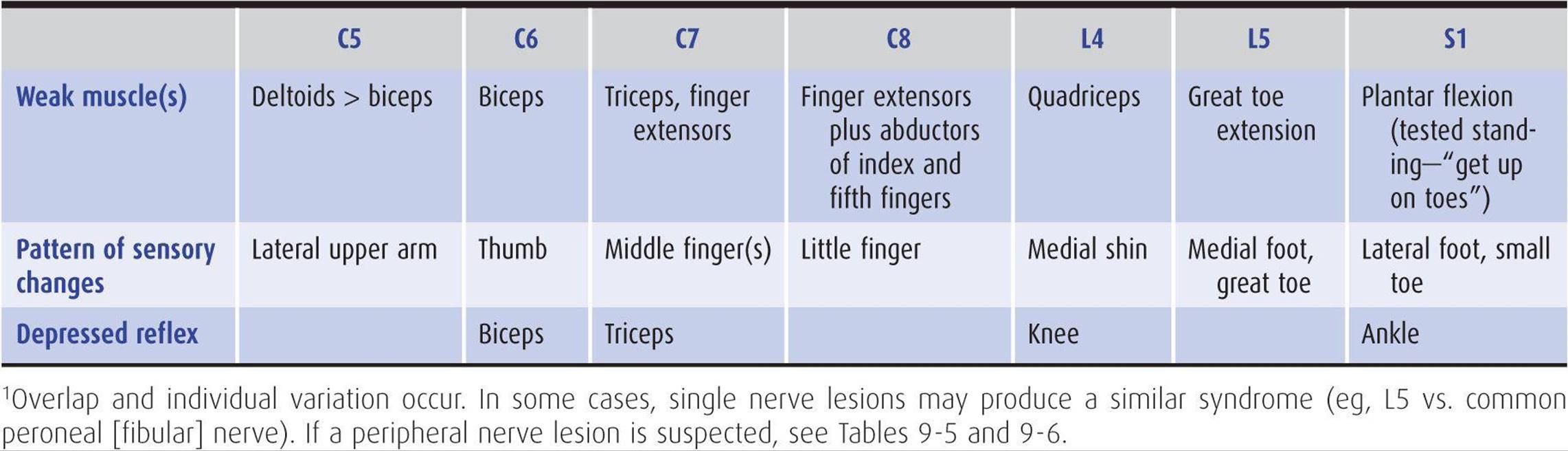
Table 9-12. The most common patterns of weakness, sensory symptoms, and reflex changes in nerve root lesions.1
Bed rest on a firm mattress for 2 or 3 days followed by gradual mobilization often permits symptoms to settle, but persisting pain, an increasing neurologic deficit, or any evidence of sphincter dysfunction should lead to CT, MRI, or CT myelography, followed by surgical treatment. Drug treatment for pain includes aspirin or acetaminophen with 30 mg of codeine, two doses three or four times daily, or other nonsteroidal analgesics such as ibuprofen or naproxen. Muscle spasm may respond to cyclobenzaprine 10 mg orally three times daily or as needed and tolerated, or diazepam 5 to 10 mg orally three times daily or as tolerated.
CERVICAL DISK PROLAPSE
Acute protrusion of a cervical disk can occur at any age, often with no preceding trauma, and leads to pain in the neck and radicular pain in the arm. The pain is exacerbated by head movement. With lateral herniation of the disk, a motor, sensory, or reflex deficit may be found in a radicular (usually C6 or C7) distribution on the affected side (Table 9-12); with more centrally directed herniations, the spinal cord may also be involved (Figure 9-9), leading to a spastic paraparesis and sensory disturbance in the legs, sometimes accompanied by impaired sphincter function. The diagnosis is confirmed by CT scanning, MRI, or CT myelography. Surgical treatment may be needed.
CERVICAL SPONDYLOSIS
This disorder has been described earlier as a cause of myelopathy.
TRAUMATIC AVULSION OF NERVE ROOTS
ERB-DUCHENNE PARALYSIS
Traumatic avulsion of the C5 and C6 roots can occur at birth as a result of traction on the head during delivery of the shoulder. It can also be the result of injuries causing excessive separation of the head and shoulder. It leads to loss of shoulder abduction and elbow flexion. In consequence, the affected arm is held internally rotated at the shoulder, with a pronated forearm and extended elbow. The biceps and brachioradialis jerks are lost, but sensory impairment is usually inconspicuous, because it is confined to a small area overlying the deltoid muscle.
KLUMPKE PARALYSIS
Involvement of the C8 and T1 roots causes paralysis and wasting of the small muscles of the hand and of the long finger flexors and extensors. Horner syndrome is sometimes an associated finding. This kind of lower plexus paralysis often follows a fall that has been arrested by grasping a fixed object with one hand or may result from traction on the abducted arm.
BRACHIAL PLEXOPATHY
NEURALGIC AMYOTROPHY (IDIOPATHIC BRACHIAL PLEXOPATHY)
This disorder, also referred to as Parsonage-Turner syndrome, typically begins with severe pain about the shoulder followed within a few days by weakness, reflex changes, and sensory disturbances in the arm, often involving the C5 and C6 segments especially. Symptoms and signs are unilateral in approximately 70% of patients. The motor deficit sometimes corresponds to the territory of an individual nerve, especially the axillary, suprascapular, or radial nerve, but in other instances appears to arise in the brachial plexus (Figure 9-10). Wasting of the affected muscles is often profound. Recurrences occur in approximately 25% of patients.

Figure 9-10. Brachial plexus. The numbers in parentheses refer to the segmental origin of the nerves depicted. (Reproduced from Waxman SG. Clinical Neuroanatomy. 26th ed. New York, NY:McGraw-Hill; 2010.)
The cause is unknown, but may be autoimmune in nature. Neuralgic amyotrophy sometimes occurs shortly after minor injury, injections, inoculations, or minor systemic infections, but whether these are of etiologic relevance is unclear. Hereditary neuralgic amyotrophy occurs occasionally as an autosomal dominant disorder characterized by recurrent symptoms. In some cases, it results from mutations in the gene (SEPT9) coding for septin 9, a cytoskeleton-interacting GTPase.
Treatment is symptomatic. The combination of a non-steroidal anti-inflammatory drug and an opiate is often effective treatment for pain in the acute phase, whereas persisting pain may respond to an anticonvulsant or tricyclic antidepressant. Recovery over the ensuing weeks and months is the rule—but it is sometimes incomplete.
CERVICAL RIB SYNDROME
The C8 and T1 roots or the lower trunk of the brachial plexus may be compressed by a cervical rib or band arising from the seventh cervical vertebra. This leads to weakness and wasting of intrinsic hand muscles, especially those in the thenar eminence, accompanied by pain and numbness in the appropriate dermatomal distribution (often like that of an ulnar nerve lesion but extending up the medial border of the forearm). The subclavian artery may also be compressed; this forms the basis of Adson test for diagnosing the disorder. The radial pulse decreases in amplitude when the seated patient turns the head to the affected side and inhales deeply. A positive Adson test, however, also can be seen in normal subjects; a supraclavicular bruit during the maneuver supports the diagnosis of subclavian artery compromise.
X-rays may show the cervical rib or a long transverse process of the seventh cervical vertebra, but normal findings do not exclude the possibility of a cervical band. Electromyography shows evidence of chronic partial denervation in the hand—in a territory beyond that of any individual peripheral nerve. Nerve conduction studies show no evidence of peripheral nerve disease, but there is a small or absent ulnar sensory nerve action potential on stimulation of the little finger. Treatment is by surgical excision of the rib or band.
OTHER CAUSES OF BRACHIAL PLEXOPATHY
Brachial plexopathy may occur in patients with neoplastic infiltration (especially by breast or lung cancer) or as a consequence of radiation therapy, following median sternotomy, and after trauma. Electrophysiologic studies are important in defining the extent and severity of involvement and localizing the lesion. The absence of electrodiagnostic abnormalities despite an adequate examination suggests an incorrect diagnosis and raises the possibility of conversion reactions, malingering, or so-called nonneurogenic thoracic outlet syndrome (a controversial and disputed entity).
LUMBOSACRAL PLEXOPATHY
A disorder similar to idiopathic brachial plexopathy occasionally affects the lumbosacral plexus. Treatment is symptomatic. As noted previously for brachial plexopathy, lumbosacral plexopathy can also be caused by neoplasms (colorectal or gynecologic cancers, sarcomas, or lymphomas) or radiation. Intrapartum maternal lumbosacral plexopathy is an uncommon but important cause of acute foot drop developing during labor. It occurs mostly in short women and relates to compression of the lumbosacral trunk by the fetal head at the pelvic brim. Most patients recover completely within 6 months.
DISORDERS OF PERIPHERAL NERVE
The term peripheral neuropathy designates a disturbance in function of one or more peripheral nerves. Several types of peripheral neuropathy are distinguishable by the extent of involvement.
Depending on the underlying cause, there may be selective involvement of motor, sensory, or autonomic fibers or more diffuse involvement of all fibers in the peripheral nerve.
The clinical deficit is usually a mixed one, and sensory symptoms and signs are often the initial—and most conspicuous—feature of peripheral nerve involvement. Further discussion of these disorders and their treatment is therefore deferred to Chapter 10, except in those instances in which presentation is typically with acute motor deficits. For convenience, however, the root and peripheral nerve supply of the major limb muscles is set forth in Tables 9-5 and 9-6. Reference to the tables should facilitate evaluation of patients presenting with focal weakness of lower motor neuron type.
POLYNEUROPATHY
In polyneuropathy, because there is symmetric and simultaneous involvement of various nerves, the deficits resulting from individual nerves cannot be recognized clinically. Polyneuropathies are discussed in Chapter 10, but brief mention is made here of those neuropathies in which patients present with acute weakness.
ACUTE INFLAMMATORY POLYRADICULONEUROPATHY (GUILLAIN-BARRÉ SYNDROME)
This disorder commonly presents with weakness that is often symmetric and most often begins in the legs. The speed and extent of progression vary, but in severe cases there is marked weakness of all limbs in addition to bilateral facial weakness. There may also be subjective sensory complaints, although objective sensory disturbances are usually far less conspicuous than motor deficits. Autonomic involvement is common and may lead to a fatal outcome, as may aspiration pneumonia or impaired respiration from weakness. Further details about this disorder are given in Chapter 10.
CRITICAL ILLNESS POLYNEUROPATHY
Patients with sepsis and multiorgan failure may develop a polyneuropathy that often first comes to attention when unexpected difficulty is encountered in weaning the patients from a mechanical ventilator. In more advanced cases, wasting and weakness of the extremities are present and the tendon reflexes are lost. Sensory abnormalities are overshadowed by the motor deficit.
Electrophysiologic studies are helpful in distinguishing the disorder from Guillain-Barré syndrome by revealing an axonal neuropathy. The underlying pathogenesis is obscure, but the polyneuropathy may relate in part to the use of neuromuscular blocking agents or corticosteroids. Treatment is supportive, with the long-term outlook being good in patients who recover from the underlying critical illness.
DIPHTHERITIC POLYNEURITIS
Infection with Corynebacterium diphtheriae can occur either in the upper respiratory tract or by infection of a skin wound, and neuropathy results from a neurotoxin that is released by the organism. Diphtheria toxin kills cells by inactivating eukaryotic elongation factor-2 and thereby blocking protein synthesis.
Palatal weakness may develop 2 to 3 weeks after infection of the throat, and cutaneous diphtheria may be followed by focal weakness of neighboring muscles after a similar interval. Impaired pupillary responses to accommodation may occur approximately 4 to 5 weeks after infection and a generalized sensorimotor polyneuropathy after 1 to 3 months. The polyneuropathy may follow a biphasic course, with further deterioration occurring 5 to 6 weeks after onset. The weakness may be asymmetric and is often more marked proximally than distally. The tendon reflexes may be depressed or absent. Respiratory paralysis occurs in severe cases. Recovery usually occurs over the following 2 to 3 months but may take longer in severe cases.
In patients with diphtheritic polyneuritis, CSF protein content is usually increased, and there may be a mild pleocytosis. Electrophysiologic studies show a slowing of nerve conduction velocity, but this is often not manifest until the patient has begun to improve clinically. Treatment consists of early administration of equine diphtheria antitoxin without awaiting the results of bacterial culture, provided the patient is not hypersensitive to horse serum. A 2-week course of penicillin or erythromycin will usually eradicate the infection but does not alter the incidence of serious complications. In patients with marked weakness, supportive measures, including ventilatory assistance, are necessary.
PARALYTIC SHELLFISH POISONING
Mussels and clams found on the East and West Coasts of the United States may be dangerous to eat, especially in the summer months. They feed on poisonous varieties of plankton and come to contain saxitoxin, which blocks sodium channels—and therefore action potentials—in motor and sensory nerves and in muscle. A rapidly progressive acute peripheral neuropathy, with sensory symptoms and a rapidly ascending paralysis, begins within 30 minutes after eating affected shellfish and may lead to respiratory paralysis and death. There is no available antitoxin, but with proper supportive care (including mechanical ventilation if necessary) the patient recovers completely. A cathartic or enema may help remove unabsorbed toxin.
PORPHYRIA
Acute polyneuropathy may occur with the hereditary hepatic porphyrias. Attacks can be precipitated by drugs (eg, barbiturates, estrogens, sulfonamides, griseofulvin, phenytoin, and succinimides) that can induce the enzyme δ-aminolevulinic acid synthetase, or by infection, a period of fasting, or, occasionally, menses or pregnancy. They usually last for 1 to 2 weeks but can be life-threatening.
Colicky abdominal pain—sometimes also felt in the back or thighs—frequently precedes neurologic involvement, and there may also be anxiety, agitation, acute confusion or delirium, and convulsions. Weakness is the major neurologic manifestation and is due to a predominantly motor polyneuropathy that causes a symmetric disturbance that is sometimes more marked proximally than distally. It may begin in the upper limbs and progress to involve the lower limbs or trunk. Progression occurs at a variable rate and can lead to complete flaccid quadriparesis with respiratory paralysis over a few days. Sensory loss occurs also but is less conspicuous and extensive; muscle pain is sometimes prominent. The tendon reflexes may be depressed or absent. Acute attacks may be accompanied by fever, excessive sweating, persistent tachycardia, hypertension, hyponatremia (attributed to inappropriate secretion of antidiuretic hormone), and peripheral leukocytosis, and patients may become dehydrated.
The CSF may show a slight increase in protein concentration and a slight pleocytosis. The diagnosis is confirmed by demonstrating increased levels of porphobilinogen and δ-aminolevulinic acid in the urine or deficiency of porphobilinogen deaminase in red blood cells (acute intermittent porphyria) or of coproporphyrinogen oxidase in lymphocytes (hereditary coproporphyria).
Treatment is with intravenous dextrose to suppress the heme biosynthetic pathway and propranolol to control tachycardia and hypertension. Hematin (4 mg/kg by intravenous infusion over 15 minutes once daily) is also effective in improving the clinical state. The best index of progress is the heart rate. The abdominal and mental symptoms (but not the neuropathy) may be helped by chlorpromazine or another phenothiazine. Pain relief may require opiates. Patients with impaired respiratory function, depressed level of consciousness, or convulsions should be followed in an intensive care unit. Respiratory failure may necessitate tracheostomy and mechanical ventilation. Recovery from paralysis is gradual and may not be complete.
Any precipitant should be removed: Precipitating medications should be discontinued, infections should be treated, and inadequate diets corrected. Preventing future acute attacks by avoiding known precipitants is important. Identification of the responsible genetic mutation in an affected patient allows for genetic screening of other family members to prevent acute attacks in those with occult disease. Different genes have been implicated in different porphyrias: In acute intermittent porphyria, the responsible mutation is in the porphobilinogen deaminase (PBGD) gene.
ACUTE ARSENIC OR THALLIUM POISONING
Acute arsenic or thallium poisoning can produce a rapidly evolving sensorimotor polyneuropathy, often with an accompanying or preceding gastrointestinal disturbance and crampy abdominal pain. Arsenic may also cause a skin rash, with increased skin pigmentation and marked exfoliation, together with the presence of Mees lines (transverse white lines) on the nails in long-standing cases. Thallium can produce a scaly rash and hair loss. Sensory symptoms, often painful, are usually the earliest manifestation of polyneuropathy; this is followed by symmetric motor impairment, which is usually more marked distally than proximally and occurs in the legs rather than the arms.
The CSF protein may be increased, with little or no change in cell content, and the electrophysiologic findings sometimes resemble those of Guillain-Barré syndrome, especially in the acute phase of the disorder. The diagnosis of arsenic toxicity is best established by measuring the arsenic content of hair protected from external contamination (eg, hair from the pubic region). Urine also contains arsenic in the acute phase. The diagnosis of thallium poisoning is made by finding thallium in body tissues or fluids, especially in urine. The degree of neurologic recovery depends on the severity of the intoxication. Chelating agents are of uncertain value.
ORGANOPHOSPHATE POLYNEUROPATHY
Organophosphate compounds are widely used as insecticides and are also the active principles in the nerve gas of chemical warfare. They have a variety of acute toxic effects, particularly manifestations of cholinergic crisis caused by inhibition of acetylcholinesterase. Some organophosphates, however, also induce a delayed polyneuropathy that generally begins approximately 1 to 3 weeks after acute exposure.
Cramping muscle pain in the legs is usually the initial symptom of neuropathy, sometimes followed by distal numbness and paresthesias. Progressive leg weakness then occurs, along with depression of the tendon reflexes. Similar deficits may develop in the upper limbs after several days. Sensory disturbances also develop in some instances, initially in the legs and then in the arms, but these disturbances are often mild or inconspicuous.
Examination shows a distal, symmetric, predominantly motor polyneuropathy, with wasting and flaccid weakness of distal leg muscles. In some patients, involvement may be severe enough to cause quadriplegia, whereas in others the weakness is much milder. Mild pyramidal signs also may be present. Objective evidence of sensory loss is usually slight.
The acute effects of organophosphate poisoning may be prevented by the use of protective masks and clothing. Treatment after exposure includes decontamination of the skin with bleach or soap and water and administration of atropine 2 to 6 mg every 5 minutes and pralidoxime 1 g every hour for up to 3 hours, both given intramuscularly or intravenously. Atropine blocks muscarinic cholinergic receptors, and pralidoxime binds to and reactivates acetylcholinesterase. There is no treatment for the neuropathy other than supportive care. Recovery of peripheral nerve function may occur with time, but central deficits are usually permanent and may govern the extent of ultimate functional recovery.
MONONEUROPATHY MULTIPLEX
This term signifies that there is involvement of various nerves but in an asymmetric manner and at different times, so that the individual nerves involved can usually be identified until the disorder reaches an advanced stage. Comment here will be restricted to two disorders characterized by motor involvement in the absence of sensory symptoms and signs.
LEAD TOXICITY
Lead toxicity is common among persons involved in the manufacture or repair of storage batteries or other lead-containing products, the smelting of lead or lead-containing ores, and the ship breaking industry. It may also occur in persons using lead-containing paints or those who ingest contaminated alcohol. Inorganic lead can produce dysfunction of both the central and peripheral nervous systems. In children, who can develop toxicity by ingesting lead-containing paints that flake off old buildings or furniture, acute encephalopathy is the major neurologic feature.
The peripheral neuropathy is predominantly motor, and in adults it is more severe in the arms than in the legs. It typically affects the radial nerves, although other nerves may also be affected, leading to an asymmetric progressive motor disturbance. Sensory loss is usually inconspicuous or absent. There may be loss or depression of tendon reflexes. Systemic manifestations of lead toxicity include anemia, constipation, colicky abdominal pain, gum discoloration, and nephropathy. The extent to which exposed workers develop minor degrees of peripheral nerve damage as a result of lead toxicity is not clear. Similarly, there is no agreement about the lowest concentration of blood lead that is associated with damage to the peripheral nerves.
The optimal approach to treatment is not known, but intravenous or intramuscular edetate calcium disodium (EDTA) and oral penicillamine have been used, as has dimercaprol (BAL).
MULTIFOCAL MOTOR NEUROPATHY
This disorder is characterized by progressive asymmetric wasting and weakness, electrophysiologic evidence of multifocal motor demyelination with partial motor conduction block but normal sensory responses, and the presence of antiglycolipid (usually anti-GM1 IgM) antibodies in the serum of many patients. Cramps and fasciculations sometimes occur and may lead to an erroneous diagnosis of motor neuron disease unless electrophysiologic studies are performed. There is no sensory loss or evidence of upper motor neuron involvement. The disorder typically has an insidious onset and chronic course, but variants with a more acute onset have been described. For a diagnosis to be made with confidence, the motor deficit should be in the distribution of two or more named nerves and related to conduction block outside of common entrapment sites. A variant with involvement of only a single nerve has been described (monofocal motor neuropathy). The conduction block is a major consequence of demyelination, but axonal excitability changes also contribute to conduction failure. Treatment with prednisone and plasmapheresis has been disappointing, but patients may improve after treatment with cyclophosphamide 1 g/m2 intravenously once per month for 6 months or in response to human immunoglobulin 2 g/kg intravenously given over 3 to 5 days. Improvement is sometimes associated with a decrease in anti-GM1 antibody levels.
MONONEUROPATHY SIMPLEX
In mononeuropathy simplex there is involvement of a single peripheral nerve. Most of the common mononeuropathies entail both motor and sensory involvement (as discussed in Chapter 10). Accordingly, only Bell palsy, which leads primarily to a motor deficit, is discussed here.
BELL PALSY
Facial weakness of the lower motor neuron type caused by idiopathic facial (VII) nerve involvement outside the central nervous system, without evidence of more widespread neurologic disease, has been designated Bell palsy. The cause is unclear, but the disorder occurs more commonly in pregnant women and diabetics. Increasing evidence suggests that reactivation of herpes simplex virus type 1 or varicella-zoster virus infection in the geniculate ganglion may injure the facial nerve and is responsible for Bell palsy in at least some patients.
Facial weakness is often preceded or accompanied by pain about the ear. Weakness generally comes on abruptly but may progress over several hours or even a day or so. Depending on the site of the lesion, there may be associated impairment of taste, lacrimation, or hyperacusis. There may be paralysis of all muscles supplied by the affected nerve (complete palsy) or variable weakness in different muscles (incomplete palsy). Clinical examination reveals no abnormalities beyond the territory of the facial nerve.
Most patients recover completely without treatment, but this may take several days in some instances and several months in others. A poor prognosis for complete recovery is suggested by severe pain at onset and complete palsy when the patient is first seen. Even if recovery is incomplete, permanent disfigurement or some other complication affects only about 10% of patients.
There is no convincing evidence that treatment with acyclovir or other antiviral agents confers any benefit. Treatment with corticosteroids (prednisone 60 or 80 mg/d orally for 3 days, tapering over the next 7 days), beginning within 5 days after the onset of palsy, increases the proportion of patients who recover completely, significantly improving the chances of complete recovery at 3 and 9 months. Surgical procedures to decompress the facial nerve are generally of no benefit. If eye closure is not possible, the eye should be protected with lubricating drops and a patch.
Other conditions that can produce facial palsy include tumors, herpes zoster infection of the geniculate ganglion (Ramsay Hunt syndrome), Lyme disease, AIDS, sarcoidosis, or any inflammatory process involving the subarachnoid space, such as infective or neoplastic meningitis. Facial palsies that are bilateral or are associated with another cranial neuropathy merit lumbar puncture and brain MRI to search for an underlying cause.
DISORDERS OF NEUROMUSCULAR TRANSMISSION
MYASTHENIA GRAVIS
PATHOGENESIS
Myasthenia gravis is caused by variable block of neuromuscular transmission related to an immune-mediated decrease in the number of functioning nicotinic acetylcholine receptors (Figure 9-11). In approximately 80% of cases, antibodies to the skeletal muscle nicotinic acetylcholine receptor are present and lead to loss of receptor function. In patients seronegative for these antibodies, the disease is probably also immune mediated; many of these patients have antibodies against the muscle-specific receptor tyrosine kinase (MuSK) that is involved in the clustering of acetylcholine receptors during development and is also expressed in mature neuromuscular junctions. A similar disorder can occur in patients receiving penicillamine for rheumatoid arthritis; it frequently remits when the drug is discontinued.

Figure 9-11. Sites of involvement in disorders of neuromuscular transmission. At left, normal transmission involves depolarization-induced influx of calcium (Ca) through voltage-gated channels. This stimulates release of acetylcholine (ACh) from synaptic vesicles at the active zone and into the synaptic cleft. ACh binds to ACh receptors and depolarizes the postsynaptic muscle membrane. At right, disorders of neuromuscular transmission result from blockage of Ca channels (Lambert-Eaton syndrome or aminoglycoside antibiotics), impairment of Ca-mediated ACh release (botulinum toxin), or antibody-induced internalization and degradation of ACh receptors (myasthenia gravis).
CLINICAL FINDINGS
Myasthenia gravis can occur at any age and is sometimes associated with thymic tumor, thyrotoxicosis, rheumatoid arthritis, or disseminated lupus erythematosus. More common in females than males, it is characterized by fluctuating weakness and easy fatigability of voluntary muscles; muscle activity cannot be maintained, and initially powerful movements weaken readily. There is a predilection for the external ocular muscles and certain other cranial muscles, including the masticatory, facial, pharyngeal, and laryngeal muscles. Respiratory and limb muscles may also be affected.
History
Although the onset of the disease is usually insidious, the disorder is sometimes unmasked by a concurrent infection, which leads to an exacerbation of symptoms. Exacerbations may also occur in pregnancy or before the menstrual periods. Symptoms may be worsened by quinine, quinidine, procainamide, propranolol, phenytoin, lithium, tetracycline, and aminoglycoside antibiotics, which therefore should be avoided in such patients.
Myasthenia follows a slowly progressive course. Patients present with ptosis, diplopia, difficulty in chewing or swallowing, nasal speech, respiratory difficulties, or weakness of the limbs (Table 9-13). These symptoms often fluctuate in intensity during the day, and this diurnal variation is superimposed on longer-term spontaneous relapses and remissions that may last for weeks.

Table 9-13. Presenting symptoms in myasthenia gravis.
Examination
Clinical examination confirms the weakness and fatigability of affected muscles. The weakness does not conform to the distribution of any single nerve, root, or level of the central nervous system. In more than 90% of cases the extraocular muscles are involved, leading to often asymmetric ocular palsies and ptosis. Pupillary responses are not affected. The characteristic feature of the disorder is that sustained activity of affected muscles leads to temporarily increased weakness. Thus sustained upgaze for 2 minutes can lead to increased ptosis, with power in the affected muscles improving after a brief rest. In advanced cases, there may be some mild atrophy of affected muscles. Sensation is normal, and there are usually no reflex changes.
DIAGNOSIS
The diagnosis of myasthenia gravis can generally be confirmed by the benefit that follows administration of anti-cholinesterase drugs; the power of affected muscles is influenced at a dose that has no effect on normal muscles and slight, if any, effect on muscles weakened by other causes.
The most commonly used pharmacologic test is the edrophonium (Tensilon) test. Edrophonium is given intravenously in a dose of 10 mg (1 mL), of which 2 mg is given initially as a test dose and the remaining 8 mg approximately 30 seconds later if the test dose is well tolerated. In myasthenic patients, there is an obvious improvement in the strength of weak muscles that lasts for approximately 5 minutes. Alternatively, 1.5 mg of neostig-minecan be given intramuscularly, with a response that lasts for about 2 hours. Atropine sulfate (0.6 mg intravenously) should be available to counteract the muscarinic cholinergic side effects of increased salivation, diarrhea, and nausea. Atropine does not affect nicotinic cholinergic function at the neuromuscular junction. The longer-acting neostigmine reduces the incidence of false-negative evaluations.
INVESTIGATIVE STUDIES
X-rays and CT scans of the chest, with and without contrast, may reveal a coexisting thymoma. Normal studies do not exclude this possibility. Impaired neuromuscular transmission can be detected electrophysiologically by a decremental response of muscle to repetitive supramaximal stimulation (at 2 or 3 Hz) of its motor nerve, but normal findings do not exclude the diagnosis. Single-fiber electro-myography shows increased variability in the interval between two muscle fiber action potentials from the same motor unit in clinically weak muscles. Measuring serum acetylcholine receptor antibody levels is often helpful, because increased values are found in 80% to 90% of patients with generalized myasthenia gravis.
TREATMENT
Medications (referred to earlier) that impair neuromuscular transmission should be avoided. The following approaches to treatment are recommended.
Anticholinesterase Drugs
Treatment with these drugs provides symptomatic benefit without influencing the course of the underlying disease. The mainstay of treatment is pyridostigmine, at doses individually determined but usually between 30 and 180 mg (average, 60 mg) four times daily. The older drug neostigmine may still be used, in rare instances, by parenteral administration. Small doses of atropine may attenuate side effects such as bowel hypermotility or hypersalivation. Overmedication can lead to increased weakness, which, unlike myasthenic weakness, is unaffected or enhanced by intravenous edrophonium. Such a cholinergic crisis may be accompanied by pallor, sweating, nausea, vomiting, salivation, colicky abdominal pain, and miosis.
Thymectomy
Thymectomy should be performed in patients younger than 60 years of age, and considered in those older, with weakness that is not restricted to the extraocular muscles. Although thymectomy usually leads to symptomatic benefit or remission, the mechanism by which it confers benefit is unclear, and its beneficial effect may not be evident immediately.
Corticosteroids
Corticosteroids are indicated for patients who have responded poorly to anticholinesterase drugs and have already undergone thymectomy. Treatment is initiated with the patient in the hospital, as weakness may initially be exacerbated. An initial high dose of prednisone (60-100 mg/d orally) can be tapered gradually to a relatively low maintenance level (5-15 mg/d) as improvement occurs. Alternate-day treatment is helpful in reducing the incidence of side effects, which are described (as clinical findings) in the section on hyperadrenalism (Cushing syndrome) in Chapter 4.
Azathioprine
This drug can be used in patients with severe or progressive disease despite thymectomy and treatment with anticholinesterases and corticosteroids. It can also be given in place of high doses of corticosteroids to patients who show no sustained benefit with low doses. The usual dose is 2 to 3 mg/kg/d, increased from a lower initial dose.
Plasmapheresis
Plasmapheresis may be used to achieve temporary improvement in patients deteriorating rapidly or in myasthenic crisis and in certain special circumstances, such as before surgery that is likely to produce postoperative respiratory compromise.
Intravenous Immunoglobulins
Intravenous immunoglobulins also have been used to provide temporary benefit in circumstances similar to those in which plasmapheresis is used.
Mycophenolate Mofetil
This agent selectively inhibits proliferation of T and B lymphocytes and has been used as an immunosuppressant with only modest side effects, including diarrhea, nausea, abdominal pain, fever, leukopenia, and edema. Several studies indicate that many patients with myasthenia gravis improve or are able to lower their steroid intake in response to this medication (unlabeled use; 1 g twice daily by mouth), but usually after a delay of several months.
PROGNOSIS
Most patients can be managed successfully with drug treatment. The disease may have a fatal outcome because of respiratory complications such as aspiration pneumonia.
MYASTHENIC SYNDROME (LAMBERT-EATON SYNDROME)
PATHOGENESIS
This disorder has a well-recognized association with an underlying neoplasm and occasionally may be associated with such autoimmune diseases as pernicious anemia; occasionally no cause is found. In the paraneoplastic disorder, antibodies directed against tumor antigens cross-react with voltage-gated calcium channels involved in acetylcho-line release, leading to a presynaptic disturbance of neuro-muscular transmission (Figure 9-11).
CLINICAL FINDINGS
Clinically there is weakness, especially of the proximal muscles of the limbs. Unlike myasthenia gravis, however, the extraocular muscles are characteristically spared, and power steadily increases if a contraction is maintained. Autonomic disturbances, such as dry mouth, constipation, and impotence, may also occur.
DIAGNOSIS
The diagnosis is confirmed electrophysiologically by the incremental response to repetitive nerve stimulation. There is a remarkable increase in the size of the muscle response to stimulation of its motor nerve at high rates—even in muscles that are not clinically weak. The presence of autoantibodies to the P/Q subtype of voltage-gated calcium channels, found on the presynaptic membrane of the neuromuscular junction, is highly sensitive and specific to the Lambert-Eaton syndrome of any etiology.
TREATMENT
Immunosuppressive drug therapy (corticosteroids and azathioprine as described earlier for myasthenia gravis) and plasmapheresis or intravenous immunoglobulin therapy may lead to improvement. Guanidine hydrochloride (25-50 mg/kg/d in three or four divided doses) is sometimes helpful in seriously disabled patients, but adverse effects of the drug include bone marrow suppression and renal failure. The response to treatment with anticholinest-erase drugs such as pyridostigmine or neostigmine, alone or in combination with guanidine, is variable but usually disappointing. 3,4-Diaminopyridine (amifampridine), a potassium channel antagonist that enhances the release of acetylcholine at the neuromuscular junction, used in doses up to 25 mg orally four times daily, may improve weakness and autonomic dysfunction; paresthesia is a common side effect, and seizures may occur. The disease improves with treatment of the underlying condition, often a small-cell lung cancer.
BOTULISM
PATHOGENESIS
The toxin of Clostridium botulinum can cause neuromuscular paralysis. It acts by preventing the release of acetylcho-line at neuromuscular junctions and autonomic synapses (Figure 9-11). Botulism occurs most commonly after ingestion of home-canned food that is contaminated with the toxin; it occurs rarely from infected wounds. The shorter the latent period between ingestion of the toxin and the onset of symptoms, the greater the dose of toxin and the risk for further involvement of the nervous system.
CLINICAL FINDINGS
Fulminating weakness begins 12 to 72 hours after ingestion of the toxin and characteristically is manifested by diplopia, ptosis, facial weakness, dysphagia, nasal speech, and then difficulty with respiration; weakness usually appears last in the limbs. In addition to the motor deficit, blurring of vision is characteristic, the pupils being dilated and unreactive, and there may be dryness of the mouth, paralytic ileus, and postural hypotension. There is no sensory deficit, and the tendon reflexes are usually unchanged unless the involved muscles are quite weak. Symptoms can progress for several days after their onset.
In infants, enteric infection with local production of the toxin leads to a different clinical picture with hypotonia, constipation, progressive weakness, and a poor suck. This is now the most common form of botulism in the United States.
INVESTIGATIVE STUDIES
Once the diagnosis is suspected, the local health authority should be notified and samples of the patient’s serum and the contaminated food (if available) sent to be assayed for toxin. The most common types of toxin encountered clinically are A, B, and E. Electrophysiologic studies may help confirm the diagnosis, as the evoked muscle response tends to increase in size progressively with repetitive stimulation of motor nerves at fast rates.
TREATMENT
Patients should be hospitalized, because respiratory insufficiency can develop rapidly and necessitates ventilatory assistance. Treatment with trivalent antitoxin (ABE) is commenced once it is established that the patient is not allergic to horse serum, but the effect on the course of the disease is unclear.
In wound botulism, antibiotic therapy is often prescribed but is of unclear benefit; either penicillin G (3 million units IV every 4 hours in adults) or metronidazole (500 mg IV every 8 hours in patients allergic to penicillin) is typically administered, but the regimen may need to be altered depending on the results of wound culture.
Guanidine hydrochloride (25-50 mg/kg/d in divided doses), a drug that facilitates release of acetylcholine from nerve endings, is sometimes helpful in improving muscle strength; anticholinesterase drugs are generally of no value.
In infants, human-derived botulinum immune globulin should be given intravenously as soon as possible in the course of the illness. Nursing and supportive care are important.
AMINOGLYCOSIDE ANTIBIOTICS
Large doses of antibiotics such as kanamycin and gentamicin can produce a clinical syndrome rather like botulism, because the release of acetylcholine from nerve endings is prevented. This effect may be related to calcium channel blockade (Figure 9-11). Symptoms resolve rapidly as the responsible drug is eliminated from the body. Note that these antibiotics are particularly dangerous in patients with preexisting disturbances of neuromuscular transmission and are therefore best avoided in patients with myasthenia gravis.
MYOPATHIC DISORDERS
MUSCULAR DYSTROPHIES
The muscular dystrophies are a group of inherited myopathic disorders characterized by progressive muscle weakness and wasting. They are subdivided by their mode of inheritance, age at onset, distribution of involved muscles, rate of progression, and long-term outlook (Table 9-14). A more satisfactory means of classification may be on genetic grounds. A number of genes have now been associated with the different muscular dystrophies (Table 9-15). These skeletal muscle genes encode sarcolemmal (eg, sarcoglycans), cytoskeletal (eg, dystrophin), cytosolic, extracellular matrix, and nuclear membrane proteins. Abnormalities of these proteins may lead to a greater susceptibility to necrosis of muscle fibers, but the molecular mechanisms involved are not yet clear. Genetic heterogeneity for the same phenotype has led to subdivision of the main clinical disorders, but the basis for the different clinical phenotypes is unknown.


Table 9-14. The muscular dystrophies.


Table 9-15. Genetic basis of muscular dystrophies.
There is no specific treatment for the muscular dystrophies. It is important to encourage patients to lead as normal a life as possible. Deformities and contractures often can be prevented or ameliorated by physical therapy and orthopedic procedures. Prolonged bed rest must be avoided, as inactivity often leads to worsening of disability.
DUCHENNE DYSTROPHY
The most common form of muscular dystrophy, it is an X-linked disorder that predominantly affects males (Table 9-14). Symptoms begin by age 5 years, and patients are typically severely disabled by adolescence, with death occurring in the third decade. Toe walking, waddling gait, and an inability to run are early symptoms. Weakness is most pronounced in the proximal lower extremities but also affects the proximal upper extremities. In attempting to rise to stand from a supine position, patients characteristically must use their arms to climb up their bodies (Gowers sign). Pseudohypertrophy of the calves caused by fatty infiltration of muscle is common. The heart is involved late in the course, and cognitive impairment is a frequent accompaniment. Serum CK levels are exceptionally high.
No definitive treatment is available, but prednisone 0.75 mg/kg/d orally may improve muscle strength for up to 3 years. Side effects include weight gain, cushingoid appearance, and hirsutism; the long-term effects of prednisone in this disorder are uncertain. Deflazacort (0.9 mg/kg/d), an analogue of prednisone, is probably as effective as prednisone but with fewer side effects. Creatine monohydrate (5-10 g/d) may also be beneficial.
The genetic defect responsible for Duchenne dystrophy has been identified (Table 9-15) and forms the basis of a diagnostic test. The gene in question is located on the short arm of the X chromosome and codes for the protein dystrophin, which is absent or profoundly reduced in muscle from patients with the disorder. The absence of dystrophin from synaptic regions of cerebral cortical neurons may contribute to the cognitive changes in patients with Duchenne dystrophy. Gene therapy has not proven effective at this time for treating this muscular dystrophy.
BECKER DYSTROPHY
This is also X-linked and associated with a pattern of weakness similar to that observed in Duchenne dystrophy (Table 9-14). Its average onset (11 years) and age at death (42 years) are later, however. Cardiac and cognitive impairment do not occur, and serum CK levels are less strikingly elevated than in Duchenne dystrophy. In contrast to Duchenne dystrophy, dystrophin levels in muscle are normal in Becker dystrophy, but the protein is qualitatively altered. It is not clear whether steroids have any role in treatment of this dystrophinopathy.
LIMB-GIRDLE DYSTROPHY
Previously a catchall designation that probably subsumed a variety of disorders, including undiagnosed cases of other dystrophies, it is (in classic form) inherited in autosomal recessive fashion (Tables 9-14and 9-15). Patients with different genetic mutations may be clinically indistinguishable, and patients with the same mutation may show marked phenotypic variation, even within the same family. The disorder begins clinically between late childhood and early adulthood. In contrast to Duchenne and Becker dystrophies, the shoulder and pelvic girdle muscles are affected to a more nearly equal extent. Pseudohypertrophy is not seen, and serum CK levels are less elevated.
FACIOSCAPULOHUMERAL DYSTROPHY
This is an autosomal dominant disorder that usually has its onset in adolescence and is compatible with a normal life span (Table 9-14). The genetic defect involves contraction of repeated DNA sequences on chromosome 4, together with single-nucleotide polymorphisms that create a polyadenylation site for a homeobox gene, DUX4 (Table 9-15). Contraction produces a more open chromatin structure, promoting transcription of DUX4, whereas polyadenylation enhances stability of the DUX4 transcript. As a result, levels of the transcript are increased, consistent with a toxic gain of protein function. The clinical severity of this condition is highly variable. Weakness is typically confined to the face, neck, and shoulder girdle, but foot drop can occur. Winged scapulae are common. The heart is not involved, and serum CK levels are normal or only slightly elevated.
EMERY-DREIFUSS MUSCULAR DYSTROPHY
This disorder occurs in an X-linked recessive form and rarer autosomal dominant and recessive forms (Tables 9-14 and 9-15). Clinical onset in childhood is followed by slow progression, with development of contractures, weakness and wasting (particularly of triceps and biceps in the arms, and of peronei and tibialis anterior in the legs, with later spread to the girdle muscles), cardiac conduction abnormalities, and cardiomyopathy. The serum CK is usually mildly increased. Cardiac function should be monitored and a pacemaker inserted if necessary. Physical therapy is important to maintain mobility.
DISTAL MYOPATHY
The autosomal dominant variety (Table 9-15) typically presents after age 40 years, although onset may be earlier and symptoms more severe in homozygotes. Small muscles of the hands and feet, wrist extensors, and the dorsiflexors of the foot are affected (Table 9-14). The precise pattern of involvement varies in the different subtypes of the disorder. The course is slowly progressive. Distal myopathies with autosomal recessive inheritance or occurring sporadically are also described and present with progressive leg weakness in adolescents or young adults. Late-onset variants are also described; in one, there is selective involvement of the posterior calves.
OCULAR DYSTROPHY
This is typically an autosomal dominant disorder, although recessive and sporadic cases also occur (Table 9-14). Some cases are associated with deletions in mitochondrial DNA. Onset is usually before age 30 years. Ptosis is the earliest manifestation, but progressive external ophthalmoplegia subsequently develops; facial weakness is also common, and subclinical involvement of limb muscles may occur. The course is slowly progressive. The extent to which ocular dystrophy is distinct from oculopharyngeal dystrophy (see next section) is unclear in many cases.
OCULOPHARYNGEAL DYSTROPHY
An autosomal dominant disorder (Table 9-15), this is found with increased frequency in certain geographic areas, including Quebec and the southwestern United States. It most often begins in the third to fifth decade. Findings include ptosis, total external ophthalmoplegia, dysphagia, facial weakness, and often proximal limb weakness (Table 9-14). Serum CK is mildly elevated. Dysphagia is particularly incapacitating and may require nasogastric feeding or gastrostomy.
PARASPINAL DYSTROPHY
Progressive paraspinal weakness may develop after the age of 40 years in patients of either sex, some of whom may have a family history of the disorder. Back pain and a marked kyphosis (bent spine syndrome or camptocormia) are characteristic (Table 9-14). The serum CK is mildly elevated. CT scans show fatty replacement of paraspinal muscles.
CONGENITAL MYOPATHIES
The congenital myopathies are a heterogeneous group of rare and relatively nonprogressive disorders that usually begin in infancy or childhood but may not become clinically apparent until adulthood. Most are characterized by predominantly proximal muscle weakness, hypotonia, hyporeflexia, and normal serum CK; many are inherited.
Congenital myopathies are classified according to ultrastructural histopathologic features and are diagnosed by muscle biopsy. They include nemaline myopathy, characterized by rod-shaped bodies in muscle fibers, which are also seen in some patients with AIDS-related myopathy (see later); central core disease, which may be associated with malignant hyperthermia as a complication of general anesthesia; myotubular or centronuclear myopathy; and mitochondrial myopathies, such as Kearns-Sayre-Daroff syndrome, a cause of progressive external ophthalmoplegia (Chapter 7). No treatment is available for any of these disorders.
MITOCHONDRIAL MYOPATHIES
The mitochondrial myopathies are a clinically heterogeneous group of disorders caused by defective oxidative phosphorylation and accompanied by structural mitochondrial abnormalities on skeletal muscle biopsy. Their morphologic signature is the “ragged red fiber” seen with the modified Gomori stain, containing accumulations of abnormal mitochondria. Since the first reported pathogenic mutation of human mitochondrial DNA in the 1980s, a large number of other mutations have been described, including point mutations and large-scale deletions.
Patients may present with Kearns-Sayre-Daroff syndrome (progressive external ophthalmoplegia, pigmentary degeneration of the retina, and cardiomyopathy) or with limb weakness that is exacerbated or induced by activity. In other patients, the symptoms and signs are of central neurologic dysfunction and may include myoclonic epilepsy (myoclonic epilepsy, ragged red fiber syndrome [MERRF]) or the combination of mitochondrial myopathy, encephalopathy, lactic acidosis, and strokelike episodes (MELAS). These various syndromes are caused by separate abnormalities of mitochondrial DNA. Investigations may include muscle biopsy, measurement of serum and CSF lactate, and imaging studies. Treatment is generally supportive.
Mitochondrial DNA depends for its proper function on various factors encoded by nuclear DNA. Mutations in nuclear genes may thus affect mitochondrial function. This is exemplified by mutations in the gene for thymidine phosphorylate, which lead to an autosomal recessive disorder called mitochondrial neurogastrointestinal encephalomyopathy (MNGIE), manifest by gastrointestinal dysmotility and skeletal muscle abnormalities.
MYOTONIC DISORDERS
In myotonia, an abnormality of the muscle fiber membrane (sarcolemma) causes marked delay before the affected muscles can relax after a contraction; this leads to apparent muscle stiffness. On examination, it is frequently possible to demonstrate myotonia by difficulty in relaxing the hand after sustained grip or by persistent contraction after percussion of the belly of a muscle. Electromyography of affected muscles may reveal characteristic high-frequency discharges of potentials that wax and wane in amplitude and frequency, producing over the EMG loudspeaker a sound like that of a dive bomber or chain saw.
MYOTONIC DYSTROPHIES
Myotonic Dystrophy Type 1
Myotonic dystrophy type 1 (DM1) is a dominantly inherited disorder that usually is manifest in the third or fourth decade, although it may appear in early childhood. The gene defect is an expanded trinucleotide (CTG) repeat in an untranslated region of the gene coding for dystrophia myotonica protein kinase (DMPK). This expanded trinucleotide repeat forms the basis of a diagnostic test; repeat lengths greater than 34 repeats are abnormal. Prenatal testing during at-risk pregnancies can be performed. Myotonia accompanies weakness and wasting of the facial, sterno-mastoid, and distal limb muscles (Figure 9-12). There may also be cataracts, frontal baldness, testicular atrophy, diabetes mellitus, cardiac abnormalities, intellectual changes, and sudden death. A severe electrocardiographic abnormality and the presence of atrial tachyarrhythmia predict sudden death.

Figure 9-12. Photograph of a 37-year-old man with myotonic dystrophy, showing frontal baldness, bilateral ptosis, and wasting of the temporalis, facial, and sternocleidomastoid muscles. (Courtesy of R. Griggs.)
Myotonic Dystrophy Type 2
Patients with myotonic dystrophy type 2 (DM2), sometimes designated proximal myotonic myopathy, have myotonia, cataracts, primarily proximal weakness (neck and finger flexors, followed by hip flexors and extensors, and elbow extensors), and a less severe course than those with DM1. The disorder is dominantly inherited and results from an expanded CCTG repeat in a noncoding (intronic) region of the gene for zinc finger protein-9 (ZNF9). Onset is commonly in young adults. A variant with more severe muscle involvement and hearing loss has been described.
Treatment
Management includes the use of assistive devices as needed for weakness and the management of complications such as cataracts or cardiac arrhythmias. If necessary, myotonia can be treated with procainamide 0.5 to 1 g four times daily, mexiletine 150 to 200 mg three times daily, or phenytoin 100 mg three times daily. In myotonic dystrophy, phenytoin is perhaps the drug of choice, as the other drugs may have undesirable effects on cardiac conduction. There is no treatment for the weakness that occurs, and pharmacologic maneuvers do not influence the natural history.
NONDYSTROPHIC MYOTONIAS
The nondystrophic myotonias are caused by dysfunction of certain skeletal muscle ion channels. They include myotonia congenita, paramyotonia congenita, and the sodium-channel myotonias. These disorders are manifest primarily by muscle stiffness resulting from myotonia but—depending on the disorder—pain, weakness, and fatigue may also be conspicuous.
Myotonia Congenita
Myotonia congenita is usually inherited as a dominant disorder (Thomsen disease) that relates to a mutation in the CLCN1 skeletal muscle chloride channel gene. Many different mutations have been identified. Generalized myotonia without weakness is usually present from birth, but symptoms may not develop until early childhood. Muscle stiffness is enhanced by cold and inactivity and relieved by exercise. Muscle hypertrophy, sometimes pronounced, is also a feature. A recessive form with later onset (Becker disease, not to be confused with Becker muscular dystrophy) is associated with slight weakness—especially on initiating a movement—and with atrophy of distal muscles. It is also due to a CLCN1 mutation. Drugs acting specifically on the CLCN1 channel are not available, but treatment with mexiletine or phenytoin may help the myotonia.
Paramyotonia Congenita and Sodium-Channel Myotonias
These are allelic, autosomal dominant disorders caused by point mutations in the skeletal muscle voltage-gated sodium channel gene, SCN4A.
Paramyotonia is characterized by episodic muscle cramps and weakness that is exacerbated markedly by cold and exercise and becomes symptomatic in childhood. The facial, tongue, and hand muscles are most affected, and the lower limbs much less so. The myotonia can last up to several minutes, but weakness may persist for hours and even days. Muscle hypertrophy is sometimes present.
Patients with sodium-channel myotonias are typically not sensitive to cold (although this occurs in some cases), but their myotonia is worsened with potassium ingestion. Weakness does not occur. The myotonia tends to occur about 10 or more minutes after the onset of exercise and is improved with acetazolamide treatment. In some cases, myotonia is severe and may impair respiration.
Diagnosis
Various electrophysiologic test protocols can aid diagnosis. Sarcolemmal excitability is measured indirectly by changes in size of the compound muscle action potential after varying periods of exercise, and the effect of muscle cooling is noted. Distinct patterns have been recognized and have been used to direct genetic testing.
Treatment
There is insufficient evidence to make treatment recommendations for this group of disorders, but sodium-channel blockers may be of some benefit. Tocainide, initially encouraging, was withdrawn because of the risk of agranulocytosis. Flecainide, an antiarrhythmic drug, is being studied. Mexiletine is often favored, but it may cause cardiac arrhythmias and evidence of efficacy is incomplete.
INFLAMMATORY MYOPATHIES
Muscle biopsy is important in confirming the diagnosis of an inflammatory myopathy and facilitating recognition of unusual varieties such as eosinophilic, granulomatous, and parasitic myositis.
TRICHINOSIS, TOXOPLASMOSIS, & SARCOIDOSIS
These disorders may lead to an inflammatory disorder of muscle, but this is uncommon. Treatment is of the underlying cause.
POLYMYOSITIS & DERMATOMYOSITIS
Pathogenesis
Polymyositis and dermatomyositis are immune-mediated inflammatory myopathies characterized by destruction of muscle fibers and inflammatory infiltration of muscles. There are immunologic and histopathologic differences between the two disorders. Dermatomyositis is a microangiopathy affecting skin and muscle: Lysis of endomysial capillaries is caused by activation and deposition of complement, leading to muscle ischemia. In polymyositis, muscle fibers expressing major histocompatibility complex (MHC) class I antigens are invaded by clonally expanded CD8-positive cytotoxic T cells, leading to necrosis.
Clinical Findings
Polymyositis can occur at any age; it progresses at a variable rate and leads to weakness and wasting, especially of the proximal limb and girdle muscles (Table 9-16). It is often associated with muscle pain, tenderness, dysphagia, and respiratory difficulties. Raynaud phenomenon, arthralgia, malaise, weight loss, and a low-grade fever round out the clinical picture.

Table 9-16. The inflammatory myopathies.
Dermatomyositis is distinguished clinically by the presence of an erythematous rash over the eyelids, about the eyes (heliotrope rash), or on the extensor surfaces of the joints. Polymyositis/dermatomyositis has been reported in association with various autoimmune disorders, including scleroderma, lupus erythematosus, rheumatoid arthritis, and Sjögren syndrome. In addition, approximately 25% of patients with adult-onset dermatomyositis have an associated malignancy (most often ovarian, lung, gastrointestinal, or nasopharyngeal carcinoma).
Diagnosis
The serum CK is generally elevated in patients with polymyositis or dermatomyositis, sometimes to very high levels, but normal values do not exclude the diagnosis. At electromyography, an abundance of short, low-amplitude, polyphasic motor unit potentials is found, as in any myopathic process, but abnormal spontaneous activity is often conspicuous as well. Muscle biopsy usually shows muscle fiber necrosis and infiltration with inflammatory cells and is important for accurate diagnosis.
Treatment
Treatment is with anti-inflammatory drugs. Prednisone is commonly used in an initial dose of 60 or 80 mg/d, along with potassium supplements and frequent antacids if necessary. As improvement occurs and serum CK values decline, the prednisone dose is gradually tapered to maintenance levels that usually range between 10 and 20 mg/d. Patients may need to continue this regimen for 2 to 3 years, however; too rapid a reduction in dose may lead to relapse.
Intravenous immunoglobulin is efficacious in dermatomyositis and can be used with or in place of steroids. Methotrexate and azathioprine have also been used, either alone or in combination with corticosteroids; they are particularly useful in corticosteroid-resistant patients. Newer immunosuppressants with less troublesome side effects, such as mycophenolate mofetil, also may be helpful but are still under study. Physical therapy may help to prevent contractures, and, as the patient responds to anti-inflammatory drugs, active exercise may hasten recovery.
INCLUSION-BODY MYOSITIS
Epidemiology
This disorder is more common in men than women and has an insidious onset, usually after 50 years of age. Its incidence is unclear, but it is being recognized with increasing frequency.
Etiology
The etiology of the myositis is unknown, but accumulating evidence suggests a T-cell-mediated myocytotoxicity and probably a multifactorial genetic susceptibility to the disease. Associated disorders include various autoimmune disturbances, diabetes mellitus, and diffuse peripheral neuropathy.
Clinical Findings
Inclusion-body myositis produces weakness of the lower and then the upper extremities. Weakness and atrophy of the quadriceps and of the forearm flexor and extensor muscles are characteristic. The disease is progressive and is associated with early depression of the knee reflexes. Muscle pain occurs in some patients. Distal weakness also develops, but is usually less severe than proximal weakness. Dysphagia is common.
Diagnosis
Serum CK levels may be normal or increased. The EMG reveals nonspecific findings suggestive of an inflammatory myopathy. The diagnosis is confirmed by histologic examination of biopsied muscle.
Differential Diagnosis
A familial disorder with onset in young adults, characterized by slowly progressive weakness, especially of distal muscles, and with histologic features resembling inclusion-body myositis (vacuolar myopathy with inclusions) has been described. The family history helps to distinguish this hereditary inclusion-body myopathy from the sporadic disorder, as does the lack of inflammatory changes on histologic examination. Table 9-16 summarizes the differences between inclusion-body myositis, polymyositis and dermatomyositis.
Treatment
The disorder is unresponsive to immunosuppressive or immunomodulatory therapies. Conflicting reports on the utility of intravenous globulin therapy make its role unclear. Patients may eventually become chair-bound and require help with the activities of daily life.
AIDS
Several forms of myopathy can occur in patients with either symptomatic or otherwise asymptomatic HIV-1 infection (Table 9-17). These disorders can be distinguished by muscle biopsy.

Table 9-17. Myopathies related to HIV-1 infection or its treatment.
Polymyositis
This most common AIDS-related myopathy may be caused by autoimmune mechanisms triggered by HIV-1 infection. It resembles polymyositis in patients without HIV-1 infection (see earlier) and may respond to treatment with corticosteroids.
Inclusion-Body Myositis-Like Syndrome
Patients with AIDS may also develop a myopathy resembling inclusion-body myositis, apparently because the virus triggers an immune response similar to that occurring in sporadic inclusion-body myositis; there does not seem to be direct infection of the muscle.
Muscle-Wasting Syndrome
This sometimes relates to type II muscle fiber atrophy; malnutrition, cachexia, immobility, or remote effects of AIDS-related tumors may have a pathogenetic role. Proximal muscle weakness is the major finding, and serum CK is normal.
Rod-Body (Nemaline) Myopathy
Rod-body myopathy is a noninflammatory disorder characterized by rod-shaped bodies and selective loss of thick filaments. Clinical features include proximal muscle weakness and moderate elevation of serum CK. Treatment with corticosteroids or plasmapheresis may be helpful.
Vasculitic Myopathy
Vasculitic processes may involve the muscles (and nerves); treatment in AIDS patients usually involves antiretroviral and immunomodulatory agents including intravenous immunoglobulins and steroids.
Infective Myopathy
Opportunistic infections of muscle are well recognized and may present as pyomyositis; muscle toxoplasmosis may lead to a subacute painful myopathy. Treatment is directed against the offending organisms.
Mitochondrial Myopathy
A myopathy in which muscle biopsy specimens show the ragged red fibers indicative of damaged mitochondria can occur in patients receiving zidovudine for treatment of AIDS and may coexist with polymyositis. The disorder is characterized clinically by proximal muscle weakness, myalgia, and moderate to marked elevation of serum CK; it is thought to result from a toxic effect of zidovudine on muscle. Mild symptoms may be controlled with nonsteroidal anti-inflammatory drugs or corticosteroids, and more severe involvement may respond to discontinuing zidovudine. If there is no response, a muscle biopsy should be performed to look for other causes of myopathy.
Treatment-Related Myopathies
Patients with HIV-1 infection treated with highly active antiretroviral therapy (HAART) may develop a lipodystrophy; muscle biopsy (performed for unrelated reasons) then shows fatty accumulation in muscle. In the immune restoration inflammatory syndrome (IRIS), HIV-1-infected patients receiving HAART develop inflammatory responses that may lead to a myositis resembling polymyositis.
Acute Rhabdomyolysis
This sometimes occurs in patients with HIV-1 infection and causes myalgia, muscle weakness, and an elevated serum CK; it may also relate to medication or opportunistic infection.
POLYMYALGIA RHEUMATICA
Polymyalgia rheumatica, which is more common in women than in men, generally occurs in patients older than 50 years and is best regarded as a variant of giant cell arteritis. It is characterized by muscle pain and stiffness, particularly about the neck and girdle muscles. Headache, anorexia, weight loss, and low-grade fever may be conjoined, and the erythrocyte sedimentation rate is increased. Serum enzymes, electromyography, and muscle biopsy are normal.
There is usually a dramatic response to treatment with corticosteroids in low dosage (eg, prednisone 10-15 mg/d orally). Treatment is monitored by clinical parameters and sedimentation rate and may need to be continued for 1 year or more if serious complications are to be avoided, as noted for giant cell arteritis (see Chapter 6). Relapses may occur when the daily dose of prednisone is reduced to about 5 mg or less. Methotrexate is the most commonly used corticosteroid-sparing agent.
EOSINOPHILIA-MYALGIA SYNDROME
Eosinophilia-myalgia syndrome produces muscle pain and weakness associated with inflammation of skin and other soft tissues but little direct involvement of muscle. Because the prominent symptoms are sensory, this disorder is discussed in Chapter 10.
METABOLIC MYOPATHIES
HYPOKALEMIA
Proximal myopathic weakness may result from chronic hypokalemia, and once the metabolic disturbance has been corrected, power usually returns to normal within a few weeks. Acute hypo- or hyperkalemia may also lead to muscle weakness that is rapidly reversed by correcting the metabolic disturbance.
PERIODIC PARALYSES
The periodic paralysis syndromes, which may be familial (dominant inheritance), are characterized by episodes of flaccid weakness or paralysis with preserved ventilation that may be associated with abnormalities of the serum potassium level. Strength is normal between attacks. They are channelopathies in which there is abnormal, often potassium-sensitive, muscle-membrane excitability. Mutations in genes encoding three ion channels—CACNA1S, SCN4A, and KCNJ2— are responsible for most cases.
Hypokalemic Periodic Paralysis
In the hypokalemic form, sometimes associated with thyrotoxicosis, attacks tend to occur on awakening, after exercise, or after a heavy meal—and may last for several days. The disorder is due to a mutation in the gene encoding the α1S subunit of the L-type (dihydropyridine-sensitive) skeletal muscle calcium channel (CACNA1S). The clinical disorder is genetically heterogeneous and may also be caused by mutations in the α subunit of the type IV voltage-gated sodium channel (SCN4A), which is more typically associated with hyperkalemic periodic paralysis (see later).
Acetazolamide (250-750 mg daily) or oral potassium supplements may prevent attacks, as also may a low salt and low carbohydrate diet. Ongoing attacks may be arrested by potassium chloride given orally or even intravenously if the ECG can be monitored and kidney function is satisfactory. Excessive exertion should be avoided. If hyperthyroidism is associated—as is often the case, particularly in young Asian men—its treatment may prevent recurrences. All patients with suspected hypokalemic periodic paralysis should therefore be screened for thyroid disease.
Hyperkalemic Periodic Paralysis
Attacks associated with hyperkalemia also tend to come on after exercise but are usually much briefer, lasting less than 1 hour. Severe attacks may be terminated by intravenous calcium gluconate (1-2 g), intravenous diuretics (furosemide 20-40 mg), or glucose, and daily acetazolamide or chlorothiazide may help prevent further episodes. Many families with this disorder have mutations in the gene encoding the α subunit of the type IV voltage-gated sodium channel (SCN4A); several allelic mutations have been recognized and account for some phenotypic variation, such as the presence of myotonia or paramyotonia.
Cardiodysrhythmic Periodic Paralysis
This disorder, also referred to as Andersen-Tawil syndrome, results from mutations in the gene for an inwardly rectifying potassium channel (KCNJ2). The disorder is inherited in autosomal dominant fashion and is characterized by periodic paralysis, ventricular arrhythmias, and facial or skeletal deformities. Serum potassium levels may be increased, decreased, or normal at the time of paralytic attacks, which may be triggered by rest following exertion.
Paramyotonia Congenita
This is a dominantly inherited disorder, related to mutation of the SCN4A gene, discussed earlier; attacks of hyper-kalemic periodic paralysis may also occur.
Normokalemic Periodic Paralysis
This disorder is similar clinically to the hyperkalemic variety, but the plasma potassium level is normal in attacks; treatment is with acetazolamide. It is sometimes unresponsive to treatment; in severe attacks, it may be impossible to move the limbs, but respiration and swallowing are rarely affected.
OSTEOMALACIA
Proximal muscle weakness may also occur in osteomalacia, often with associated bone pain and tenderness, mild hypocalcemia, and elevated serum alkaline phosphatase. Strength improves after treatment with vitamin D.
ENDOCRINE MYOPATHIES
Myopathy may occur in association with hyper- or hypothyroidism, hyper- or hypoparathyroidism, hyper-or hypoadrenalism, hypopituitarism, and acromegaly. Treatment is that of the underlying endocrine disorder.
ALCOHOLIC MYOPATHIES
ACUTE NECROTIZING ALCOHOLIC MYOPATHY
Heavy binge drinking may result in an acute necrotizing myopathy that develops over 1 or 2 days. Presenting symptoms include muscle pain, weakness, and sometimes dysphagia. On examination, the affected muscles are swollen, tender, and weak. Weakness is proximal in distribution and may be asymmetric or focal. Serum CK is moderately to severely elevated, and myoglobinuria may occur. As hypokalemia and hypophosphatemia can produce a similar syndrome in alcoholic patients, serum potassium and phosphorus concentrations should be determined. With abstinence from alcohol and a nutritionally adequate diet, recovery can be expected over a period of weeks to months.
CHRONIC ALCOHOLIC MYOPATHY
Chronic myopathy characterized by proximal weakness of the lower limbs may develop insidiously over weeks to months in alcoholic patients. Muscle pain is not a prominent feature. Cessation of drinking and an improved diet are associated with clinical improvement over several months in most cases.
DRUG-INDUCED MYOPATHIES
Myopathy can occur in association with administration of corticosteroids, chloroquine, clofibrate, emetine, ε-aminocaproic acid, certain β-blockers, bretylium tosylate, colchicine, statins (HMG-CoA reductase inhibitors), zidovudine, or drugs that cause potassium depletion. Common variants in the gene for a solute carrier organic anion transporter (SLCO1B1) are strongly associated with an increased risk of statin-induced myopathy. Symptoms of drug-induced myopathy vary from an asymptomatic increase in serum CK levels to acute rhabdomyolysis, depending on the causal agent and the individual patient. Necrotizing myopathies are due mainly to lipid-lowering drugs, and mitochondrial myopathies to antiretroviral nucleoside analogues. Corticosteroid myopathy is particularly common. Drug-induced myopathies are usually reversible if the causal agent is discontinued.
CRITICAL ILLNESS MYOPATHY
Patients in the intensive care unit may develop weakness both proximally and distally in the limbs from primary muscle involvement. Histopathologic examination reveals a diffuse nonnecrotizing cachectic myopathy, a selective loss of thick (myosin) filaments, or an acute necrotizing myopathy. The myopathy may coexist with a neuropathy. Its cause is uncertain, but it is associated with the use of nondepolarizing neuromuscular blockers and corticosteroids. Serum CK is sometimes increased, especially if muscle necrosis has occurred. Electrophysiologic findings may be suggestive of muscle involvement and help to distinguish myopathy from neuropathy or disorders of the neuromuscular junction. Muscle biopsy may be definitive but is not always revealing. Prognosis is good except when muscle necrosis is conspicuous. Treatment is supportive. Sepsis must be treated aggressively.
MYOGLOBINURIA
This can result from muscle injury or ischemia (irrespective of its cause) and leads to a urine that is dark red. The following causes are important:
1. Excessive unaccustomed exercise, leading to muscle necrosis (rhabdomyolysis) and thus to myoglobinuria, sometimes on a familial basis
2. Crush injuries
3. Muscle infarction
4. Prolonged tonic-clonic convulsions
5. Polymyositis
6. Chronic potassium depletion
7. An acute alcoholic binge
8. Certain viral infections associated with muscle weakness and pain
9. Hyperthermia
10. Metabolic myopathies (eg, muscle glycogen phosphorylase deficiency [McArdle disease])
Serum CK levels are elevated, often greatly. Myoglobin can be detected in the urine by the dipstick test for heme pigment; a positive test indicates the presence of myoglobin in the urine unless red blood cells are present. In severe cases, myoglobinuria may lead to renal failure, and peritoneal dialysis or hemodialysis may then be necessary. Otherwise, treatment consists of increasing the urine volume by hydration. The serum potassium level must be monitored, as it may rise rapidly.
MOTOR-UNIT HYPERACTIVITY STATES
Disorders affecting the central or peripheral nervous system at a variety of sites can produce abnormal, increased activity in the motor unit (Table 9-18).

Table 9-18. Motor-unit hyperactivity states.
CENTRAL NERVOUS SYSTEM DISORDERS
STIFF-PERSON SYNDROME
This is a rare, usually sporadic, and slowly progressive disorder manifested by tightness, stiffness, and rigidity of axial and proximal limb muscles with superimposed painful spasms.
Pathogenesis
An immune-mediated defect in central GABAergic transmission has been proposed as the cause of the disorder. In approximately 60% of patients, the blood and cerebro-spinal fluid contain autoantibodies against glutamic acid decarboxylase (GAD), which is involved in synthesis of the neurotransmitter γ-aminobutyric acid (GABA), and is concentrated in pancreatic β-cells and GABAergic neurons of the central nervous system. Another 10% of patients have an associated neoplasm and circulating autoantibodies against the synaptic vesicle-associated protein, amphiphysin. In these patients, symptoms may improve after removal of the tumor. Antibodies against postsynaptic markers at GABAergic synapses—GABAA receptor-associated protein and gephyrin—have also been identified in some patients. No clinical difference has been found between patients with and without autoantibodies.
Clinical Findings
Stiff-person syndrome sometimes has an autoimmune basis, and it may be associated with other autoimmune disorders such as thyroiditis, myasthenia gravis, and pernicious anemia. Many patients have type 1 diabetes mellitus.
Patients with stiff person syndrome have muscle rigidity caused by sustained, involuntary contraction of agonist and antagonist muscles. Axial and proximal limb muscles are affected in particular, resulting in abnormal posture and gait and often falling. Rigidity typically disappears during sleep. Examination may show tight muscles, a slow or cautious gait, and hyperreflexia.
Painful spasms are often provoked by sudden movement, startle, or emotional upset and may be accompanied by hyperhidrosis and increased blood pressure. Paroxysmal dysautonomia may be manifest also by tachypnea and even respiratory arrest—perhaps due to stiffness of the respiratory muscles—and sometimes leads to sudden death. Some patients also have focal or generalized seizures.
Diagnosis
Electromyography reveals continuous motor-unit activity in the paraspinal or leg muscles that varies with the clinical state.
Differential Diagnosis
Stiff-person syndrome can be distinguished from tetanus (discussed later) by its more gradual onset, the absence of trismus (lockjaw), and its rapid response to diazepam.
Treatment
Treatment is with drugs that enhance GABAergic transmission, such as diazepam 5 to 75 mg orally four times daily. Baclofen, vigabatrin, sodium valproate, and gabapentin also may be helpful for relieving symptoms in some patients. Treatment with corticosteroids is helpful in refractory or severe cases, and intravenous immunoglobulin therapy is sometimes effective in patients who are otherwise unresponsive to treatment.
TETANUS
Tetanus, a disorder of central inhibitory neurotransmission caused by a toxin produced by Clostridium tetani, is discussed earlier in this chapter.
STARTLE SYNDROMES
In hyperekplexia, startle reflexes to an unexpected stimulus are excessive; startle-induced falls may occur and relate to a transient generalized stiffness that follows the startle reflex for a few seconds. In such patients, continuous stiffness is present in the neonatal period, resolving with time. The disorder may occur on a sporadic or familial (autosomal dominant or recessive) basis, associated usually with mutations in the α1 subunit of the glycine receptor gene (GLRA1). Autosomal recessive forms may also relate to mutations in a glycine transporter (SLC6A5) or the gene encoding the beta-subunit of the glycine receptor (GLRB), whereas mutations in the postsynaptic anchoring protein gephyrin (GPHN) or a Rho-like GTPase (ARHGEF9) have been observed in sporadic cases. The abnormal startle reflex seems to originate in the brainstem, but a cortical basis has sometimes been suggested. In some patients with a minor form of the disorder, excessive startle reflexes are the only abnormality; the genetic and pathophysiologic bases of this form are unknown. Patients with hereditary or sporadic hyperekplexia often respond favorably to clonazepam, which potentiates effects of the inhibitory transmitter GABA. In occasional patients, hyper-ekplexia is symptomatic and relates to diffuse acquired cerebral or brainstem pathology.
Neuropsychiatric startle syndromes (eg, Jumping Frenchman of Maine, latah, miryachit) are also described and, in some instances, involve mimicking behaviors and cultural or familial factors.
PERIPHERAL NERVE DISORDERS
CRAMPS
These involuntary and typically painful contractions of a muscle or portion of a muscle are thought to arise distally in the motor neuron. Palpable knot-like hardening of the muscle may occur. Cramps are characteristically relieved by passive stretching of the affected muscle. They usually represent a benign condition and are common at night or during or after exercise.
However, cramps may also be a manifestation of motor neuron disease or polyneuropathy, metabolic disturbances (pregnancy, uremia, hypothyroidism, adrenal insufficiency), or fluid or electrolyte disorders (dehydration, hemodialysis). If a reversible underlying cause cannot be found, daytime cramps may respond to treatment with phenytoin 300 to 400 mg/d orally or carbamazepine 200 to 400 mg orally three times a day; other drugs sometimes used are baclofen and oxcarbazepine. For none of these medications is there convincing evidence of efficacy. Open-label studies suggest that levetiracetam and gabapentin are occasionally helpful.
Nocturnal cramps may respond to a single oral bedtime dose of quinine sulfate (325 mg), phenytoin (100-300 mg), carbamazepine (200-400 mg), or diazepam (5-10 mg). However, because it may uncommonly cause serious hematologic abnormalities such as hemolytic uremic syndrome–thrombotic thrombocytopenia purpura, disseminated intravascular coagulation, and a bleeding diathesis, quinine should not be prescribed routinely unless the cramps are disabling and fail to respond to other approaches.
NEUROMYOTONIA
Neuromyotonia (Isaacs syndrome) is a rare, sporadic disorder that produces continuous muscle stiffness, rippling muscle movements (myokymia), and delayed relaxation after muscle contraction. Some cases have an autosomal dominant mode of inheritance; in others, the neuromyotonia occurs as a paraneoplastic disorder, or in association with other autoimmune diseases or with hereditary motor and sensory neuropathies. It may also follow irradiation of the nervous system. In acquired neuromyotonia, antibodies against voltage-gated potassium channels are often found in the serum and CSF. Symptoms may be controlled with phenytoin 300 to 400 mg/d orally or carbamazepine 200 to 400 mg orally three times a day.
TETANY
Tetany—not to be confused with tetanus (see earlier)—is a hyperexcitable state of peripheral nerves usually associated with hypocalcemia, hypomagnesemia, or alkalosis. Signs of tetany (Chvostek sign, Trousseau sign, carpopedal spasm) are described in the section on hypocalcemia in Chapter 4. Treatment is by correction of the underlying electrolyte disorder.
HEMIFACIAL SPASM
Hemifacial spasm is characterized by repetitive, involuntary contractions of some or all of the muscles supplied by one facial (VII) nerve. Symptoms often commence in the orbicularis oculi and then spread to the cheek and levator anguli oris muscles. The contractions initially are brief but become more sustained as the disorder progresses; they may be provoked by blinking or voluntary activity. Slight facial weakness may also be found on examination. The disorder commonly relates to the presence of an anomalous blood vessel compressing the intracranial facial nerve, but MRI should be performed to exclude other structural lesions. The involuntary movements have been attributed to ephaptic transmission and ectopic excitation of demyelinated fibers in the compressed segment, to altered excitability of the facial nerve nucleus in the brainstem, or to both mechanisms. Treatment with carbamazepine or phenytoin is sometimes helpful, and injection of botulinum toxin into the affected muscles suppresses the contractions temporarily. Microvascular decompressive procedures are often curative.
MUSCLE DISORDERS
MYOTONIA
Disorders that produce myotonia are discussed earlier.
MALIGNANT HYPERTHERMIA
Susceptibility to this disorder, which is often inherited in autosomal dominant fashion, may result from a mutation in the ryanodine receptor gene (RYR1) or less commonly in the α1S subunit of the L-type (dihydropyridine-sensitive) skeletal muscle calcium channel (CACNA1S) or unidentified genes at other sites. The clinical abnormality is thought to result from abnormal excitation-contraction coupling in skeletal muscle. Symptoms are usually precipitated by administration of neuromuscular blocking agents (eg, succinylcholine) or inhalational anesthetics. Clinical features include rigidity, hyperthermia, metabolic acidosis, and myoglobinuria. Mortality rates as high as 70% have been reported. The disorder must be distinguished from neuroleptic malignant syndrome (Table 9-19 and Chapter 11), which is manifested by rigidity, fever, altered mental status, and autonomic dysfunction.

Table 9-19. Distinction between neuroleptic malignant syndrome, malignant hyperthermia, and heat stroke.
Treatment includes prompt cessation of anesthesia, administration of the excitation-contraction uncoupler dantrolene (1-2 mg/kg intravenously every 5-10 minutes as needed, to a maximum dose of 10 mg/kg), reduction of body temperature, and correction of acidosis with intravenous bicarbonate. Patients who require surgery and are known or suspected to have malignant hyperthermia should be pretreated with dantrolene (four 1-mg/kg oral doses) on the day before surgery. Preoperative administration of atropine (which can also cause hyperthermia) should be avoided, and the anesthetics used should be restricted to those known to be safe in this condition (nitrous oxide, opiates, barbiturates, droperidol).
REFERENCES
General
Aminoff MJ. Electromyography in Clinical Practice: Clinical and Electrodiagnostic Aspects of Neuromuscular Disease. 3rd ed. New York, NY: Churchill Livingstone; 1998.
Aminoff MJ (ed). Neurology and General Medicine. 4th ed. New York, NY: Churchill Livingstone Elsevier; 2008.
Bolton CF. Neuromuscular manifestations of critical illness. Muscle Nerve. 2005;32:140-163.
Cannon SC. Pathomechanisms in channelopathies of skeletal muscle and brain. Ann Rev Neurosci. 2006;29: 387-415.
Engel AG (ed). Neuromuscular Junction Disorders. Handbook of Clinical Neurology. Vol 91. Edinburgh, UK: Elsevier; 2008.
Engel AG, Franzini-Armstrong C (eds). Myology: Basic and Clinical. New York, NY: McGraw-Hill; 2004.
Gilchrist JM, Sachs GM. Electrodiagnostic studies in the management and prognosis of neuromuscular disorders. Muscle Nerve. 2004;29:165-190.
Gracies JM. Pathophysiology of spastic paresis. I: Paresis and soft tissue changes. Muscle Nerve. 2005;31:535-551.
Gracies JM. Pathophysiology of spastic paresis. II: Emergence of muscle overactivity. Muscle Nerve. 2005;31:552-571.
Greenberg SA, Walsh RJ. Molecular diagnosis of inheritable neuromuscular disorders. Part II: application of genetic testing in neuromuscular disease. Muscle Nerve. 2005;31: 431-451.
Layzer RB. Neuromuscular Manifestations of Systemic Disease. Vol 25 of Contemporary Neurology Series. Philadelphia, PA: F.A. Davis; 1984.
Olney RK. Neuromuscular syndromes associated with human immunodeficiency virus infection. Adv Neurol. 2002;88: 315-324.
Rosenfeld MR, Dalmau J. Update on paraneoplastic and autoimmune disorders of the central nervous system. Semin Neurol. 2010;30:320-331.
Ross MA. Intravenous immunoglobulin therapy for neuromuscular disorders. Semin Neurol. 2007;27:340-346.
Sax TW, Rosenbaum RB. Neuromuscular disorders in pregnancy. Muscle Nerve. 2006;34:559-571.
Thompson PD. Higher level gait disorders. Curr Neurol Neurosci Rep. 2007;7:290-294.
Spinal Cord Disorders—General
Biering-Sørensen F, Nielsen JB, Klinge K. Spasticity-assessment: a review. Spinal Cord. 2006;44:708-722.
Harkey HL, White EA, Tibbs RE, Haines DE. A clinician’s view of spinal cord injury. Anat Rec. 2003;271B:41-48.
Simpson DM, Blitzer A, Brashear A, et al. Botulinum neurotoxin for the treatment of spasticity (an evidence-based review): report of the Therapeutics and Technology Assessment Subcommittee of the American Academy of Neurology. Neurology. 2008;70:1691-1698.
Tsai EC, Tator CH. Neuroprotection and regeneration strategies for spinal cord repair. Curr Pharm Des. 2005;11:1211-1222.
Spinal Cord Disorders—Traumatic Myelopathy
Furlan JC, Fehlings MG. The impact of age on mortality, impairment, and disability among adults with acute traumatic spinal cord injury. J Neurotrauma. 2009;26: 1707-1717.
Hagen EM, Lie SA, Rekand T, Gilhus NE, Gronning M. Mortality after traumatic spinal cord injury: 50 years of follow-up. J Neurol Neurosurg Psychiat. 2010;81:368-373.
Sayer FT, Kronvall E, Nilsson OG. Methylprednisolone treatment in acute spinal cord injury: the myth challenged through a structured analysis of published literature. Spine J. 2006;6:335-343.
Tator CH. Review of treatment trials in human spinal cord injury: issues, difficulties, and recommendations. Neurosurgery. 2006;59:957-982.
Spinal Cord Disorders—Demyelinating Myelopathies
Casetta I, Iuliano G, Filippini G. Azathioprine for multiple sclerosis. Cochrane Database Syst Rev. 2007(17):CD003982.
Cohen JA, Barkhof F, Comi G, et al. Oral fingolimod or intramuscular interferon for relapsing multiple sclerosis. N Engl J Med. 2010;362:402-415.
Cohen JA, Chun J. Mechanisms of fingolimod’s efficacy and adverse effects in multiple sclerosis. Ann Neurol. 2011;69: 759-777.
Comi G, Martinelli V, Rodegher M, et al. Effect of glatiramer acetate on conversion to clinically definite multiple sclerosis in patients with clinically isolated syndrome (PreCISe study): a randomised, double-blind, placebo-controlled trial. Lancet. 2009;374:1503-1511.
Conway D, Cohen JA. Combination therapy in multiple sclerosis. Lancet Neurol. 2010;9:299-308.
Cree BA, Lamb S, Morgan K, Chen A, Waubant E, Genain C. An open label study of the effects of rituximab in neuromyelitis optica. Neurology. 2005;64:1270-1272.
Fox EJ. Management of worsening multiple sclerosis with mitoxantrone: a review. Clin Ther. 2006;28:461-474.
Giovannoni G, Comi G, Cook S, et al. A placebo-controlled trial of oral cladribine for relapsing multiple sclerosis. N Engl J Med. 2010;362:416-426.
Hemmer B, Hartung HP. Toward the development of rational therapies in multiple sclerosis: what is on the horizon? Ann Neurol. 2007;62:314-326.
International Multiple Sclerosis Genetics Consortium. Risk alleles for multiple sclerosis identified by a genomewide study. N Engl J Med. 2007;357:851-862.
Jacob A, Weinshenker BG, Violich I, et al. Treatment of neuromyelitis optica with rituximab: retrospective analysis of 25 patients. Arch Neurol. 2008;65:1443-1448.
Jacob A, Matiello M, Weinshenker BG, et al. Treatment of neuromyelitis optica with mycophenolate mofetil: retrospective analysis of 24 patients. Arch Neurol. 2009;66:1128-1133.
Kappos L, Radue EW, O’Connor P, et al. A placebo-controlled trial of oral fingolimod in relapsing multiple sclerosis. N Engl J Med. 2010;362:387-401.
Mar S, Lenox J, Benzinger T, Brown S, Noetzel M. Long-term prognosis of pediatric patients with relapsing acute disseminated encephalomyelitis. J Child Neurol. 2010;25:681-688.
Matiello M, Lennon VA, Jacob A, et al. NMO-IgG predicts the outcome of recurrent optic neuritis. Neurology. 2008;70: 2197-2200.
Matiello M, Jacob A, Wingerchuk DM, Weinshenker BG. Neuromyelitis optica. Curr Opin Neurol. 2007;20:255-260.
McFarland HF, Martin R. Multiple sclerosis: a complicated picture of autoimmunity. Nat Immunol. 2007;8:913-919.
Menge T, Hemmer B, Nessler S, et al. Acute disseminated encephalomyelitis: an update. Arch Neurol. 2005;62:1673-1680.
Menge T, Kieseier BC, Nessler S, Hemmer B, Hartung HP, Stüve O. Acute disseminated encephalomyelitis: an acute hit against the brain. Curr Opin Neurol. 2007;20:247-254.
Miller DH, Leary SM. Primary-progressive multiple sclerosis. Lancet Neurol. 2007;6:903-912.
Neuhaus O, Kieseier BC, Hartung HP. Immunosuppressive agents in multiple sclerosis. Neurotherapeutics. 2007;4:654-660.
Nilsson P, Sandberg-Wollheim M, Norrving B, Larsson EM. The role of MRI of the brain and spinal cord, and CSF examination for the diagnosis of primary progressive multiple sclerosis. Eur J Neurol. 2007;14:1292-1295.
Noseworthy JH, Lucchinetti C, Rodriguez M, Weinshenker BG. Multiple sclerosis. N Engl J Med. 2000;343:938-952.
Polman CH, Reingold SC, Edan G, et al. Diagnostic criteria for multiple sclerosis: 2005 revisions to the “McDonald Criteria.” Ann Neurol. 2005;58:840-846.
Reder AT, Ebers GC, Traboulsee A, et al. Cross-sectional study assessing long-term safety of interferon-beta-1b for relapsing-remitting MS. Neurology. 2010;74:1877-1885.
Siffrin V, Vogt J, Radbruch H, Nitsch R, Zipp F. Multiple sclerosis: candidate mechanisms underlying CNS atrophy. Trends Neurosci. 2010;33:202-210.
Tenembaum S, Chitnis T, Ness J, et al. Acute disseminated encephalomyelitis. Neurology. 2007;68 (suppl 2):S23-36.
Thurnher MM, Cartes-Zumelzu F, Mueller-Mang C. Demyelinating and infectious diseases of the spinal cord. Neuroimaging Clin N Am. 2007;17:37-55.
Wingerchuk, Lennon VA, Lucchinetti CF, Pittock SJ, Weinshenker BG. The spectrum of neuromyelitis optica. Lancet Neurol. 2007;6:805-815.
Zivadinov R, Cox JL. Neuroimaging in multiple sclerosis. Int Rev Neurobiol. 2007;79:449-474.
Spinal Cord Disorders—Other Infective or Inflammatory Myelopathies
Bassin SL. Tetanus. Curr Treat Options Neurol. 2004;6:25-34.
Biswas HH, Engstrom JW, Kaidarova Z, et al. Neurologic abnormalities in HTLV-I- and HTLV-II-infected individuals without overt myelopathy. Neurology. 2009;73:781-789.
Darouiche RO. Spinal epidural abscess. N Engl J Med. 2006;355:2012-2020.
Engstrom JW. HTLV-I infection and the nervous system. In: Aminoff MJ (ed). Neurology and General Medicine. 4th ed. New York, NY: Churchill Livingstone Elsevier; 2008: 887-897.
Frohman EM, Wingerchuk DM. Transverse myelitis. N Engl J Med. 2010;363:564-572.
Mallick IH, Winslet MC. A review of the epidemiology, pathogenesis and management of tetanus. Int J Surg. 2004;2:109-112.
Miranda-Filho D de B, Ximenes RA, Barone AA, et al. Randomised controlled trial of tetanus treatment with antitetanus immunoglobulin by the intrathecal or intramuscular route. BMJ. 2004;328:615.
Orland JR, Engstrom J, Fridey J, et al. Prevalence and clinical features of HTLV neurologic disease in the HTLV Outcomes Study. Neurology. 2003;61:1588-1594.
Polman CH, O’Connor PW, Havrdova E, et al. A randomized, placebo-controlled trial of natalizumab for relapsing multiple sclerosis. N Engl J Med. 2006;354:899-910.
Santos ML, Mota-Miranda A, Alves-Pereira A, Gomes A, Correia J, Marçal N. Intrathecal baclofen for the treatment of tetanus. Clin Infect Dis. 2004;38:321-328.
Thwaites CL, Yen LM, Loan HT, et al. Magnesium sulphate for treatment of severe tetanus: a randomised controlled trial. Lancet. 2006;368:1436-1443.
Spinal Cord Disorders—AIDS
Eggers C, German Neuro-AIDS Working Group. HIV-1 associated encephalopathy and myelopathy. J Neurol. 2002;249: 1132-1136.
Geraci A, Di Rocco A, Liu M, et al. AIDS myelopathy is not associated with elevated HIV viral load in cerebrospinal fluid. Neurology. 2000;55:440-442.
McArthur JC, Brew BJ, Nath A. Neurological complications of HIV infection. Lancet Neurol. 2005;4:543-555.
Spinal Cord Disorders—Vascular Myelopathies
Aminoff MJ. Spinal vascular disease. In: Critchley E, Eisen A (eds). Spinal Cord Disease. New York, NY: Springer; 1997: 423-442.
Jellema K, Tijssen CC, van Gijn J. Spinal dural arteriovenous fistulas: a congestive myelopathy that initially mimics a peripheral nerve disorder. Brain. 2006;129:3150-3164.
Krings T, Lasjaunias PL, Hans FJ, et al. Imaging in spinal vascular disease. Neuroimaging Clin N Am. 2007;17:57-72.
Lonser RR, Oldfield EH. Spinal cord hemangioblastomas. Neurosurg Clin N Am. 2006;17:37-44.
Novy J, Carruzzo A, Maeder P, Bogousslavsky J. Spinal cord ischemia: clinical and imaging patterns, pathogenesis, and outcomes in 27 patients. Arch Neurol. 2006;63:1113-1120.
Ropper AH, Ayata C, Adelman L. Vasculitis of the spinal cord. Arch Neurol. 2003;60:1791-1794.
Zevgaridis D, Medele RJ, Hamburger C, Steiger HJ, Reulen HJ. Cavernous haemangiomas of the spinal cord. A review of 117 cases. Acta Neurochir. 1999;141:237-245.
Ziai WC, Ardelt AA, Llinas RH. Brainstem stroke following uncomplicated cervical epidural steroid injection. Arch Neurol. 2006;63:1643-1646.
Spinal Cord Disorders—Cervical Spondylosis
Binder AI. Cervical spondylosis and neck pain. BMJ. 2007;334:527-531.
Mazanec D, Reddy A. Medical management of cervical spondylosis. Neurosurgery. 2007;60(supp1 1):S43-S50.
Sampath P, Bendebba M, Davis JD, Ducker TB. Outcome of patients treated for cervical myelopathy. A prospective, multi-center study with independent clinical review. Spine. 2000;25:670-676.
Spinal Cord Disorders—Tumors
Flanagan EP, McKeon A, Lennon VA, et al. Paraneoplastic isolated myelopathy: clinical course and neuroimaging clues. Neurology. 2011;76:2089-2095.
Forsyth PA, Roa WH. Primary central nervous system tumors in adults. Curr Treat Options Neurol. 1999;1:377-394.
Gezen F, Kahraman S, Canakci Z, Bedük A. Review of 36 cases of spinal cord meningioma. Spine. 2000;25:727-731.
Graber JJ, Nolan CP. Myelopathies in patients with cancer. Arch Neurol. 2010;67:298-304.
Schiff D. Spinal cord compression. Neurol Clin. 2003;21:67-86.
Anterior Horn Cell Disease—Amyotrophic Lateral Sclerosis
Andersen PM, Sims KB, Xin WW, et al. Sixteen novel mutations in the gene encoding CuZn-superoxide dismutase in ALS. Amyotroph Lateral Scler Other Motor Neuron Disord. 2003;4: 62-73.
Armon C. Environmental risk factors for amyotrophic lateral sclerosis. Neuroepidemiology. 2001;20:2-6.
Bento-Abreu A, Van Damme P, Van Den Bosch L, Robberecht W. The neurobiology of amyotrophic lateral sclerosis. Eur J Neurosci. 2010;31:2247-2265.
Duffy LM, Chapman AL, Shaw PJ, Grierson AJ. Review: The role of mitochondria in the pathogenesis of amyotrophic lateral sclerosis. Neuropathol Appl Neurobiol. 2011;37:336-352.
Ilieva H, Polymenidou M, Cleveland DW. Non–cell autonomous toxicity in neurodegenerative disorders: ALS and beyond. J Cell Biol. 2009;187:761-772.
Kiernan MC, Vucic S, Cheah BC, et al. Amyotrophic lateral sclerosis. Lancet. 2011;377:942-955.
Lomen-Hoerth C. Amyotrophic lateral sclerosis from bench to bedside. Semin Neurol. 2008;28:205-211.
Miller RG, Mitchell JD, Lyon M, Moore DH. Riluzole for amyotrophic lateral sclerosis (ALS)/motor neuron disease (MND). Cochrane Database Syst Rev. 2007;CD001447.
Miller RG, Jackson CE, Kasarskis EJ, et al. Practice parameter update: the care of the patient with amyotrophic lateral sclerosis: drug, nutritional, and respiratory therapies (an evidence-based review): report of the Quality Standards Subcommittee of the American Academy of Neurology. Neurology. 2009;73:1218-1236.
Miller RG, Jackson CE, Kasarskis EJ, et al. Practice parameter update: the care of the patient with amyotrophic lateral sclerosis: multidisciplinary care, symptom management, and cognitive/behavioral impairment (an evidence-based review): report of the Quality Standards Subcommittee of the American Academy of Neurology. Neurology. 2009;73:1227-1233.
Mitchell JD, Borasio GD. Amyotrophic lateral sclerosis. Lancet. 2007;369:2031-2041.
Neusch C, Bähr M, Schneider-Gold C. Glia cells in amyotrophic lateral sclerosis: new clues to understanding an old disease? Muscle Nerve. 2007;35:712-724.
Papadeas ST, Maragakis NJ. Advances in stem cell research for amyotrophic lateral sclerosis. Curr Opin Biotechnol. 2009;20:545-551.
Phukan J, Pender NP, Hardiman O. Cognitive impairment in amyotrophic lateral sclerosis. Lancet Neurol. 2007;6:994-1003.
Traub R, Mitsumoto H, Rowland LP. Research advances in amyotrophic lateral sclerosis, 2009 to 2010. Curr Neurol Neurosci Rep. 2011;11:67-77.
Van Damme P, Veldink JH, van Blitterswijk M, et al. Expanded ATXN2 CAG repeat size in ALS identifies genetic overlap between ALS and SCA2. Neurology. 2011;76:2066-2072.
Vender RL, Mauger D, Walsh S, Alam S, Simmons Z. Respiratory systems abnormalities and clinical milestones for patients with amyotrophic lateral sclerosis with emphasis upon survival. Amyotroph Lateral Scler. 2007;8:36-41.
World Federation of Neurology Research Group on Neuromuscular Diseases. El Escorial World Federation of Neurology criteria for the diagnosis of amyotrophic lateral sclerosis. J Neurol Sci. 1994;124(suppl):96-107.
Zinman L, Cudkowicz M. Emerging targets and treatments in amyotrophic lateral sclerosis. Lancet Neurol. 2011;10: 481-490.
Anterior Horn Cell Disease—Other Noninfective Disorders
Federici T, Boulis NM. Gene-based treatment of motor neuron diseases. Muscle Nerve. 2006;33:302-323.
Finsterer J. Perspectives of Kennedy’s disease. J Neurol Sci. 2010;15;298:1-10.
Kolb SJ, Kissel JT. Spinal muscular atrophy: a timely review. Arch Neurol. 2011;68:979-984.
Ranganathan S, Fischbeck KH. Therapeutic approaches to spinal and bulbar muscular atrophy. Trends Pharmacol Sci. 2010; 31:523-527.
Rhodes LE, Freeman BK, Auh S, et al. Clinical features of spinal and bulbar muscular atrophy. Brain. 2009;132:3242-3251.
Singer MA, Statland JM, Wolfe GI, Barohn RJ. Primary lateral sclerosis. Muscle Nerve. 2007;35:291-302.
Sumner CJ. Molecular mechanisms of spinal muscular atrophy. J Child Neurol. 2007;22:979-989.
Van Den Bosch L, Timmerman V. Genetics of motor neuron disease. Curr Neurol Neurosci Rep. 2006;6:423-431.
Wee CD, Kong L, Sumner CJ. The genetics of spinal muscular atrophies. Curr Opin Neurol. 2010;23:450-458.
Wirth B, Brichta L, Hahnen E. Spinal muscular atrophy: from gene to therapy. Semin Pediatr Neurol. 2006;13:121-131.
Younger DS. Motor neuron disease and malignancy. Muscle Nerve. 2000;23:658–660.
Anterior Horn Cell Disease—Infective Disorders
Davis LE, DeBiasi R, Goade DE, et al. West Nile virus neuroinvasive disease. Ann Neurol. 2006;60:286-300.
Kramer LD, Li J, Shi PY. West Nile virus. Lancet Neurol. 2007;6:171-181.
Solomon T, Willison H. Infectious causes of acute flaccid paralysis. Curr Opin Infect Dis. 2003;16:375-381
Nerve Root or Plexus Lesions
Ferrante MA. Brachial plexopathies. Muscle Nerve. 2004;30: 547-568.
Ferrante MA, Wilbourn AJ. Electrodiagnostic approach to the patient with suspected brachial plexopathy. Neurol Clin. 2002;20:423-450.
Katirji B, Wilbourn AJ, Scarberry SL, Preston DC. Intrapartum maternal lumbosacral plexopathy. Muscle Nerve. 2002;26: 340-347.
Sureka J, Cherian RA, Alexander M, Thomas BP. MRI of brachial plexopathies. Clin Radiol. 2009;64:208-218.
Tavee J, Mays M, Wilbourn AJ. Pitfalls in the electrodiagnostic studies of sacral plexopathies. Muscle Nerve. 2007;35:725-729.
van Alfen N. Clinical and pathophysiological concepts of neuralgic amyotrophy. Nat Rev Neurol. 2011;10:315-322.
Watts GD, O’Briant KC, Borreson TE, Windebank AJ, Chance PF. Evidence for genetic heterogeneity in hereditary neuralgic amyotrophy. Neurology. 2001;56:675-678.
Wilbourn AJ:. Plexopathies. Neurol Clin. 2007;25:139-171.
Disorders of Peripheral Nerves—Polyneuropathy
Amato AA, Collins MP. Neuropathies associated with malignancy. Semin Neurol. 1998;18:125-144.
Bedlack RS, Vu T, Hammans S, et al. MNGIE neuropathy: five cases mimicking chronic inflammatory demyelinating polyneuropathy. Muscle Nerve. 2004;29:364-368.
Bolton CF, Young GB. Neurological complications in critically ill patients. In: Aminoff MJ (ed). Neurology and General Medicine. 4th ed. New York, NY: Churchill Livingstone Elsevier; 2008: 981-997.
Burns TM, et al. Vasculitic neuropathies. Neurol Clin. 2007;25: 89-113.
Cats EA, van der Pol WL, Piepers S, et al. Correlates of outcome and response to IVIg in 88 patients with multifocal motor neuropathy. Neurology. 2010;75:818-825.
Donofrio PD. Immunotherapy of idiopathic inflammatory neuropathies. Muscle Nerve. 2003;28:273-292.
Dyck PJ, Thomas PK (eds). Peripheral Neuropathy. 4th ed. Philadelphia, PA: Saunders Elsevier; 2005.
Krishnan AV, Kiernan MC. Uremic neuropathy: clinical features and new pathophysiological insights. Muscle Nerve. 2007;35: 273-290.
Kuwabara S, Mori M, Ogawara K, et al. Intravenous immunoglobulin therapy for Guillain-Barré syndrome with IgG anti-GM1 antibody. Muscle Nerve. 2001;24:54-58.
Logina I, Donaghy M. Diphtheritic polyneuropathy: a clinical study and comparison with Guillain-Barré syndrome. J Neurol Neurosurg Psychiatry. 1999;67:433-438
Manji H. Neuropathy in HIV infection. Curr Opin Neurol. 2000;13:589-592.
Nicholson GA. The dominantly inherited motor and sensory neuropathies: clinical and molecular advances. Muscle Nerve. 2006;33:589-597.
Ouvrier RA, McLeod JG, Pollard JD. Peripheral Neuropathy in Childhood. London, UK: MacKeith Press; 1999.
Puy H, Gouya L, Deybach JC. Porphyrias. Lancet. 2010;375:924-937.
Quarles RH, Weiss MD. Autoantibodies associated with peripheral neuropathy. Muscle Nerve. 1999;22:800-822.
Ropper AH, Gorson KC. Neuropathies associated with paraproteinemia. N Engl J Med. 1998;338:1601-1607.
Van der Meche FG, Van Doorn PA. Guillain-Barré syndrome. Curr Treat Options Neurol. 2000;2:507-516.
Vucic S, et al. Guillain-Barré syndrome: an update. J Clin Neurosci. 2009;16:733-741.
Yuki N. Ganglioside mimicry and peripheral nerve disease. Muscle Nerve. 2007;35:691-711.
Zochodne DW. Diabetes mellitus and the peripheral nervous system: manifestations and mechanisms. Muscle Nerve. 2007;36:144-166.
Disorders of Peripheral Nerves—Mononeuropathy Multiplex
Kaji R. Physiology of conduction block in multifocal motor neuropathy and other demyelinating neuropathies. Muscle Nerve. 2003;27:285-296.
Lacomis D, Zivkovi SA. Approach to vasculitic neuropathies. J Clin Neuromuscul Dis. 2007; 9:265-276.
Lefaucheur JP, Gregson NA, Gray I, von Raison F, Bertocchi M, Créange A. A variant of multifocal motor neuropathy with acute, generalised presentation and persistent conduction blocks. J Neurol Neurosurg Psychiatry. 2003;74:1555-1561.
Nobile-Orazio E, Cappellari A, Priori A. Multifocal motor neuropathy: current concepts and controversies. Muscle Nerve. 2005;31:663-680.
Olney RK, Lewis RA, Putnam TD, et al. Consensus criteria for the diagnosis of multifocal motor neuropathy. Muscle Nerve. 2003;27:117-121.
Rosenbaum R. Neuromuscular complications of connective tissue disease. Muscle Nerve. 2001;24:154-169.
Sederholm BH. Treatment of chronic immune-mediated neuropathies: chronic inflammatory demyelinating polyradiculoneuropathy, multifocal motor neuropathy, and the Lewis-Sumner syndrome. Semin Neurol. 2010;30: 443-456.
Taylor BV, Wright RA, Harper CM, Dyck PJ. Natural history of 46 patients with multifocal motor neuropathy with conduction block. Muscle Nerve. 2000;23:900-908.
Thomson RM, Parry GJ. Neuropathies associated with excessive exposure to lead. Muscle Nerve. 2006;33:732-741.
Van den Berg-Vos RM, Franssen H, Wokke JH, Van Es HW, Van den Berg LH. Multifocal motor neuropathy: diagnostic criteria that predict the response to immunoglobulin treatment. Ann Neurol. 2000;48:919-926.
Disorders of Peripheral Nerves—Mononeuropathy Simplex
Baima J, Krivickas L. Evaluation and treatment of peroneal neuropathy. Curr Rev Musculoskelet Med. 2008:1:147-153.
Bodénez C, Bernat I, Willer JC, Barré P, Lamas G, Tankéré F. Facial nerve decompression for idiopathic Bell’s palsy: report of 13 cases and literature review. J Laryngol Otol. 2010;124: 272-278.
De Diego JI, Prim MP, De Sarriá MJ, Madero R, Gavilán J. Idiopathic facial paralysis: a randomized, prospective, and controlled study using single-dose prednisone versus acyclovir three times daily. Laryngoscope. 1998;108:573-575.
Hato N, Matsumoto S, Kisaki H, et al. Efficacy of early treatment of Bell’s palsy with oral acyclovir and prednisolone. Otol Neurotol. 2003;24:948-951.
Quant EC, Jeste SS, Muni RH, Cape AV, Bhussar MK, Peleg AY. The benefits of steroids versus steroids plus antivirals for treatment of Bell’s palsy: a meta-analysis. BMJ. 2009;339:b3354.
Robinson LR. Traumatic injury to peripheral nerves. Muscle Nerve. 2000;23:863-873.
Sullivan FM, Swan IR, Donnan PT, et al. A randomised controlled trial of the use of aciclovir and/or prednisolone for the early treatment of Bell’s palsy: the BELLS study. Health Technol Assess. 2009;13:iii-iv, ix-xi 1-130.
Disorders of Neuromuscular Transmission
Benatar M, Kaminski HJ; Quality Standards Subcommittee of the American Academy of Neurology. Evidence report: the medical treatment of ocular myasthenia (an evidence-based review): report of the Quality Standards Subcommittee of the American Academy of Neurology. Neurology. 2007;68:2144-2149.
Chalk C, Benstead TJ, Keezer M. Medical treatment for botulism. Cochrane Database Syst Rev. 2011;3:CD008123.
Chaudhuri A, Behan PO. Myasthenic crisis. QJM. 2009;102: 97-107.
Engel AG (ed). Myasthenia Gravis and Myasthenic Disorders. Oxford: Oxford University Press; 1999.
Farrugia ME, Vincent A. Autoimmune mediated neuromuscular junction defects. Curr Opin Neurol. 2010;23:489-495.
Giometto B, Grisold W, Vitaliani R, et al. Paraneoplastic neurologic syndrome in the PNS Euronetwork database. Arch Neurol. 2010;67:330-335.
Hehir MK, Burns TM, Alpers J, Conaway MR, Sawa M, Sanders DB. Mycophenolate mofetil in AChR-antibody-positive myasthenia gravis: outcomes in 102 patients. Muscle Nerve. 2010;41:593-598.
Hughes BW, Kusner LL, Kaminski HJ. Molecular architecture of the neuromuscular junction. Muscle Nerve. 2006;33:445-461.
Jani-Acsadi A, Lisak RP. Myasthenic crisis: guidelines for prevention and treatment. J Neurol Sci. 2007;261:127-133.
Maddison P, Lang B, Mills K, Newsom-Davis J. Long term outcome in Lambert-Eaton myasthenic syndrome without lung cancer. J Neurol Neurosurg Psychiatry. 2001;70:212-217.
Meriggioli MN, Sanders DB. Autoimmune myasthenia gravis: emerging clinical and biological heterogeneity. Lancet Neurol. 2009;8:475-490.
Newsom-Davis J. Lambert-Eaton myasthenic syndrome. Curr Treat Options Neurol. 2001;3:127-131.
Okumura M, Inoue M, Kadota Y, et al. Biological implications of thymectomy for myasthenia gravis. Surg Today. 2010;40: 102-107.
Pourmand R. Lambert-Eaton myasthenic syndrome. Front Neurol Neurosci. 2009;26:120-125.
Quartel A, Turbeville S, Lounsbury D. Current therapy for Lambert-Eaton myasthenic syndrome: development of 3,4-diaminopyridine phosphate salt as first-line symptomatic treatment. Curr Med Res Opin. 2010;26:1363-1375.
Sanders DB, Evoli A. Immunosuppressive therapies in myasthenia gravis. Autoimmunity. 2010;43:428-435.
Spillane J, Beeson DJ, Kullmann DM. Myasthenia and related disorders of the neuromuscular junction. J Neurol Neurosurg Psychiatry. 2010;81:850-857.
Vincent A, Beeson D, Lang B. Molecular targets for autoimmune and genetic disorders of neuromuscular transmission. Eur J Biochem. 2000;267:6717-6728.
Myopathic Disorders—Muscular Dystrophies
Angelini C. The role of corticosteroids in muscular dystrophy: a critical appraisal. Muscle Nerve. 2007;36:424-435.
Bushby K. Diagnosis and management of the limb girdle muscular dystrophies. Pract Neurol. 2009;9:314-323.
Bushby K, Finkel R, Birnkrant DJ, et al. Diagnosis and management of Duchenne muscular dystrophy, part 1: diagnosis, and pharmacological and psychosocial management. Lancet Neurol. 2010; 9:77-93.
Bushby K, Lochmuller H, Lynn S, Straub V. Interventions for muscular dystrophy: molecular medicines entering the clinic. Lancet. 2009;374:1849-1856.
Cohn RD, Campbell KP. Molecular basis of muscular dystrophies. Muscle Nerve. 2000;23:1456-1471.
Gregorevic P, Chamberlain JS. Gene therapy for muscular dystrophy—a review of promising progress. Expert Opin Biol Ther. 2003;3:803-814.
Guglieri M, Straub V, Bushby K, Lochmüller H. Limb-girdle muscular dystrophies. Curr Opin Neurol. 2008;21:576-584.
Hermans MC, Pinto YM, Merkies IS, de Die-Smulders CE, Crijns HJ, Faber CG. Hereditary muscular dystrophies and the heart. Neuromuscul Disord. 2010;20:479-492.
Kapsa R, Kornberg AJ, Byrne E. Novel therapies for Duchenne muscular dystrophy. Lancet Neurol. 2003;2:299-310.
Katz JS, Rando TA, Barohn RJ, et al. Late-onset distal muscular dystrophy affecting the posterior calves. Muscle Nerve. 2003;28:443-448.
Kirschner J, Bonnemann CG. The congenital and limb-girdle muscular dystrophies. Arch Neurol. 2004;61:189-199.
Lemmers RJLF, van der Vliet PJ, Klooster R, et al. A unifying genetic model for facioscapulohumeral muscular dystrophy. Science. 2010;329:1650-1653.
Lenoir T, Guedj N, Boulu P, Guigui P, Benoist M. Camptocormia: the bent spine syndrome, an update. Eur Spine J. 2010;19: 1229-1237.
Manzur AY, Muntoni F. Diagnosis and new treatments in muscular dystrophies. J Neurol Neurosurg Psychiatry. 2009;80:706-714.
Meola G. Clinical and genetic heterogeneity in myotonic dystrophies. Muscle Nerve. 2000;23:1789-1799.
Miyagoe-Suzuki Y, Takeda S. Gene therapy for muscle disease. Exp Cell Res. 2010;316:3087-3092.
Muntoni F. Cardiomyopathy in muscular dystrophies. Curr Opin Neurol. 2003;16:577-583.
Muntoni F, Torelli S, Ferlini A. Dystrophin and mutations: one gene, several proteins, multiple phenotypes. Lancet Neurol. 2003;2:731-740.
Rodino-Klapac LR, Chicoine LG, Kaspar BK, Mendell JR. Gene therapy for Duchenne muscular dystrophy: expectations and challenges. Arch Neurol. 2007;64:1236-1241.
Sewry CA. Muscular dystrophies: an update on pathology and diagnosis. Acta Neuropathol. 2010;120:343-358.
Tawil R, Van Der Maarel SM. Facioscapulohumeral muscular dystrophy. Muscle Nerve. 2006;34:1-15.
Trollet C, Athanasopoulos T, Popplewell L, Malerba A, Dickson G. Gene therapy for muscular dystrophy: current progress and future prospects. Expert Opin Biol Ther. 2009;9: 849-866.
Myopathic Disorders—Congenital Myopathies
Oldfors A, Tulinius M. Mitochondrial encephalomyopathies. J Neuropathol Exp Neurol. 2003;62:217-227.
Rahman S, Hanna MG. Diagnosis and therapy in neuromuscular disorders: diagnosis and new treatments in mitochondrial diseases. J Neurol Neurosurg Psychiatry. 2009;80:943-953.
Schmiedel J, Jackson S, Schäfer J, Reichmann H. Mitochondrial cytopathies. J Neurol. 2003;250:267-277.
Testai FD, Gorelick PB. Inherited metabolic disorders and stroke part 1: Fabry disease and mitochondrial myopathy, encephalopathy, lactic acidosis, and strokelike episodes. Arch Neurol. 2010;67:19-24.
Vu TH, Hirano M, DiMauro S. Mitochondrial diseases. Neurol Clin. 2002;20:809-839.
Myopathic Disorders—Myotonic Disorders
Fournier E, Viala K, Gervais H, et al. Cold extends electromyography distinction between ion channel mutations causing myotonia. Ann Neurol. 2006; 60:356-365.
Groh WJ, Groh MR, Saha C, et al. Electrocardiographic abnormalities and sudden death in myotonic dystrophy type 1. N Engl J Med. 2008;358:2688-2697.
Logigian EL, Martens WB, Moxley RT 4th, et al. Mexiletine is an effective antimyotonia treatment in myotonic dystrophy type 1. Neurology. 2010;74:1441-1448.
Matthews E, Fialho D, Tan SV, et al. The non-dystrophic myotonias: molecular pathogenesis, diagnosis and treatment. Brain. 2010;133:9-22.
Michel P, Sternberg D, Jeannet PY, et al. Comparative efficacy of repetitive nerve stimulation, exercise, and cold in differentiating myotonic disorders. Muscle Nerve. 2007;36:643-350.
Miller TM. Differential diagnosis of myotonic disorders. Muscle Nerve. 2008;37:293-299.
Raja Rayan DL, Hanna MG. Skeletal muscle channelopathies: nondystrophic myotonias and periodic paralysis. Curr Opin Neurol. 2010;23:466-476.
Ranum LPW, Rasmussen PF, Benzow KA, Koob MD, Day JW. Genetic mapping of a second myotonic dystrophy locus. Nat Genet. 1998;19:196-198.
Thornton CA, Griggs RC, Moxley RT 3rd. Myotonic dystrophy with no trinucleotide repeat expansion. Ann Neurol. 1994;35: 269-272.
Trip J, Drost G, van Engelen BG, Faber CG. Drug treatment for myotonia. Cochrane Database Syst Rev. 2006(1):CD004762.
Turner C, Hilton-Jones D. The myotonic dystrophies: diagnosis and management. J Neurol Neurosurg Psychiatry. 2010;81: 358-367.
Verkman AS, Galietta LJ. Chloride channels as drug targets. Nat Rev Drug Discov. 2009;8:153-171.
Myopathic Disorders—Inflammatory Myopathies
Amato AA, Barohn RJ. Inclusion body myositis: old and new concepts. J Neurol Neurosurg Psychiatry. 2009;80: 1186-1193.
Authier FJ, Chariot P, Gherardi RK. Skeletal muscle involvement in human immunodeficiency virus (HIV)-infected patients in the era of highly active antiretroviral therapy (HAART). Muscle Nerve. 2005;32:247-260.
Blijham PJ, Hengstman GJ, Ter Laak HJ, Van Engelen BG, Zwarts MJ. Muscle-fiber conduction velocity and electromyography as diagnostic tools in patients with suspected inflammatory myopathy: a prospective study. Muscle Nerve. 2004;29:46-50.
Carmeli E, Moas M, Reznick AZ, Coleman R. Matrix metalloproteinases and skeletal muscle: a brief review. Muscle Nerve. 2004;29:191-197.
Dalakas MC. Therapeutic approaches in patients with inflammatory myopathies. Semin Neurol. 2003;23:199-206.
Dalakas MC. Inflammatory, immune, and viral aspects of inclusion-body myositis. Neurology. 2006;66(suppl 1): S33-S38.
Dalakas MC, Hohlfeld R. Polymyositis and dermatomyositis. Lancet. 2003;362:971-982.
Evans JM, Hunder GG. Polymyalgia rheumatica and giant cell arteritis. Rheum Dis Clin North Am. 2000;26:493-515.
Figarella-Branger D, Civatte M, Bartoli C, Pellissier JF. Cytokines, chemokines, and cell adhesion molecules in inflammatory myopathies. Muscle Nerve. 2003;28:659-682.
Giometto B, Grisold W, Vitaliani R, Paraneoplastic neurologic syndrome in the PNS Euronetwork database. Arch Neurol. 2010;67:330-335.
Grogan PM, Katz JS. Inflammatory myopathies. Curr Treat Options Neurol. 2004;6:155-161.
Hernández-Rodríguez J, Cid MC, López-Soto A, Espigol-Frigolé G, Bosch X. Treatment of polymyalgia rheumatica: a systematic review. Arch Intern Med. 2009;169:1839-1850.
Khan S, Christopher-Stine L. Polymyositis, dermatomyositis, and autoimmune necrotizing myopathy: clinical features. Rheum Dis Clin North Am. 2011;37:143-158.
Lloyd TE. Novel therapeutic approaches for inclusion body myositis. Curr Opin Rheumatol. 2010;22:658-664.
Mastaglia FL. Treatment of autoimmune inflammatory myopathies. Curr Opin Neurol. 2000;13:507-509.
Mastaglia FL, Garlepp MJ, Phillips BA, Zilko PJ. Inflammatory myopathies: clinical, diagnostic and therapeutic aspects. Muscle Nerve. 2003;27:407-425.
Needham M, Mastaglia FL, Garlepp MJ. Genetics of inclusion-body myositis. Muscle Nerve. 2007;35:549-561.
Peng A, Koffman BM, Malley JD, Dalakas MC. Disease progression in sporadic inclusion body myositis: observations in 78 patients. Neurology. 2000;55:296-298.
Phillips BA, Zilko P, Garlepp MJ, Mastaglia FL. Frequency of relapses in patients with polymyositis and dermatomyositis. Muscle Nerve. 1998;21:1668-1672.
Rosenbaum R. Neuromuscular complications of connective tissue disease. Muscle Nerve. 2001;24:154-169.
Simpson DM, Citak KA, Godfrey E, Godbold J, Wolfe DE. Myopathies associated with human immunodeficiency virus and zidovudine: can their effects be distinguished? Neurology. 1993;43:971-976.
Walter MC, Lochmüller H, Toepfer M, et al. High-dose immunoglobulin therapy in sporadic inclusion body myositis: a double-blind, placebo-controlled study. J Neurol. 2000;247:22-28.
Watts GDJ, Wymer J, Kovach MJ, et al. Inclusion body myopathy associated with Paget disease of bone and frontotemporal dementia is caused by mutant valosin-containing protein. Nat Genet. 2004;36:377-381.
Zahr ZA, Baer AN. Malignancy in myositis. Curr Rheumatol Rep. 2011;13:208-215.
Myopathic Disorders—Metabolic Myopathies
Alkaabi JM, Mushtaq A, Al-Maskari FN, Moussa NA, Gariballa S. Hypokalemic periodic paralysis: a case series, review of the literature and update of management. Eur J Emerg Med. 2010;17:45-47.
Barahona MJ, Vinagre I, Sojo L, Cubero JM, Pérez A. Thyrotoxic periodic paralysis: a case report and literature review. Clin Med Res. 2009;7:96-98.
Fontaine B. Periodic paralysis. Adv Genet. 2008;63:3-23.
Platt D, Griggs R. Skeletal muscle channelopathies: new insights into the periodic paralyses and nondystrophic myotonias. Curr Opin Neurol. 2009;22:524-531.
Myopathic Disorders—Endocrine Myopathies
Chawla J. Stepwise approach to myopathy in systemic disease. Front Neurol. 2011;2:49.
Myopathic Disorders—Drug-Induced Myopathies
Bannwarth B. Drug-induced myopathies. Expert Opin Drug Saf. 2002;1:65-70.
Dalakas MC. Toxic and drug-induced myopathies. J Neurol Neurosurg Psychiatry. 2009;80:832-838.
Fernandez-Solà J, Preedy VR, Lang CH, et al. Molecular and cellular events in alcohol-induced muscle disease. Alcohol Clin Exp Res. 2007;31:1953-1962.
Search Collaborative Group, SLCO1B1 variants and statin-induced myopathy—a genomewide study. N Engl J Med. 2008;359:789-799.
Thompson PD, Clarkson P, Karas RH. Statin-associated myopathy. JAMA. 2003;289:1681-1690.
Valiyil R, Christopher-Stine L. Drug-related myopathies of which the clinician should be aware. Curr Rheumatol Rep. 2010;12:213-220.
Myopathic Disorders—Critical Illness Myopathy
Lacomis D, Zochodne DW, Bird SJ. Critical illness myopathy. Muscle Nerve. 2000;23:1875-1878.
Pati S, Goodfellow JA, Iyadurai S, Hilton-Jones D. Approach to critical illness polyneuropathy and myopathy. Postgrad Med J. 2008;84:354-360.
Myopathic Disorders—Myoglobinuria
Cervellin G, Comelli I, Lippi G. Rhabdomyolysis: historical background, clinical, diagnostic and therapeutic features. Clin Chem Lab Med. 2010;48:749-756.
Lucia A, Nogales-Gadea G, Pérez M, Martín MA, Andreu AL, Arenas J. McArdle disease: what do neurologists need to know? Nat Clin Pract Neurol. 2008;4:568-577.
Motor-Unit Hyperactivity States
Alexopoulos H, Dalakis MC. A critical update on the immunopathogenesis of stiff person syndrome. Eur J Clin Invest. 2010;40: 1018-1025.
Auger RG. Diseases associated with excess motor unit activity. Muscle Nerve. 1994;17:1250-1263.
Bakker MJ, van Dijk JG, van den Maagdenberg AM, Tijssen MA. Startle syndromes. Lancet Neurol. 2006;5:513-524.
Brown P, Marsden CD. The stiffman and stiffman-plus syndromes. J Neurol Neurosurg Psychiatry. 1999;246:648-652.
Duddy ME, Baker MR. Stiff person syndrome. Front Neurol Neurosci. 2009;26:147-165.
Gerschlager W, Brown P. Effect of treatment with intravenous immunoglobulin on quality of life in patients with stiff-person syndrome. Mov Disord. 2002;17:590-593.
Hart IK. Acquired neuromyotonia: a new autoantibody-mediated neuronal potassium channelopathy. Am J Med Sci. 2000;319:209-216.
Harvey RJ, Topf M, Harvey K, Rees MI. The genetics of hyperekplexia: more than startle! Trends Genet. 2008;24:439-447.
Holmøy T, Geis C. The immunological basis for treatment of stiff person syndrome. J Neuroimmunol. 2011;231:55-60.
Katzberg HD, Khan AH, So YT. Symptomatic treatment for muscle cramps (an evidence-based review). Report of the Therapeutics and Technology Assessment Subcommittee of the American Academy of Neurology. Neurology. 2010;74:691-696.
Lohmann T, Londei M, Hawa M, Leslie RD. Humoral and cellular autoimmune responses in stiff person syndrome. Ann N Y Acad Sci. 2003;998:215-222.
Maddison P. Neuromyotonia. Pract Neurol. 2002;2:225-229.
Meinck HM, Thompson PD. Stiff man syndrome and related conditions. Mov Disord. 2002;17:853-866.
Miller TM, Layzer RB. Muscle cramps. Muscle Nerve. 2005;32: 431-442.
Newsom-Davis J, Buckley C, Clover L, et al. Autoimmune disorders of neuronal potassium channels. Ann N Y Acad Sci. 2003;998:202-210.
Tan NC, Chan LL, Tan EK. Hemifacial spasm and involuntary facial movements. QJM. 2002;95:493-500.
Vernino S, Lennon VA. Ion channel and striational antibodies define a continuum of autoimmune neuromuscular hyperexcitability. Muscle Nerve. 2002;26:702-707.
Vincent A. Understanding neuromyotonia. Muscle Nerve. 2000;23:655-657.
Yaltho TC, Jankovic J. The many faces of hemifacial spasm: Differential diagnosis of unilateral facial spasms. Mov Disord. 2011;26:1582-1592.