
Zohar Keidar • Gad Abikhzer • Ron Epelbaum
INTRODUCTION
Cancer of unknown primary (CUP) is defined as a metastatic malignant disease whose primary site cannot be identified after a standard diagnostic workup. CUP accounts for 2% to 8% of all malignant tumors, with an estimated 31,000 people diagnosed in the United States in 2012. It is the seventh to eighth most common malignancy and the fourth most frequent cause of cancer death.1,2 However, the actual number of CUP patients might be greater, because some of them are classified and treated under known primary categories, after a primary site is suspected based on clinical and pathologic features, but still remains uncertain. On the other hand, improving diagnostic technologies might decrease the number of CUP patients in the future as more of them are diagnosed.3 It is important to note that even after autopsy studies, 20% to 50% of patients will not have an identifiable primary tumor.4,5 Although rare, some primary cancers may have undergone spontaneous regression after the seeding of metastases.6
The pathophysiology of CUP is poorly understood. Although representing a group of metastatic tumors with unidentified primaries, the association of clinically undetectable small dormant primary tumor with a propensity for early dissemination with an unpredictable metastatic pattern might define CUP as a biologically distinct entity but no pivotal aberration characterizing this entity has been found thus far in the search for a CUP molecular signature. Frequent expression of oncoproteins, lack of activating epidermal growth factor receptor/c-Kit mutations or amplification, uncommon presence of tumor- or metastasis-suppressor gene mutations, and highly active angiogenesis have been reported but these findings are similar to the molecular signature of known primary cancers.5,7 Specific risk factors for CUP are difficult to identify in such a diverse group. However, it has been shown that smoking increases the likelihood of identifying primary tumors in autopsy studies of CUP, particularly pancreatic and lung cancer. More than half of the patients with CUP have a history of smoking. Other common risk factors for some gastrointestinal tumors eventually found to be the primary tumors of CUP include older age, diet, alcohol, and obesity. Exposure to sunlight may be a risk factor for melanoma, another source of CUP.8
Patients with CUP have a wide and heterogeneous clinical presentation, natural history, and response to therapy. Most have a short period of symptoms and signs associated with the metastatic sites, such as a lump, pain or cough, or nonspecific symptoms of anorexia, weight loss, and fatigue. Most common sites of metastatic involvement in CUP are the liver, lungs, lymph nodes, and bones. Prognosis for these patients is poor, as they have an aggressive clinical course, refractory to systemic therapy. The median survival is about 6 to 9 months and 1-year survival ranges from 15% to 35%. About 20% of CUP patients have a better prognosis. The favorable subset of patients are more responsive to chemotherapy and have longer survivals.3 Patients with CUP should undergo a systematic diagnostic evaluation to identify the primary in a subset of patients who might respond to therapy. The minimal standard clinical workup includes a thorough medical history and physical examination, blood indices and biochemistry survey, urinalysis, fecal occult blood test and CT scan of thorax, abdomen and pelvis, or the head and neck in patients presenting with cervical adenopathy.
The standard pathologic evaluation includes light microscopy examination and immunohistochemical (IHC) staining using a series of monoclonal and polyclonal antibodies directed against cellular markers, enzymes, structural and cell-specific components, hormones and hormone receptor, and oncofetal antigens. The goal is to determine the broad tumor type, whether carcinoma, lymphoma, melanoma, or sarcoma. For carcinoma, the major CUP histologic diagnosis, the second step is to detect whether it is one of the five major histologic subtypes of CUP which include well- or moderate-differentiated adenocarcinoma (60%), poorly or undifferentiated adenocarcinoma (30%), squamous cell carcinoma (SCC) (5%), poorly or undifferentiated carcinoma (5%) and neuroendocrine tumors (NETs) of unknown primary (1%), or one of several other solid cancers, such as germ cell, renal cell, or thyroid tumors.1,3,9,10
Based on clinical and pathologic characteristics, CUP has been categorized into two major prognostic groups. The group of cancers that has a favorable prognosis includes9–11 the following:
• Poorly differentiated carcinoma with midline distribution, mainly affecting young men. It involves mediastinal and retroperitoneal lymph nodes and occasionally peripheral nodes and lung parenchyma. It resembles extragonadal germ-cell tumor. Serum β-HCG and AFP are elevated in approximately 20% of cases.
• Serous papillary adenocarcinoma of the peritoneal cavity in women, with a clinical picture that resembles advanced-stage ovarian cancer. The disease predominantly involves the peritoneal, mesenteric, and omental surfaces. IHC expression is also similar to ovarian cancer.
• Isolated axillary nodal adenocarcinoma in women. The clinical picture resembles that of stage II breast cancer. Mean age of diagnosis is about 50 years and most patients are postmenopausal. Occult breast carcinoma can be identified in 72% of patients following mastectomy.
• SCC with mostly unilateral involvement of cervical lymph nodes, which usually occurs in elderly male patients.
• Isolated SCC in inguinal lymph nodes, probably originated in genitalia or anal regions.
• NET, either low-grade malignancies with an indolent course or high-grade tumors with an aggressive behavior.
• Osteoblastic bone metastases and elevated serum prostate-specific antigen (PSA) in men. Histology is adenocarcinoma with positive immunohistochemistry staining for PSA.
• Single small, potentially resectable tumor in various sites, with different histopathologies.
• Adenocarcinoma with a colon cancer profile—metastatic adenocarcinoma or poorly differentiated adenocarcinoma in liver, peritoneum, or other sites.
The group that has an unfavorable prognosis includes9–11 the following:
• Multiple adenocarcinoma metastases to liver, lymph nodes, lungs, bones, and brain.
• Malignant ascites of nonpapillary serous adenocarcinoma with diffuse carcinomatosis of the peritoneal surface, usually of gastrointestinal tract origin.
• Multiple cerebral adenocarcinoma or SCC metastases.
• Multiple lung or pleural adenocarcinoma metastases.
• Adenocarcinoma with multiple bone lesions.
Patients with CUP require specific workup with additional procedures to identify the primary tumor according to these clinicopathologic subsets. For example, women with axillary adenopathy proceed to mammography or breast MRI and measurement of blood CA 15-3. Other tumor markers should be measured in some cases, that is, α-fetoprotein and β-human chorionic gonadotropin in males with midline metastatic poorly differentiated carcinoma, PSA in males with metastatic adenocarcinoma to the bone, and CA-125 in women with serous peritoneal adenocarcinoma. Endoscopies are performed based on the presence of relevant symptoms or signs, or according to site-specific (IHC) profile. Panels of cytokeratins and other IHC markers, such as CK-7, CK-20, CDX2, and TTF1 create phenotypic expressions that correlate with specific tumor sites and can therefore predict or identify the primary site of the adenocarcinoma. Altogether, these techniques can accurately predict the primary site in 30% to 40% of CUP patients.10,12
A further increase in the detection rate of tissues of origin is the result of gene expression profiling. Several gene expressing profile assays have been developed using real-time quantitative reverse transcription–polymerase chain reaction (qRT–PCR) (10- and 92-gene assays) or microarray approach (1,500 and 1,900 genes). Another form of molecular profiling is miRview®, based on the presence of 48 microRNAs (miRNAs). Retrospective studies correlated the molecular assays with the clinical and pathologic findings and found a correct prediction of the primary tumor site in 61% to 95% of cases.3,10
Treatment of patients with CUP should be tailored according to their clinicopathologic subset. Patients belonging to the favorable group have specific treatable clinical syndromes, even if the primary site is not identified. Patients with poorly differentiated carcinoma with midline distribution are treated with cisplatin-based chemotherapy, similar to germ-cell tumor regimens, with a response rate of about 50%, and almost 20% enjoy long-term disease-free survival. Women with peritoneal carcinomatosis are treated, similar to advanced stage ovarian cancer, with surgical cytoreduction followed by taxane and platinum chemotherapy, with a response rate of 80% and a median survival of 3 years. The recommended approach in women with axillary lymph node metastases is similar to early stage breast carcinoma, including mastectomy or breast irradiation, axillary lymph node dissection, and adjuvant treatment. Prognosis is similar to stage II to III breast cancer. The approach in patients with SCC involving cervical lymph nodes should be similar to locally advanced squamous carcinoma of the head and neck, with neck dissection and/or combined chemoradiotherapy achieving long-term disease control in 50% to 60% of cases. Isolated SCC involving inguinal lymph nodes is treated as primary tumor of the anorectal or genital areas. Inguinal dissection approach, with or without radiation or chemotherapy, results in a 5-year survival of 15% to 20%. Treatment of NETs is related to their aggressiveness, being mainly platinum-based chemotherapy in poorly differentiated tumors and somatostatin or radiolabeled somatostatin analoges for the low-grade malignancies. Men with elevated serum PSA or PSA tumor staining and osteoblastic bone metastases are treated as having metastatic prostate cancer. Patients with only one small metastatic site receive local therapy including resection with or without radiation therapy and may enjoy a prolonged disease-free survival. Patients with a colon cancer profile respond very well to standard colon cancer therapy with a median survival of 20 to 24 months. The median survival of patients in all favorable CUP subgroups was found to exceed 24 months.1,11 In patients with CUP in the unfavorable subsets, empiric chemotherapy of various types has been administered along the years, with median survival of 6 to 13 months, and a 2-year survival of less than 20%. Although there is currently no standard chemotherapy for these patients, administration of platinum-based therapies has been associated with improved response rates and survival.1,13
IMAGING IN ASSESSMENT OF CARCINOMA OF UNKNOWN PRIMARY
Imaging studies are of crucial importance in the standard workup of patients with CUP. Patients are classified as CUP when a primary tumor is not identified after undergoing standard imaging procedures such as a chest x-ray and CT. These imaging procedures often define the extent of disease and more importantly may reveal the primary site. These findings will subsequently dictate treatment and provide information regarding the expected outcome and overall prognosis. Cases in which imaging studies have assisted in the clinical evaluation and defined the primary site are managed according to their defined tumor type. A body CT study can detect 32% to 46% of sites of unknown primary tumors in patients with CUP14,15 and CT of the neck detects 25% of primary lesions in cases presenting with metastatic cervical lymphadenopathy.16
Whole-body (WB) turbo short tau inversion recovery (STIR) MRI has been shown to have a high accuracy in a small group of patients with CUP.17 More recently, WB diffusion-weighted MRI with fat suppression by STIR detected 83% of primary tumors in 29 CUP patients.18 Breast MRI has a definite role in the evaluation of patients with metastatic axillary lymph nodes. Breast MRI has a high sensitivity to detect primary breast cancer in patients with negative mammograms, although false-positive results have been reported.19,20
Prior to the advent of 18F-fluorodeoxyglucose (FDG) positron emission tomography (PET), very few studies looked at the role of nuclear medicine in the workup of CUP. One study compared Thallium-201 (Tl-201) single photon emission tomography (SPECT) and CT/MRI in the detection of CUP of the head and neck. The authors reported similar diagnostic accuracy in 32 patients who underwent Tl-201 SPECT and 29 patients examined with CT or MRI, 69% and 72% respectively.21 Gallium-67 has also been used to evaluate patients with CUP. Only modest results were reported in a single study performed specifically to search for a primary tumor.22 These tracers are rarely used currently for oncologic imaging and have been replaced by FDG PET/CT (where available).
FDG PET/CT for Assessment of CUP
FDG uptake reflects glucose metabolic rate which is increased in metabolically active cells and is therefore useful to detect increased glycolytic rates in malignancies. Integrated PET/CT provides both functional and anatomical information. Metabolic imaging with the PET component can evaluate the biologic characteristics of a tumor with high lesion-to-background ratio. The high-resolution CT component can be used to determine tumor localization and the extent of metastatic spread. 18FDG PET/CT is now widely used for diagnosis, staging, determining prognosis, and assessing response to therapy in a large variety of malignancies.23,24 18FDG PET/CT is included in the National Comprehensive Cancer Network (Category 2B recommendation) list of procedures for the initial evaluation of CUP.9 The overall aim of performing 18FDG PET/CT in patients with CUP is to
• search for the occult primary tumor.
• guide biopsy to evaluate the extent of disease prior to multimodality therapy.
• confirm solitary site of disease when surgical resection or radiation therapy is considered.
• assess response to therapy.25
18FDG PET/CT Imaging Protocol in Patients with CUP
A standard departmental protocol for 18FDG PET/CT imaging should be also used in patients with CUP. Based on the known origin of most CUP in the head and neck, lungs, and solid abdominal organs, it is reasonable not to include the lower extremities in the routine protocol for 18FDG PET/CT imaging of CUP. An example of an exception to this statement is metastatic melanoma. More specific protocols should be considered on a case-by-case basis. For patients with cervical adenopathy, a dedicated separate acquisition of the region of the head and neck can be performed. A prospective study aimed at detection of an unknown primary in the head and neck performed two separate PET/CT acquisitions. A first study acquired images in the region from the thoracic inlet to the upper thighs, for 4 minutes per field of view (FOV) in patients weighing between 127 and 150 kg, and 3 minutes per FOV in patients weighing less than127 kg. This study was followed by a dedicated acquisition of the neck with 5 to 6 minutes per FOV. A low-dose CT for attenuation correction and localization was then performed, followed by a contrast-enhanced diagnostic CT scan of the neck and chest.26 The added value of PET/CT compared to PET alone in patients with CUP is now clearly established.27–29
TABLE 26.1
THE USE OF FDG PET OR PET/CT IN THE ASSESSMENT OF CUP OUTSIDE THE HEAD AND NECK REGION

The use of intravenous (IV) contrast-enhanced PET/CT to increase the diagnostic accuracy is performed routinely in many facilities, with excellent results for the detection of occult primary head and neck tumors.27 The added value of contrast-enhanced PET/CT for extracervical CUP is less evident. In a study investigating the prognostic and diagnostic accuracy of 18FDG PET/CT in 190 patients with CUP, mainly in extracervical sites, a contrast-enhanced CT component was performed in about half of the study population. There was no statistically significant difference in the detection of the primary tumor site between the group with contrast- and noncontrast-enhanced 18FDG PET/CT.30
Performance of 18FDG PET/CT in Assessment of Non–Head and Neck CUP
The detection rate of 18FDG imaging in CUP studies that did not exclusively include head and neck patients is 22% to 63% (Table 26.1).28–40 The location of the most common primary lesions is presented in Figure 26.1 for patients with non-head and neck CUP. A primary lung cancer represented the origin of the majority of CUP, which were diagnosed by 18FDG imaging even in patients with CT scans of the chest that were considered normal or inconclusive.41The most recent meta-analysis reported an overall diagnostic accuracy, sensitivity, and specificity of 37%, 84%, and 84%, respectively.42 There are many explanations for the variability of primary tumor detection rate between the studies. With respect to the study design, different trials have used different definitions for CUP. In addition, the workup prior to performance of 18FDG imaging varies greatly among studies. There is a wide variability in imaging-related parameters among studies, with early studies performed with PET only instruments, often without attenuation correction. Results have improved with PET/CT.29,38 The initial injection-to-imaging time of 45 minutes was shorter than current standards of 60 to 90 minutes, with longer uptake times having been shown to increase tumor uptake.43,44
During 18FDG PET/CT interpretation, special attention needs to be placed toward the most common sites of primary tumors based on the known histologic subtype and location of the metastases in the individual. Interpretation of studies in patients presenting with metastatic melanoma should focus both on the skin as well as additional common sites of primary tumor location, such as the vulva or the eyes. Patients with left supraclavicular lymph node metastases (Virchow’s node) should be interpreted with particular attention toward a potential primary gastrointestinal tract or pelvic tumor.45
The definition of false-negative cases varies. Although some studies include patients in whom the primary tumor was not identified by any modality or follow-up,46 most reports defined false-negative cases only when the primary was detected by other modalities. It is important to realize that even a perfect imaging modality may not detect all occult primary tumors as even autopsy studies identify the primary in up to 55% of cases.47 Therefore, the reported results for 18FDG PET/CT are comparable to autopsy studies. False-negative studies occur when a primary tumor may be located in sites such as the oropharynx, breast, and lungs (Figs. 26.2 and 26.3). The histology of the suspected specific malignancy known to be less 18FDG avid needs also to be considered when searching for a primary tumor. 18FDG imaging has limited sensitivity for detection of certain histologic subtypes of breast cancer such as ductal carcinoma in situ, tubular carcinoma, and invasive lobular carcinoma,48 as well as for mucinous adenocarcinoma and prostate adenocarcinoma which can show minimal or no 18FDG uptake.49 False-negative 18FDG PET/CT results may also be related to small size of the primary tumor, below the resolution of the imaging device, or to the merging of 18FDG uptake in primary and metastatic lesions in close proximity. Physiologic tracer uptake in various organs may limit the ability of 18FDG imaging to detect and characterize primary tumors in these sites. Urinary excretion of the radiotracer may obscure visualization of primary renal tumors or those located in the ureter and bladder. Focal uptake in the colon is often indicative of a malignant or premalignant lesion but is also a frequent site of false-positive uptake,50 with uptake in the colon accounting for 58% of false-positive 18FDG PET studies in patients with CUP according to one meta-analysis.51 The use of dual time point imaging of focal sites of colon uptake has been suggested to reduce false-positive interpretations as 47% of focal sites of uptake will no longer be present or change in location on delayed images.52

FIGURE 26.1. Distribution of primary sites in 216 patients with non–head and neck cancer of unknown primary presented in Table 26.1. “Others” category includes patients diagnosed with the primary tumor in the following sites: Anus, mediastinum, urethra, thymus, esophagus, and vagina.

FIGURE 26.2. Distribution of 18 reported false-negative sites on FDG imaging in patients with head and neck cancer of unknown primary presented in Table 26.2. “Others” category includes a single case in each of the following sites: scalp (melanoma), esophagus, salivary gland, and nasopharynx.

FIGURE 26.3. Distribution of 40 reported false-negative sites on PET or PET/CT in patients with non–head and neck cancer of unknown primary presented in Table 26.1. “Others” category includes a single case in each of the following sites: Neuroectodermal, testis, prostate, bowel, thymoma, esophagus, cutaneous epidermoid carcinoma, myeloma, larynx, pancreas, adrenal, and cholangiocarcinoma.
Performance of 18FDG PET/CT in Assessment of Head and Neck CUP
The detection rate of 18FDG imaging in patients presenting with cervical metastases and CUP varies from 28% to 63% depending on the inclusion criteria (Table 26.2).26,46,53–63 For patients with SCC metastases in cervical lymph nodes, particular attention should be directed to the tonsils and base of tongue, as these are the sites of most common head and neck unknown primaries (Figs. 26.4 and 26.5). In addition to demonstrating the value of 18FDG PET/CT in SCC-type cervical adenopathy CUP which represent the majority of cases, 18FDG PET/CT has been assessed in patients with cervical metastases of nonsquamous histology; 50% of primary tumors are detected.59 The WB 18FDG PET/CT can detect unsuspected synchronous primary tumors that are frequent in this population.64,65 In patients presenting with SCC cervical node metastases and CUP, panendoscopy is usually performed and in the absence of suspicious findings, blind biopsies are taken from the nasopharynx, the base of tongue and the tonsils. The yield of these blind invasive procedures is low. Several studies have looked at the change in yield of directed panendoscopy guided by 18FDG imaging results. Additional studies have assessed whether negative 18FDG PET/CT studies can predict a negative endoscopy. In a study of 13 patients with metastatic cervical lymph nodes and unknown primary after physical examination, CT and/or MRI of the neck, both WB 18FDG imaging and panendoscopy were performed.54 18FDG studies correctly identified the primary tumor site in 3/13 patients in the region of the head and neck and 1 lung cancer. The initial panendoscopy was negative in the three patients, with primary head and neck cancer, and only a repeat procedure guided by the 18FDG results was able to identify the primary tumor site. Endoscopy identified a base of tongue cancer that was not seen on 18FDG imaging.54 An additional study in patients with SCC cervical lymph nodes with unknown primary found that 18FDG PET/CT identified 55% of primary tumor sites whereas panendoscopy identified 25%.26 These results were further confirmed in another series showing that 18FDG imaging identified tumors not seen by CT, MRI, or endoscopy.55 Several studies present conflicting results suggesting that a negative 18FDG study does not preclude negative panendoscopy and the invasive diagnostic procedure should still be performed.27,61 Stoeckli et al.56 found that panendoscopy identified additional sites of primary tumors that were not identified by 18FDG imaging, whereas 18FDG PET did not detect any tumors not identified by panendoscopy.
TABLE 26.2
THE USE OF FDG PET OR PET/CT IN THE ASSESSMENT OF HEAD AND NECK CUP


FIGURE 26.4. A 49-year-old male presenting with metastatic squamous cell cervical lymphadenopathy of unknown primary. FDG PET/CT (transaxial PET slice left; fused image right) demonstrates a tracer-avid primary lesion in the left lingual tonsil (arrow ), in addition to intense uptake in left cervical lymphadenopathy (level IIA, arrowhead ).

FIGURE 26.5. Distribution of primary sites in 145 patients with head and neck cancer of unknown primary presented in Table 26.2. “Others” category includes up to two patients diagnosed with the primary tumor in the following sites: Epiglottis, esophagus, maxillary sinus, prostate, axilla, bone, rectum, sarcoma of neck, skin (melanoma).
The value of 18FDG PET/CT in patients with cervical lymph node metastases and CUP is not limited to the detection of the primary tumor in the head and neck region. Cervical lymph node metastases can also arise from lung cancer, one of the most common sites of unknown primary tumors, and supraclavicular lymph node metastases may also originate from the gastrointestinal tract. Furthermore, patients with cervical lymph node metastases from unknown primary head and neck tumor often present with advanced disease. WB 18FDG imaging detects unsuspected metastases in mediastinal lymph nodes, lungs, and bones in approximately 20% of patients with CUP in the head and neck.27 These patients can be spared curative-intent extended field radiotherapy with its associated morbidity. It appears reasonable that 18FDG PET/CT should be performed prior to invasive panendoscopy in patients presenting with metastatic cervical SCC.
Panendoscopy should be directed to suspicious head and neck sites found on 18FDG PET/CT. Panendoscopy can probably be omitted in patients whose primary tumor is identified outside the head and neck region as well as in those who are diagnosed with distant metastatic disease in spite of not identifying the site of the primary tumor.
False-positive studies can be the result of prominent lymphoid tissue uptake in Waldeyer’s ring that can be asymmetric, especially in the presence of an infectious or inflammatory process. Asymmetric physiologic uptake can also mimic malignancy.66 With the introduction of PET/CT technology for clinical use and the gradual replacement of the older PET cameras, PET/CT scanners equipped with 4, 16, or 64 slice CT scans have become the standard. The additional images provided by the CT component of the PET/CT reduce false positives.28,57 Physiologic sites of uptake or normal variants are localized better and recognized as nonmalignant findings. Inflammatory and infectious causes of 18FDG uptake such as pneumonia, hypermetabolic atheromas, and degenerative changes are recognized, avoiding misinterpretation of these sites as possible sites of primary tumors. The location of the most common sites of false positives is presented in Figures 26.6 and 26.7 for patients with both cervical and extracervical CUP.
Impact of 18FDG PET/CT on Clinical Management and Prognosis of Patients with CUP
18FDG PET or PET/CT has been reported to affect patient management in 10% to 60% of patients with cervical CUP and in 10% to 49% of patients in studies with metastatic sites, the head and neck as well as other regions of the body (Tables 26.1, 26.2).

FIGURE 26.6. Distribution of 30 reported false positive PET or PET/CT sites in patients with head and neck cancer of unknown primary presented in Table 26.2. “Others” category includes a single finding in each of the following sites: Maxillary sinus, esophagus, floor of mouth, salivary glands, larynx, rectum, retromolar, mediastinum, and hypopharynx.
The role of 18FDG imaging in CUP is not limited to the detection of the primary tumor site; it often detects additional metastatic lesions and thus, the extent of disease (Fig. 26.8). 18FDG PET/CT spares unnecessary surgery in patients previously considered to be candidates for curative therapy. In patients with localized disease, 18FDG imaging confirms the absence of widespread dissemination and thus can guide therapy with curative intent (Fig. 26.9).
18FDG imaging studies are also of prognostic significance in patients with CUP. In patients with bilateral cervical lymph node metastases and an unknown primary, the 5-year survival rate is 17% to 28%.46 In contrast, in SCC of the head and neck presenting with bilateral lymph node metastases, 5-year survival rates of 55% are reported,67 highlighting the importance of detecting occult primaries in the head and neck. In patients with localized disease in whom 18FDG PET/CT detected the primary tumor site followed by an appropriate change in management, 1-year survival was reported at 100%. In cases where 18FDG imaging was the only diagnostic modality to detect additional metastases, 1-year survival was 47%. Even if 18FDG PET/CT failed to detect the primary tumor site and demonstrated only local disease, 1-year survival improved to 73%.35 Curative local therapy may be possible in up to 28% of patients with CUP, following WB staging with 18FDG PET/CT which demonstrated the presence of isolated disease.30,35 An additional study showed that a positive 18FDG PET/CT showing 18FDG uptake in either the primary malignancy or the metastasis was associated with poorer survival. Fencl et al.30 studied 190 patients with 18FDG PET/CT performed for the workup of CUP. Of these 190 patients, 82 had histologically proven metastatic disease and 108 had clinical suspicion of malignancy that only partially meets the definition of CUP. The presence of a positive 18FDG PET/CT study in both patient populations was associated with a statistically significant lower overall survival after a mean follow-up of 5 months (range: 1 to 23 months) than in patients with negative 18FDG PET/CT studies.30 Based on these study results, 18FDG PET/CT has a prognostic role in CUP. Follow-up 18FDG PET/CT has been also used to monitor the response to therapy in patients with CUP. Three of seven patients had a complete response and four had either stable or progressive disease, resulting in a change in chemotherapy regimen (Fig. 26.10).40The change in management that results after 18FDG PET/CT allows more appropriate and targeted therapy for patients. Alternatively, inappropriate and unnecessary surgery, as well as expensive chemotherapy or radiation therapy can be avoided when 18FDG PET/CT detects additional sites of unsuspected metastatic disease.62 The detection of false-positive sites of increased 18FDG uptake can lead to additional investigations and procedures and their additional costs need to be taken into consideration in a cost-effectiveness evaluation.

FIGURE 26.7. Distribution of 34 reported false-positive PET or PET/CT sites in patients with non–head and neck cancer of unknown primary presented in Table 26.1. “Others” category includes a single finding in each of the following sites: Cardiac, renal, uterus, esophagus, and nasopharynx.

FIGURE 26.8. A 50-year-old male, presenting with liver metastases from adenocarcinoma of unknown origin. FDG PET/CT transaxial slices (upper left, PET; lower left, fused image) demonstrates focal uptake in the pancreatic tail, the primary tumor site (arrows). Multiple, previously unsuspected bone metastases are also demonstrated (right, selected sagittal fused image).

FIGURE 26.9. A 50-year-old female, post oophorectomy, with histology demonstrating metastatic adenocarcinoma (Krukenberg tumor). FDG PET/CT (left, transaxial slices, fused image left; center, PET; right, CT) demonstrates intense focal uptake (arrow) in a thickened midesophagus (arrowhead) representing the primary tumor. PET/CT indicated that the primary esophageal cancer was the only site of disease once the ovarian metastasis had been resected and the patient was referred for therapy with curative intent.
Cost Effectiveness of 18FDG PET/CT in Patients with CUP
The multiple diagnostic procedures obtained in patients with metastatic disease and an unknown primary site can be costly. The average cost of the diagnostic evaluation in a group of 56 patients who underwent a variety of 410 diagnostic tests, revealing only four primary sites, with the majority not surviving for more than 1 year, was $17,973 per patient. Identification of the primary sites did not result in a change of therapy with curative intent.68
The use of 18FDG PET/CT as an initial diagnostic test may reduce the number of investigations performed in search of the unknown primary with an overall cost savings. In a group of 42 patients with CUP, a median of seven invasive and noninvasive diagnostic procedures (range: 3 to 11) were performed prior to 18FDG imaging. Despite the extensive workup, 18FDG imaging detected 62% of unsuspected primary sites. Furthermore, follow-up studies were unable to detect the primary tumor site in all patients with negative 18FDG studies.35 A study by AAssar et al.53 showed the superiority of 18FDG imaging which detected 47% of primary tumors in patients with metastatic head and neck cancer, compared to 33% with CT or MRI. A primary tumor was not identified in all 18FDG negative patients with a mean follow-up of 29 months. These results suggest that 18FDG PET should be used as an initial diagnostic procedure in patients with CUP and that further investigation with other diagnostic modalities is of little yield, with cost savings because of the reduction in the number of diagnostic procedures performed in the search for an unknown primary.33

FIGURE 26.10. A 49-year-old male, presented with malignant ascites. Peritoneal tap showed adenocarcinoma. Maximal intensity projection (MIP) images (left) of initial FDG PET/CT do not reveal the primary site of tumor but demonstrate extensive peritoneal disease and abdominal lymphadenopathy. Repeat FDG PET/CT study after chemotherapy (right, MIP image) demonstrates complete response to therapy.
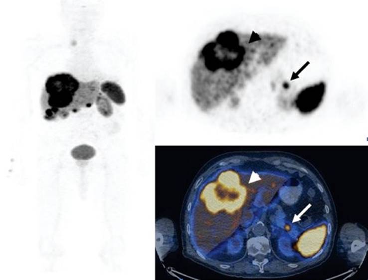
FIGURE 26.11. An 84-year-old male, presented with recurrent hypoglycemia and multiple liver metastases. Liver biopsy revealed metastatic neuroendocrine tumor. Ga-68-DOTA-NOC PET/CT (left,maximal intensity projection, transaxial slices; upper right, PET; lower right, fused image) demonstrates multiple liver metastases (arrowhead ) and focal, intense uptake in the pancreatic tail representing the primary malignancy (arrow). Final diagnosis was metastatic insulinoma.
Metastatic Neuroendocrine Tumor of Unknown Primary
Metastatic NET disease from unknown primary is also frequent. 18FDG PET/CT has a limited role in the evaluation of NET, especially in the well-differentiated subtypes.69 In-111 octreotide (pentetreotide) has traditionally been the tracer used to image NET, with excellent results.70 More recently, Ga-68-DOTA-NOC has provided images that are superior to In-111 octreotide71 and it is also more cost effective.72 In-111 octreotide was found to detect 35% of occult primary NET73 whereas Ga-68-DOTA-NOC detected 59% of occult primary tumor in a group of 59 patients with NET. CT had a detection rate of only 20%. Where available, it now appears that Ga-68-DOTA-NOC PET/CT is the tracer of choice to image patients with NET (Fig. 26.11).74 A study comparing Ga-68-DOTA-NOC PET/CT and WB MRI to stage NET found comparable overall detection rates for metastatic disease but PET/CT was superior for lymph node and pulmonary disease and WB MRI for liver and bone metastases. Hybrid PET/MR, an emerging technology, may become the comprehensive examination of the future.75
FUTURE DIRECTIONS FOR ASSESSMENT OF CUP
Current PET/CT devices have improved reconstruction, scatter and attenuation correction algorithms, which, together with the implementation of time of flight (TOF), produce low-noise PET images.76 These devices have a spatial resolution of 4 to 6 mm and are also equipped with helical 64-slice CT with a resolution of 0.5 mm.77 These devices have the potential to increase the detection rate of small primary tumors which has been the basis of many false negatives in patients with CUP.
MRI provides excellent soft tissue contrast and high spatial resolution without ionizing radiation.77 The recent development of WB hybrid PET/MR systems combines the high sensitivity of MRI with the specificity of FDG PET, with more precise assessment of the head, neck, breast, and abdominal organs. Review of the MR component of a PET/MR examination may allow the detection of non–18FDG-avid tumors where CT is of limited value, such as prostate cancer or certain ovarian cancers. However, based on current available evidence, it does not seem that the fusion of these two modalities will replace PET/CT in patients with CUP, mainly because of the high frequency of occult lung primaries. Some of the current limitations of MRI may affect patients with CUP, including the use of sequences with high sensitivity at the cost of low specificity78 and the low sensitivity for detection of peripheral skin and lung lesions.79 In a study population of 98 oncology patients, 18FDG PET/CT was superior to WB MRI in a subgroup of 12 patients with CUP and 13 with head and neck tumors.80 Extensive studies will be necessary to assess the potential clinical added value with PET/MR in the search for CUP when compared to PET/CT.
Detection of very small occult primary breast tumors may significantly alter therapy, offering the possibility of breast conserving surgery.81 The combination of positron emission mammography (PEM) and MRI has been shown to provide the highest sensitivity for detection of malignant lesions, while improving on the low specificity of MRI.82 Dedicated breast PET/MRI devices are technically feasible and integration of the strengths of both modalities can theoretically be of added value.83
CONCLUSIONS
CUP presents with various signs, symptoms, locations, and histologies. The PET/CT detection rate of the primary tumor varies greatly and ranges from 22% to 63%. It is increasingly accepted that 18FDG imaging should be a part of the standard workup of this spectrum of malignancies especially when other modalities failed to reveal the origin of the metastatic disease. It should be taken into consideration that false-positive and false-negative rates are not negligible and can contribute significantly to inaccurate 18FDG PET/CT reports when assessing patients with CUP. Improved molecular imaging technologies are expected to improve the assessment of patients presenting with metastatic CUP.
REFERENCES
1. Greco FA, Hainsworth J. Cancer of unknown primary site. In: DeVita VT, Lawrence TS, Rosenberg SA, eds. DeVita, Hellman, and Rosenberg’s Cancer: Principles & Practice of Oncology. 9th ed. Philadelphia, PA: Lippincott Williams & Wilkins; 2011.
2. Siegel R, Naishadham D, Jemal A. Cancer statistics, 2012. CA Cancer J Clin. 2012; 62:10–29.
3. Greco FA, Oien K, Erlander M, et al. Cancer of unknown primary: Progress in the search for improved and rapid diagnosis leading toward superior patient outcomes. Ann Oncol. 2012;23:298–304.
4. Bugat R, Bataillard A, Lesimple T, et al. Summary of the standards, options and recommendations for the management of patients with carcinoma of unknown primary site (2002). Br J Cancer. 2003;89(suppl 1):S59–S66.
5. Pentheroudakis G, Briasoulis E, Pavlidis N. Cancer of unknown primary site: Missing primary or missing biology? Oncologist. 2007;12:418–425.
6. Papac RJ. Spontaneous regression of cancer: Possible mechanisms. In Vivo. 1998; 12:571–578.
7. van de Wouw AJ, Jansen RL, Speel EJ, et al. The unknown biology of the unknown primary tumour: A literature review. Ann Oncol. 2003;14:191–196.
8. American Cancer Society. Cancer Facts & Figures 2012. Atlanta, GA: American Cancer Society; 2012.
9. National Comprehensive Cancer Network. NCCN Clinical Practice Guidelines in Oncology (NCCN Guidelines™) Occult Primary (Cancer of Unknown Primary), Version 1.2012. 2011.
10. Pavlidis N, Pentheroudakis G. Cancer of unknown primary site. Lancet. 2012; 379:1428–1435.
11. Pavlidis N, Petrakis D, Golfinopoulos V, et al. Long-term survivors among patients with cancer of unknown primary. Crit Rev Oncol Hematol. 2012; 84(1):85–92.
12. Briasoulis E, Pavlidis N, Felip E. Cancers of unknown primary site: ESMO clinical recommendations for diagnosis, treatment and follow-up. Ann Oncol. 2009; 20(suppl 4):154–155.
13. Amela EY, Lauridant-Philippin G, Cousin S, et al. Management of “unfavourable” carcinoma of unknown primary site: Synthesis of recent literature. Crit Rev Oncol Hematol. 2012;84(2):213–223.
14. Karsell PR, Sheedy PF 2nd, O’Connell MJ. Computed tomography in search of cancer of unknown origin. JAMA. 1982;248:340–343.
15. McMillan JH, Levine E, Stephens RH. Computed tomography in the evaluation of metastatic adenocarcinoma from an unknown primary site. A retrospective study. Radiology. 1982;143:143–146.
16. Muraki AS, Mancuso AA, Harnsberger HR. Metastatic cervical adenopathy from tumors of unknown origin: The role of CT. Radiology. 1984;152:749–753.
17. Eustace S, Tello R, DeCarvalho V, et al. Whole body turbo STIR MRI in unknown primary tumor detection. J Magn Reson Imaging. 1998;8:751–753.
18. Liang X, Congxin W, Yi Z, et al. Whole-body diffusion-weighted MR imaging with fat suppression by using STIR: Clinical utility for detection of primary malignancies in patients initially presented with metastasis. Eur J Radiol. 2011;79:359–362.
19. McMahon K, Medoro L, Kennedy D. Breast magnetic resonance imaging: An essential role in malignant axillary lymphadenopathy of unknown origin. Australas Radiol. 2005;49:382–389.
20. Orel SG, Weinstein SP, Schnall MD, et al. Breast MR imaging in patients with axillary node metastases and unknown primary malignancy. Radiology. 1999; 212:543–549.
21. van Veen SA, Balm AJ, Valdes Olmos RA, et al. Occult primary tumors of the head and neck: Accuracy of thallium 201 single-photon emission computed tomography and computed tomography and/or magnetic resonance imaging. Arch Otolaryngol Head Neck Surg.2001;127:406–411.
22. Lentle BC, Jackson FI, Dierich H. Gallium-67-citrate scintiscanning in the search for occult primary malignant tumours. Can Med Assoc J. 1976;114:1113–1138.
23. Rusthoven KE, Koshy M, Paulino AC. The role of fluorodeoxyglucose positron emission tomography in cervical lymph node metastases from an unknown primary tumor. Cancer. 2004;101:2641–2649.
24. Even-Sapir E, Keidar Z, Bar-Shalom R. Hybrid imaging (SPECT/CT and PET/CT)–improving the diagnostic accuracy of functional/metabolic and anatomic imaging. Semin Nucl Med. 2009;39:264–275.
25. Varadhachary GR, Greco FA. Overview of patient management and future directions in unknown primary carcinoma. Semin Oncol. 2009;36:75–80.
26. Rudmik L, Lau HY, Matthews TW, et al. Clinical utility of PET/CT in the evaluation of head and neck squamous cell carcinoma with an unknown primary: A prospective clinical trial. Head Neck. 2011;33:935–940.
27. Keller F, Psychogios G, Linke R, et al. Carcinoma of unknown primary in the head and neck: Comparison between positron emission tomography (PET) and PET/CT. Head Neck. 2011;33:1569–1575.
28. Gutzeit A, Antoch G, Kuhl H, et al. Unknown primary tumors: Detection with dual-modality PET/CT–initial experience. Radiology. 2005;234:227–234.
29. Nanni C, Rubello D, Castellucci P, et al. Role of 18F-FDG PET-CT imaging for the detection of an unknown primary tumour: Preliminary results in 21 patients. Eur J Nucl Med Mol Imaging. 2005;32:589–592.
30. Fencl P, Belohlavek O, Skopalova M, et al. Prognostic and diagnostic accuracy of [18F]FDG-PET/CT in 190 patients with carcinoma of unknown primary. Eur J Nucl Med Mol Imaging. 2007;34:1783–1792.
31. Kole AC, Nieweg OE, Pruim J, et al. Detection of unknown occult primary tumors using positron emission tomography. Cancer. 1998;82:1160–1166.
32. Lassen U, Daugaard G, Eigtved A, et al. 18F-FDG whole body positron emission tomography (PET) in patients with unknown primary tumours (UPT). Eur J Cancer. 1999;35:1076–1082.
33. Lonneux M, Reffad A. Metastases from unknown primary tumor. PET-FDG as initial diagnostic procedure? Clin Positron Imaging. 2000;3:137–141.
34. Mantaka P, Baum RP, Hertel A, et al. PET with 2-[F-18]-fluoro-2-deoxy-D-glucose (FDG) in patients with cancer of unknown primary (CUP): Influence on patients’ diagnostic and therapeutic management. Cancer Biother Radiopharm. 2003;18:47–58.
35. Rades D, Kuhnel G, Wildfang I, et al. Localised disease in cancer of unknown primary (CUP): The value of positron emission tomography (PET) for individual therapeutic management. Ann Oncol. 2001;12:1605–1609.
36. Alberini JL, Belhocine T, Hustinx R, et al. Whole-body positron emission tomography using fluorodeoxyglucose in patients with metastases of unknown primary tumours (CUP syndrome). Nucl Med Commun. 2003;24:1081–1086.
37. Joshi U, van der Hoeven JJ, Comans EF, et al. In search of an unknown primary tumour presenting with extracervical metastases: The diagnostic performance of FDG-PET. Br J Radiol. 2004;77:1000–1006.
38. Ambrosini V, Nanni C, Rubello D, et al. 18F-FDG PET/CT in the assessment of carcinoma of unknown primary origin. Radiol Med. 2006;111:1146–1155.
39. Pelosi E, Pennone M, Deandreis D, et al. Role of whole body positron emission tomography/computed tomography scan with 18F-fluorodeoxyglucose in patients with biopsy proven tumor metastases from unknown primary site. Q J Nucl Med Mol Imaging. 2006;50:15–22.
40. Yapar Z, Kibar M, Yapar AF, et al. The value of 18F-fluorodeoxyglucose positron emission tomography/computed tomography in carcinoma of an unknown primary: Diagnosis and follow-up. Nucl Med Commun. 2010;31:59–66.
41. Seve P, Sawyer M, Hanson J, et al. The influence of comorbidities, age, and performance status on the prognosis and treatment of patients with metastatic carcinomas of unknown primary site: A population-based study. Cancer. 2006; 106:2058–2066.
42. Kwee TC, Kwee RM. Combined FDG-PET/CT for the detection of unknown primary tumors: Systematic review and meta-analysis. Eur Radiol. 2009;19:731–744.
43. Kumar R, Loving VA, Chauhan A, et al. Potential of dual-time-point imaging to improve breast cancer diagnosis with (18)F-FDG PET. J Nucl Med. 2005;46: 1819–1824.
44. Demura Y, Tsuchida T, Ishizaki T, et al. 18F-FDG accumulation with PET for differentiation between benign and malignant lesions in the thorax. J Nucl Med. 2003;44:540–548.
45. Nasuti JF, Mehrotra R, Gupta PK. Diagnostic value of fine-needle aspiration in supraclavicular lymphadenopathy: A study of 106 patients and review of literature. Diagn Cytopathol. 2001;25:351–355.
46. Bohuslavizki KH, Klutmann S, Kroger S, et al. FDG PET detection of unknown primary tumors. J Nucl Med. 2000;41:816–822.
47. Blaszyk H, Hartmann A, Bjornsson J. Cancer of unknown primary: Clinicopathologic correlations. APMIS. 2003;111:1089–1094.
48. Lim HS, Yoon W, Chung TW, et al. FDG PET/CT for the detection and evaluation of breast diseases: Usefulness and limitations. Radiographics. 2007;27(suppl 1): S197–S213.
49. Fletcher JW, Djulbegovic B, Soares HP, et al. Recommendations on the use of 18F-FDG PET in oncology. J Nucl Med. 2008;49:480–508.
50. Israel O, Yefremov N, Bar-Shalom R, et al. PET/CT detection of unexpected gastrointestinal foci of 18F-FDG uptake: Incidence, localization patterns, and clinical significance. J Nucl Med. 2005;46:758–762.
51. Seve P, Billotey C, Broussolle C, et al. The role of 2-deoxy-2-[F-18]fluoro-D-glucose positron emission tomography in disseminated carcinoma of unknown primary site. Cancer. 2007;109:292–299.
52. Kim JS, Lim ST, Jeong YJ, et al. The clinical value of dual point F-18 FDG PET/CT imaging for the differentiation of colonic focal uptake lesions. Nucl Med Mol Imaging. 2009;43:309–316.
53. AAssar O, Fischbein NJ, Caputo GR, et al. Metastatic head and neck cancer: Role and usefulness of FDG PET in locating occult primary tumors. Radiology. 1999; 210:177–181.
54. Braams JW, Pruim J, Kole AC, et al. Detection of unknown primary head and neck tumors by positron emission tomography. Int J Oral Maxillofac Surg. 1997; 26:112–115.
55. Regelink G, Brouwer J, de Bree R, et al. Detection of unknown primary tumours and distant metastases in patients with cervical metastases: Value of FDG-PET versus conventional modalities. Eur J Nucl Med Mol Imaging. 2002;29:1024–1030.
56. Stoeckli SJ, Mosna-Firlejczyk K, Goerres GW. Lymph node metastasis of squamous cell carcinoma from an unknown primary: Impact of positron emission tomography. Eur J Nucl Med Mol Imaging. 2003;30:411–416.
57. Freudenberg LS, Fischer M, Antoch G, et al. Dual modality of 18F-fluorodeoxyglucose-positron emission tomography/computed tomography in patients with cervical carcinoma of unknown primary. Med Princ Pract. 2005;14:155–160.
58. Nassenstein K, Veit-Haibach P, Stergar H, et al. Cervical lymph node metastases of unknown origin: Primary tumor detection with whole-body positron emission tomography/computed tomography. Acta Radiol. 2007:1–8.
59. Paul SA, Stoeckli SJ, von Schulthess GK, et al. FDG PET and PET/CT for the detection of the primary tumour in patients with cervical non-squamous cell carcinoma metastasis of an unknown primary. Eur Arch Otorhinolaryngol. 2007;264:189–195.
60. Johansen J, Buus S, Loft A, et al. Prospective study of 18FDG-PET in the detection and management of patients with lymph node metastases to the neck from an unknown primary tumor. Results from the DAHANCA-13 study. Head Neck. 2008;30:471–478.
61. Miller FR, Karnad AB, Eng T, et al. Management of the unknown primary carcinoma: Long-term follow-up on a negative PET scan and negative panendoscopy. Head Neck. 2008;30:28–34.
62. Wartski M, Le Stanc E, Gontier E, et al. In search of an unknown primary tumour presenting with cervical metastases: Performance of hybrid FDG-PET-CT. Nucl Med Commun. 2007;28:365–371.
63. Yabuki K, Tsukuda M, Horiuchi C, et al. Role of 18F-FDG PET in detecting primary site in the patient with primary unknown carcinoma. Eur Arch Otorhinolaryngol. 2010;267:1785–1792.
64. Fleming AJ Jr, Smith SP Jr, Paul CM, et al. Impact of [18F]-2-fluorodeoxyglucose-positron emission tomography/computed tomography on previously untreated head and neck cancer patients. Laryngoscope. 2007;117:1173–1179.
65. Strobel K, Haerle SK, Stoeckli SJ, et al. Head and neck squamous cell carcinoma (HNSCC)–detection of synchronous primaries with (18)F-FDG-PET/CT. Eur J Nucl Med Mol Imaging. 2009;36:919–927.
66. Blodgett TM, Fukui MB, Snyderman CH, et al. Combined PET-CT in the head and neck: Part 1. Physiologic, altered physiologic, and artifactual FDG uptake. Radiographics. 2005;25:897–912.
67. Clarke RW, Jones AS. Neck dissection for non-squamous malignancy. Clin Otolaryngol Allied Sci. 1992;17:540–544.
68. Schapira DV, Jarrett AR. The need to consider survival, outcome, and expense when evaluating and treating patients with unknown primary carcinoma. Arch Intern Med. 1995;155:2050–2054.
69. Adams S, Baum R, Rink T, et al. Limited value of fluorine-18 fluorodeoxyglucose positron emission tomography for the imaging of neuroendocrine tumours. Eur J Nucl Med. 1998;25:79–83.
70. Krenning EP, Kwekkeboom DJ, Bakker WH, et al. Somatostatin receptor scintigraphy with [111In-DTPA-D-Phe1]- and [123I-Tyr3]-octreotide: The Rotterdam experience with more than 1000 patients. Eur J Nucl Med. 1993;20:716–731.
71. Buchmann I, Henze M, Engelbrecht S, et al. Comparison of 68Ga-DOTATOC PET and 111In-DTPAOC (Octreoscan) SPECT in patients with neuroendocrine tumours. Eur J Nucl Med Mol Imaging. 2007;34:1617–1626.
72. Schreiter NF, Brenner W, Nogami M, et al. Cost comparison of 111In-DTPA-octreotide scintigraphy and 68Ga-DOTATOC PET/CT for staging enteropancreatic neuroendocrine tumours. Eur J Nucl Med Mol Imaging. 2012;39:72–82.
73. Savelli G, Lucignani G, Seregni E, et al. Feasibility of somatostatin receptor scintigraphy in the detection of occult primary gastro-entero-pancreatic (GEP) neuroendocrine tumours. Nucl Med Commun. 2004;25:445–449.
74. Prasad V, Ambrosini V, Hommann M, et al. Detection of unknown primary neuroendocrine tumours (CUP-NET) using (68)Ga-DOTA-NOC receptor PET/CT. Eur J Nucl Med Mol Imaging. 2009;37:67–77.
75. Pfannenberg C, Schraml C, Schwenzer N, et al. Comparison of [68Ga]DOTATOC-PET/CT and whole-body MRI in staging of neuroendocrine tumors. Cancer Imaging. 2011;11(Spec No A):S38–S39.
76. Pichler BJ, Kolb A, Nagele T, et al. PET/MRI: Paving the way for the next generation of clinical multimodality imaging applications. J Nucl Med. 2010;51:333–336.
77. Pichler BJ, Wehrl HF, Judenhofer MS. Latest advances in molecular imaging instrumentation. J Nucl Med. 2008;49(suppl 2):5S–23S.
78. Ratib O, Beyer T. Whole-body hybrid PET/MRI: Ready for clinical use? Eur J Nucl Med Mol Imaging. 2011;38:992–995.
79. Schmidt GP, Haug AR, Schoenberg SO, et al. Whole-body MRI and PET-CT in the management of cancer patients. Eur Radiol. 2006;16:1216–1225.
80. Antoch G, Vogt FM, Freudenberg LS, et al. Whole-body dual-modality PET/CT and whole-body MRI for tumor staging in oncology. JAMA. 2003;290:3199–3206.
81. de Bresser J, de Vos B, van der Ent F, et al. Breast MRI in clinically and mammographically occult breast cancer presenting with an axillary metastasis: A systematic review. Eur J Surg Oncol. 2010;36:114–119.
82. Berg WA, Madsen KS, Schilling K, et al. Breast cancer: Comparative effectiveness of positron emission mammography and MR imaging in presurgical planning for the ipsilateral breast. Radiology. 2011;258:59–72.
83. Moadel RM. Breast cancer imaging devices. Semin Nucl Med. 2011;41:229–241.