
Gregory Barsh, MD, PhD
Mechanisms of cellular and tissue dysfunction in genetic diseases are as varied as the organs they affect. To some extent, these mechanisms are similar to those that occur in nonheritable disorders. For example, a fracture resulting from decreased bone density in osteoporosis heals in much the same way as one caused by a defective collagen gene in osteogenesis imperfecta, and the response to coronary atherosclerosis in most individuals does not depend on whether they have inherited a defective low-density lipoprotein (LDL) receptor. Thus, the pathophysiologic principles that distinguish genetic disease focus not so much on the affected organ system as on the mechanisms of mutation, inheritance, and molecular pathways from genotype to phenotype.
This chapter begins with a discussion of the terminology used to describe inherited conditions, the prevalence of genetic disease, and some major principles and considerations in medical genetics. Important terms and key words used throughout the chapter are defined in Table 2-1.
TABLE 2-1 Glossary of terms and keywords.


Next, a group of disorders caused by mutations in collagen genes is discussed (ie, osteogenesis imperfecta). Although osteogenesis imperfecta is often considered a single entity, different mutations and different genes subject to mutation lead to a wide spectrum of clinical phenotypes. The different types of osteogenesis imperfecta exhibit typical patterns of autosomal dominant or autosomal recessive inheritance and are, therefore, examples of so-called mendelian conditions. To show how environmental factors can influence the relationship between genotype and phenotype, I discuss another mendelian condition, phenylketonuria. This serves as a paradigm for newborn screening programs and treatment of genetic disease. Several genetic conditions have been found to depend not only on the gene being inherited but also on the phenotype or the sex of the parent. As an example of a condition that exhibits nontraditional inheritance, fragile X–associated mental retardation syndrome is discussed. This syndrome not only is the most common inherited cause of mental retardation but also illustrates how different types of mutations can explain the perplexing phenomenon of genetic anticipation, where the severity of a mendelian syndrome appears to progress with every generation of inheritance. Another group of disorders that depend on the phenotype and sex of the parent consists of those that affect the mitochondrial genome. As examples, Leber hereditary optic neuropathy (LHON) and myoclonic epilepsy with ragged red fibers (MERRF) are considered. These illustrate the principles of mitochondrial inheritance and its pathophysiology. Aneuploidy is discussed as one of the most common types of human genetic disease that does not affect DNA structure but instead alters the normal chromosome content per cell. The example that is considered, Down syndrome, has had a major impact on reproductive medicine and reproductive decision making and serves to illustrate general principles that apply to many aneuploid conditions. Finally, I consider how genome sequences and sequencing are improving our understanding of pathophysiology for many diseases. With the completion of the human genome sequence and technological advances that allow individual genomes to be sequenced rapidly and inexpensively, prospects are at hand to identify genetic components of any human phenotype and to provide medical care that is truly personalized.
UNIQUE PATHOPHYSIOLOGIC ASPECTS OF GENETIC DISEASES
Although the phenotypes of genetic diseases are diverse, their causes are not. The primary cause of any genetic disease is a change in the sequence or cellular content of DNA that ultimately deranges gene expression. Most genetic diseases are caused by an alteration in DNA sequence that alters the synthesis of a single gene product. However, some genetic diseases are caused by (1) structural rearrangements that result in deletion or duplication of a group of closely linked genes or (2) abnormalities during mitosis or meiosis that result in an abnormal number of chromosomes per cell. In most genetic diseases, every cell in an affected individual carries the mutated gene or genes as a consequence of its inheritance via a mutant egg or sperm cell (gamete). However, mutation of the gametic cell may have arisen during its development, in which case somatic cells of the parent do not carry the mutation and the affected individual is said to have a “new mutation.” In addition, some mutations may arise during early embryogenesis, in which case tissues of the affected individual contain a mixture, or mosaic, of mutant and nonmutant cells. Depending on the time of embryogenesis and cell type in which a new mutation arises, an individual may carry the mutation in some but not all of their germ cells (germline mosaicism), some but not all of their somatic cells (somatic mosaicism), or both.
It is helpful to begin with a brief review of terms that are commonly used in discussing genetic disease with patients and their families. Although genes were recognized and studied long before the structure of DNA was known, it has become common usage to regard a gene as a short stretch of DNA, usually but not always <100,000 base pairs (bp) in length, that encodes a product (usually protein) responsible for a measurable trait. DNA length is typically measured in base pairs, kilobase pairs (kb), or megabase pairs (Mb); chromosomes vary in length from about 46 Mb to 245 Mb. The locus is the place where a particular gene lies on its chromosome. A gene’s DNA sequence nearly always shows slight differences when many unrelated individuals are compared, and the variant sequences are described as alleles. A mutation is a biochemical event such as a nucleotide change, deletion, or insertion that has produced a new allele. Many changes in the DNA sequence of a gene, such as those within introns or at the third “wobble” position of codons for particular amino acids, do not affect the structure or expression of the gene product; therefore, although all mutations result in a biochemical or molecular biologic phenotype (ie, a change in DNA), only some of them result in a clinically abnormal phenotype.
At the molecular level, variant alleles are usually recognized by DNA sequencing and are referred to as a single nucleotide polymorphism (SNP) if a single base pair change has occurred. As originally coined, the word polymorphism referred to an allele present in 1% or more of a population; today, the terminology tends to be less rigid and is often described qualitatively, ie, rare and common variants. At the clinical level, variant alleles are recognized by their effect on a phenotype such as human leukocyte antigen (HLA) type or hair color. For an autosomal gene (those that lie on chromosomes 1–22, carried in two copies per cell), individuals carrying identical copies are homozygous, whereas individuals whose two copies differ from each other are heterozygous. These terms—homozygous and heterozygous—can apply to the DNA sequence, the protein product, or the clinical phenotype. In other words, an individual may be heterozygous for a SNP that does not alter the protein product, heterozygous for a deletion that causes a genetic disease, or heterozygous for a DNA sequence alteration that causes a change in protein structure but does not cause disease.
This discussion helps to illustrate the use of the word phenotype, which refers simply to any characteristic that can be measured, with the type of measurement depending on the characteristic. Hair color and height are phenotypes readily apparent to a casual observer that are not obviously associated with disease, diabetes and coronary artery disease are disease phenotypes that typically require clinical investigation to be recognized, whereas restriction fragment length polymorphisms (RFLPs), simple sequence length polymorphisms (SSLPs), and SNPs are molecular biologic phenotypes that can be detected only with a laboratory test.
PENETRANCE & EXPRESSIVITY
One of the most important principles of human genetics is that two individuals with the same mutated gene may have different phenotypes. For example, in the autosomal dominant condition called type I osteogenesis imperfecta, pedigrees may occur in which there is both an affected grandparent and an affected grandchild even though the obligate carrier parent is asymptomatic (Figure 2-1). Given a set of defined criteria, recognition of the condition in individuals known to carry the mutated gene is described as penetrance. In other words, if 7 of 10 individuals older than 40 with the type I osteogenesis imperfecta mutation have an abnormal bone density scan, the condition is said to be 70% penetrant by that criterion. Penetrance may vary both with age and according to the set of criteria being used; for example, type I osteogenesis imperfecta may be 90% penetrant at age 40 when the conclusion is based on a bone density scan in conjunction with laboratory tests for abnormal collagen synthesis. Reduced penetrance or age-dependent penetrance is a common feature of dominantly inherited conditions that have a relatively high fitness(the extent to which individuals carrying a mutant allele produce offspring relative to individuals who do not carry a mutant allele); Huntington disease and polycystic kidney disease are examples.

FIGURE 2-1 Penetrance and expressivity in type I osteogenesis imperfecta. In this schematic pedigree of the autosomal dominant condition type I osteogenesis imperfecta, nearly all of the affected individuals exhibit different phenotypic features that vary in severity (variable expressivity). As is shown, type I osteogenesis imperfecta is fully penetrant, because every individual who transmits the mutation is phenotypically affected to some degree. However, if mild short stature in the individual indicated with the arrow had been considered to be a normal variant, then the condition would have been nonpenetrant in this individual. Thus, in this example, judgments about penetrance or nonpenetrance depend on the criteria for normal and abnormal stature.
When the same mutated gene gives rise to a different spectrum of phenotypes, the situation is referred to as variable expressivity. For example, blue scleras and short stature may be the only manifestations of type I osteogenesis imperfecta in a particular individual, whereas a sibling who carries the identical mutation may be confined to a wheelchair as a result of multiple fractures and deformities. The mutation is penetrant in both individuals, but its expression is variable. Both reduced penetrance and variable expressivity may occur in individuals who carry the same mutated allele; therefore, phenotypic differences between these individuals must be due to the effects of other “modifier” genes, to environmental interactions, or to chance.
MECHANISMS OF MUTATION & INHERITANCE PATTERNS
Mutations can be characterized both by their molecular nature—nucleotide deletion, insertion, substitution—and by their effects on gene activity (ie, no effect [neutral or silent], complete loss of function [amorphic mutation], partial loss of function [hypomorphic mutation], gain of function [hypermorphic mutation], or acquisition of a new property [neomorphic mutation]). Geneticists who study experimental organisms frequently use specific deletions to ensure that a mutated allele causes a loss of function, but human geneticists rely on biochemical or cell culture studies. Amorphic and hypomorphic mutations are probably the most frequent type of mutation in human genetic disease because there are many ways to interfere with a protein’s function.
For autosomal genes, the fundamental difference between dominant and recessive inheritance is that, with dominant inheritance, the disease state or trait being measured is apparent when one copy of the mutated allele and one copy of the normal allele are present. With recessive inheritance, two copies of the mutated allele must be present for the disease state or trait to be apparent. However, for genes that lie on the X chromosome, the situation is slightly different because females have two X chromosomes and males have only one. X-linked dominant inheritance occurs when one copy of a mutant gene causes the disease phenotype (in males and females); X-linked recessive inheritance occurs when two copies of a mutant gene cause the disease phenotype (in females). Because most mutations are amorphic or hypomorphic, however, one copy of an X-linked mutant allele in males is not “balanced” with a nonmutant allele, as it would be in females; therefore, in X-linked recessive inheritance, one copy of a mutant allele is sufficient to produce a disease phenotype in males, a situation referred to as hemizygosity.
RECESSIVE INHERITANCE & LOSS-OF-FUNCTION MUTATIONS
Most recessive mutations are due to loss of function of the gene product, which can occur from a variety of different causes, including failure of the gene to be transcribed or translated and failure of the translated gene product to function correctly. There are two general principles to keep in mind when considering loss-of-function mutations. First, because expression from the nonmutant allele usually does not change (i.e., there is no dosage compensation), gene expression in a heterozygous carrier of a loss-of-function allele is reduced to 50% of normal. Second, for most biochemical pathways, a 50% reduction in enzyme concentration is not sufficient to produce a disease state. Thus, most diseases resulting from enzyme deficiencies such as phenylketonuria (Table 2-2) are inherited in a recessive fashion.
TABLE 2-2 Phenotype, inheritance, and prevalence of selected genetic disorders.

DOMINANT INHERITANCE & LOSS-OF-FUNCTION MUTATIONS
If 50% of a particular product is not enough for the cell or tissue to function normally, then a loss-of-function mutation in this gene produces a dominantly inherited phenotype. Such mutations often occur in structural proteins; an example is type I osteogenesis imperfecta, which is considered in detail later. Most dominantly inherited phenotypes are actually semidominant, which means that an individual who carries two copies of the mutant allele is affected more severely than someone who carries one mutant and one normal copy. However, for most dominantly inherited conditions, homozygous mutant individuals are rarely observed. For example, inheritance of achondroplasia, the most common genetic cause of very short stature, is usually described as autosomal dominant. However, rare matings between two affected individuals have a 25% probability of producing offspring with two copies of the mutant gene. This results in homozygous achondroplasia, a condition that is very severe and usually fatal in the perinatal period; thus, achondroplasia exhibits semidominant inheritance. Huntington disease, a dominantly inherited neurologic disease, is the only known human condition in which the homozygous mutant phenotype is identical to the heterozygous mutant phenotype (sometimes referred to as a “true dominant”).
DOMINANT NEGATIVE GENE ACTION
A special kind of pathophysiologic mechanism, referred to as dominant negative, occurs frequently in human genetic diseases that involve proteins that form oligomeric or polymeric complexes. In these disorders, the mutant allele gives rise to a structurally abnormal protein that interferes with the function of the normal allele. Note that any molecular lesion (ie, deletion, nonsense, missense, or splicing) can produce a loss-of-function allele. However, only molecular lesions that yield a protein product (ie, splicing, missense, or nonsense mutations) can result in a dominant negative allele. Type II osteogenesis imperfecta, described later, is an example of a dominant negative mutation.
Although the terms “dominant” and “recessive” are occasionally used to describe specific mutations, a DNA sequence alteration itself cannot, strictly speaking, be dominant or recessive. The terms are instead appropriate to the effect of a mutation on a particular trait. Therefore, in characterizing a particular mutation as “recessive,” one is referring to the effect of the mutation on the trait being studied.
MUTATION RATE & THE PREVALENCE OF GENETIC DISEASE
At the level of DNA sequence, nucleotide mutations (substitutions, small insertions, or small deletions) in humans occur at a rate of approximately 2 × 10⁻⁸ per nucleotide per human generation, or 150 new mutations per diploid genome. However, only about 5% of the human genome is functional, so most new mutations have no effect. Still, with approximately 23,000 genes in the human genome and an estimated deleterious “per locus” mutation rate of 10−5 per generation, the chance of a new deleterious mutation in any one individual is about 20%. Furthermore, assuming 10 billion new births in the last millennium, every gene in the human genome has probably been mutated (in a deleterious manner) about 100,000 different times. However, from a clinical perspective, only about 5000 single-gene disorders have been recognized to cause a human disease. In considering possible explanations for this disparity, it seems likely that deleterious mutations of many single genes are lethal very early in development and thus not clinically apparent, whereas deleterious mutations in other genes do not cause an easily recognizable phenotype. The overall frequency of disease attributable to defects in single genes (ie, mendelian disorders) is approximately 1% of the general population.
Table 2-2 lists the major symptoms, genetic mechanisms, and prevalence of the diseases considered in this chapter as well as of several others. The most common conditions, such as neurofibromatosis, cystic fibrosis, and fragile X–associated mental retardation syndrome, will be encountered at some time by most health care professionals regardless of their field of interest. Other conditions such as Huntington disease and adenosine deaminase deficiency, although of intellectual and pathophysiologic interest, are not likely to be seen by most practitioners.
Many common conditions such as atherosclerosis and breast cancer that do not show strictly mendelian inheritance patterns have a genetic component evident from familial aggregation or twin studies. These conditions are usually described as multifactorial, which means that the effects of one or more mutated genes and environmental differences all contribute to the likelihood that a given individual will manifest the phenotype.
ISSUES IN CLINICAL GENETICS
Most patients with genetic disease present during early childhood with symptoms that ultimately give rise to a diagnosis such as fragile X–associated mental retardation or Down syndrome. The major clinical issues at presentation are arriving at the correct diagnosis and counseling the patient and family regarding the natural history and prognosis of the condition. It is important to assess the likelihood that the same condition will occur again in the family and determine whether it can be diagnosed prenatally. These issues are the subject matter of genetic counseling by medical geneticists and genetic counselors.
Understanding the pathophysiology of genetic diseases that interfere with specific metabolic pathways—so-called inborn errors of metabolism—has led to effective treatments for selected conditions such as phenylketonuria, maple syrup urine disease, and homocystinuria. Many of these diseases are rare, but efforts are underway to develop treatments for common single-gene disorders such as Duchenne muscular dystrophy, cystic fibrosis, and hemophilia. Some forms of therapy are directed at replacing the mutant protein, whereas others are directed at ameliorating its effects.
CHECKPOINT
1. Define gene, locus, allele, mutation, heterozygosity, hemizygosity, polymorphism, and phenotype.
2. How is it possible for two individuals with the same mutation to have differences in the severity of an abnormal phenotype?
3. Explain the pathophysiologic difference between mutations that act via loss of function and those that act via dominant negative gene action.
PATHOPHYSIOLOGY OF SELECTED GENETIC DISEASES
OSTEOGENESIS IMPERFECTA
Osteogenesis imperfecta is a condition inherited in mendelian fashion that illustrates many principles of human genetics. It is a heterogeneous and pleiotropic group of disorders characterized by a tendency toward fragility of bone. Advances in the last two decades demonstrate two genetically different groups: the “classical” group, in which more than 90% of cases are caused by a mutation of the COL1A1 or COL1A2 genes, which encode the subunits of type I collagen, proα1(I) and proα2(I), respectively, and a newer group, caused by loss-of-function mutations in proteins required for proper folding, processing, and secretion of collagen. More than 100 different mutant alleles have been described for osteogenesis imperfecta; the relationships between different DNA sequence alterations and the type of disease (genotype-phenotype correlations) illustrate several pathophysiologic principles in human genetics.
Clinical Manifestations
The clinical and genetic characteristics of osteogenesis imperfecta are summarized in Table 2-3, in which the timing and severity of fractures, radiologic findings, and presence of additional clinical features help to distinguish four different subtypes. This classification was presented more than 30 years ago. Over the past decade, it has become clear that there are more than a dozen different genes in which mutations can cause osteogenesis imperfecta, and that the utility of alternative or more extended nosologic approaches depends on whether the condition is being considered from the perspective of patients, caregivers, or molecular geneticists.
TABLE 2-3 Subtypes of dominant osteogenesis imperfecta.

All forms of osteogenesis imperfecta are characterized by increased susceptibility to fractures (“brittle bones”), but there is considerable phenotypic heterogeneity, even within individual subtypes. Individuals with type I or type IV osteogenesis imperfecta present in early childhood with one or a few fractures of long bones in response to minimal or no trauma; x-ray films reveal mild osteopenia, little or no bony deformity, and often evidence of earlier subclinical fractures. However, most individuals with type I or type IV osteogenesis imperfecta do not have fractures in utero. Type I and type IV osteogenesis imperfecta are distinguished by the severity (less in type I than in type IV) and by scleral hue, which indicates the thickness of this tissue and the deposition of type I collagen. Individuals with type I osteogenesis imperfecta have blue scleras, whereas the scleras of those with type IV are normal or slightly gray. In type I, the typical number of fractures during childhood is 10–20; fracture incidence decreases after puberty, and the main features in adult life are mild short stature, a tendency toward conductive hearing loss, and occasionally dentinogenesis imperfecta. Individuals with type IV osteogenesis imperfecta generally experience more fractures than those with type I and have significant short stature caused by a combination of long bone and spinal deformities, but they often are able to walk independently. Approximately one fourth of the cases of type I or type IV osteogenesis imperfecta represent new mutations; in the remainder, the history and examination of other family members reveal findings consistent with autosomal dominant inheritance.
Type II osteogenesis imperfecta presents at or before birth (diagnosed by prenatal imaging) with multiple fractures, bony deformities, increased fragility of nonbony connective tissue, and blue scleras and usually results in death in infancy. Two typical radiologic findings are the presence of isolated “islands” of mineralization in the skull (wormian bones) and a beaded appearance to the ribs. Nearly all cases of type II osteogenesis imperfecta represent a new dominant mutation, and there is no family history. Death usually results from respiratory difficulties.
Type III osteogenesis imperfecta presents at birth or in infancy with progressive bony deformities, multiple fractures, and blue scleras. It is intermediate in severity between types II and IV; most affected individuals will require multiple corrective surgeries and lose the ability to ambulate by early adulthood. Unlike other forms of osteogenesis imperfecta, which are nearly always due to mutations that act dominantly, type III may be inherited, very rarely, in a recessive manner.
Although different subtypes of osteogenesis imperfecta can often be distinguished biochemically, the classification presented in Table 2-3 is primarily clinical rather than molecular, and the disease phenotypes for each subtype show a spectrum of severities that overlap one another. For example, a few individuals diagnosed with type II osteogenesis imperfecta based on the presence of severe bony deformities in utero will survive for many years and thus overlap the type III subtype. Similarly, some individuals with type IV osteogenesis imperfecta have fractures in utero and develop deformities that lead to loss of ambulation. Distinguishing this presentation from type III osteogenesis imperfecta may be possible only if other affected family members exhibit a milder course.
Additional subtypes of osteogenesis imperfecta have been suggested for individuals that do not match types I–IV, and there are additional disorders associated with congenital fractures that are usually not considered to be “classic” osteogenesis imperfecta. In particular, work over the past several years has identified 10 additional genes in which mutations can cause autosomal recessive osteogenesis imperfecta and has provided additional insight into the genetic pathophysiology. In general, recessively inherited osteogenesis imperfect is caused by loss-of-function mutations in genes whose protein product is required for proper protein folding, intracellular processing, and trafficking of type I collagen.
Pathophysiology
Osteogenesis imperfecta is a disease of type I collagen, which constitutes the major extracellular protein in the body. It is the major collagen in the dermis, the connective tissue capsules of most organs, and the vascular and gastrointestinal (GI) adventitia and is the only collagen in bone. A mature type I collagen fibril is a rigid structure that contains multiple type I collagen molecules packed in a staggered array and stabilized by intermolecular covalent cross-links. Each mature type I collagen molecule contains two α1 chains and one α2 chain, encoded by the COL1A1 and COL1A2 genes, respectively (Figure 2-2). The α1 and α2 chains are synthesized as larger precursors with amino and carboxyl terminal “propeptide” extensions, assemble with each other inside the cell, and are ultimately secreted as a heterotrimeric type I procollagen molecule. During intracellular assembly, the three chains wind around each other in a triple helix that is stabilized by interchain interactions between hydroxylated proline and adjacent carbonyl residues. There is a dynamic relationship between the posttranslational action of prolyl hydroxylase and assembly of the triple helix, which begins at the carboxyl terminal end of the molecule. Increased levels of hydroxylation result in a more stable helix, but helix formation prevents further prolyl hydroxylation. The nature of the triple helix causes the side chain of every third amino acid to point inward, and steric constraints allow only a proton in this position. Thus, the amino acid sequence of virtually all collagen chains in the triple-helical portion is (Gly-X-Y)n, where Y is proline about one third of the time.

FIGURE 2-2 Molecular assembly of type I pro-collagen. Type I procollagen is assembled in the endoplasmic reticulum from three proα chains that associate with each other beginning at their carboxyl terminals. An important requirement for proper assembly of the triple helix is the presence of a glycine residue at every third position in each of the proα chains. After secretion, the amino and carboxyl terminal propeptides are proteolytically cleaved, leaving a rigid triple helical collagen molecule with very short non–triplehelical domains at both ends. (Modified and reproduced, with permission, from Alberts BA. Molecular Biology of the Cell, 3rd ed. Garland Science, 1994.)
The fundamental defect in most individuals with type I osteogenesis imperfecta is reduced synthesis of type I collagen resulting from loss-of-function mutations in COL1A1. In most cases, the mutant COL1A1allele gives rise to greatly reduced (partial loss-of-function) or no (complete loss-of-function) mRNA. Because the nonmutant COL1A1 allele continues to produce mRNA at a normal rate (ie, there is no dosage compensation), heterozygosity for a complete loss-of-function mutation results in a 50% reduction in the total rate of proα1(I) mRNA synthesis, whereas heterozygosity for a partial loss-of-function mutation results in a less severe reduction. A reduced concentration of proα1(I) chains limits the production of type I procollagen, leading to (1) a reduced amount of structurally normal type I collagen and (2) an excess of unassembled proα2(I) chains, which are degraded inside the cell (Figure 2-3).

FIGURE 2-3 Molecular pathogenesis of type I and type II osteogenesis imperfecta (OI). The COL1A1 gene normally produces twice as many proα chains as the COL1A2 gene. Therefore, in nonmutant cells, the ratio of proα1 to proα2 chains is 2:1, which corresponds to the ratio of α1 and α2 chains in intact collagen molecules. In type I osteogenesis imperfecta, a mutation (X) in one of the COL1A1 alleles (*) results in failure to produce proα1 chains, leading to a 50% reduction in the total number of proα1 chains, a 50% reduction in the production of intact type I collagen molecules, and an excess of unassembled proα2 chains, which are degraded inside the cell. In type II osteogenesis imperfecta, a mutation in one of the COL1A1 alleles results in a structural alteration that blocks triple-helix formation and secretion of partially assembled collagen molecules containing the mutant chain. (Adapted from Thompson MW et al. Genetics in Medicine, 5th ed. Saunders, 1991.)
There are several potential molecular defects responsible for COL1A1 mutations in type I osteogenesis imperfecta, including alterations in a regulatory region leading to reduced transcription, splicing abnormalities leading to reduced steady state levels of RNA, and deletion of the entire COL1A1 gene. However, in many cases, the underlying defect is a single base pair change that creates a premature stop codon (also known as a “nonsense mutation”) in an internal exon. In a process referred to as nonsense-mediated decay, partially synthesized mRNA precursors that carry the nonsense codon are recognized and degraded by the cell. With collagen and many other genes, production of a truncated protein (as might be predicted from a nonsense mutation) would be more damaging to the cell than production of no protein at all. Thus, nonsense-mediated decay, which has been observed to occur for mutations in many different multiexon genes, serves as a protective phenomenon and is an important component of the genetic pathophysiology.
An example of these principles is apparent from considering type II osteogenesis imperfecta, which is caused by structurally abnormal forms of type I collagen and is more severe than type I osteogenesis imperfecta. Mutations in type II osteogenesis imperfecta can be caused by defects in either COL1A1 or COL1A2 and usually are missense alterations of a glycine residue that allow the mutant peptide chain to bind to normal chains in the initial steps of trimer assembly (Figure 2-3). However, triple-helix formation is ineffective, often because amino acids with large side chains are substituted for glycine. Ineffective triple-helix formation leads to increased post-translational modification by prolyl hydroxylase, a reduced rate of secretion, and activation of the unfolded protein stress response. These appear to be critical events in the cellular pathogenesis of type II osteogenesis imperfecta, because glycine substitutions toward the carboxyl terminal end of the molecule are generally more severe than those at the amino terminal end.
These considerations help to explain why type II osteogenesis imperfecta is more severe than type I and exemplify the principle of dominant negative gene action. The effects of an amino acid substitution in a proα1(I) peptide chain are amplified at the levels of both triple-helix assembly and fibril formation. Because every type I procollagen molecule has two proα1(I) chains, only 25% of type I procollagen molecules will contain two normal proα1(I) chains even though only one of the two COL1A1 alleles is mutated. Furthermore, activation of the unfolded protein stress response appears to be a key event in the pathophysiology of the disease, as discussed further below. Finally, because each molecule in a fibril interacts with several others, incorporation of an abnormal molecule can have disproportionately large effects on fibril structure and integrity.
Collagen mutations that cause type III and type IV osteogenesis imperfecta are diverse and include glycine substitutions in the amino terminal portion of the collagen triple helix, a few internal deletions of COL1A1 and COL1A2 that do not significantly disturb triple helix formation, and some unusual alterations in the non–triple-helical extensions at the amino and carboxyl terminals of proα chains.
Recessively inherited osteogenesis imperfect can be caused by loss of function for a key prolyl hydroxylase encoded by the PLOD2 gene, one of three genes, CRTAP, LEPRE1, PPIB, that encode members of a protein complex that resides within the rough endoplasmic reticulum and facilitates the folding and processing of type I collagen, as well as several additional genes whose protein products are required for intracellular trafficking and secretion of type I collagen. A common pathway for all types of osteogenesis imperfect involves a combination of reduced production of type I collagen in the extracellular matrix and/or dysfunctional intracellular collagen processing and maturation.
Genetic Principles
As already described, most cases of type I osteogenesis imperfecta are caused by partial or complete loss-of-function mutations in COL1A1. However, in approximately one-third of affected individuals, the disease is caused by a new mutation; in addition, there are many ways in which DNA sequence alterations can reduce gene expression. Consequently, there is a wide range of mutant alleles (ie, allelic heterogeneity), which represents a challenge for the development of molecular diagnostic tests. In a family in which type I osteogenesis imperfecta is known to occur clinically and a proband seeks a diagnostic test for the purposes of reproductive planning, it is possible in most cases to use linkage analysis at the COL1A1 locus. In this approach, one distinguishes between chromosomes that carry the mutant and nonmutant COL1A1 alleles using closely linked DNA-based polymorphisms, even though the causative molecular defect is not known. Once this information is established for a particular family, inheritance of the mutant allele can be predicted in future pregnancies.
For types III and IV osteogenesis imperfecta, mutations can occur in COL1A1 or COL1A2 (ie, locus heterogeneity), and in this situation, linkage analysis is more difficult because one cannot be sure which locus is abnormal.
For both type I and type IV osteogenesis imperfecta, the most important question in the clinical setting often relates to the natural history of the illness. For example, reproductive decision making in families at risk for osteogenesis imperfecta is influenced greatly by the relative likelihood of producing a child who will never walk and will require multiple orthopedic operations versus a child whose major problems will be a few long bone fractures and an increased risk of mixed sensorineural and conductive hearing loss in childhood and adulthood. As evident from the prior discussion, different mutant genes and different mutant alleles, as well as other genes that modify the osteogenesis imperfecta phenotype, can contribute to this phenotypic heterogeneity.
In type II osteogenesis imperfecta, a single copy of the mutant allele causes the abnormal phenotype and, therefore, has a dominant mechanism of action. Although the type II phenotype itself is never inherited, there are rare situations in which a phenotypically normal individual harbors a COL1A1 mutant allele among his or her germ cells. These individuals with so-called gonadal mosaicism can produce multiple offspring with type II osteogenesis imperfecta (Figure 2-4), a pattern of segregation that can be confused with recessive inheritance. In fact, many other mutations, including Duchenne muscular dystrophy, which is X linked, and type 1 neurofibromatosis, which is autosomal dominant, also occasionally show unusual inheritance patterns explained by gonadal mosaicism.

FIGURE 2-4 Gonadal mosaicism for type II osteogenesis imperfecta. In this idealized pedigree, the phenotypically normal father (indicated with the arrow) has had two children by different mates, each of whom is affected with autosomal dominant type II osteogenesis imperfecta (OI). Analysis of the father showed that some of his spermatozoa carried a COL1A1 mutation, indicating that the explanation for this unusual pedigree is germline mosaicism. (Adapted from Cohn DH et al. Recurrence of lethal osteogenesis imperfecta due to parental mosaicism for a dominant mutation in a human type I collagen gene [ COL1A1 ]. Am J Hum Genet. 1990;46:591.)
CHECKPOINT
4. When and how does type II osteogenesis imperfecta present? To what do these individuals succumb?
5. What are two typical radiologic findings in type II osteogenesis imperfecta?
6. Explain how nonsense-mediated decay can help protect individuals affected by a genetic disease.
PHENYLKETONURIA
Phenylketonuria presents one of the most dramatic examples of how the relationship between genotype and phenotype can depend on environmental variables. Phenylketonuria was first recognized as an inherited cause of mental retardation in 1934, and systematic attempts to treat the condition were initiated in the 1950s. The term “phenylketonuria” denotes elevated levels of urinary phenylpyruvate and phenylacetate, which occur when circulating phenylalanine levels, normally between 0.06 and 0.1 mmol/L, rise above 1.2 mmol/L. Thus, the primary defect in phenylketonuria is hyperphenylalaninemia, which itself has a number of distinct genetic causes.
The pathophysiology of phenylketonuria illustrates several important principles in human genetics. Hyperphenylalaninemia itself is caused by substrate accumulation, which occurs when a normal intermediary metabolite fails to be eliminated properly and its concentrations become elevated to levels that are toxic. As described later, the most common cause of hyperphenylalaninemia is deficiency of the enzyme phenylalanine hydroxylase, which catalyzes the conversion of phenylalanine to tyrosine. Individuals with mutations in phenylalanine hydroxylase usually do not suffer from the absence of tyrosine because this amino acid can be supplied to the body by mechanisms that are independent of phenylalanine hydroxylase. In other forms of phenylketonuria, however, additional disease manifestations occur as a result of end-product deficiency, which occurs when the downstream product of a particular enzyme is required for a key physiologic process.
A discussion of phenylketonuria also helps to illustrate the rationale for, and application of, population-based screening programs for genetic disease. More than 10 million newborn infants per year are tested for phenylketonuria, and the focus today in treatment has shifted in several respects. First, “successful” treatment of phenylketonuria by dietary restriction of phenylalanine is, in general, accompanied by subtle neuropsychologic defects that have been recognized only in the last decade. Thus, current investigations focus on alternative treatment strategies such as somatic gene therapy as well as on the social and psychologic factors that affect compliance with dietary management. Second, a generation of females treated for phenylketonuria are now bearing children, and the phenomenon of maternal phenylketonuria has been recognized in which in utero exposure to maternal hyperphenylalaninemia results in congenital abnormalities regardless of fetal genotype. The number of pregnancies at risk has risen in proportion to the successful treatment of phenylketonuria and represents a challenge to public health officials, physicians, and geneticists in the future.
Clinical Manifestations
The incidence of hyperphenylalaninemia varies among different populations. In African Americans, it is about 1:50,000; in Yemenite Jews, about 1:5000; and in most Northern European populations, about 1:10,000. Post-natal growth retardation, moderate-to-severe mental retardation, recurrent seizures, hypopigmentation, and eczematous skin rashes constitute the major phenotypic features of untreated phenylketonuria. However, with the advent of widespread newborn screening programs for hyperphenylalaninemia, the major phenotypic manifestations of phenylketonuria today occur when treatment is partial or when it is terminated prematurely during late childhood or adolescence. In these cases, there is usually a slight but significant decline in IQ, an array of specific performance and perceptual defects, and an increased frequency of learning and behavioral problems.
Newborn screening for phenylketonuria is performed on a small amount of dried blood obtained at 24–72 hours of age. From the initial screen, there is about a 1% incidence of positive or indeterminate test results, and a more quantitative measurement of plasma phenylalanine is then performed before 2 weeks of age. In neonates who undergo a second round of testing, the diagnosis of phenylketonuria is ultimately confirmed in about 1%, providing an estimated phenylketonuria prevalence of 1:10,000, although there is great geographic and ethnic variation (see prior discussion). The false-negative rate of phenylketonuria newborn screening programs is approximately 1:70; phenylketonuria in these unfortunate individuals is usually not detected until developmental delay and seizures during infancy or early childhood prompt a systematic evaluation for an inborn error of metabolism.
Infants in whom a diagnosis of phenylketonuria is confirmed are usually placed on a dietary regimen in which a semisynthetic formula low in phenylalanine can be combined with regular breast feeding. This regimen is adjusted empirically to maintain a plasma phenylalanine concentration at or below 1 mmol/L, which is still several times greater than normal but similar to levels observed in so-called benign hyperphenylalaninemia (see later discussion), a biochemical diagnosis which is not associated with phenylketonuria and has no clinical consequences. Phenylalanine is an essential amino acid, and even individuals with phenylketonuria must consume small amounts to avoid protein starvation and a catabolic state. Most children require 25–50 mg/kg/d of phenylalanine, and these requirements are met by combining natural foods with commercial products designed for phenylketonuria treatment. When dietary treatment programs were first implemented, it was hoped that the risk of neurologic damage from the hyperphenylalaninemia of phenylketonuria would have a limited window and that treatment could be stopped after childhood. However, it now appears that even mild hyperphenylalaninemia in adults (>1.2 mmol/L) is associated with neuropsychologic and cognitive deficits; therefore, dietary treatment of phenylketonuria should probably be continued indefinitely.
As an increasing number of treated females with phenylketonuria reach childbearing age, a new problem—fetal hyperphenylalaninemia via intrauterine exposure—has become apparent. Newborn infants in such cases exhibit microcephaly and growth retardation of prenatal onset, congenital heart disease, and severe developmental delay regardless of the fetal genotype. Rigorous control of maternal phenylalanine concentrations from before conception until birth reduces the incidence of fetal abnormalities in maternal phenylketonuria, but the level of plasma phenylalanine that is “safe” for a developing fetus is 0.12–0.36 mmol/L—significantly lower than what is considered acceptable for phenylketonuria-affected children or adults on phenylalanine-restricted diets.
Pathophysiology
The normal metabolic fate of free phenylalanine is incorporation into protein or hydroxylation by phenylalanine hydroxylase to form tyrosine (Figure 2-5). Because tyrosine, but not phenylalanine, can be metabolized to produce fumarate and acetoacetate, hydroxylation of phenylalanine can be viewed both as a means of making tyrosine a nonessential amino acid and as a mechanism for providing energy via gluconeogenesis during states of protein starvation. In individuals with mutations in phenylalanine hydroxylase, tyrosine becomes an essential amino acid. However, the clinical manifestations of the disease are caused not by absence of tyrosine (most people get enough tyrosine in the diet in any case) but by accumulation of phenylalanine. Transamination of phenylalanine to form phenylpyruvate normally does not occur unless circulating concentrations exceed 1.2 mmol/L, but the pathogenesis of central nervous system (CNS) abnormalities in phenylketonuria is related more to phenylalanine itself than to its metabolites. In addition to a direct effect of elevated phenylalanine levels on energy production, protein synthesis, and neurotransmitter homeostasis in the developing brain, phenylalanine can also inhibit the transport of neutral amino acids across the blood-brain barrier, leading to a selective amino acid deficiency in the cerebrospinal fluid. Thus, the neurologic manifestations of phenylketonuria are felt to be due to a general effect of substrate accumulation on cerebral metabolism. The pathophysiology of the eczema seen in untreated or partially treated phenylketonuria is not well understood, but eczema is a common feature of other inborn errors of metabolism in which plasma concentrations of branched-chain amino acids are elevated. Hypopigmentation in phenylketonuria is probably caused by an inhibitory effect of excess phenylalanine on the production of dopaquinone in melanocytes, which is the rate-limiting step in melanin synthesis.

FIGURE 2-5 Metabolic fates of phenylalanine. Because catabolism of phenylalanine must proceed via tyrosine, the absence of phenylalanine hydroxylase leads to accumulation of phenylalanine. Tyrosine is also a biosynthetic precursor for melanin and certain neurotransmitters, and the absence of phenylalanine hydroxylase causes tyrosine to become an essential amino acid.
Approximately 90% of infants with persistent hyperphenylalaninemia detected by newborn screening have typical phenylketonuria caused by a defect in phenylalanine hydroxylase (see later discussion). Of the remainder, most have benign hyperphenylalaninemia, in which circulating levels of phenylalanine are between 0.1 mmol/L and 1 mmol/L. However, approximately 1% of infants with persistent hyperphenylalaninemia have defects in the metabolism of tetrahydrobiopterin (BH4), which is a stoichiometric cofactor for the hydroxylation reaction (Figure 2-6). Unfortunately, BH4 is required not only for phenylalanine hydroxylase but also for tyrosine hydroxylase and tryptophan hydroxylase. The products of these latter two enzymes are catecholaminergic and serotonergic neurotransmitters; thus, individuals with defects in BH4 metabolism suffer not only from phenylketonuria (substrate accumulation) but also from absence of important neurotransmitters (end-product deficiency). Affected individuals develop a severe neurologic disorder in early childhood manifested by hypotonia, inactivity, and developmental regression and are treated not only with dietary restriction of phenylalanine but also with dietary supplementation with BH4, dopa, and 5-hydroxytryptophan.

FIGURE 2-6 Normal and abnormal phenylalanine metabolism. Tetrahydrobiopterin (BH4) is a cofactor for phenylalanine hydroxylase, tyrosine hydroxylase, and tryptophan hydroxylase. Consequently, defects in the biosynthesis of BH4 or its metabolism result in a failure of all three hydroxylation reactions. The absence of phenylalanine hydroxylation has phenotypic effects because of substrate accumulation, but the absence of tyrosine or tryptophan hydroxylation has phenotypic effects as a result of end-product deficiency. (6-PTS, 6-pyruvoyltetrahydrobiopterin synthetase; qBH2, quinonoid dihydrobiopterin.)
Genetic Principles
Phenylketonuria is one of several mendelian conditions that have a relatively high incidence, others being cystic fibrosis, Duchenne muscular dystrophy, neurofibromatosis type I, and sickle cell anemia (Table 2-2). These conditions share no single feature: Some are recessive, some dominant, some autosomal, and some X linked, some are lethal in early childhood, but others have very little effect on reproduction (and transmission of mutant genes to subsequent generations). In fact, the incidence of a mendelian condition is determined by a balance of factors, including the rate at which new mutations occur, and the likelihood that an individual carrying a mutation will transmit it to his or her offspring. The latter characteristic—the probability, compared with the general population, of transmitting one’s genes to the next generation—is called fitness. Reduced fitness exhibited by many genetic conditions such as Duchenne muscular dystrophy or type 1 neurofibromatosis is balanced by an appreciable new mutation rate, so that the incidence of the condition remains constant in successive generations.
For recessive conditions like phenylketonuria or sickle cell anemia (or X-linked recessive conditions such as Duchenne muscular dystrophy), another factor that can influence disease incidence is whether heterozygous carriers experience a selective advantage or disadvantage compared with homozygous nonmutant individuals. For example, the relatively high incidence of sickle cell anemia in individuals of West African ancestry is due in part to heterozygote advantage, conferring resistance to malaria. Because the detrimental effects of homozygosity for the hemoglobin B sickle allele (HBBS) are balanced by the beneficial effects of heterozygosity, the overall frequency of the HBBS allele has increased over time in populations where malaria is endemic.
A final factor that may contribute to the high incidence of a mendelian disease is genetic drift, which refers to the fluctuation of gene frequencies due to random sampling over many generations. The extent of fluctuation is greatest in very small populations. A related phenomenon is the founder effect, which occurs when a population founded by a small number of ancestors has, by chance, a high frequency of a deleterious gene. A founder effect and genetic drift can operate together to produce large changes in the incidence of mendelian diseases, especially in small populations founded by a small number of ancestors. In the case of phenylketonuria, the fitness of affected individuals has until recently been very low, and new mutations are exceedingly rare; however, population genetic studies provide evidence for both a founder effect and heterozygote advantage.
Phenylketonuria is also representative of a class of mendelian conditions for which efforts are under way to develop gene therapy, such as hemophilia and ornithine transcarbamoylase deficiency. A thorough understanding of the pathophysiology of these conditions is an important prerequisite to developing treatments. Each of these conditions is caused by loss of function for an enzyme expressed specifically in the liver; therefore, attempts to deliver a normal gene to affected individuals have focused on strategies to express the gene in hepatocytes. However, as is the case for benign hyperphenylalaninemia, individuals with very low levels of enzymatic activity are clinically normal, and successful gene therapy might, therefore, be accomplished by expressing the target gene in only a small proportion of hepatic cells.
CHECKPOINT
7. What are the primary defects in phenylketonuria?
8. Why is dietary modification a less than satisfactory treatment of this condition?
9. Explain how strategies of dietary treatment for inborn errors of metabolism depend on whether the pathophysiology is caused by substrate accumulation or end-product deficiency.
10. Explain the phenomenon of maternal phenylketonuria.
FRAGILE X–ASSOCIATED MENTAL RETARDATION SYNDROME
Fragile X–associated mental retardation syndrome produces a combination of phenotypic features that affect the CNS, the testes, and the cranial skeleton. These features were recognized as a distinct clinical entity more than 50 years ago. A laboratory test for the syndrome was developed during the 1970s, when it was recognized that most affected individuals exhibit a cytogenetic abnormality of the X chromosome: failure of the region between bands Xq27 and Xq28 to condense at metaphase. Instead, this region appears in the microscope as a thin constriction that is subject to breakage during preparation, which accounts for the designation “fragile X.” Advances in the last decade have helped to explain both the presence of the fragile site and the unique pattern of inheritance exhibited by the syndrome. In some respects, fragile X–associated mental retardation syndrome is similar to other genetic conditions caused by X-linked mutations: Affected males are impaired more severely than affected females, and the condition is never transmitted from father to son. However, the syndrome breaks the rules of mendelian transmission in that at least 20% of carrier males manifest no signs of it. Daughters of these nonpenetrant but “transmitting males” are themselves nonpenetrant but produce affected offspring, male and female, with frequencies close to mendelian expectations (Figure 2-7). About a third of carrier females (those with one normal and one abnormal X chromosome) exhibit a significant degree of mental retardation. These unusual features of the syndrome were explained when the subchromosomal region spanning the fragile site was isolated and shown to contain a segment in which the triplet sequence CGG was repeated many times: (CGG)n. The number of triplet repeats is very polymorphic but normally less than 60. A repeat size between 60 and 200 does not cause a clinical phenotype or a cytogenetic fragile site but is unstable and subject to additional amplification, leading to typical features of the syndrome (Figures 2–8 and 2–9).

FIGURE 2-7 Likelihood of fragile X–associated mental retardation syndrome in an artificial pedigree. The percentages shown indicate the likelihood of clinical manifestation according to position in the pedigree. Because individuals carrying the abnormal X chromosome have a 50% chance of passing it to their offspring, penetrance is twice that of the values depicted. Penetrance increases with each successive generation owing to the progressive expansion of a triplet repeat element (see text). Expansion is dependent on maternal inheritance of the abnormal allele; thus, daughters of normal transmitting males (indicated with a T in II-4) are nonpenetrant. Obligate carrier females are indicated with a central dot. (Reproduced, with permission, from Nussbaum and Ledbetter. Fragile X syndrome: a unique mutation in man. Annu Rev Genet. 1986;20:109.)

FIGURE 2-8 Molecular genetics of fragile X–associated mental retardation syndrome. The cytogenetic fragile site at Xq27.3 is located close to a small region of DNA that contains a CpG island (see text) and the FMR1 gene. Within the 5′ untranslated region of the FMR1 gene lies an unstable segment of repetitive DNA 5′–(CGG)n–3′. The table shows the methylation status of the CpG island, the size of the triplet repeat, and whether the FMR1 mRNA is expressed depending on the genotype of the X chromosome. Note that the inactive X chromosome in nonmutant females has a methylated CpG island and does not express the FMR1 mRNA. The methylation and expression status of FMR1 in premutation and full mutation alleles applies to males and to the active X chromosome of females; premutation and full mutation alleles on the inactive X chromosome of females exhibit methylation of the CpG island and fail to express the FMR1 mRNA.

FIGURE 2-9 Transmission and amplification of the fragile X–associated mental retardation triplet repeat. The heavy arrows show expansion of the triplet repeat, which is thought to occur postzygotically after the pre-mutation or full mutation is transmitted through the female germline. The dashed arrows represent potential phenotypic consequences. Daughters with the full mutation may not express the fragile X–associated mental retardation phenotype, depending on the proportion of cells in which the mutant allele happens to lie on the inactive X chromosome. (Adapted from Tarleton JC et al. Molecular genetic advances in fragile X syndrome. J Pediatr. 1993;122:169.)
Clinical Manifestations
Fragile X–associated mental retardation syndrome is usually recognized in affected boys because of developmental delay apparent by 1–2 years of age, small joint hyperextensibility, mild hypotonia, and a family history of mental retardation in maternally related males. Affected females generally have either mild mental retardation or only subtle impairments of visuospatial ability, and the condition may not be evident or diagnosed until it is suspected after identification of an affected male relative. In late childhood or early adolescence, affected males begin to exhibit large testes and characteristic facial features, including mild coarsening, large ears, a prominent forehead and mandible, a long face, and relative macrocephaly (considered in relation to height). The syndrome is extremely common and affects about 1:1500–1:1000 males. Virtually all affected males are born to females who are either affected or carry the premutation, and there are no well-recognized cases of new premutations in males or females.
The inheritance of fragile X–associated mental retardation syndrome exhibits several unusual features and is often described in terms of empiric risk figures (Figure 2-7). In particular, the likelihood that an individual carrying an abnormal chromosome will manifest clinical features depends on the number of generations through which the abnormal chromosome has been transmitted and the sex of the transmitting parent. For example, nonpenetrant transmitting males tend to occur in the same sibship with each other and with nonpenetrant carrier females. This is reflected in low risk figures for brothers and sisters of transmitting males: 9% and 5%, respectively, compared with 40% and 16% for their maternal grandsons and granddaughters. This latter observation, in which the penetrance or expressivity (or both) of a genetic disease seems to increase in successive generations, is sometimes referred to more generally as genetic anticipation.
Genetic anticipation in fragile X–associated mental retardation syndrome is caused by progressive expansion of the triplet repeat. A similar phenomenon occurs in several neurodegenerative disorders such as Huntington disease and spinocerebellar ataxia (ie, grandchildren are affected more severely than grandparents). The neurodegenerative disorders are caused by production of abnormal proteins; fragile X–associated mental retardation is caused by failure to produce a normal protein. Although the biochemical mechanisms are different, the underlying molecular causes of genetic anticipation are identical and involve progressive expansion of an unstable triplet repeat.
In addition to triplet repeat expansion, genetic anticipation can be caused by bias of ascertainment, which occurs when a mild or variably expressed condition first diagnosed in grandchildren from a three generation pedigree is then easily recognized in siblings of the grandchildren who are available for examination and testing. In contrast to genetic anticipation caused by expansion of a triplet repeat, anticipation caused by bias of ascertainment affects the apparent rather than the actual penetrance.
Pathophysiology
Amplification of the (CGG)n repeat at the fraXq27.3 site affects both methylation and expression of the FMR1 gene. This gene and the unstable DNA responsible for the expansion were isolated on the basis of their proximity to the cytogenetic fragile site in Xq27.3. FMR1 encodes an RNA-binding protein that regulates translation of mRNA molecules carrying a characteristic sequence in which four guanine residues can form intramolecular bonds, a so-called G quartet structure.
The (CGG)n repeat is located in the 5′ untranslated region of the FMR1 gene (Figure 2-8). This segment is highly variable in length; the number of repeats, n, is equal to about 30 ± 25 in individuals who are neither affected with nor carriers for fragile X–associated mental retardation syndrome. In transmitting males and in unaffected carrier females, the number of repeats is usually between 70 and 100. Remarkably, alleles with fewer than 50 repeats are very stable and almost always transmitted without a change in repeat number. However, alleles with 55 or more repeats are unstable and often exhibit expansion after maternal transmission; these individuals are said to carry a premutation. Although premutation carriers do not develop a typical fragile X–associated mental retardation syndrome, studies indicate that female premutation carriers exhibit a 20% incidence of premature ovarian failure, whereas male premutation carriers are at increased risk for a tremor/ataxia syndrome. In both cases, the mechanism is likely to be explained by somatic expansion of the premutation (see later discussion). The degree of expansion is related to the number of repeats; premutation alleles with a repeat number less than 60 rarely are amplified to a full mutation, but premutation alleles with a repeat number greater than 90 are usually amplified to a full mutation. The number of repeats in the full mutation—observed both in affected males and in affected females—is always greater than 200 but is generally heterogeneous, suggesting that once this threshold is reached, additional amplification occurs frequently in somatic cells.
Expansion from a premutation to a full mutation has two important effects: FMR1 gene transcription is shut off, and DNA surrounding the transcriptional start site of the FMR1 gene becomes methylated. The clinical phenotype is caused by failure to produce FMR1; in addition, methylation of surrounding DNA has important implications for molecular diagnosis. Methylation occurs in a so-called CpG island, a several hundred base pair segment just upstream of the FMR1 transcriptional start site that contains a high frequency of 5′-CG-3′ dinucleotides compared with the rest of the genome. Methylation of the CpG island and expansion of the triplet repeat can be easily detected with molecular biologic techniques, which are the basis of the common diagnostic tests for individuals at risk.
Genetic Principles
In addition to the tendency of (CGG)n premutation alleles to undergo further amplifications in length, the molecular genetics of fragile X–associated mental retardation syndrome exhibits several unusual features. As described previously, each phenotypically affected individual carries a full mutation defined by a repeat number greater than 200, but the exact repeat number exhibits considerable heterogeneity in different cells and tissues.
Diagnostic testing for the number of CGG repeats is usually performed on lymphocytes taken from a small amount of peripheral blood. In individuals who carry a repeat number less than 50, each cell has the same number of repeats. However, in phenotypically affected males or females (ie, those with a repeat number greater than 200), many of the cells may have a different number of repeats. This situation, often referred to as somatic mosaicism, indicates that at least some of the amplification is postzygotic, meaning that it occurs in cells of the developing embryo after fertilization. In addition to the DNA methylation associated with an abnormal FMR1 gene, methylation of many genes is a normal process during development and differentiation that helps to regulate gene expression. Cells in which a particular gene should not be expressed frequently shut off that gene’s expression by alterations to chromatin structure that include modification of DNA by methylation and modification of histones by methylation and/or acetylation. For example, globin should be expressed only in reticulocytes; albumin should be expressed only in hepatocytes; and insulin should be expressed only by pancreatic B cells. During gametogenesis and immediately after fertilization, specific patterns of chromatin modification characteristic of differentiated cells are erased, only to be reestablished in fetal development. In this way, DNA methylation and other chromatin modifications provide a reversible change in gene structure that can be inherited during mitosis of differentiated cells yet erased during meiosis and early development. This type of alteration—a heritable phenotypic change that is not determined by DNA sequence—is broadly referred to as epigenetic.
Analysis of fragile X–associated mental retardation syndrome pedigrees reveals that one of the most important factors influencing whether a premutation allele is subject to postzygotic expansion is the sex of the parent who transmits the premutation allele (Figures 2-7 and 2-9). As discussed, a premutation allele transmitted by a female expands to a full mutation with a likelihood proportionate to the length of the premutation. Premutation alleles with a repeat number between 52 and 60 rarely expand to a full mutation, and those with a repeat number greater than 90 nearly always expand. In contrast, a premutation allele transmitted by a male rarely if ever expands to a full mutation regardless of the length of the repeat number.
The notion that alleles of the same DNA sequence can behave very differently depending on the sex of the parent who transmitted them is closely related to the concept of gametic imprinting, which is used to describe the situation that occurs when expression of a particular gene depends on the sex of the parent who transmitted it. Gametic imprinting affects a handful of genes involved in fetal or placental growth, including insulin-like growth factor 2 (IGF2) and the type 2 IGF receptor (IGF2R); for example, the IGF2 gene is expressed only on the paternally derived chromosome, whereas in some individuals the IGF2R gene is expressed only on the maternally derived chromosome. The mechanisms responsible for gametic imprinting depend on biochemical modifications to the chromosome that occur during gametogenesis; these modifications do not affect the actual DNA sequence but are stably transmitted for a certain number of cell divisions (ie, they are epigenetic and contribute to the pathogenesis of certain types of cancer).
CHECKPOINT
11. Explain why fragile X syndrome exhibits an unusual pattern of inheritance.
12. What is genetic anticipation? What are two explanations for it?
13. What is an epigenetic change?
LEBER HEREDITARY OPTIC NEUROPATHY, MITOCHONDRIAL ENCEPHALOMYOPATHY WITH RAGGED RED FIBERS, & OTHER MITOCHONDRIAL DISEASES
In nearly every cell in the body, the indispensable job of turning nutrients into energy takes place in mitochondria, ubiquitous subcellular organelles with their own genomes and unique rules of gene expression. Over the past decade, defects in mitochondrial function have become increasingly recognized as important human causes of diseases, from rare conditions whose study has led to a deeper understanding of pathophysiologic mechanisms to common conditions such as diabetes and deafness. On one level, the consequences of defective mitochondrial function are predictable and nonspecific: Inability to generate sufficient adenosine triphosphate (ATP) leads to accumulation of lactic acid, weakness, and, eventually, cell death. However, every mitochondrion contains multiple mitochondrial genomes; every cell contains multiple mitochondria; the requirements for energy production vary from one tissue to another; and, most importantly, mutations in mitochondrial DNA affect only a fraction of mitochondrial genomes within a given individual. Because of these characteristics, defects in mitochondrial function present clinically with symptoms and signs that are both specific and protean. In addition, mitochondrial DNA is transmitted by eggs but not by sperm, leading to a unique and characteristic pattern of inheritance.
Clinical Manifestations
First described by a German physician in 1871, Leber hereditary optic neuropathy (LHON) presents as painless bilateral loss of vision that occurs in young adults, more commonly in males. Loss of vision can be sudden and complete or subacute and progressive, proceeding from central scotomas to blindness over 1–2 years and usually affecting both eyes within 1–2 months. The condition is occasionally associated with neurologic findings, including ataxia, dysarthria, or symptoms of demyelinating disease, and may be associated also with cardiac conduction abnormalities. Ophthalmologic examination shows peripapillary telangiectasia, microangiopathy, and vascular tortuosity; in patients with neurologic findings (and some without), CNS imaging studies may reveal abnormalities of the basal ganglia and corpus striatum.
By contrast to LHON, mitochondrial encephalomyopathy with ragged red fibers (MERRF) was recognized as a distinct clinical entity relatively recently. The presenting symptoms are usually periodic jerking and progressive skeletal weakness, but the onset and severity of the symptoms are variable. The term “ragged red fibers” refers to the histologic appearance of muscle from affected individuals, in which abnormal mitochondria accumulate and aggregate in individual muscle fibers. Other symptoms may include sensorineural hearing loss, ataxia, cardiomyopathy, and dementia.
Pathophysiology
The central energy-producing machinery of the mitochondria, complexes I–V of the electron transport chain, contains approximately 90 polypeptides. The majority are encoded by the nuclear genome and, like proteins required for replication, transcription, and translation of the mitochondrial genome, are imported into the mitochondria after translation. The mitochondrial genome itself (mtDNA) is 16,569 bp in length and encodes 13 polypeptides that are transcribed and translated in mitochondria; mtDNA also encodes mitochondrial ribosomal RNA and 22 mitochondrial tRNA species. Complexes 1, III, IV, and V of the electron transport chain contain subunits encoded by both mtDNA and the nuclear genome, whereas the proteins that form complex II are encoded entirely in the nuclear genome.
LHON and MERRF are both caused by mutations in mtDNA; LHON is caused by mutations in a component of the electron transport chain, whereas MERRF is caused by mutations of mitochondrial tRNA, usually tRNALys. Thus, from a biochemical perspective, LHON is caused by a specific inability to generate ATP, whereas MERRF is caused by a general defect in mitochondrial protein synthesis. However, the pathophysiologic mechanisms that lead from defective mitochondrial function to specific organ abnormalities are not completely understood. In general, organ systems affected by mitochondrial diseases are those in which ATP production plays a critical role, such as skeletal muscle and the CNS. In addition, defects in electron transport can cause excessive production of toxic free radicals, leading to oxidative damage and cell death, and may contribute to age-related dementia. Finally, several proteins that normally reside within mitochondria play key roles in the control of apoptosis; thus, primary abnormalities in mitochondrial integrity can contribute to disease both by decreasing energy production and by increasing programmed cell death.
Genetic Principles
For mitochondrial proteins encoded by the nuclear genome and imported into mitochondria after translation, defects that cause disease are inherited in a typical mendelian fashion. mtDNA, however, is transmitted by the egg and not the sperm, in part because the egg contains more than 1000 times more mtDNA molecules than the sperm. Therefore, for diseases like LHON and MERRF caused by defects in mtDNA, the conditions show a characteristic pattern of maternal inheritance (Figure 2-10) in which all offspring of an affected female are at risk but affected males never transmit the condition.
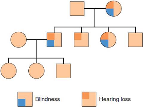
FIGURE 2-10 Maternal inheritance. An idealized pedigree illustrating maternal inheritance, which occurs in disease caused by mutations in mitochondrial DNA. Mothers transmit the mutated mtDNA to all of their offspring, but fathers do not. Variable expressivity and reduced penetrance are a consequence of different levels of heteroplasmy.
A second unique feature of diseases caused by mutations in mtDNA is the mosaic nature of the mutation within individual cells. Typically, a single cell contains 10–100 separate mtDNA molecules; in the case of an mtDNA mutation, only a fraction of the molecules carry the mutation, a situation referred to as heteroplasmy. The levels of heteroplasmy may vary considerably among different individuals and among different tissues; furthermore, a female primordial germ cell with a mixture of normal and mutated mtDNA molecules can transmit different proportions to daughter eggs (Figure 2-11). For both LHON and MERRF, levels of mutant mtDNA may vary from about 50% to about 90%; in general, the severity of the condition correlates with the extent of heteroplasmy.

FIGURE 2-11 Heteroplasmy and variable expressivity. The fraction of mutated mtDNA molecules within a cell is determined by a combination of random chance and selection at the cellular level during embryonic development. Adult tissues are mosaic for cells with different fractions of mutated mtDNA molecules, which helps to explain why mitochondrial dysfunction can produce different phenotypes and different levels of severity.
A final principle that is apparent from the pathophysiology of mitochondrial diseases is genetic interaction between the nuclear and mitochondrial genomes. One of the best examples is the sex difference in LHON, which affects four to five times as many males as females. This observation suggests that there may be a gene on the X chromosome that modifies the severity of a mitochondrial tRNALys mutation and underscores the observation that, even though mtDNA itself encodes for a set of key mitochondrial components, most mitochondrial proteins are encoded by the nuclear genome.
DOWN SYNDROME
The clinical features of Down syndrome were described over a century ago. Although the underlying cause—an extra copy of chromosome 21—has been known for more than 4 decades, the nearly complete DNA sequence of chromosome 21—some 33,546,361 base pairs—was determined only 4 years ago, and the relationship of genotype to phenotype is just beginning to be understood. Down syndrome is broadly representative of aneuploid conditions, or those that are caused by a deviation from the normal chromosome complement (euploidy). Chromosome 21, which contains a little less than 2% of the total genome, is one of the acrocentric autosomes (the others are 13, 14, 15, and 22), which means one in which nearly all the DNA lies on one side of the centromere. In general, aneuploidy may involve part or all of an autosome or sex chromosome. Most individuals with Down syndrome have 47 chromosomes (ie, one extra chromosome 21, or trisomy 21) and are born to parents with normal karyotypes. This type of aneuploidy is usually caused by nondisjunction during meiotic segregation, which means failure of two homologous chromosomes to separate (disjoin) from each other at anaphase. In contrast, aneuploid conditions that affect part of an autosome or sex chromosome must at some point involve DNA breakage and reunion. DNA rearrangements are an infrequent but important cause of Down syndrome and are usually evident as a karyotype with 46 chromosomes in which one chromosome 21 is fused via its centromere to another acrocentric chromosome. This abnormal chromosome is described as a robertsonian translocation and can sometimes be inherited from a carrier parent (Figure 2-12). Thus, Down syndrome may be caused by a variety of different karyotypic abnormalities, which have in common a 50% increase in gene dosage for nearly all of the genes on chromosome 21.

FIGURE 2-12 Mechanisms leading to Down syndrome. A pedigree in which the mother is phenotypically normal yet is a balanced carrier for a 14;21 robertsonian translocation. She transmits both the translocation chromosome and a normal chromosome 21 to her son, who also inherits a normal chromosome 21 from his father. Three copies of chromosome 21 in the son cause Down syndrome. (Adapted from Thompson MW et al. Genetics in Medicine, 5th ed. Saunders, 1991.)
Clinical Manifestations
Down syndrome occurs approximately once in every 700 live births and accounts for approximately one-third of all cases of mental retardation. The likelihood of conceiving a child with Down syndrome is related exponentially to increasing maternal age. Historically, because screening programs were offered to pregnant women older than 35 years (Figure 2-13), most children with Down syndrome have been born to women younger than 35 years. Recent advances in noninvasive prenatal testing, however, have led most obstetricians to offer prenatal testing for Down syndrome and other aneuploidies to all women. When not identified prenatally, Down syndrome is usually suspected shortly after birth from the presence of characteristic facial and dysmorphic features such as brachycephaly, epicanthal folds, small ears, transverse palmar creases, and hypotonia (Table 2-4). Approximately 50% of affected children have congenital heart defects that come to medical attention in the immediate perinatal period because of cardiorespiratory problems. Strong suspicion of the condition on clinical grounds is usually confirmed by molecular testing within 2–3 days.
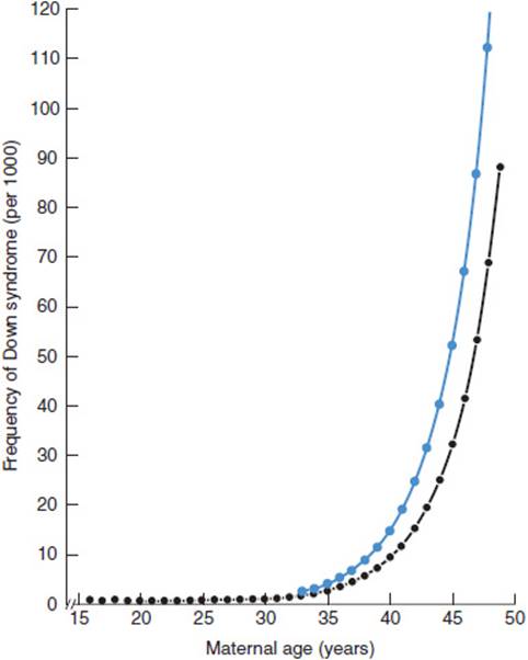
FIGURE 2-13 Relationship of Down syndrome to maternal age. The frequency of Down syndrome rises exponentially with increasing maternal age. The frequency at amniocentesis (blue symbols) is slightly higher than in live-born infants (black symbols) because miscarriages are more likely in fetuses with Down syndrome. (Data from Scriver CR et al, eds. The Metabolic and Molecular Bases of Inherited Disease, 8th ed. McGraw-Hill, 2001.)
TABLE 2-4 Phenotypic features of trisomy 21.1

A great many minor and major abnormalities occur with increased frequency in Down syndrome, yet two affected individuals rarely have the same set of abnormalities, and many single abnormalities can be seen in unaffected individuals. For example, the incidence of a transverse palmar crease in Down syndrome is about 50%, ten times that in the general population, yet most individuals in whom transverse palmar creases are the only unusual feature do not have Down syndrome or any other genetic disease.
The natural history of Down syndrome in childhood is characterized mainly by developmental delay, growth retardation, and immunodeficiency. Developmental delay is usually apparent by 3–6 months of age as failure to attain age-appropriate developmental milestones and affects all aspects of motor and cognitive function. The mean IQ is between 30 and 70 and declines with increasing age. However, there is a considerable range in the degree of mental retardation in adults with Down syndrome, and many affected individuals can live semi-independently. In general, cognitive skills are more limited than affective performance, and only a minority of affected individuals are severely impaired. Retardation of linear growth is moderate, and most adults with Down syndrome have statures 2–3 standard deviations below that of the general population. In contrast, weight growth in Down syndrome exhibits a mild proportionate increase compared with that of the general population, and most adults with Down syndrome are overweight. Although increased susceptibility to infections is a common clinical feature at all ages, the nature of the underlying abnormality is not well understood, and laboratory abnormalities can be detected in both humoral and cellular immunity.
One of the most prevalent and dramatic clinical features of Down syndrome—premature onset of Alzheimer disease—is not evident until adulthood. Although frank dementia is not clinically detectable in all adults with Down syndrome, the incidence of typical neuropathologic changes—senile plaques and neurofibrillary tangles—is nearly 100% by age 35. The major causes of morbidity in Down syndrome are congenital heart disease, infections, and leukemia. Life expectancy depends to a large extent on the presence of congenital heart disease; survival to ages 10 and 30 years is approximately 60% and 50%, respectively, for individuals with congenital heart disease and approximately 85% and 80%, respectively, for individuals without congenital heart disease.
Pathophysiology
The advent of molecular markers for different portions of chromosome 21 provided considerable information about when and how the extra chromosomal material arises in Down syndrome; and the Human Genome Project has provided a list of the approximately 230 genes found on chromosome 21. In contrast, much less is known about why increased gene dosage for chromosome 21 should produce the clinical features of Down syndrome.
For trisomy 21 (47,XX+21 or 47,XY+21), cytogenetic or molecular markers that distinguish between the maternal and paternal copies of chromosome 21 can be used to determine whether the egg or the sperm contributed the extra copy of chromosome 21. There are no obvious clinical differences between these two types of trisomy 21 individuals, which suggests that gametic imprinting does not play a significant role in the pathogenesis of Down syndrome. If both copies of chromosome 21 carried by each parent can be distinguished, it is usually possible to determine whether the nondisjunction event leading to an abnormal gamete occurred during anaphase of meiosis I or meiosis II (Figure 2-14). Studies such as these show that approximately 75% of cases of trisomy 21 are caused by an extra maternal chromosome, that approximately 75% of the nondisjunction events (both maternal and paternal) occur in meiosis I, and that both maternal and paternal nondisjunction events increase with advanced maternal age.

FIGURE 2-14 Nondisjunction has different consequences depending on whether it occurs at meiosis I or meiosis II. The abnormal gamete has two copies of a particular chromosome. When nondisjunction occurs at meiosis I, each of the copies originates from a different chromosome; however, when nondisjunction occurs at meiosis II, each of the copies originates from the same chromosome. Both cytogenetic and molecular polymorphisms can be used to determine the stage and the parent in which nondisjunction occurred. (Reproduced, with permission, from Thompson MW et al. Genetics in Medicine, 5th ed. Saunders, 1991.)
Several theories have been proposed to explain why the incidence of Down syndrome increases with advanced maternal age (Figure 2-13). Most germ cell development in females is completed before birth; oocytes arrest at prophase of meiosis I (the dictyotene stage) during the second trimester of gestation. One proposal suggests that biochemical abnormalities that affect the ability of paired chromosomes to disjoin normally accumulate in these cells over time and that, without a renewable source of fresh eggs, the proportion of eggs undergoing nondisjunction increases with maternal age. However, this hypothesis does not explain why the relationship between the incidence of trisomy 21 and advanced maternal age holds for paternal as well as maternal nondisjunction events.
Another hypothesis proposes that structural, hormonal, and immunologic changes that occur in the uterus with advanced age produce an environment less able to reject a developmentally abnormal embryo. Thus, an older uterus would be more likely to support a trisomy 21 conceptus to term regardless of which parent contributed the extra chromosome. This hypothesis can explain why paternal nondisjunction errors increase with advanced maternal age. However, it does not explain why the incidence of Down syndrome resulting from chromosomal rearrangements (see later discussion) does not increase with maternal age.
These and other hypotheses are not mutually exclusive, and it is possible that a combination of factors is responsible for the relationship between the incidence of trisomy 21 and advanced maternal age. A number of environmental and genetic factors have been considered as possible causes of Down syndrome, including exposure to caffeine, alcohol, tobacco, radiation, and the likelihood of carrying one or more genes that would predispose to nondisjunction. Although it is difficult to exclude all of these possibilities from consideration as minor factors, there is no evidence that any of these factors play a role in Down syndrome.
The recurrence risk for trisomy 21 is not altered significantly by having had previous affected children. However, approximately 5% of Down syndrome karyotypes have 46 rather than 47 chromosomes as a result of robertsonian translocations that usually involve chromosomes 14 or 22. As described, this type of abnormality is not associated with increased maternal age; however, in about 30% of such individuals, cytogenetic evaluation of the parents reveals a so-called balanced rearrangement such as 45,XX,+t(14q;21q). Because the robertsonian translocation chromosome can pair with both of its component single acrocentric chromosomes at meiosis, the likelihood of segregation leading to unbalanced gametes is significant (Figure 2-15), and the recurrence risk to the parent with the abnormal karyotype is much higher than for trisomy 21 (Table 2-5). Approximately 1% of Down syndrome karyotypes show mosaicism in which some cells are normal and some abnormal. Somatic mosaicism for trisomy 21 or other aneuploid conditions may initially arise either prezygotically or postzygotically, corresponding to nondisjunction in meiosis or mitosis, respectively. In the former case (one in which a zygote is conceived from an aneuploid gamete), the extra chromosome is then presumably lost mitotically in a clone of cells during early embryogenesis. The range of phenotypes seen in mosaic trisomy 21 is great, ranging from mild mental retardation with subtle dysmorphic features to “typical” Down syndrome, and does not correlate with the proportion of abnormal cells detected in lymphocytes or fibroblasts. Nonetheless, on average, mental retardation in mosaic trisomy 21 is generally milder than in nonmosaic trisomy 21.

FIGURE 2-15 Types of gametes produced at meiosis by a carrier of a robertsonian translocation. In a balanced carrier for a robertsonian translocation, different types of segregation at meiosis lead to several different types of gametes, including ones that are completely normal (A), ones that would give rise to other balanced translocation carriers (B), and ones that would give rise to aneuploid progeny (C).
TABLE 2-5 Risk for Down syndrome depending on parental sex and karyotype.1

Genetic Principles
A fundamental question in understanding the relationship between an extra chromosome 21 and the clinical features of Down syndrome is whether the phenotype is caused by abnormal gene expression or an abnormal chromosomal constitution. An important principle derived from studies directed at this question is that of gene dosage, which states that the amount of a gene product produced per cell is proportionate to the number of copies of that gene present. In other words, the amount of protein produced by all or nearly all genes that lie on chromosome 21 is 150% of normal in trisomy 21 cells and 50% of normal in monosomy 21 cells. Thus, unlike the X chromosome, there is no mechanism for dosage compensation that operates on autosomal genes.
Experimental evidence generally supports the view that the Down syndrome phenotype is caused by increased expression of specific genes and not by a nonspecific detrimental effect of cellular aneuploidy. Rarely, karyotypic analysis of an individual with Down syndrome reveals a chromosomal rearrangement (usually an unbalanced reciprocal translocation) in which only a very small portion of chromosome 21 is present in three copies per cell (Figure 2-16). These observations suggest that there may be a critical region of chromosome 21, which, when present in triplicate, is both sufficient and necessary to produce Down syndrome.

FIGURE 2-16 Down syndrome (DS) critical region. Rarely, individuals with Down syndrome will have chromosomal rearrangements that cause trisomy for just a portion of chromosome 21. The APP, SOD1, ETS2, CRYA1, and CBSgenes encode proteins (amyloid precursor, superoxide dismutase, the Ets2 transcription factor, crystallin, and cystathionine beta-synthase, respectively) that may play a role in the pathogenesis of Down syndrome. Analysis of two sets of individuals (indicated by the two vertical lines) suggests that the genes responsible for Down syndrome lie in the region of overlap. (Reproduced, with permission, from Thompson MW et al. Genetics in Medicine, 5th ed. Saunders, 1991.)
The idea that altered gene dosage of a group of closely linked genes can produce a distinct clinical phenotype is also supported by the observation that an increasing number of congenital anomaly syndromes have been found to be caused by so-called copy number or structural variants, often mediated by homologous segments of DNA that lie at both ends of deletion and/or insertion breakpoints. Such structural variants, which can be easily detected with molecular genetic techniques, result in an increase and/or decrease in gene copy number for one or more genes. Contiguous gene syndromes, described in Table 2-6, are generally rare, but they have played important roles in understanding the pathophysiology of aneuploid conditions.
TABLE 2-6 Phenotype and karyotype of some contiguous gene syndromes.

Carriers for robertsonian translocations that involve chromosome 21 can produce several different types of unbalanced gametes (Figure 2-15). However, the empiric risk for such a carrier bearing an infant with Down syndrome is higher than for other aneuploid conditions, in part because embryos with other types of aneuploidies are likely to result in miscarriages early in development. Thus, the consequences of trisomy for embryonic and fetal development are proportionate to the number of genes expressed to 150% of their normal levels. Because monosomy for chromosome 21 (and other autosomes) is virtually never seen in live-born infants, a similar line of reasoning suggests that a 50% reduction in gene expression is more severe than a 50% increase. Finally, female robertsonian translocation carriers exhibit much higher empiric recurrence risks than male carriers, which suggests that (1) selective responses against aneuploidy can operate on gametic as well as somatic cells and (2) spermatogenesis is more sensitive to aneuploidy than oogenesis.
CHECKPOINT
14. What are the common features of the variety of different karyotypic abnormalities resulting in Down syndrome?
15. What are the major categories of abnormalities in Down syndrome, and what is their natural history?
16. Explain why trisomy 21 is associated with such a wide range of phenotypes from mild mental retardation to that of “typical” Down syndrome.
IMPACT OF THE HUMAN GENOME PROJECT AND GENOME SEQUENCING ON PATHOPHYSIOLOGY
The major goal of the Human Genome Project is to determine the identity and gain understanding of all the genes of human beings and to apply this information to the diagnosis and treatment of human disease. An international collaboration, in which U.S. efforts were coordinated by the National Human Genome Research Institute, achieved a primary milestone in 2003 when the approximately 3 billion nucleotide human genome DNA sequence was determined.
Understanding the function of all of the genes of human beings has been facilitated by determining genome sequences for other living organisms. Some are closely related to humans on an evolutionary time scale, such as the chimpanzee, whose genome is approximately 98% the same as humans, and whose last common ancestor with humans lived approximately 6 million years ago. Others are more distantly related such as the laboratory mouse, the fruitfly, or bakers’ yeast, but nonetheless serve as valuable model organisms for experimental biologists. Even the laboratory mouse, whose last common ancestor with humans lived approximately 100 million years ago, shares more than 95% of its genes with the human genome. These considerations underscore the important genetic principle that the processes of evolution have left valuable molecular footprints that can be used to learn more about human biology.
One important advance of the Human Genome Project in the last decade has been a catalog of common human genetic variation, usually referred to as the HapMap (for Haplotype Map), in which more than 3 million SNPs have been genotyped among individuals of diverse genetic ancestry, including populations from Asia, Africa, the Americas, and Europe. Because common genetic differences are a major determinant of susceptibility to conditions such as diabetes mellitus, hypertension, obesity, and schizophrenia, a principle goal of the HapMap is to develop a molecular understanding of those determinants. Importantly, the HapMap catalog of common human genetic variation makes it possible to predict DNA sequence variation for specific segments of the genome, even when that sequence has not been measured directly. The underlying reason is that in most cases, closely linked SNPs are not independently distributed among humans but are nonrandomly associated in clusters known as haplotype blocks. For example, if two closely linked SNPs are each found at a frequency of 30%, chromosomes that carry both SNPs may exist at a frequency considerably different from 9%, which would be the prediction if the two SNPs were completely independent. This phenomenon, referred to as allelic association or linkage disequilibrium, is due to human evolutionary and population history; the extent to which new SNPs (that arise by mutation) become separated from closely adjacent SNPs (by recombination) depends on the distance between adjacent SNPs and the effects of population history on the chances for recombination.
The idea that measuring human genetic variation on a genome-wide scale could provide insight into common diseases such as hypertension, schizophrenia, and cancer underscores the perspective that there is a spectrum of genetic disease from rare conditions inherited in a mendelian fashion (which have been the major subject of this chapter) to so-called complex genetic or multifactorial conditions, for which the incidence of the disease is influenced by a combination of genes, environment, and chance. Identifying genetic components of multifactorial conditions is an important goal of the field of genetic epidemiology, in which epidemiology-based study designs are applied to populations whose familial structure is uncertain or unknown, and the measurement of SNPs in candidate genes are treated as hypothetical risk factors. For example, the epsilon 4 allele of the apolipoprotein E gene (APOE4), is found in approximately 15% of the population and increases the risk of both atherosclerosis and late-onset Alzheimer disease. However, APOE4 is just one of many genes that influence susceptibility to these important conditions, and a major goal of the HapMap is to identify and characterize those genes, both to develop new treatments, and to provide as much information as possible to physicians and their patients regarding disease susceptibility as a function of genetics.
Indeed, there is much excitement today about the potential of personalized genetic medicine, in part due to recent advances in several different areas. First, technological advances now make it possible to efficiently measure variation at millions of SNPs in individual patient samples as a routine laboratory test. These kinds of tests have been applied to thousands of individuals in so-called case-control studies, to identify particular SNPs that occur more or less frequently in cases vs. controls. Second, advances in the design and analysis of this type of approach, known as a genomewide association study (GWAS), have been very successful in identifying new genetic determinants for obesity, diabetes, inflammatory bowel disease, coronary artery disease, and other common conditions.
A second very important advance in the Human Genome Project has been the drive to develop new technological approaches for efficient and inexpensive DNA sequencing. So-called next-generation sequencing instruments use an innovative combination of molecular biologic, computational, and optical principles and have revolutionized our approach to biomedical research and medical care. The scale of technological advance is staggering: Sequencing the first human genome cost several billion dollars and required the effort of several thousand scientists over a decade; today, a single laboratory technician can sequence a genome on a benchtop instrument for a few thousand dollars.
The availability and low cost of genome sequencing are having an enormous impact on our approach to the diagnosis and pathophysiologic understanding of genetic disease. For example, the ability to compare entire genome sequences (or partial sequences of the protein-coding regions, or exomes) of individuals affected with rare syndromes is rapidly leading to the identification of mutations that cause thousands of different conditions, including recessively inherited forms of osteogenesis imperfecta, many unexplained syndromes that involve intellectual disability, and neuropsychiatric conditions such as autism. In addition, the ability to compare genome sequences of different tissues or biopsy samples from the same individual allows unprecedented insight into the pathophysiology of many cancers, identifying, for example, a catalog of DNA sequence alterations that have occurred and in some cases have helped to drive the progression of blood cancers, brain tumors, breast cancer, prostate cancer, and melanoma.
The future of genetic medicine will be greatly informed by these advances; many scientists envision that powerful but inexpensive laboratory tests that measure genetic variation across the entire genome will soon be used routinely to predict individual susceptibility to common and rare diseases and take appropriate steps to intervene and/or modify the course of those conditions. For example, individuals at high risk for certain types of cancer may benefit from aggressive screening programs.
Genetic differences may also help identify subgroups of patients whose course is likely to be more or less severe and who may respond to a particular treatment. The latter approach is part of the larger field of pharmacogenomics, in which sequence variation in the hundreds of genes that influence drug absorption, metabolism, and excretion is a major determinant of the balance between pharmacologic efficacy and toxicity. One might imagine, for example, that tests for specific nucleotide differences in a set of genes unique to a particular situation might be used to help predict the pathophysiologic response to alcoholic liver damage, type of regimen used to treat leukemia, and course of infectious diseases like tuberculosis or HIV infection.
CASE STUDIES
Yeong Kwok, MD
(See Chapter 25, p. 695 for Answers)
CASE 1
A 4-year-old boy is brought in with pain and swelling of the right thigh after a fall in the home. An x-ray film reveals an acute fracture of the right femur. Questioning of the mother reveals that the boy has had two other known fractures—left humerus and left tibia—both with minimal trauma. The family history is notable for a bone problem during childhood in the boy’s father that got better as he grew into adulthood. A diagnosis of osteogenesis imperfecta is entertained.
Questions
A. What are the four types of osteogenesis imperfecta? How are they genetically transmitted?
B. Which two types are most likely in this patient? How might they be distinguished clinically?
C. Further workup results in a diagnosis of type I osteogenesis imperfecta. What clinical features may the boy expect in adult life?
D. What is the pathogenesis of this patient’s disease?
CASE 2
A newborn girl tests positive for phenylketonuria (PKU) on a newborn screening examination. The results of a confirmatory serum test done at 2 weeks of age are also positive, establishing the diagnosis of PKU.
Questions
A. What are the metabolic defects in persons with PKU?
B. How do these defects lead to clinical disease in persons not treated with dietary restrictions appropriate for PKU?
C. What is the genetic pattern of inheritance, and what are some possible explanations for why the gene for the condition has persisted in the gene pool despite the obvious disadvantages for the affected individuals?
CASE 3
A young woman is referred for genetic counseling. She has a 3-year-old boy with developmental delay and small joint hyperextensibility. The pediatrician has diagnosed fragile X–associated mental retardation. She is currently pregnant with her second child at 14 weeks of gestation. The family history is unremarkable.
Questions
A. What is the genetic mutation responsible for fragile X–associated mental retardation? How does it cause the clinical syndrome of developmental delay, joint hyperextensibility, large testes, and facial abnormalities?
B. Which parent is the probable carrier of the genetic mutation? Explain why this parent and the grandparents are phenotypically unaffected.
C. What is the likelihood that the unborn child will be affected?
CASE 4
A 16-year-old boy presents with worsening vision for the past 2 months. He first noticed that he was having trouble with central vision in his right eye, seeing a dark spot in the center of his visual field. The dark spot had gotten larger over time, and he had also developed a central loss of vision in his left eye. Two of his maternal uncles had loss of vision, but his mother and another maternal uncle and two maternal aunts had no visual difficulties. No one on his father’s side was affected. Physical examination reveals microangiopathy and vascular tortuosity of the retina. Genetic testing confirms the diagnosis of Leber hereditary optic neuropathy.
Questions
A. What is the central defect in Leber hereditary optic neuropathy (LHON)?
B. How is this disorder inherited, and what is the principle of heteroplasmy?
C. What explains the fact that males are much more likely to be affected than females?
CASE 5
A 40-year-old woman, recently married and pregnant for the first time, comes to the clinic with a question about the chances of having “a Down syndrome baby.”
Questions
A. What is the rate of occurrence of Down syndrome in the general population? What are some of the common clinical features?
B. What major genetic abnormalities are associated with Down syndrome? How might these abnormalities lead to the clinical features of the syndrome?
C. How might this woman’s age contribute to her risk of having a child with Down syndrome?
REFERENCES
Osteogenesis Imperfecta
Byers PH et al. Recessively inherited forms of osteogenesis imperfecta. Annu Rev Genet. 2012;46:475–97. [PMID: 23145505]
Forlino A et al. New perspectives on osteogenesis imperfecta. Nat Rev Endocrinol. 2011 Jun 14;7(9):540–57. [PMID: 21670757]
Ishikawa Y et al. A molecular ensemble in the rER for procollagen maturation. Biochim Biophys Acta. 2013 Apr 18. 2013 Nov;1833:2479–91. [PMID: 23602968]
Phenylketonuria
Flydal M et al. Phenylalanine hydroxylase: function, structure, and regulation. IUBMB Life. 2013 Apr;65(4):341–9. [PMID: 23457044]
Giovannini M et al. Phenylketonuria: nutritional advances and challenges. Nutr Metab (Lond). 2012 Feb 3;9(1):7. [PMID: 22305125]
Hartnett C et al. Long-term outcomes of blood phenylalanine concentrations in children with classical phenylketonuria. Mol Genet Metab. 2013 Apr;108(4):255–8. [PMID: 23465864]
Prick BW et al. Maternal phenylketonuria and hyperphenylalaninemia in pregnancy: pregnancy complications and neonatal sequelae in untreated and treated pregnancies. Am J Clin Nutr. 2012 Feb;95(2):374–82. [PMID: 22205310]
Fragile X–Associated Mental Retardation
Bhakar AL et al. The pathophysiology of fragile X (and what it teaches us about synapses). Annu Rev Neurosci. 2012;35:417–43. [PMID: 22483044]
Lee HY et al. Fragile X syndrome: mechanistic insights and therapeutic avenues regarding the role of potassium channels. Curr Opin Neurobiol. 2012 Oct;22(5):887–94. [PMID: 22483378]
Mclennan Y et al. Fragile X syndrome. Curr Genomics. 2011 May;12(3):216–24. [PMID: 22043169]
Wang T et al. New perspectives on the biology of fragile X syndrome. Curr Opin Genet Dev. 2012 Jun;22(3):256–63. [PMID: 22382129]
LHON, MERFF, & Mitochondrial Diseases
Davis RL et al. The genetics of mitochondrial disease. Semin Neurol. 2011 Nov;31(5):519–30. [PMID: 22266889]
Finsterer J. Inherited mitochondrial disorders. Adv Exp Med Biol. 2012;942:187–213. [PMID: 22399423]
Kirches E. LHON: mitochondrial mutations and more. Curr Genomics. 2011 Mar;12(1):44–54. [PMID: 21886454]
Down Syndrome
Haydar TF et al. Trisomy 21 and early brain development. Trends Neurosci. 2012 Feb;35(2):81–91. [PMID: 22169531]
Letourneau A et al. Genomic determinants in the phenotypic variability of Down syndrome. Prog Brain Res. 2012;197:15–28. [PMID: 22541286]
Mersy E et al. Noninvasive detection of fetal trisomy 21: systematic review and report of quality and outcomes of diagnostic accuracy studies performed between 1997 and 2012. Hum Reprod Update. 2013 Jul–Aug;19(4):318–29. [PMID: 23396607]
The Human Genome Project & Human Genetic Variation
Alkan C et al. Genome structural variation discovery and genotyping. Nat Rev Genet. 2011 May;12(5):363–76. [PMID: 21358748]
Bamshad MJ et al. Exome sequencing as a tool for Mendelian disease gene discovery. Nat Rev Genet. 2011 Sep 27;12(11):745–55. [PMID: 21946919]
Connolly JJ et al. The impact of genomics on pediatric research and medicine. Pediatrics 2012 Jun;129(6):1150–60. [PMID: 22566424]
Gonzaga-Jauregui C et al. Human genome sequencing in health and disease. Annu Rev Med. 2012;63:35–61. [PMID: 22248320]
Shendure J et al. The expanding scope of DNA sequencing. Nat Biotechnol. 2012 Nov;30(1):1084–94. [PMID: 23138308]
Vandeweyer G et al. Detection and interpretation of genomic structural variation in health and disease. Expert Rev Mol Diagn. 2013 Jan;13(1):61–82. [PMID: 23256704]