
Melissa M. Meier, MD, & Timothy H. McCalmont, MD
NORMAL SKIN
The skin is the most accessible organ of the human body. Its most basic function is simply a protective one. As a barrier, the skin holds off desiccation and disease by keeping moisture in and pathogens out. Nevertheless, characterization of the skin as mere “plastic wrap” is a gross underestimation of the anatomic and physiologic complexity of this vital structure.
Unlike parenchymal organs, end-organ dysfunction or failure is not a prerequisite for the diagnosis of a skin disease, because all skin diseases can be observed clinically irrespective of their functional effects. Among the spectacular array of neoplastic, inflammatory, infectious, and genetic cutaneous disorders, some elicit only trivial aberrations in skin structure or function, whereas others lead to profound and morbid consequences.
ANATOMY
The integumentary system consists of a layer of tissue, 1–4 mm thick, that covers all exposed surfaces of the body. The skin merges uninterruptedly with the structurally similar envelope of the mucous membranes, but skin is distinct from mucosa in that it contains adnexal structures such as the eccrine units that exude sweat and the folliculosebaceous units that produce hairs and oils. There is considerable variation in skin thickness and composition, depending on the requirements of a particular body site. For example, the thinnest skin overlies the eyelids, where delicacy and mobility are essential. The thickest skin is present on the upper trunk, where sturdiness exceeds mobility in importance. The surfaces of the palms and soles are characterized by a high density of eccrine sweat units, reflecting the importance of this region in regulation of temperature; an absence of hairs, which would interfere with sensation; and accentuation of the cornified layer (see later discussion), contributing to the tackiness needed to handle objects deftly. The size of the structures between sites can also vary greatly, best illustrated by the contrast between large terminal hair follicles found on the scalp, bearded areas, and genital skin and the small vellus hair follicles found at most other sites.
HISTOLOGY
Using a light microscope, two important skin layers are easily identifiable: a stratified squamous epithelium, the epidermis; and a layer of connective tissue, the dermis. The subjacent adipose tissue is considered as a third layer by some and is referred to as the subcutis.
The epidermis consists of keratinocytes arrayed in four distinct substrata: the basal, spinous, granular, and cornified layers (Figure 8-1). Basal keratinocytes include the proliferative pool of keratinocytes. These cells divide, giving rise to progeny that are displaced toward the skin surface. As the keratinocytes move outwardly, they progressively flatten and accumulate keratin filaments within their cytoplasm. Individual keratinocytes are tightly bound together by intracellular junctions called desmosomes (Figure 8-2). The desmosomal junctions appear as delicate “spines” between cells in conventional microscopic sections and are most conspicuous in the epidermal spinous layer (Figure 8-3). Keratin filaments are linked intracellularly and are also attached to the desmosomes, forming a network that is vital to structural integrity.

FIGURE 8-1 Although the epidermis biologically displays a gradient of differentiation, four distinct layers are recognized on the basis of microscopic appearance. Cuboidal germinative keratinocytes serve as a foundation in the basal layer; cells with ample cytoplasm and prominent desmosomes constitute the spinous layer; cells with cytoplasmic granularity resulting from an accumulation of keratin complexes and other structural proteins are found in the granular layer; and anucleate, flattened keratinocytes comprise the tough, membrane-like cornified layer. (Redrawn, with permission, from Orkin M et al, eds. Dermatology. Originally published by Appleton & Lange. Copyright © 1991 by The McGraw-Hill Companies, Inc.)

FIGURE 8-2 In an ultrastructural view of a human keratinocyte (A), numerous desmosomes (B) appear as plaques that tightly bind two cell membranes together. With very high magnification (C), the attachment of cytoplasmic keratin filaments (F) to the desmosomes can be appreciated. (Reproduced, with permission, from Junqueira LO et al. Basic Histology, 10th ed. McGraw-Hill, 2003.)

FIGURE 8-3 With conventional light microscopy, the numerous desmosomes of the spinous layer appear as delicate attachments (“spines”) between individual keratinocytes.
Melanocytes and Langerhans cells are dendritic cells that are intercalated among the keratinocytes of the epidermis. Melanocytes, which are positioned in the basal layer, synthesize a reddish-brown biochrome, melanin, and dispense it to numerous adjacent keratinocytes through their dendrites (Figure 8-4). This distribution system permits melanin to provide a dispersed screen against the potentially harmful ultraviolet rays of the sun. Langerhans cells share a similar arborized morphology but are positioned in the midspinous layer. Langerhans cells are bone marrow–derived antigen-presenting cells (see also Chapter 3).

FIGURE 8-4 The human melanocyte displays a branching morphology, and the dendrites of the cell maintain contact with 35–40 adjacent keratinocytes in a multicellular structure termed the epidermal melanin unit. The function of the unit is the effective dispersion of melanin pigment, packaged in organelles known as melanosomes, across a broad surface area. (Redrawn, with permission, from Junqueira LO et al. Basic Histology, 10th ed. McGraw-Hill, 2003.)
The epidermal-dermal junction, or basement membrane zone, is a structure that welds the epidermis to the dermis and contributes to the skin barrier. The juncture of the epidermis and dermis is arrayed in an undulating fashion to increase the surface area of binding between the two structures and to resist shearing forces. The downward projections of the epidermis are referred to as rete ridges, and the upward projections of the superficial dermis are called dermal papillae (Figure 8-5). Although the basement membrane comprises a thin eosinophilic (pink) band beneath the basal cells in conventional microscopic sections, it has a sophisticated multilayered structure that reaches from the hemidesmosomes of the basal keratinocytes to the collagen bundles of the superficial dermis (Figure 8-6). The lamina densa and lamina lucida are two of the layers of the basement membrane zone and are so named because of their electron-dense and electron-lucid appearance when viewed ultrastructurally.

FIGURE 8-5 The undulating configuration of the epidermal-dermal junction consists of downward extensions of the epidermis, known as rete ridges, and upward extensions of the dermis, known as dermal papillae.

FIGURE 8-6 Schematic diagram of the basement membrane zone of human epidermis. (Redrawn, with permission, from Orkin M et al, eds. Dermatology. Originally published by Appleton & Lange. Copyright © 1991 by The McGraw-Hill Companies, Inc.)
The dermis consists of a connective tissue gel composed largely of proteins and mucopolysaccharides (so-called ground substance). This matrix serves as the scaffolding that supports the complex neurovascular networks, which course through the skin, and also supports the eccrine (sweat gland) and follicular (hair) adnexal structures. The vast majority of the fibrous structural proteins of the dermis are composed of collagen types I and III, and a network of elastic microfibrils is also woven throughout the full dermal thickness. Fibrocytes, the synthetic units of the structural proteins, are ubiquitous, and there are also mast cells and dendritic immune cells arrayed throughout the dermis. A discussion of the fine structure of the dermis—the dermal vascular and neural networks and the adnexal structures—is beyond the scope of this chapter.
OVERVIEW OF SKIN DISEASES
In the broadest and simplest sense, there are two types of skin diseases: growths and rashes. A skin growth is a cyst, a malformation, or a benign or malignant neoplasm, something that presents clinically as a bump on the skin. A rash is, with rare exception, a nonneoplastic skin disease; it is more precisely referred to as an inflammatory skin condition or a dermatitis. The pathophysiologic aspects of the huge number of described growths and rashes exceed the scope of this chapter, and our discussion will focus on nine prototypical rashes.
TYPES OF SKIN LESIONS
Physicians interested in the skin learned decades ago that the accurate diagnosis and classification of the many patterns of dermatitis were dependent on a standardized nomenclature for the description and documentation of rashes. When used in association with a few well-chosen adjectives, the terms used to describe the prototypical types of inflammatory skin lesions (so-called primary lesions) permit vivid description of a rash. To illustrate the importance of terminology, imagine trying to describe a patient’s condition over the telephone to another physician. Talking about a red raised rash may truthfully describe the eruption in some sense, but the mental image evoked could be any one of dozens of skin diseases. The only way to accurately characterize an eruption is by the use of precisely defined terms.
The most important types of primary lesions include macules and patches, papules and plaques, vesicles and bullae, pustules, and nodules. The terms macule and patch denote flat areas of discoloration without any discernible change in texture. Macules are 1 cm or less in diameter, whereas patches exceed 1 cm in size. Papules and plaques are elevated, palpable skin lesions in which the breadth of the lesion exceeds its thickness. A papule is small, 1 cm or less in diameter, whereas a plaque exceeds 1 cm in size. Vesicles and bullae are fluid-filled spaces within the skin. Vesicles are less than 1 cm in diameter, whereas bullae exceed 1 cm in size. A vesicle or bulla containing purulent fluid is known as a pustule. A nodule is a solid, rounded skin lesion in which diameter and thickness are roughly equal.
TYPES OF INFLAMMATORY SKIN DISEASES
Different inflammatory processes involve different structures within the skin and display different microscopic patterns. Experience has shown that pattern analysis can serve as a useful means of diagnosis and classification. Pattern analysis is dependent on accurate recognition of the distribution of inflammation within the skin as well as recognition of the specific structures affected by the inflammatory reaction. There are nine distinct patterns of dermatitis (Table 8-1; Figure 8-7). Eight of these patterns and some of the diseases that produce them are discussed in detail next.
TABLE 8-1 Patterns of inflammatory skin disease.


FIGURE 8-7 Nine patterns of inflammatory skin disease. (See also Table 8-1.)
CHECKPOINT
1. What are the two most basic barrier functions of skin?
2. How is skin distinct from mucosa? Why are these terms important?
3. What are the main primary lesions? Why are these terms important?
4. What are the main patterns of inflammatory skin disease?
5. What is the value of knowing the microscopic pattern of inflammation of a skin lesion? What additional information is needed for this information to be most useful?
PATHOPHYSIOLOGY OF SELECTED SKIN DISEASES
PATTERN: PSORIASIFORM DERMATITIS
Example: Psoriasis
Clinical Presentation
Psoriasis is a common chronic, persistent or relapsing, scaling skin condition. Individual lesions are distinctive in their classic form: sharply marginated and erythematous and surmounted by silvery scales (Figure 8-8). Most patients with psoriasis have a limited number of fixed plaques, but there is great variation in clinical presentation.

FIGURE 8-8 Classic plaque-type psoriasis (psoriasis vulgaris)consisting of sharply marginated scaling plaques. (Image used with permission from Dr. Timothy Berger.)
Epidemiology and Etiology
Psoriasis affects between 1% and 2% of individuals of both sexes in most ethnic groups. The most common age at onset is the third decade, but psoriasis can develop soon after birth, and psoriasis of new onset has been documented in a centenarian.
Several lines of evidence have established that genetic factors contribute to the development of psoriasis. There is a high rate of concordance for psoriasis in monozygotic twins and an increased incidence in relatives of affected individuals. The gene products of specific class I alleles of the major histocompatibility complex (MHC) are overexpressed in patients with psoriasis. Psoriasis is not merely a genetic disorder, however, because some susceptible individuals never develop characteristic lesions. In other predisposed individuals, a number of environmental factors, including infection, physical injury, stress, and drugs, can serve as triggers for the development of psoriasis (Table 8-2).
TABLE 8-2 Factors that induce or exacerbate psoriasis.

Histopathology and Pathogenesis
Psoriasis is the prototypical form of psoriasiform dermatitis, a pattern of inflammatory skin disease in which the epidermis is thickened as a result of elongation of rete ridges (Figures 8–7 and 8–9). In psoriatic lesions, epidermal thickening reflects excessive epidermopoiesis (epidermal proliferation). The increase in epidermopoiesis is reflected in shortening of the duration of the keratinocyte cell cycle and doubling of the proliferative cell population. Because of these alterations, lesional skin contains up to 30 times as many keratinocytes per unit area as normal skin. Evidence of excessive proliferation is also manifest microscopically as numerous intraepidermal mitotic figures.

FIGURE 8-9 Histopathologic features of psoriasis at low magnification. The rete ridges are strikingly and evenly elongated, and the overlying cornified layer contains cells with retained nuclei (parakeratosis), a pattern that reflects the increased epidermal turnover.
During normal keratinocyte maturation, nuclei are eliminated as cells enter the cornified layer and condense to form a semipermeable envelope. In psoriasis, the truncation of the cell cycle leads to an accumulation of cells within the cornified layer with retained nuclei, a pattern known as parakeratosis. As parakeratotic cells accumulate, neutrophils migrate to the cornified layer. Histopathologically, the silvery scale of psoriatic plaques consists of a thick layer of parakeratotic keratinocytes with numerous intercalated neutrophils. At times, the number of neutrophils in the stratum corneum is so great that lesions assume a pustular appearance.
Psoriasis also induces endothelial cell hyperproliferation that yields pronounced dilation, tortuosity, and increased permeability of capillaries in the superficial dermis (Figure 8-10). The vascular alterations contribute to the bright erythema seen clinically. The capillary changes are most pronounced at the advancing margins of psoriatic plaques.

FIGURE 8-10 In a psoriatic plaque at high magnification, dilated capillaries are evident in an edematous portion of the superficial dermis.
After years of research, a large number of immunologic abnormalities that involve both innate and adaptive immunity have been documented in psoriatic skin. Antigenic stimuli are thought to activate the innate immune response, leading to the production of cytokines, such as interferon, tumor necrosis factor (TNF), interleukin-23 (IL-23), and IL-12, by macrophages, dendritic cells, and neutrophils. This leads to attraction, activation, and differentiation of T cells. These T cells, most importantly T helper 1 and T helper 17 cells, produce cytokines that lead to epidermal hyperplasia, recruitment of inflammatory cells, and ultimately a positive feedback loop that perpetuates the pathologic process.
Clinical Manifestations
The cardinal features of the plaques of psoriasis include sharp margination, bright erythema, and nonconfluent whitish or silvery scales. Lesions can occur at any site, but the scalp, the extensor surfaces of the extremities, and the flexural surfaces are often involved. Psoriasis commonly affects the nail bed and matrix, yielding pitted or markedly thickened dystrophic nails. Mucosal surfaces are spared.
The only extracutaneous manifestation of psoriasis is psoriatic arthritis, a deforming, asymmetric, oligoarticular arthritis that can involve small or large joints. The distal interphalangeal joints of the fingers and toes are characteristically involved. Psoriatic arthritis is classified as one of the seronegative spondyloarthropathies, distinguishable from rheumatoid arthritis by a lack of circulating autoantibodies (so-called rheumatoid factors) or circulating immune complexes and by linkage with specific MHC class I alleles, including HLA-B27.
There are many variants of psoriasis, all of which are histopathologically similar but which differ greatly in clinical distribution (Table 8-3).
TABLE 8-3 Variants of psoriasis.

CHECKPOINT
6. What evidence supports a genetic role in the development of psoriasis? An environmental role?
7. Which cell types hyperproliferate in psoriasis?
8. What immunologic defects have been identified in psoriasis?
PATTERN: INTERFACE DERMATITIS
Example: Lichen Planus
Clinical Presentation
Lichen planus is a distinctive itchy eruption that usually presents with numerous small papules. Individual lesions have angulate borders, flat tops, and a violaceous hue, attributes that form the basis of their alliterative description as pruritic polygonal purple papules (Figure 8-11). The individual papules of lichen planus sometimes coalesce to form larger plaques. Minute whitish streaks, barely visible to the naked eye and known as Wickham striae, are characteristically found on the surfaces of lesions.

FIGURE 8-11 Pruritic polygonal flat-topped papules of lichen planus.
Epidemiology and Etiology
Lichen planus generally develops in adulthood and affects women slightly more commonly than men. Although the factors that trigger lichen planus remain obscure in many patients, it is clear that the rash represents a cell-mediated immune reaction that directly or indirectly damages basal keratinocytes of the epidermis. Observations that suggest a cell-mediated mechanism include the occurrence of lichen planus-like eruptions as a manifestation of graft-versus-host disease after bone marrow transplantation and the development of a lichen planus–like eruption in mice after injection of sensitized, autoreactive T cells. Although most lichen planus is idiopathic, drugs are one established cause of lichen planus or lichen planus–like reactions. Therapeutic gold and antimalarial agents are the medications most closely linked to the development of lichenoid eruptions, but a long list of other agents has accumulated (Table 8-4).
TABLE 8-4 Medications that induce lichenoid (lichen planus–like) reactions.

Histopathology and Pathogenesis
Lichen planus is a form of lichenoid interface dermatitis, a type of inflammatory skin disease in which a dense infiltrate of lymphocytes occupies the papillary dermis and the superficial dermis immediately subjacent to the epidermis, in association with vacuolization of the lower epidermis (Figure 8-12). The papillary dermal infiltrate is composed largely, if not entirely, of T lymphocytes. Some of the T cells are also found within the epidermis, where adjacent vacuolated, injured keratinocytes are found. Dense eosinophilic (pink) globules, known as colloid bodies, are also identifiable within the epidermis and the infiltrate (Figure 8-13). Colloid bodies represent condensed, anucleate keratinocytes that have succumbed to the inflammatory reaction. Although the keratinocytes bear the brunt of the lymphocyte attack, melanocytes may be coincidentally destroyed in the reaction as “innocent bystanders.” Free melanin pigment is released as melanocytes are damaged, and the pigment is phagocytosed by dermal macrophages known as melanophages.

FIGURE 8-12 Histopathologic features of lichen planus at low magnification. There is a bandlike infiltrate of lymphocytes that impinges on the epidermal-dermal junction, and some keratinocytes adjacent to the infiltrate show cytoplasmic vacuolization.

FIGURE 8-13 Necrotic keratinocytes (so-called colloid bodies) in a lesion of lichen planus appear as rounded globules along the epidermal-dermal junction.
In incipient lesions of lichen planus, CD4 helper T lymphocytes predominate, and some of the cells have been found in proximity to macrophages and Langerhans cells (see also Chapter 3). In contrast, CD8 cytotoxic T cells comprise the bulk of the infiltrate in mature lesions. This shift in infiltrating T-cell composition is thought to reflect the afferent and efferent aspects of lesional development. In the afferent phase, causative antigens are processed and presented to helper T cells, probably in the context of specific HLA determinants. The stimulated CD4 lymphocytes then elaborate specific cytokines that lead to the recruitment of cytotoxic lymphocytes. Cell-mediated cytotoxicity and cytokines such as interferon-α and TNF are then thought to contribute to the vacuolization and necrosis of keratinocytes as a secondary event.
The clinical appearance of lichen planus lesions reflects several synchronous alterations in the skin. The dense array of lymphocytes in the superficial dermis yields the elevated, flat-topped appearance of each papule or plaque. The chronic inflammatory reaction induces accentuation of the cornified layer (hyperkeratosis) of the epidermis, which contributes to the superficial whitish coloration perceived as Wickham striae. Although the many melanophages that accumulate in the papillary dermis hold a brownish black pigment, the fact that the pigmented cells are embedded in a colloidal matrix such as the skin permits extensive scattering of light, a phenomenon known as the Tyndall effect. Thus, the human eye interprets a lesion of lichen planus as dusky or violaceous despite the fact that the pigment that serves as the basis for the coloration is melanin.
Clinical Manifestations
Lichen planus affects both skin and mucous membranes. Papules are generally distributed bilaterally and symmetrically. The sites most commonly involved include the flexor surfaces of the extremities, the genital skin, and the mucous membranes. Rarely, lichen planus may involve the mucosa of internal organs, such as the esophagus. Cutaneous lesions are virtually never seen on the palms, soles, or face.
In general, lichen planus variants can be grouped into three categories.
A. Lichen Planus Papules Arrayed in an Unusual Configuration— In these variants, typical individual papules of lichen planus are grouped in a distinctive larger pattern. In annular lichen planus, small lichenoid papules coalesce to form a larger ring. Linear and zosteriform patterns of lichen planus have also been observed. When lichen planus presents in an unusual configuration, it is prone to be underdiagnosed or misdiagnosed.
B. Lichen Planus Papules Arrayed at Distinctive Sites— Although most lichen planus is widespread, at times papules are restricted to a specific body site, such as the mouth (oral lichen planus) or genitalia. Nearly 25% of all lichen planus patients have disease limited to the mucous membranes.
C. Lichen Planus Papules with Unusual Clinical Morphology—Some examples of lichen planus defy clinical recognition because the appearance of the individual lesions is atypical. Erosive, vesiculobullous, atrophic, and hypertrophic lesions can be seen. In erosive lichen planus, the interface reaction that is directed against the epidermis is so profound that the entire epidermis becomes necrotic and ulceration ensues. The closely related entity vesiculobullous lichen planus is also characterized by an intense interface reaction that yields necrosis of the epidermal junctional zone across a broad front. As a result of basal layer necrosis, the epidermis becomes detached from its dermal attachments and a blister develops. In atrophic lichen planus, the rate of destruction of keratinocytes by the lichenoid interface reaction exceeds the rate of epidermal regeneration, and the epidermis becomes attenuated as a result. In contrast, in hypertrophic lichen planus, the rate of epidermal regeneration triggered by the interface reaction exceeds the rate of destruction, and thick, verrucous, hyperkeratotic lesions develop. All of these variants are histopathologically similar with the exception of the foci of ulceration seen in erosive lichen planus.
CHECKPOINT
9. What skin cells are damaged by cell-mediated immune reactions in lichen planus?
10. Which drugs have been most commonly implicated in licheniform eruptions?
11. What synchronous alterations in the skin are reflected in the clinical appearance of lichen planus?
Example: Erythema Multiforme
Clinical Presentation
Erythema multiforme is an acute cutaneous eruption that presents with a wide spectrum of clinical severity. The eruption is commonly brief and self-limited, but repetitive or generalized attacks can be disabling or even life threatening. As the name implies, variation in lesional morphology can be seen, but most patients present with a monomorphous pattern in a given bout. The prototypical lesion is a red macule or thin papule that expands centrifugally and develops a dusky or necrotic center, creating a target-like pattern (Figure 8-14).

FIGURE 8-14 Target lesions—a characteristic pattern seen in erythema multiforme—consist of a papule or plaque with a central zone of epidermal necrosis surrounded by a rim of erythema. (Image used with permission from Dr. Timothy Berger.)
Epidemiology and Etiology
Erythema multiforme is an uncommon but distinctive skin disease that afflicts men and women in nearly equal numbers. The peak incidence is in the second to fourth decades of life, and onset during infancy or early childhood is a rarity. Like lichen planus, erythema multiforme represents a cell-mediated immune reaction that eventuates in necrosis of epidermal keratinocytes. Herpes simplex viral infection and reactions to medications have been established as the most common causes of erythema multiforme. Other known causes include Mycoplasma infection, contact dermatitis, drugs, and radiation.
Histopathology and Pathogenesis
Erythema multiforme is a prototypical form of vacuolar interface dermatitis. In contrast to lichen planus, which typically presents with a dense obscuring lichenoid infiltrate within the superficial dermis, in erythema multiforme the inflammatory infiltrate is sparse. Thus, the vacuolated keratinocytes that are widely distributed within the epidermal basal layer are conspicuous in the face of a sparse infiltrate, and the damaged keratinocytes serve as the basis for the name of this pattern of inflammatory skin disease.
The dermal infiltrate in erythema multiforme is composed of a mixture of CD4 and CD8 T lymphocytes. CD8 cytotoxic cells are also found within the epidermis, in proximity to vacuolated and necrotic keratinocytes. Keratinocytes that are killed in the course of the inflammatory reaction become anucleate and are manifest microscopically as round, dense, eosinophilic bodies similar to the colloid bodies of lichen planus (Figure 8-15).

FIGURE 8-15 Histopathologic features of erythema multiforme, a type of vacuolar interface dermatitis. There is a modest infiltrate of lymphocytes in the vicinity of the epidermaldermal junction where vacuolated and necrotic keratinocytes are conspicuous.
Although lichen planus and erythema multiforme are clinically, microscopically, and etiologically distinct, both appear to share a common pathogenetic pathway in which specific inciting agents recruit effector lymphocytes into the epidermis and papillary dermis. After this recruitment, keratinocytes are injured and killed by the combined negative influences of cytotoxicity and cytokines, such as interferon-? and TNF.
Many cases of so-called erythema multiforme minor are triggered by herpes simplex viral infection. A relationship between erythema multiforme and herpetic infection had long been suspected based on the documentation of preceding herpes simplex lesions in patients with erythema multiforme. The relationship was strengthened after antiherpetic drug therapy, in the form of oral acyclovir, was shown to suppress the development of erythema multiforme lesions in some individuals. Molecular studies have substantiated the relationship by confirming the presence of herpes simplex DNA within skin from erythema multiforme lesions. Herpesvirus DNA is also demonstrable within peripheral blood lymphocytes and within lesional skin after resolution but not within nonlesional skin. These findings suggest that viral DNA is disseminated from the primary infection in the peripheral blood and becomes integrated into the skin at specific target sites. The herpetic genomic fragments then contribute to the development of a cytotoxic effector response in their chosen target tissue, the skin.
The target-like clinical appearance of many erythema multiforme lesions reflects zonal differences in the intensity of the inflammatory reaction and its deleterious effects. At the periphery of an erythema multiforme lesion, only sparse inflammation, slight edema, and subtle vacuolization of the epidermis are apparent in the outer erythematous halo. In contrast, the dusky “bull’s eye” often shows pronounced epidermal vacuolization, with areas of near-complete epidermal necrosis.
Clinical Manifestations
Erythema multiforme is generally limited to the skin and mucous membranes. The lesions develop rapidly in crops and are initially distributed on acral surfaces, although proximal spread to the trunk and face occurs not uncommonly. Mucosal erosions and ulcers are seen in roughly 25% of cases, and mucositis can be the sole presenting feature of the disease. Although erythema multiforme is an epithelial disorder, nonspecific constitutional symptoms such as malaise can also occur.
Although the spectrum of erythema multiforme exists as a continuum, a given patient is usually classified as having minor or major disease. The disorder is referred to as erythema multiforme minor when there are scattered lesions confined to the skin or when skin lesions are observed in association with limited mucosal involvement. A diagnosis of erythema multiforme major is based on the presence of prominent involvement of at least two of three mucosal sites: oral, anogenital, or conjunctival. Many examples of erythema multiforme major also display severe, widespread cutaneous involvement. Although Stevens-Johnson syndrome had classically been used to describe severe cases of erythema multiforme, consensus classification has separated Stevens-Johnson syndrome from erythema multiforme and added it to the spectrum of toxic epidermal necrolysis. These two entities, Steven-Johnson syndrome and toxic epidermal necrolysis, are now considered to represent variant dermatologic manifestations of severe idiosyncratic reactions. Most often the result of medications, these entities involve vast regions of skin and mucosal necrosis (Figure 8-16) with secondary vesiculation. Pathologically, the findings are similar to those of a severe burn in that the integrity of a patient’s skin fails, resulting in an increased risk of infectious and metabolic sequelae.

FIGURE 8-16 Toxic epidermal necrolysis. Generalized maculopapular erythema of the trunk and extremities is followed by extensive desquamation, as illustrated on this patient’s trunk, resulting from epidermal necrosis. Patients are often admitted to a burn unit for acute care. (Image used with permission from Dr. Timothy Berger.)
CHECKPOINT
12. What is the prototypical lesion in erythema multiforme?
13. In what ways is erythema multiforme similar to and different from lichen planus?
14. What are some complications of toxic epidermal necrolysis?
PATTERN: VESICULOBULLOUS DERMATITIS
Example: Bullous Pemphigoid
Clinical Presentation
Bullous pemphigoid is a blistering disease in which tense fluid-filled spaces develop within erythematous, inflamed skin. The blisters in bullous pemphigoid develop because of detachment of the epidermis from the dermis (subepidermal vesiculation) as the result of a specific inflammatory reaction directed against structural proteins. The term “pemphigoid” reflects the clinical similarity of bullous pemphigoid to pemphigus, another form of blistering skin disease that is characterized by intraepidermal rather than subepidermal vesiculation. The distinction between bullous pemphigoid and pemphigus is an important one, because bullous pemphigoid has a more favorable prognosis.
Epidemiology and Etiology
Bullous pemphigoid is generally a disorder of the elderly. There are rare reports of bullous pemphigoid in children and young adults, but the vast majority of patients are older than 60 years. There is no sex predilection.
It has been known for years that immunoglobulins and complement are deposited along the epidermal-dermal junction in bullous pemphigoid. The deposited antibodies are specific for antigens within the basement membrane zone (BP180 and BP230), and bullous pemphigoid thus represents a form of autoimmune skin disease. The specific factors that induce autoantibody production have not been identified.
Histopathology and Pathogenesis
Microscopically, biopsies from fully developed bullous pemphigoid lesions show a subepidermal cleft containing lymphocytes, eosinophils, and neutrophils as well as eosinophilic (pink) material that represents extravasated macromolecules such as fibrin (Figure 8-17). An inflammatory infiltrate of eosinophils, neutrophils, and lymphocytes is also evident in the dermis beneath the cleft. These findings represent the aftermath of an inflammatory reaction centered on the basement membrane zone.

FIGURE 8-17 Histopathologic features of bullous pemphigoid. There is a subepidermal cleft that contains numerous eosinophils and lymphocytes, and a similar infiltrate is present in the superficial dermis. Ultrastructurally, the separation is within the lamina lucida of the basement membrane zone, at the level of the bullous pemphigoid antigen (see Figure 8-6 ).
Insights into this reaction can be obtained from direct immunofluorescence microscopy, in which fluorochrome-labeled anti-immunoglobulin G (IgG), anti-IgA, anti-IgM, and anticomplement antibodies are incubated with lesional skin. Using an ultraviolet microscope to localize the fluorochrome, tagged antibodies that are specific for IgG and complement component C3 are found in a linear distribution along the epidermal-dermal junction (Figure 8-18). Circulating IgG that binds to the basement membrane zone of human epidermis is also identifiable in bullous pemphigoid patients. These antibodies are capable of complement fixation, and pathogenicity has been confirmed by injection into laboratory animals, in whom the antibodies bind to the junctional zone and induce blisters.

FIGURE 8-18 Direct immunofluorescence findings in lesional skin from a bullous pemphigoid patient. When fluorochrome-stained sections are viewed through an ultraviolet microscope, a bright linearn band, signifying deposition of immunoglobulin G, is evident along the epidermal-dermal junction. (Image used with permission from Dr. Kari Connolly.)
The autoantibodies (IgG) in bullous pemphigoid are directed against hemidesmosomal proteins, namely bullous pemphigoid antigen 180 and bullous pemphigoid antigen 230. The binding of these autoantibodies to the basement membrane zone leads to an inflammatory cascade with activation of the classic complement cascade (Chapter 3). Complement fragments induce mast cell degranulation and attract neutrophils. The presence of eosinophils in the infiltrate of bullous pemphigoid is probably a reflection of mast cell degranulation, because mast cell granules contain eosinophil chemotactic factors. Numerous enzymes are released by granulocytes and mast cells during the reaction, and enzymatic digestion is thought to be the primary mechanism behind the separation of the epidermis from the dermis with formation of tense bullae. It is also possible that the bullous pemphigoid antigen plays a vital structural role that is compromised by autoantibody binding, leading to cleavage. Quantifiable titers of bullous pemphigoid antigen correlate to disease activity.
Clinical Manifestations
Patients with bullous pemphigoid present with large, tense blisters on an erythematous base (Figure 8-19). Lesions are most commonly distributed on the extremities and lower trunk, but blisters can develop at any site. Most patients experience considerable pruritus in association with their blisters, possibly triggered by the many eosinophils in the dermal infiltrate. Mucous membrane lesions develop in up to one-third of patients and are usually clinically innocuous.

FIGURE 8-19 Large tense bullae on erythematous bases are distributed over the lower trunk. (Image used with permission from Dr. Timothy Berger.)
Some patients with bullous pemphigoid present with itchy, erythematous plaques, with no blistering for an extended period of time, but blisters eventually develop in most patients. This pattern is known as preeruptive or urticarial bullous pemphigoid. Immunofluorescence and histopathologic examination of biopsies from such patients reveals junctional deposition of autoantibodies and complement in association with an eosinophil-rich infiltrate, implying that the inflammatory reaction is identical to that of conventional bullous pemphigoid. The explanation for the delayed blistering seen in these patients is not presently known.
Bullous pemphigoid is a disease of the skin and mucous membranes only, and systemic involvement has never been documented. Some patients with bullous pemphigoid have developed skin lesions synchronously with a diagnosis of malignancy, but careful studies with age-matched controls have not demonstrated an increased incidence of bullous pemphigoid in cancer patients.
CHECKPOINT
15. How do pemphigus and pemphigoid differ and why is the distinction important?
16. How does immunoglobulin binding to the bullous pemphigoid antigen cause blistering in lesions of bullous pemphigoid?
17. Is there a connection between bullous pemphigoid and cancer?
PATTERN: VASCULITIS
Example: Leukocytoclastic Vasculitis
Clinical Presentation
Leukocytoclastic vasculitis is an inflammatory disorder affecting small blood vessels of the skin that typically presents as an eruption of reddish or violaceous papules, a pattern known as palpable purpura(Figure 8-20). The lesions develop in crops, and individual papules persist for a few days or weeks and generally less than a month. Although each individual lesion is transient, the duration of the eruption can vary from weeks to months, and in exceptional cases crops can develop over a period of years.

FIGURE 8-20 Purpuric papules are scattered on the lower extremity in leukocytoclastic vasculitis. (Image used with permission from Dr. Timothy Berger.)
Epidemiology and Etiology
Leukocytoclastic vasculitis can develop at any age, and the incidence is equal in both sexes. The most common precipitants include infections and medications. Bacterial, mycobacterial, and viral infections can all trigger bouts, but poststreptococcal and poststaphylococcal eruptions are most common.
A wide variety of drugs have been established as leukocytoclastic vasculitis elicitors, including antibiotics, thiazide diuretics, and nonsteroidal anti-inflammatory agents. Among antibiotics, penicillin derivatives are the foremost offenders.
Histopathology and Pathogenesis
The name of this disorder conveys its chief pathological attributes, namely an inflammatory reaction involving blood vessels in association with an accumulation of necrotic nuclear (leukocytoclastic) debris. The key steps that contribute to this pattern include the accumulation of triggering molecules within the walls of small blood vessels, subsequent stimulation of the complement cascade with the elaboration of chemoattractants, and entry of neutrophils with oxidative enzyme release, eventuating in cellular destruction and nuclear fragmentation. The molecules that trigger leukocytoclastic vasculitis are immune complexes, consisting of antibodies bound to exogenous antigens that are usually derived from microbial proteins or medications. Circulating immune complexes have been documented by laboratory assays of serum from patients with active leukocytoclastic vasculitis, and the presence of circulating complexes can also be deduced based on the finding of low serum complement levels during exacerbations. The exact factors that lead to preferential deposition of immune complexes within small cutaneous vessels (venules) remain unknown, but the fact that venules exhibit relatively high permeability in the face of a relatively low flow rate is probably contributory. The deposited complexes are detectable within vessel walls by direct immunofluorescence testing (Figure 8-21).

FIGURE 8-21 Direct immunofluorescence microscopy localizes complement component C3 within the walls of small cutaneous vessels. The complement fragments are present after activation of the complement cascade by immune complexes. Immunoglobulin deposition within vessel walls is detectable by the same method.(Image used with permission from Dr. Kari Connolly.)
After becoming trapped in tissue, immune complexes activate the complement cascade, and localized production of chemotactic fragments (such as C5a) and vasoactive molecules ensues (Chapter 3). Chemoattractants draw neutrophils out of vascular lumens and into vascular walls, where release of neutrophilic enzymes results in destruction of the immune complexes, the neutrophils, and the vessel. Microscopically, this stage is characterized by an infiltrate of neutrophils, neutrophilic nuclear dust, and protein (fibrin) in the vessel wall, a pattern that has historically been called “fibrinoid necrosis” (Figure 8-22). Throughout the inflammatory reaction, the integrity of the channel is progressively compromised. As cellular interstices widen, erythrocytes and fibrin exude through the vessel wall and enter the surrounding dermis.

FIGURE 8-22 Histopathologic features of leukocytoclastic vasculitis, a form of small-vessel vasculitis. Neutrophils, neutrophilic nuclear debris, and amorphous protein deposits are present within the expanded wall of a cutaneous venule.
Leukocytoclastic vasculitis lesions are raised and papular because lesional skin is altered and expanded by an intense vasocentric infiltrate containing numerous neutrophils. The erythematous or purpuric quality of leukocytoclastic vasculitis is attributable to the numerous extravasated erythrocytes that accumulate in the dermis of fully developed lesions. In patients with repetitive or persistent leukocytoclastic vasculitis, extravasated erythrocyte debris is metabolized into hemosiderin, which accumulates within macrophages (siderophages) in the deep dermis. The dermal hemosiderin can contribute to a dusky, violaceous clinical appearance, clinically similar to but pathologically distinct from the pigmentary changes seen in lichen planus. After resolution of the eruption, the hyperpigmentation resolves slowly over a period of weeks to months as the hemosiderin is resorbed.
Clinical Manifestations
Lesions of leukocytoclastic vasculitis can develop at any site but are usually distributed on the lower extremities or in dependent areas. Although purpuric lesions comprise the most common clinical pattern, a variety of other morphologic patterns, including vesicopustules, necrotic papules, and ulcers, can develop. These patterns often reflect secondary ischemic changes that are superimposed on the primary vasculitic papule. Vesicopustules develop after ischemic necrosis of the epidermis results in subepidermal separation or after massive dermal accumulation of neutrophils secondary to immune complex deposition. Necrotic papules, eschars, and ulcers are end-stage lesions that develop after total necrosis of the epidermis and superficial dermis. In essence, these lesions represent vasculitic infarcts.
Leukocytoclastic vasculitis is not merely a dermatitis but often part of a systemic vasculitis involving small vessels. In such cases, the vascular eruption is accompanied by arthralgias, myalgias, and malaise. Arthralgias and myalgias are probably attributable to vasculitic changes in small vessels in joint capsules and soft tissue. Vasculitic involvement of the kidneys, liver, and gastrointestinal tract can also occur. Such involvement of abdominal organ systems often presents clinically as abdominal pain. Laboratory studies are important to evaluate possible renal or hepatic impairment.
CHECKPOINT
18. Why are leukocytoclastic vasculitis lesions papular?
19. What are the most common precipitants of leukocytoclastic vasculitis?
20. When leukocytoclastic vasculitis is part of a systemic vasculitis, what additional symptoms are typically observed?
PATTERN: SPONGIOTIC DERMATITIS
Example: Allergic Contact Dermatitis
Clinical Presentation
Allergic contact dermatitis is an eruption, usually pruritic, caused by a specific immune-mediated reaction to a substance that has touched the skin. The acute phase is characterized by erythematous papules, vesicles, and bullae confined to the area of primary contact of the “allergen” (Figure 8-23). Often the blisters break down and result in weeping and formation of a yellowish crust.

FIGURE 8-23 Allergic contact dermatitis. Confluent, linear, eruptive vesicles with surrounding erythema. (Image used with permission from Dr. Timothy Berger.)
Epidemiology and Etiology
Reliable data on the incidence of allergic contact dermatitis are impossible to gather because of the vast number of people affected, including those with mild disease who do not come to medical attention. However, the disorder has been estimated to cost millions annually in occupation-related direct medical costs and lost productivity.
The factors that determine which individuals will react to which substances are not known, although HLA types are thought to play a role. Some animal models of allergic contact dermatitis demonstrate autosomal inheritance patterns.
Histopathology and Pathogenesis
As the term “spongiotic dermatitis” implies, spongiosis is the pathologic hallmark of this category of skin disease. The term “spongiosis” refers to edema of the epidermis, which separates keratinocytes from one another. Microscopically, edema makes visible the normally indiscernible “spines,” or desmosomes, which interconnect the keratinocytes (Figures 8–7 and 8–24). Spongiosis may be slight and barely perceptible microscopically or so massive that it is evident clinically as a blister. Spongiotic dermatitis is accompanied by a variable amount of perivascular inflammation that may be around the superficial vascular plexus or the superficial and deep vascular plexuses or perivascular and interstitial in distribution (Figure 8-25). The infiltrate is typically composed of lymphocytes, but eosinophils are often concurrently present in significant numbers in spongiotic dermatitis.

FIGURE 8-24 Allergic contact dermatitis. Intercellular edema has made the “spines” (desmosomes) between keratinocytes visible.

FIGURE 8-25 Histopathologic features of allergic contact dermatitis, a type of acute spongiotic dermatitis. There is a perivascular and interstitial infiltrate of inflammatory cells. The paleness of the papillary dermis is due to edema.
The series of events leading to the development of allergic contact dermatitis has been and continues to be intensively studied, because the mechanism of development of contact hypersensitivity in the skin is analogous to cell-mediated rejection of organs used for transplantation. Delayed-type (type IV) hypersensitivity reactions consist of two phases: induction (sensitization/afferent) and elicitation (efferent). In the induction phase, the allergen that has come into contact with an individual who is naive to that allergen binds to an endogenous protein and alters it to make it appear foreign. This protein-allergen complex is then intercepted by the immunosurveillance cells of the skin: the Langerhans cells. Langerhans cells are bone marrow–derived dendritic cells that reside in the epidermis and form a network at the interface of the immune system with the environment. They engulf the complex, partially degrade (“process”) it, migrate to the lymph nodes, and present antigenic fragments on the cell surface in conjunction with an MHC-II molecule. The Langerhans cells with antigen–MHC-II complexes on the surface contact naive T cells possessing T-cell receptors that specifically recognize the MHC-II–allergen complex. The binding of the T-cell receptors to the MHC-II–allergen complex in the context of important co-stimulatory molecules on the surface of the Langerhans cells stimulates clonal expansion of reactive T cells. This process progresses over a period of days. If the allergen exposure is transient, the first exposure often does not result in a reaction at the exposure site. However, a contingent of “armed and ready” memory T cells is now policing the skin, waiting for the allergen to reappear. The individual is said to be sensitized.
The elicitation phase begins once the sensitized individual encounters the antigen again. Memory T cells from the prior exposure have been policing the skin constantly. The Langerhans cells again process antigen and migrate to lymph nodes, but presentation and T-cell proliferation also occur at the site of contact with the allergen. Nonspecific T cells in the vicinity are recruited and stimulated by the inflammatory cytokines released by the specifically reactive T cells, and an amplification loop ensues, eventuating in clinically recognizable dermatitis. This complex series of events takes time to develop, resulting in the 24- to 48-hour delay between reexposure and eruption. Many individuals have experienced this delay in their own personal experience with poison ivy or poison oak. The onset of these disorders never occurs while the yardwork is being completed or during the hike but always a day or two later.
Delayed-type hypersensitivity serves the organism’s need for defense against noxious invaders such as viruses; responding T cells recognize virally infected cells and selectively kill them. The development of contact allergy represents an aberrance of this protective mechanism, and the allergen invokes a somewhat nonselective onslaught of T cells that damage the epidermis and result in spongiotic dermatitis histopathologically and a pruritic erythematous blistering eruption clinically.
Clinical Manifestations
Few skin diseases are as well embedded in the lay lexicon as poison ivy and poison oak, which are among the most common causes of allergic contact dermatitis. Although there are many causes of allergic contact dermatitis, a number of airborne allergens are frequently identifiable in occupational settings. For those who have been unfortunate enough to experience a full-blown case of poison ivy or oak (so-called Rhus dermatitis, after the genus of plant involved), the salient features of the eruption are well known, manifest as an extremely pruritic erythematous eruption on areas of skin exposed directly to the allergenic plant leaves. The eruption consists of erythematous papules, papulovesicles, vesicles, or bullae, often in a linear pattern where the offending leaf was drawn across the skin. Linear streaks, although characteristic, are not always noted because the eruption will assume the pattern of the exposure: a hand covered in allergen that then touches the face may result in a rash in a nonlinear configuration.
A common misconception regarding Rhus dermatitis is that blister fluid from broken blisters (or even touching the blistered area) causes the eruption to spread. In fact, once the eruption has developed, the allergen has been irreversibly bound to other proteins or has been so degraded that it cannot be transferred to other sites. Apparent spread of the eruption to other sites can be accounted for by several possible scenarios. First, the Rhus allergen is tremendously stable and can persist on unwashed clothing and remain capable of inducing allergic contact dermatitis for up to 1 year. Inadvertent contact with contaminated clothes or other surfaces may induce new areas of dermatitis that are often thought to represent spread and not additional contact. (Washing the skin with soap and water soon after contact with the offending sap will usually abort development of the eruption.) Second, intense allergic contact dermatitis can induce an eruption on skin that was never contacted by allergen. This poorly understood phenomenon is termed “autosensitization.” The autosensitization eruption consists of erythematous papules or papulovesicles that are often confined to the hands and feet but may be generalized. The pattern of individual lesions is not linear or geometric, as it is at the original site of allergic contact dermatitis.
Importantly, Rhus is but one cause of allergic contact dermatitis. The list of known antigens numbers in the thousands, and there are countless ways for these substances to come into contact with the skin. Often an unnatural geometric pattern of an eruption is the clue to an “outside-in” disease, caused by a contactant. Importantly, a contact eruption does not develop immediately on contact but only after a delay of 24–48 hours. This sometimes makes identification of the offending agent difficult as the connection between exposure and eruption is obscured by the time delay. Patch testing is a useful clinical technique for helping to pinpoint a possible cause when an unknown contactant is suspected as the origin of a persistent or recurrent eruption. In patch testing, a panel of small amounts of standardized antigens are applied in an array to unaffected skin (typically on the back) and left in place for 48 hours. The patches are then removed and the skin is inspected for development of erythema or vesiculation; wherever a reaction is present, the substance that induced the reaction is noted. Readings are performed again at 96 hours to detect long-delayed reactions. To be useful clinically, positive patch test reactions must be correlated with the pattern of the original eruption and the overall clinical context.
CHECKPOINT
21. What is spongiosis?
22. What are the two phases of development of allergic contact dermatitis? What steps are involved in each?
23. What is the role of patch testing in patients with suspected allergic contact dermatitis?
PATTERN: PANNICULITIS
Example: Erythema Nodosum
Clinical Presentation
Panniculitis is an inflammatory process that occurs in the fat of the subcutis. Erythema nodosum is the most common form of panniculitis, presenting most often with tender red nodules on the anterior lower legs (Figure 8-26). The number of lesions is variable, but typically a dozen or more lesions may be present at onset.

FIGURE 8-26 Erythema nodosum on the lower legs of a woman. The lesions are firm, painful, red or red-brown plaques and nodules. Lesion borders are indistinct. (Image used with permission from Dr. Timothy Berger.)
Because the infiltrate in panniculitis occurs deeply in the skin, demarcation of individual lesions is often indistinct. Fever and constitutional symptoms—in particular arthralgias—may accompany the onset of erythema nodosum. The duration of the eruption is typically a few weeks to a few months.
Epidemiology and Etiology
Erythema nodosum is a common condition, although precise data regarding its prevalence are not available. Women seem especially susceptible to its development, and there is an adult female/male predominance of 3:1. This is not true in childhood cases, in which boys and girls are equally affected. Erythema nodosum represents a final common pathway of inflammation that may develop in response to any one of a number of general causes, including infection, medication, hormones (including pregnancy), and inflammatory disease. Streptococcal pharyngitis, sulfonamide-containing drugs, estrogen-containing oral contraceptives, and inflammatory bowel disease are well-known inducers of the disorder.
Histopathology and Pathogenesis
Panniculitis can be separated into two broad categories based on the distribution of inflammation: mostly septal panniculitis and mostly lobular panniculitis (Figure 8-7). The septa are the fibrous divisions between fat compartments and contain the neurovascular bundles. The lobules are the conglomerations of adipocytes demarcated by septa. The modifier “mostly” is meant to convey that the inflammatory process is not strictly confined to a single compartment but, in fact, will frequently spill over from one to the other. An important step in making a specific histopathologic diagnosis is deciding where the majority of the inflammatory response is located.
In the case of erythema nodosum, the inflammatory response occurs in the septal compartment and consists of lymphocytes, histiocytes, and granulocytes (neutrophils and eosinophils) (Figure 8-27). Multinucleated histiocytes within the septa are a finding of considerable diagnostic value (Figure 8-28). The septa are thickened and may become fibrotic depending on the density of the infiltrate and the duration of the reaction. Even though the infiltrate is largely confined to subcutaneous septa, there is commonly an element of fat necrosis at the edges of the subcutaneous lobules in erythema nodosum. Evidence of fat necrosis may be seen in the form of an infiltrate of foamy (lipid-laden) macrophages at the periphery of subcutaneous lobules or in the form of small stellate clefts within multinucleate macrophages, indicating an element of lipomembranous fat necrosis.

FIGURE 8-27 Histopathologic features of erythema nodosum, a form of septal panniculitis. The septa are thickened and inflamed. There is little inflammation of the fat lobules.

FIGURE 8-28 Erythema nodosum. There are multiple large multinucleated giant cells in this septum. Note the prominent fibrous background with increased cellularity.
The favored hypothesis regarding the mechanism of development of erythema nodosum is that of a delayed-type hypersensitivity reaction occurring in the septal fat. Immune complex deposition has not been found in the lesions. It is not yet known why systemic hypersensitivity is localized to the fat in such microscopically distinctive fashion.
Clinical Manifestations
As mentioned, erythema nodosum presents as tender, deep-seated, red to red-brown nodules. As the lesions age, they evolve to more “bruise-like” patches or thin plaques. Erythema nodosum tends to occur on the anterior shins but may involve the thighs, the extensor forearms, and, rarely, the trunk. Because the lesions represent a hypersensitivity response to some inciting stimulus, they may persist or continue to develop in crops for as long as the stimulus is present. In the case of streptococci-associated erythema nodosum, the lesions will probably resolve within a few weeks after successful antibiotic treatment of the primary infection. A prolonged course of erythema nodosum should prompt a search for persistent infection and possible other causes. Erythema nodosum may also be the presenting sign of sarcoidosis (see the following discussion).
CHECKPOINT
24. What are the two general categories of panniculitis?
25. Which category of panniculitis does erythema nodosum fit into? What are the features of erythema nodosum clinically? Histopathologically?
26. What are some common precipitators of erythema nodosum?
PATTERN: NODULAR DERMATITIS
Example: Cutaneous Sarcoidosis
Clinical Presentation
Sarcoidosis is an enigmatic systemic disease with a hugely variable clinical spectrum ranging from mild asymptomatic skin papules to life-threatening lung disease. Lesions are often red-brown dermal papules or nodules that may occur anywhere on the cutaneous surface but have a special predilection for the face (Figure 8-29). Similar nodular granulomas can occur in the pulmonary tree and other viscera.

FIGURE 8-29 Reddish-brown papule near the nose, typical of sarcoidosis. (Image used with permission from Dr. Timothy Berger.)
Epidemiology and Etiology
Sarcoidosis can affect patients of any age or ethnic background but does occur more frequently in young adults and, in the United States, is more common in people of black African descent. Among this population, estimates of disease incidence range from 35.5 to 64 cases per 100,000 compared with 10–14 cases per 100,000 in whites. In Europe, Irish and Scandinavian populations are at increased risk.
Numerous causes of sarcoidosis have been proposed, including infectious agents. Among these, Mycobacterium species (especially M tuberculosis) have been favored suspects, although investigation has yielded contradictory results. Other proposed etiologic agents include Histoplasma, viruses, and minute systematized foreign particles (which may incite a reactive process in susceptible individuals), although no solid evidence supporting these suspected causes exists. One report found polarizable foreign material in diseased skin of patients with sarcoidosis, but the authors emphasized that this finding probably reflects the propensity of sarcoidal lesions to develop around a nidus of foreign material in affected patients and does not imply that sarcoidosis is directly caused by foreign detritus. The extent to which genetic heritage determines the susceptibility of an individual to sarcoidosis is not clear, although a higher than expected incidence of sarcoidosis among siblings of affected patients is suggestive of a genetic role. Although both HLA and non-HLA genes (such as the TNF gene) have been implicated in sarcoidosis, alterations in these genes and their interaction with environmental factors continue to be areas of investigation.
Histopathology and Pathogenesis
Sarcoidosis is manifest microscopically as collections of tissue macrophages (ie, histiocytes), known as granulomas, situated within the dermis (Figures 8–30 and 8–31). Unlike tuberculoid granulomas of tuberculosis, sarcoidal granulomas are noncaseating and do not show central coagulation necrosis. Multinucleated histiocytes formed by the fusion of individual cells are a common finding (Figure 8-32). The characteristic microscopic appearance of sarcoidal granulomas is of small numbers of lymphocytes around the granulomas (“naked granulomas”). This appearance contrasts with the dense lymphocytic infiltrate that blankets the granulomas in many other granulomatous disorders, including tuberculosis. Sarcoidal granulomas can occupy almost the entire dermis in affected skin or may occur only in relatively small foci that are widely spaced. Histochemical stains for infectious organisms are generally negative.

FIGURE 8-30 Histopathologic features of sarcoidosis, a nodular dermatitis. Note the nodular collections of histiocytes scattered throughout the dermis.

FIGURE 8-31 Sarcoidosis. Pale-staining histiocytes form nodular aggregates among the collagen of the dermis.

FIGURE 8-32 Sarcoidosis. Multinucleated giant cells such as the one seen here in the center of the field are common in sarcoidal granulomas.
Just as the cause of sarcoidosis remains unknown, the mechanisms of granuloma formation in sarcoidosis are not completely understood. In general, certain antigenic stimuli elicit a T-cell reaction (see prior discussion regarding pathogenesis of allergic contact dermatitis). Antigens presented in the proper context induce the responding T cells to release various cytokines. The specific cytokines monocyte chemotactic factor and migration inhibitory factor, along with a host of others, recruit macrophages to the site and direct the cells to remain there. Even though lymphocytes are a small component of sarcoidal granulomas microscopically, they are believed to be crucial to the pathogenesis of the disease.
Studies of the organization of sarcoidal granulomas suggest a pattern of lymphocyte arrangement similar to that of tuberculoid leprosy, a condition in which a potent immune response keeps the M lepraeorganisms in relative check. In these conditions, the lymphocytes present within the centers of the granulomas are CD4 positive, whereas CD8-positive cells are arranged at the periphery. This structure may allow the CD4 helper cells to direct the immune response to center around an offending antigen while the CD8 suppressor cells limit the extent of the response. Granulomas are not organized in this fashion in lepromatous leprosy, and the lack of an effective suppressive reaction permits uncontrolled proliferation of M leprae bacilli.
Clinical Manifestations
The clinical picture in sarcoidosis is quite broad. The spectrum of symptoms in an individual patient depends on which tissues are involved and to what extent. There are several prototypical presentations. One consists of bilateral pulmonary hilar lymphadenopathy (resulting from sarcoidal granulomas in perihilar lymph nodes) and acute erythema nodosum, a combination known as Löfgren syndrome. Fever, arthralgias, uveitis, and lung parenchymal involvement are common in Löfgren syndrome. Another variant of sarcoidosis involves the nose, with beadlike papules at the rims of the nares (Figure 8-29). This presentation is known as lupus pernio, a term of some antiquity that still enjoys widespread use in dermatology. More recently, the designation “nasal rim sarcoidosis” has been proposed for this variant. This cutaneous finding usually indicates significant involvement of the tracheobronchial tree or lung parenchyma.
Skin disease occurs in systemic sarcoidosis in only one third of cases, although about 80% of patients with sarcoidosis of the skin have concurrent systemic disease. The lungs are commonly involved, and the possibility of lung involvement should always be investigated in any case of sarcoidosis. Cutaneous sarcoidosis has been termed “the great imitator,” as the clinical morphology can be variable, including skin-colored to red-brown papules, plaques, and nodules, hair loss (alopecia) on the scalp or other sites, pigmentary alteration, ulcers, and numerous other patterns. New dermal papules or nodules arising within tattoos that have been present even for many years are a well-recognized phenomenon in sarcoidosis. This should not be surprising because tattoo pigment is a foreign body that is phagocytosed by tissue macrophages and probably serves as a nidus for the development of lesions of sarcoidosis. New dermal papules have also been described in association with scars.
Diagnosing sarcoidosis may be difficult. It is often a diagnosis of exclusion. Only when the clinical spectrum is consistent with sarcoidosis and standard investigations have failed to uncover a clear origin (infectious or otherwise) can a diagnosis of sarcoidosis be issued with confidence. Helpful studies include chest x-ray and bone radiographs with findings suggestive of sarcoidosis or a biopsy of skin or other involved tissue showing the noncaseating granulomas characteristic of the disease.
CHECKPOINT
27. Who gets sarcoidosis? How common is it?
28. What pattern of inflammatory skin disease does sarcoidosis exhibit?
29. How does the pathology of skin lesions of sarcoidosis correspond to clinical lesions?
PATTERN: FOLLICULITIS & PERIFOLLICULITIS
Example: Acne
Clinical Presentation
Acne most commonly presents as follicle-based comedones, inflammatory papules, or pustules on the face, neck, chest, and back (Figure 8-33). Teenagers are stereotypically afflicted, but neonatal acne and adult acne are common also. Disfiguring nodulocystic acne with resulting severe scarring does not occur before puberty.
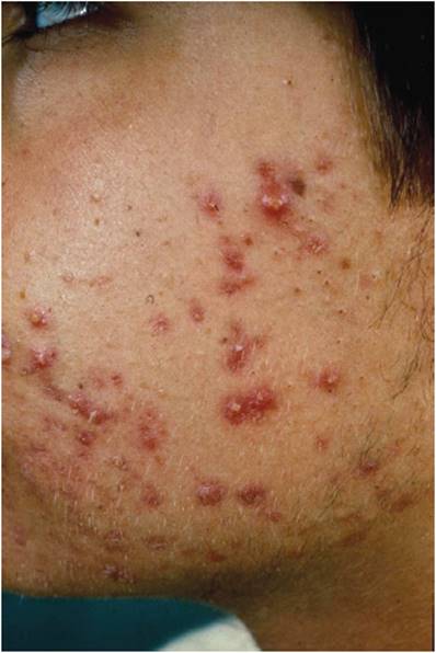
FIGURE 8-33 Acne vulgaris. There are numerous inflamed pustules and papules with central black plugs termed open comedones or “blackheads.” (Image used with permission from Dr. Timothy Berger.)
Epidemiology
Acne vulgaris is so common that it is said by some authors to affect practically everyone at some point in their lives. The peak incidence is at 18 years of age, although adults can also have acne. There are studies showing that 3% of men and 5% of women have acne between 40 and 49 years of age.
Histopathology and Pathogenesis
Histopathologically, comedonal acne is manifest as a widened follicle with a dense keratin plug within its infundibulum. If the follicular orifice is patulous, the acne lesion is said to be an open comedone. If the orifice is normal and the follicle is expanded below the skin surface, the lesion is termed a closed comedone. Secondary inflammatory changes occur commonly within plugged follicular units. Neutrophils may accompany the keratinous plug with the follicular canal, creating a pustular lesion. Inflammatory acne lesions are a consequence of follicles that have ruptured with resultant spillage of keratinous debris into the perifollicular dermis, evoking a dense inflammatory reaction with a mixture of neutrophils, lymphocytes, and histiocytes (Figure 8-34).

FIGURE 8-34 Histopathologic features of acne. There is a follicle with a central keratin plug. Surrounding the follicle, there is lymphocytic inflammatory infiltrate. This lesion would correspond to an erythematous papule seen in inflammatory acne (see Figure 8-33).
Understanding of the evolution of acne lesions has led to therapies that are effective for the vast majority of cases. There are four essential components to the development of acne lesions: (1) plugging of the folliculosebaceous unit; (2) sebum production; (3) overgrowth of the bacterium Propionibacterium acnes within the plugged follicle; and (4) a secondary inflammatory response. The formation of keratin plugs within follicles is a complex process that is thought to be genetically controlled on a cellular level. Keratinocytes become sticky and fail to slough appropriately, yielding follicular plugging. Contrary to a commonly held belief, being “dirty” does not cause acne, and vigorous or frequent cleansing does not improve the condition. However, some exogenous substances such as oily cosmetics or petrolatum-based hair care products may promote comedone formation and thus exacerbate acne.
Plugged follicles alone would never become more than comedones, however, if it were not for sebum production and P acnes overgrowth. P acnes is a commensal organism of the skin. However, with ample sebum as a food source within the well-protected environment of a plugged follicle, P acnes overgrowth occurs. The sebum becomes broken down to constituent lipids and free fatty acids. The failure of keratinous debris and sebum to exit the follicle freely expands the follicular canal. The bacteria release factors chemotactic for neutrophils, and their infiltration of the follicle results in pustule formation. Neutrophilic enzymes weaken the follicle wall and follicular rupture occurs, releasing large amounts of inflammatory reactants into the dermis. Lymphocytes, macrophages, and more neutrophils respond, and the comedonal lesion is transformed into an inflamed papule, pustule, or nodule of acne. Follicular rupture and an intense secondary inflammatory reaction may eventuate with profound scarring in some hosts.
Clinical Manifestations
The spectrum of acne severity is quite broad. In the neonate, maternal androgens stimulate enlargement of and sebum overproduction from sebaceous glands. The presence of sebum promotes P acnesovergrowth, and acne ensues until the maternal androgens have cleared and the sebaceous glands atrophy to a normal neonatal size. Significant sebum production does not begin again until puberty. Under the stimulation of androgens at puberty, sebaceous glands enlarge once again and produce sebum in the sebaceous areas of the body, namely the face, neck, chest, and back (the same areas affected most by acne). Onset may be gradual or rapid, and severity may range from primarily comedonal to inflammatory papules and pustules to highly inflammatory, painful nodules. Severe scarring variants may be explosive in onset and present with systemic symptoms of fever and arthralgias. Age at onset and family history are predictors of the severity of acne.
Acne may present as a component of a syndrome, as in polycystic ovary disease (ie, Stein-Leventhal syndrome) or so-called SAPHO syndrome (synovitis, acne, palmoplantar pustulosis, hyperostosis, and osteitis). At least in polycystic ovary disease, there may be hormonal influences that predispose to the acne lesion development.
CHECKPOINT
30. Why do some infants develop acne? What factors explain its spontaneous resolution?
31. What is the pathophysiology of lesion development in acne?
32. What are some broad treatment categories for acne, and which aspect of acne pathogenesis does each address?
CASE STUDIES
Yeong Kwok, MD
(See Chapter 25, p. 711 for Answers)
CASE 34
A 25-year-old woman presents with a complaint of rash that has developed over the past several weeks and seems to be progressing. On examination, she is noted to have several plaque-like lesions over the extensor surfaces of both upper and lower extremities as well as similar lesions on her scalp. The plaques are erythematous, with silvery scales, and are sharply marginated.
Questions
A. What is the likely diagnosis? Is this skin disease genetic, environmental, or both? Based on what evidence?
B. What are the pathophysiologic mechanisms behind the development of the plaques, scale, and erythema characteristic of this disorder?
C. What immunologic defects have been implicated in patients with this skin disease?
CASE 35
A 35-year-old woman who recently returned from Africa presents to the clinic with complaints of a rash. During her trip, she developed an itchy rash on both arms. She has an unremarkable medical history. Medications recently taken include chloroquine for malaria prophylaxis. Examination discloses multiple small violaceous papules on the flexor surfaces of the arms. The lesions have angular borders and flat tops. Some of the lesions have minute white streaks on the surface, barely visible to the naked eye.
Questions
A. What is the likely diagnosis? What is the possible underlying cause?
B. What is the pathophysiologic mechanism by which these skin lesions are formed?
C. What histopathologic changes in the skin are responsible for the appearance of these lesions as violaceous papules with minute white striae?
CASE 36
A 27-year-old woman presents to the urgent care clinic complaining of a red, itchy rash developing suddenly the day before on her arms and legs and spreading to the trunk. She denies ulcers in the mouth or genital area. Her medical history is unremarkable except for occasional episodes of genital herpes. The most recent outbreak was approximately 2 weeks ago. She generally takes oral acyclovir on such occasions, but her prescription has run out and so she did not take any with her last bout. On physical examination, she has multiple erythematous papules over the arms, legs, and trunk. Many of the papules have a central area of duskiness or clearing, such that the lesions resemble targets. There is no evidence of mucosal involvement.
Questions
A. What is the likely diagnosis?
B. What is the pathophysiologic mechanism by which these skin lesions are formed? In what ways is this disease similar to and different from lichen planus?
C. What factors may have triggered this rash? What evidence supports this link?
D. What is responsible for the target-like appearance of these lesions, and what does the histopathology show?
CASE 37
A 65-year-old man presents to the dermatology clinic with a complaint of blisters developing on his abdomen and extremities over the past week. The lesions consisted initially of red patches followed by blister formation. They are pruritic but not painful. The patient has no other complaints and denies mucous membrane involvement. Examination shows only multiple large, tense blisters with an erythematous base over the lower trunk and extremities. The clinical picture is felt to be most consistent with bullous pemphigoid.
Questions
A. What is the major differential diagnosis for multiple bullae? How do these diseases differ, and why is the distinction important?
B. What is the most likely diagnosis, and what would one expect histologic examination to show?
C. What would one expect to find on direct immunofluorescence microscopy?
D. What is the presumed mechanism by which blister formation occurs in bullous pemphigoid?
CASE 38
A 60-year-old man presents to the clinic with complaints of a recurring rash. He states that for the past 2–3 months, he has had several episodes of a painless, nonpruritic rash over his distal lower extremities. The lesions are described as purple and raised. His medical history is remarkable for hepatitis C—with no history of cirrhosis—and peripheral neuropathy. The patient has recently been treated for otitis media with amoxicillin. He has taken no other medications. Physical examination is notable only for multiple reddish-purple papules over the distal lower extremities (palpable purpura). The underlying skin is hyperpigmented. Biopsy reveals neutrophils, neutrophilic debris, and amorphous protein deposits involving the small blood vessels, consistent with fibrinoid necrosis.
Questions
A. What is the likely dermatologic diagnosis? What are some possible precipitants of that disease in this patient?
B. What is the underlying pathogenetic mechanism by which the lesions are formed?
C. What histologic characteristics are responsible for the appearance of the lesions as papular and purpuric?
D. What additional symptoms should this patient be asked about? Should any laboratory tests be ordered?
CASE 39
A 30-year-old woman presents to the clinic complaining that she has “an itchy rash all over the place.” She noticed that her legs became red, itchy, and blistered about 2 days after she had been hiking in a heavily wooded area. She says that scratching broke the blisters and afterward the rash became much worse and spread all over. She is convinced that the rash could not be poison ivy because once before she was exposed to that plant and did not develop a rash. On examination, there are erythematous vesicles and bullae in linear streaks on both of her legs. Some areas are weepy, with a yellowish crust. There are ill-defined erythematous plaques studded with papulovesicles on the trunk and arms.
Questions
A. What is the likely diagnosis? What feature on the physical examination is the cardinal sign?
B. What made the eruption spread?
C. How do you explain the diagnosis to the patient in light of the fact that she did not develop a rash after known exposure to poison ivy in the past? Why didn’t the rash appear until 2 days after the apparent exposure?
CASE 40
A 45-year-old woman presents to clinic with a rash on her legs for 2 months. She notes that the rash started soon after babysitting her niece, who had “strep throat.” She initially had a sore throat herself, but it stopped hurting after she took 2 days’ worth of antibiotics she had left over from a previous prescription. On examination, on the anterior lower legs, she has several scattered ill-defined erythematous nodules, which are tender to palpation.
Questions
A. What is the likely diagnosis? What is the probable cause? What might explain why the eruption has persisted?
B. What are some other common causes of this condition?
C. What is the pathophysiologic mechanism of skin lesion formation?
D. What are the histopathologic findings of this disease?
CASE 41
A 52-year-old African American man presents to the clinic with a rash that has been worsening for several months. Review of systems is notable for a chronic cough. Examination reveals multiple red-brown dermis-based papules on the trunk, arms, and face. Several lesions are clustered near the nares. The examination is otherwise unremarkable.
Questions
A. What is the likely diagnosis? What information is necessary to confirm the diagnosis?
B. What organ system (in addition to the skin) is at risk for disease involvement based on the clinical examination?
C. What are the histopathologic features of this disease?
D. How does it present clinically?
CASE 42
A 15-year-old girl presents to the clinic complaining of “pimples” for 6 months. She has been using an over-the-counter face wash four times a day to keep the oil and dirt off, but it has not helped. Examination reveals several dozen erythematous papules and pustules over the forehead and central face with scattered open and closed comedones. A diagnosis of moderate inflammatory acne is entertained.
Questions
A. Why has her meticulous cleansing practice not helped her condition? What advice do you give her regarding facial cleansing?
B. What is the life cycle of an inflammatory papule of acne?
C. What are some general categories of acne treatment, and what component of lesion development does each address?
REFERENCES
General
Ackerman AB et al. The Lives of Lesions: Chronology in Dermatopathology. Masson, 1984.
Bolognia J et al, eds. Dermatology, 3rd ed. Saunders, 2012.
James W et al. Andrews’ Diseases of the Skin, 11th ed. Elsevier, 2001.
Psoriasis
Meier M et al. Clinical spectrum and severity of psoriasis. Curr Probl Dermatol. 2009;38:1–20. [PMID: 19710547]
Naldi L et al. The clinical spectrum of psoriasis. Clin Dermatol. 2007 Nov–Dec;25(6):510–8. [PMID: 18021886]
Nickoloff BJ et al. The cytokine and chemokine network in psoriasis. Clin Dermatol. 2007 Nov–Dec;25(6):568–73. [PMID: 18021894]
Tonel G et al. Cutting edge: a critical functional role for IL-23 in psoriasis. J Immunol. 2010 Nov 15:185(10):5688–91. [PMID: 20956338]
Yoo IS et al. T-helper 17 cells: the driving force of psoriasis and psoriatic arthritis. Int J Rheum Dis. 2012 Dec;15(6):531–7. [PMID: 23253236]
Lichen Planus
Cheng S et al. Interventions for erosive lichen planus affecting mucosal sites. Cochrane Database Syst Rev. 2012 Feb 15;2:CD008092. [PMID: 22336835]
Ismail SB et al. Oral lichen planus and lichenoid reactions: etiopathogenesis, diagnosis, management and malignant transformation. J Oral Sci. 2007 Jun;49(2):89–106. [PMID: 17634721]
Lage D et al. Lichen planus and lichenoid drug-induced eruption: a histological and immunohistochemical study. Int J Dermatol. 2012 Oct;51(10):1199–205. [PMID: 22416968]
Le Cleach L et al. Clinical practice. Lichen planus. N Engl J Med. 2012 Feb 23;366(8):723–32. [PMID: 22356325]
Roopashree MR et al. Pathogenesis of oral lichen planus—a review. J Oral Pathol Med. 2010 Nov;39(10):729–34. [PMID: 20923445]
Erythema Multiforme
Abe R. Toxic epidermal necrolysis and Stevens-Johnson syndrome: soluble Fas ligand involvement in the pathomechanisms of these diseases. J Dermatol Sci. 2008 Dec;52(3):151–9. [PMID: 18657400]
Chung WH et al. Recent advances in the genetics and immunology of Stevens-Johnson syndrome and toxic epidermal necrosis. J Dermatol Sci. 2012 Jun;66(3):190–6. [PMID: 22541332]
Gerull R et al. Toxic epidermal necrolysis and Stevens-Johnson syndrome: a review. Crit Care Med. 2011 Jun;39(6):1521–32. [PMID: 21358399]
Sokumbi O et al. Clinical features, diagnosis, and treatment of erythema multiforme: a review for the practicing dermatologist. Int J Dermatol. 2012 Aug;51(8):889–902. [PMID: 22788803]
Wetter DA et al. Recurrent erythema multiforme: clinical characteristics, etiologic associations, and treatment in a series of 48 patients at Mayo Clinic, 2000 to 2007. J Am Acad Dermatol. 2010 Jan;62(1):45–53. [PMID: 19665257]
Bullous Pemphigoid
Feng S et al. Serum levels of autoantibodies to BP180 correlate with disease activity in patients with bullous pemphigoid. Int J Dermatol. 2008 Mar;47(3):225–8. [PMID: 18289320]
Fine JD. Prevalence of autoantibodies to bullous pemphigoid antigens within the normal population. Arch Dermatol. 2010 Jan;146(1):74–5. [PMID: 20083697]
Kasperkiewicz M et al. The pathophysiology of bullous pemphigoid. Clin Rev Allergy Immunol. 2007 Oct;33(1–2):67–77. [PMID: 18094948]
Schmidt E et al. Clinical features and practical diagnosis of bullous pemphigoid. Immunol Allergy Clin North Am. 2012 May;32(2):217–32. [PMID: 22560135]
Wieland CN et al. Anti-bullous pemphigoid 180 and 230 antibodies in a sample of unaffected subjects. Arch Dermatol. 2010 Jan;146(1):21–5. [PMID: 20083688]
Leukocytoclastic Vasculitis
Carlson JA. The histological assessment of cutaneous vasculitis. Histopathology. 2010 Jan;56(1):3–23. [PMID: 20055902]
Kawakami T. New algorithm (KAWAKAMI algorithm) to diagnose primary cutaneous vasculitis. J Dermatol. 2010 Feb;37(2):113–24. [PMID: 20175844]
Allergic Contact Dermatitis
Cavani A et al. Allergic contact dermatitis: novel mechanisms and therapeutic perspectives. Curr Drug Metab. 2010 Mar;11(3):228–33. [PMID: 20406191]
Fyhrquist-Vanni N et al. Contact dermatitis. Dermatol Clin. 2007 Oct;25(4):613–23. [PMID: 17903620]
Lepoittevin JP et al, eds. Allergic Contact Dermatitis: The Molecular Basis. Springer, 2011.
Martin SF. Contact dermatitis: from pathomechanisms to immunotoxicology. Exp Dermatol. 2012 May;21(5):382–9. [PMID: 22509837]
Martin SF et al. Mechanisms of chemical-induced innate immunity in allergic contact dermatitis. Allergy. 2011 Sep;66(9):1152–63. [PMID: 21599706]
McFadden JP et al. Why does allergic contact dermatitis exist? Br J Dermatol. 2013 Apr;168(4):692–9. [PMID: 23383741]
Swinnen I et al. An update on airborne contact dermatitis: 2007–2011. Contact Dermatitis. 2013 Apr;68(4):232–8. [PMID: 23343440]
Erythema Nodosum
Gilchrist H et al. Erythema nodosum and erythema induratum (nodular vasculitis): diagnosis and management. Dermatol Ther. 2010 Jul–Aug;23(4):320–7. [PMID: 20666819]
Kisacik B et al. Multiclinical experiences in erythema nodosum: rheumatology clinics versus dermatology and infection diseases clinics. Rheumatol Int. 2013 Feb;33(2):315–8. [PMID: 22441968]
Papagrigoraki A et al. Erythema nodosum: etiological factors and relapses in a retrospective cohort study. Eur J Dermatol. 2010 Nov–Dec;20(6):773–7. [PMID: 21030339]
Thrash B et al. Cutaneous manifestations of gastrointestinal disease: part II. J Am Acad Dermatol. 2013 Feb;68(2):211.e1–33. [PMID: 23317981]
Sarcoidosis
Chen ES et al. Sarcoidosis—scientific progress and clinical challenges. Nat Rev Rheumatol. 2011 Jul 12;7(8):457–67. [PMID: 21750528]
Haimovic A. Sarcoidosis: a comprehensive review and update for the dermatologist: part I. Cutaneous disease. J Am Acad Dermatol. 2012 May;66(5):699.e1–18. [PMID: 22507585]
Tchernev G. Cutaneous sarcoidosis: the “great imitator”: etiopathogenesis, morphology, differential diagnosis, and clinical management. Am J Clin Dermatol. 2006;7(6):375–82. [PMID: 17173472]
Acne
Bhate K et al. Epidemiology of acne vulgaris. Br J Dermatol. 2013 Mar;168(3):474–85. [PMID: 23210645]
McInturff JE et al. The role of toll-like receptors in the pathophysiology of acne. Semin Cutan Med Surg. 2005 Jun;24(2):73–8. [PMID: 16092794]
Webster GF. The pathophysiology of acne. Cutis. 2005 Aug;76(2 Suppl): 4–7. [PMID: 16164150]
Williams HC et al. Acne vularis. Lancet. 2012 Jan 28;379(9813):361–72. Erratum in: Lancet. 2012 Jan 28;379(9813):314. [PMID: 21880356]