
Hueylan Chern
Emily Finlayson
Presentation
A 19-year-old otherwise healthy woman presents with abdominal pain and diarrhea. The abdominal pain is described as sharp and constant in the right lower quadrant. She denies melena or bloody diarrhea. She reports postprandial nausea and bloating without emesis. She denies recent travel history and her family history is unremarkable. Her menstrual cycle has been regular. On abdominal examination, her right lower quadrant is tender to palpation.
Differential Diagnosis
Differential diagnoses for right lower-quadrant abdominal pain include appendicitis, inflammatory bowel disease, Meckel’s diverticulum, urinary tract infection, and infectious enterocolitis. Campylobacterand Yersinia infection from poor handling of food can affect the ileocolic region and mimic Crohn’s ileocolic disease. In women, gynecologic causes such as ovarian torsion, tubal ovarian abscess, pelvic inflammatory disease, and ectopic pregnancy need to be considered as well.
Workup
The patient undergoes CT scan of the abdomen and pelvis with IV and oral contrast that reveals an inflamed, narrowed segment of terminal ileum (Figure 1). Serum laboratory values include hemoglobin of 11 g/dL, an albumin level of 3.3 g/dL, and a C-reactive protein level of 86 g/dL.

FIGURE 1 • Terminal ileum stricture.
Discussion
Crohn’s disease (CD) is a chronic inflammatory intestinal disease that can be unremitting and is incurable. It can affect any part of the gastrointestinal tract. The distribution is usually discontinuous with segments of uninvolved intestine. The inflammation in CD is transmural involving the full thickness of the bowel wall from the mucosa to serosa. The etiology for CD remains uncertain. CD has a bimodal age distribution with the first peak occurring between the ages of 15 to 30 years and the second between 60 and 80 years. The most common anatomic pattern in patients with CD is ileocolic disease occurring in 40%, followed by small intestinal disease, isolated colonic disease, and gastroduodenal disease. The disease behavior is classified into three categories: inflammatory, stricturing, and fistulizing. However, the anatomic distribution and behavior can change in any given Crohn’s patient over time.
The keys to evaluation of CD are determining extent and location of involved intestines and obtaining tissue for diagnosis. Several imaging modalities such as contrast studies, computed tomography (CT), and magnetic resonance imaging (MRI) have been used to evaluate CD. In the acute setting, CT of the abdomen and pelvic with contrast is a good choice to look for acute complication such as abscess, obstruction, or perforation and also to eliminate other causes for acute abdomen. CT is also helpful in looking for thickened intestine, stricture, adjacent organ involvement, fistulas, phlegmon, and abscess (Figure 2A). CD complicated by enterovesical or colovesical fistulas will present with air or oral contrast within the bladder (Figure 2B).
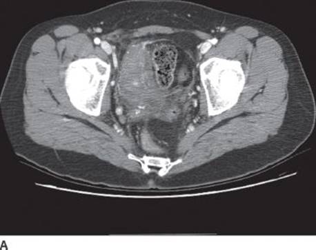
FIGURE 2A • Right lower-quadrant phlegmon.
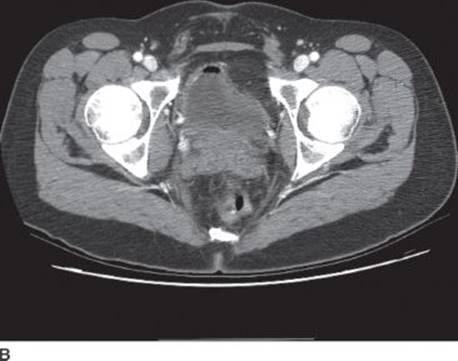
FIGURE 2B • Air in the bladder indicating fistula to the gastrointestinal tract.
Contrast studies such as small bowel follow through are helpful for evaluation of Crohn’s when small bowel proximal to terminal ileum is involved. CT enterography, MRI enterography, and capsule endoscopy are other modalities used to diagnose proximal small bowel disease. MRI enterography has recently gained popularity because it avoids radiation exposure in a typically young patient requiring imaging over lifetime for followup or diagnosis. Furthermore, unlike contrast studies, it has the ability demonstrate both extraluminal and intraluminal pathology.
Endoscopy is critical in workup, surveillance, and management of CD. Both colonoscopy and esopha-gogastroduodenoscopy are essential for demonstrating mucosal inflammation and obtaining tissue for diagnosis. The terminal ileum should be intubated and examined whenever possible during colonoscopy. The classic endoscopic findings for Crohn’s include aphthous ulcers, patchy erythema, linear serpiginous ulcers, deep “bear claw” ulceration, and strictures. However, the risk of perforation from colonoscopy in acute inflammation is high. Therefore, colonoscopy is generally avoided in an acute setting. A limited endoscopy such as flexible sigmoidoscopy can be considered if results will alter management in the acute setting.
Diagnosis and Treatment
Medical Therapy
In this patient with newly diagnosed CD and a stricture that is likely inflammatory in nature, medical therapy is the most appropriate initial approach. In general, medical treatment is the initial and primary therapy for CD. Medication frequently used includes aminosalicylates, antibiotics, steroids, thiopurines, cyclosporines, and antibodies to tumor necrosis factor. Very few patients will require surgery at initial disease presentation. Patients presenting with obstructive symptoms can be managed with bowel rest and nasogastric tube. The fact that this patient has an elevated C-reactive protein level suggests that her stricture is most likely inflammatory in nature (not fibrostenotic). Intravenous steroid can be used to treat the acute inflammation. Patients presenting with an associated intra-abdominal abscess can undergo percutaneous drainage. Surgery if indicated for persistent symptoms can then be delayed and performed in an elective setting when inflammation is not as severe and overall condition more stable. The goal of staged resection is to avoid bleeding associated with acute inflammation and to preserve bowel length.
Surgical Therapy
If this patient has persistent pain and obstructive symptoms despite medical therapy, surgical resection is indicated. In general, surgical therapy is reserved for failed medical therapy or an acute, severe complication of CD (Table 1). Because CD is not curable, operative intervention is only intended to address complication and alleviate symptoms to improve quality of life.
TABLE 1. Indications for Surgery
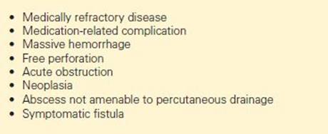
The most common indication for operation is obstruction from stricture. Several options exist including resection with or without anastomosis, strictureplasty, and bypass. What operation to perform depends on factors such as nutritional status, number of prior bowel resections, the length and number of strictures, surrounding inflammation, and immunosuppression status. Resection, strictureplasty, and bypass techniques may be used in one operation to treat multiple strictures. In this patients with a long, isolated segment of strictured terminal ileum, ileocolic resection is the recommended option.
Surgical Approach for Ileocolic Crohn’s Disease
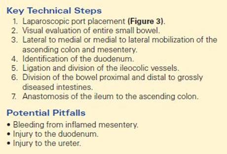
Laparoscopic resection by experienced surgeons can be done safely in inflammatory bowel disease (Table 2). Preoperative imaging is essential to assess the entire gastrointestinal tract. If performed laparoscopically, one camera port and three working ports are typically necessary (Figure 3). The entire small bowel is examined to assess the full extent of disease. Nondiseased intestines may be drawn into the inflammatory process, and they should be freed and preserved. A medial to lateral approach can be sometimes be difficult in ileocolic Crohn’s because of associated inflammation, phlegmon, or abscess. However, if the duodenum can be identified and the ileocolic vessels can be appreciated, scoring underneath the ileocolic vessels may allow entry into the avascular plane between the mesentery and the retroperitoneum. If not, a lateral to medial dissection can be started by dividing along the line of Toldt. The terminal ileum, the right colon, and the hepatic flexure need to completely mobilized. Once the ileocolic vessels are ligated and divided, grossly abnormal bowel is resected. The bowel is divided and an anastomosis is constructed ensuring correct orientation, without tension, and with good blood supply. The anastomosis can be performed with stapled technique, handsewn technique in a side-to-side or an end-to-end fashion. The abdomen cavity is washed out and the fascia closed.
TABLE 2. Key Technical Steps and Potential Pitfalls of Ileocolic Resection
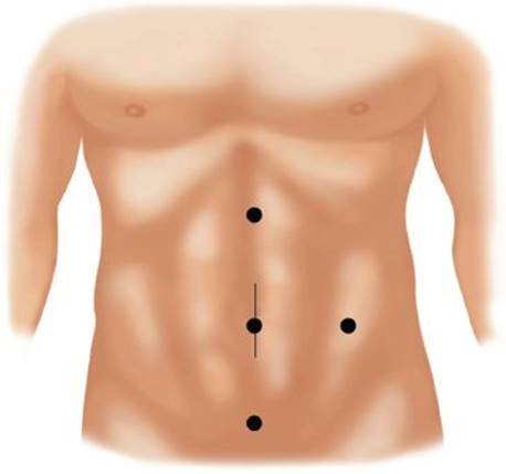
FICURE 3 • Common port placement for a laparoscopic ileocolic resection.
Intraoperative Considerations
When operating for CD, caution should be exercised in dividing the mesentery. Mesentery in CD is typically thickened with fat deposit and lymphadenopathy making division difficult. Rapid spread of mesenteric hematoma can result in further loss of intestinal length. Nondiseased intestine can also be drawn into the diseased process and involved in fistula formation or inflammatory adhesion. Care should be taken to preserve all normal intestines. Primary closure of the fistula after wedge resection in the unaffected intestine is usually sufficient. Wider margins do not decrease postoperative recurrence, and it is not necessary to achieve microscopic negative margins. Therefore, to preserve bowel length, the margins can be determined by resection to macroscopically normal intestine.
Risk of Recurrence, Postop Surveillance, and Treatment
After resection, endoscopic recurrence can be as high as 80% at 1 year. Twenty percent of the patients may experience clinical relapse at 1 year. The risk of developing disease complications requiring surgery approaches 50% at 10 years. Aminosalicylates, antibiotics, and thiopurines are only modestly effective in preventing disease recurrence. The antitumor necrosis factor agent has been shown to be most effective in preventing recurrence and therefore should be considered in patients at high risk for recurrence. Smoking is a well-known independent risk factor for recurrence. Therefore, smoking cessation is encouraged.
Case Conclusion
A CT of the abdomen and pelvis was done and showed inflamed terminal ileum. She, however, continued to have significant obstructive symptoms on maximal medical treatment and eventually required a laparoscopic ileocolic resection.
TAKE HOME POINTS
· Crohn’s disease (CD) is a chronic inflammatory panintestinal disease that relies on medical treatment and surgery is not curable.
· Emergent surgery is rarely necessary because acute complication such as obstruction and abscess can often be managed nonoperatively.
· Preservation of healthy bowel is essential when operating on patients with CD.
· Surgery is intended to address complications and alleviate symptoms to improve quality of life.
SUGGESTED READINGS
Lichtenstein GR, Hanauer SB, Sandborn WJ; Practice Parameters Committee of American College of Gastroenterology. Management of Crohn’s disease in adults. Am J Gastroenterol. 2009;104(2):465–483.
Strong SA, Koltun WA, Hyman NH, et al. Standards Practice Task force of The American Society of Colon and Rectal Surgeons. Practice parameters for the surgical management of Crohn’s disease. Dis Colon Rectum. 2007;50:1735–1746.
Yamamoto T, Fazio VW, Tekkis PP. Safety and efficacy of strictureplasty for Crohn’s disease: a systematic review and meta-analysis. Dis Colon Rectum. 2007;50(11):1968–1986.