
1.1 Terms of Location and Direction, Cardinal Planes and Axes
– All locational and directional terms used in anatomy, and in medical practice, refer to the human body in the anatomic position, in which the body is upright, arms at the side, with the eyes, palms of the hands, and feet directed forward (Fig. 1.1, Table 1.1).

Fig. 1.1  Anatomic position
Anterior view.
TABLE 1.1  General Terms of Location and Direction
|
Upper body (head, neck, and trunk) |
|
|
Term |
Explanation |
|
Cranial |
Pertaining to, or located toward, the head |
|
Caudal |
Pertaining to, or located toward, the tail |
|
Anterior |
Pertaining to, or located toward, the front Synonym: Ventral (used for all animals) |
|
Posterior |
Pertaining to, or located toward, the back Synonym: Dorsal (used for all animals) |
|
Superior |
Upper or above |
|
Inferior |
Lower or below |
|
Axial |
Pertaining to the axis of a structure |
|
Transverse |
Situated at right angles to the long axis of a structure |
|
Longitudinal |
Parallel to the long axis of a structure |
|
Horizontal |
Parallel to the plane of the horizon |
|
Vertical |
Perpendicular to the plane of the horizon |
|
Medial |
Toward the median plane |
|
Lateral |
Away from the median plane (toward the side) |
|
Median |
Situated in the median plane or midline |
|
Peripheral |
Situated away from the center |
|
Superficial |
Situated near the surface |
|
Deep |
Situated deep beneath the surface |
|
External |
Outer or lateral |
|
Internal |
Inner or medial |
|
Apical |
Pertaining to the top or apex |
|
Basal |
Pertaining to the bottom or base |
|
Sagittal |
Situated parallel to the sagittal suture |
|
Coronal |
Situated parallel to the coronal suture (pertaining to the crown of the head) |
|
Limbs |
|
|
Term |
Explanation |
|
Proximal |
Close to, or toward, the trunk, or toward the point of origin |
|
Distal |
Away from the trunk (toward the end of the limb), or away from the point of origin |
|
Radial |
Pertaining to the radius or the lateral side of the forearm |
|
Ulnar |
Pertaining to the ulna or the medial side of the forearm |
|
Tibial |
Pertaining to the tibia or the medial side of the leg |
|
Fibular |
Pertaining to the fibula or the lateral side of the leg |
|
Palmar (volar) |
Pertaining to the palm of the hand |
|
Plantar |
Pertaining to the sole of the foot |
|
Dorsal |
Pertaining to the back of the hand or top of the foot |
– Three perpendicular cardinal planes and three axes based on the three spatial coordinates can be drawn through the body (Fig. 1.2).
• The sagittal plane passes through the body from front to back, dividing it into right and left sides.
• The coronal plane passes through the body from side to side, dividing it into front (anterior) and back (posterior) parts.
• The transverse (axial, horizontal, cross-sectional) plane divides the body into upper and lower parts. A particular transverse section is often given the designation of the corresponding vertebral level, such as T4, which passes through the 4th thoracic vertebra.
• The longitudinal axis passes along the height of the body in a craniocaudal direction.
• The sagittal axis passes from the front to the back (or the back to the front) of the body in an anteroposterior direction.
• The transverse (horizontal) axis passes through the body from side to side.
1.2 Landmarks and Reference Lines
– In surface anatomy, palpable structures or visible markings on the surface of the body are used to identify the location of underlying structures. Reference lines are vertical or transverse planes that connect palpable structures or markings (Tables 1.2, 1.3, and 1.4; see also Fig. 1.4A and B).

Fig. 1.2  Cardinal planes and axes
Neutral position, left anterolateral view.
TABLE 1.2  Anterior and Lateral Reference Lines on Trunk
|
Anterior midline |
Passes through the center of the sternum |
|
Sternal line |
Passes along the lateral border of the sternum |
|
Midclavicular line |
Passes through the midpoint of the clavicle |
|
Parasternal line |
Passes through a point midway between the sternal and midclavicular lines |
|
Anterior axillary line |
Marks the anterior axillary fold formed by the pectoralis major muscle |
|
Posterior axillary line |
Marks the posterior axillary fold formed by the teres major muscle |
|
Midaxillary line |
Marks the midpoint between the anterior and posterior axillary lines |
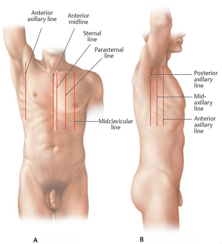
A Anterior view.
B Right lateral view.
TABLE 1.3 Landmarks and Transverse Planes on the Anterior Trunk
|
Jugular notch |
Marks the superior border of the manubrium |
|
Sternal angle |
Marks the junction of manubrium and body of the sternum |
|
Transpyloric plane |
Passes through the midpoint between the jugular notch and pubic symphysis |
|
Subcostal plane |
Marks the lowest level of the thoracic cage, the 10th costal cartilage |
|
Supracrestal plane |
Connects the top of the iliac crests |
|
Intertubercular plane |
Passes through the iliac tubercles |
|
Interspinal plane |
Connects the anterior superior iliac crests |
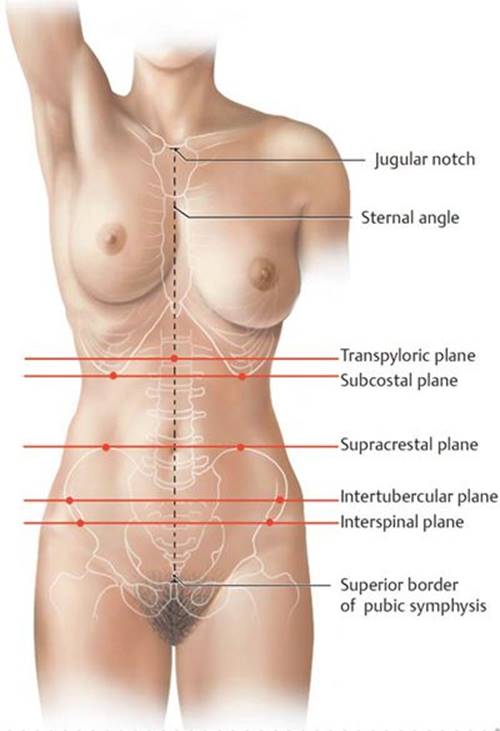
TABLE 1.4  Vertebral Spinous Processes and Posterior Landmarks
|
C7 |
The vertebra prominens |
|
T3 |
Level of the medial edge of spines of the scapulae |
|
T7 |
Level of the inferior angles of the scapulae |
|
T12 |
Level of the lower limit of the thoracic cavity |
|
L4 |
Level of the iliac crests |
|
S2 |
Level of the posterior superior iliac spine |

1.3 The Integumentary System
The skin (integument), the largest organ of the body, protects underlying tissue from biologic, mechanical, and chemical injury; regulates body temperature; and participates in metabolic processes, such as the synthesis of vitamin D.
– The skin is composed of
• an outer waterproof avascular layer, the epidermis, which has a superficial layer of keratinized cells that shed continuously and a deep basal layer of regenerating cells, and
• an inner richly vascularized and innervated layer, the dermis, which supports the epidermis and contains hair follicles.
1.4 Fascia
Fascia is a sheet of connective tissue that lies between the skin and underlying muscles and bone (Fig. 1.3).
– Superficial fascia, a layer of varying thickness that lies deep to the skin, is composed of loose connective tissue and fat. Superficial nerves and vessels traverse this layer.
– Deep fascia, a dense connective tissue layer, lies under (deep to) the superficial fascia and is devoid of fat. It forms an investing layer, which envelops neurovascular structures and muscles of the limbs, trunk wall, head, and neck. Invaginations of the deep fascia of the limbs form intermuscular septa that separate limb musculature into functional groups (compartments).

Fig. 1.3  Fascia
Cross section through the right arm, proximal view.
1.5 The Skeletal System
The bones and cartilages of the body, which make up the skeletal system, provide leverage for muscles and protect the internal organs. Bone is also the site for calcium storage and blood cell production.
– There are two anatomic divisions of the skeleton (Fig. 1.4A and B):
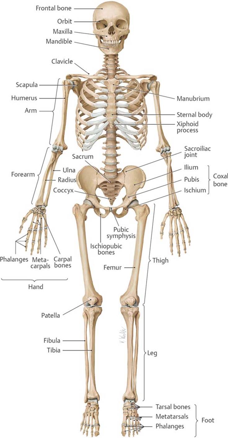
Fig. 1.4  Human skeleton
Left forearm is pronated, and both feet are in plantar flexion.
A Anterior view.

B Posterior view.
• The axial skeleton, which consists of the skull, vertebrae, sactum, coccyx, ribs, and sternum
• The appendicular skeleton, which includes the clavicle and scapula of the pectoral girdle, the coxal bones of the pelvic girdle, and the bones of the upper and lower limbs
– Periosteum is a thin layer of fibrous connective tissue that coats the outer surface of each bone (Fig. 1.5). Perichondrium forms a similar layer around cartilaginous structures. These tissues nourish and assist in the healing of the underlying bone.
– All bones have a superficial layer of dense compact (cortical) bone that surrounds a less dense cancellous (spongy) bone. In some areas of the bone, a medullary cavity contains yellow (fatty) or red (blood cell or platelet-forming) bone marrow.
– Bones develop from mesenchyme (embryonic connective tissue) through two processes of ossification (bone formation).
• The clavicle and some bones of the skull develop by membranous ossification, in which the bones form through direct ossification of mesenchymal templates that are set down during the embryonic period.
• Most bones, including the long bones of the limbs, develop by endochondral ossification, in which a cartilaginous template, formed from mesenchyme, is laid down during the fetal period. Over the first and second decades of life, bone replaces most of the cartilage.
○ Within each bone undergoing endochondral ossification, bone formation occurs first at a primary ossification center, which is in the diaphysis (shaft) of the long bones. Secondary ossification centersappear later at the epiphyses (growing ends) of the bones.
– Long bones of the skeleton increase in length through growth of the epiphyses and diaphysis on either side of the epiphyseal plate, an intervening cartilaginous area. During childhood and adolescence the epiphyseal plates gradually shorten as they are replaced by bone. In the adult these areas are completely ossified, and only thin epiphyseal lines remain.
– Apophyses, bony outgrowths that lack their own growth center, serve as attachment sites for ligaments or tendons. Specific apophyses are referred to as condyles, tubercles, spines, crests, trochanters, or processes.
– Ligaments are connective tissue bands that connect bones to each other or to cartilage. (Within the body cavities, the term ligament refers to folds or condensations of a serous or fibrous membrane that support visceral structures.)
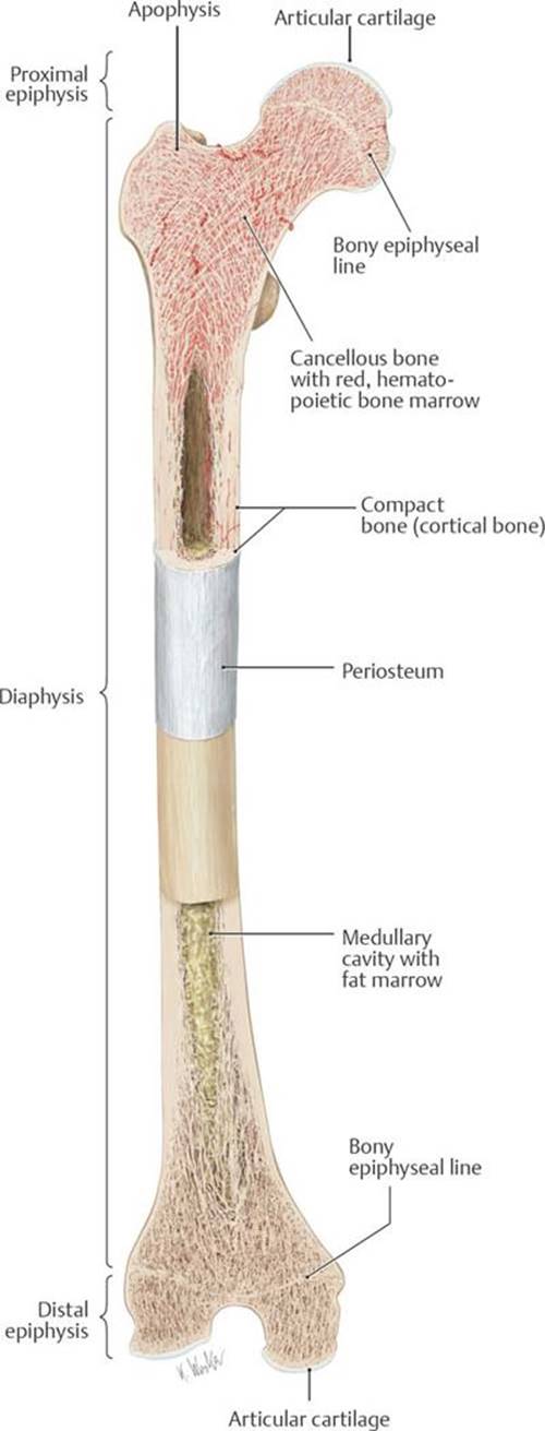
Fig. 1.5 Structure of a typical long bone
Illustrated for the femur. Coronal cuts through the proximal and distal parts of an adult femur.
– Joints are classified according to the type of tissue that connects the bones.
• Syndesmoses (fibrous joints), such as those found in the sutures of the skull and interosseous membrane of the forearm, are united by fibrous tissue and allow minimal movement (Fig. 1.6A and B).
• Synchondroses (cartilaginous joints) are united either by fibrocartilaginous segments, such as the costal cartilages of the ribs, intervertebral disks, and pubic symphysis (Fig. 1.7A and B), or by articular cartilage, often found in temporary joints, such as those that join the ilium, ischium, and pubis of the hip bone (Fig. 1.7C). Subsequent fusion of these temporary joints creates synostoses (sites of bony fusion) (Fig. 1.8).
• Synovial joints, the most common type of joint, allow free movement (Fig. 1.9) and typically have
○ a joint cavity that is enclosed by a fibrous joint capsule and lined by a synovial membrane, which secretes a thin film of lubricating synovial fluid;
○ articulating ends of the bones that are covered by articular (hyaline) cartilage; and
○ extrinsic ligaments on the outer surface, which reinforce the joints.
○ Some synovial joints also contain intrinsic (intra-articular) ligaments and intervening fibrocartilaginous disks (such as the menisci of the knee joint).
– Bursae are closed sacs that contain a thin film of fluid and are lined with a synovial membrane. Commonly found around joints of the limbs, bursae cushion prominent bony processes from external pressure and prevent friction where tendons cross bony surfaces.

Fig. 1.6 Syndesmoses
A Interosseous membrane of the forearm, anterior view.
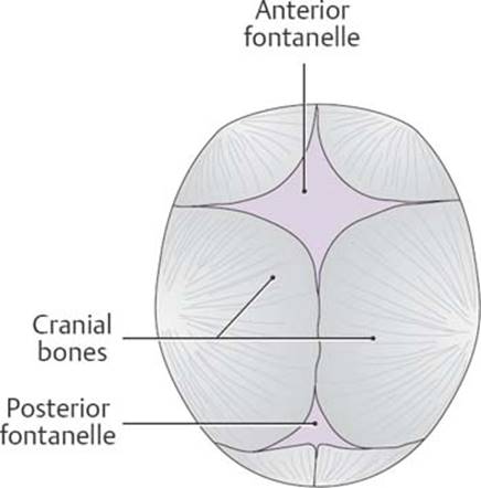
B Skull of a neonate showing open fontanelles, superior view.
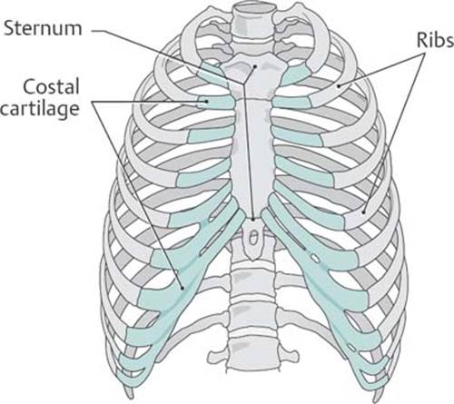
Fig. 1.7 Synchondroses
A Costal cartilages.

B Pubic symphysis and intervertebral disks.

C Hip bone before closure of the growth plates.
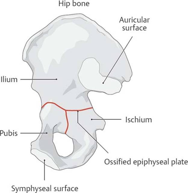
Fig. 1.8 Synostoses
Hip bone (fusion of the ischium, ilium, and pubis).

Fig. 1.9  Structure of a synovial joint
1.6 The Muscular System
The muscular system is composed of muscles and their tendons, which produce movement through contraction of muscle cells.
– Muscle cells are the structural units of the muscular system. Connective tissue binds muscle cells (fibers) together to form bundles, which in turn are bound together to form muscles (Fig. 1.10).
– Somatic muscles move the skeleton and are found in the neck, trunk wall, and limbs; visceral muscles alter the shape of internal structures, such as the heart and gastrointestinal tract.
– Muscle tissue is classified by location (somatic or visceral), appearance (striated or nonstriated), and innervation (voluntary or involuntary).
• Skeletal muscle, the most prevalent type of muscle, which is found in the limbs and body wall, is somatic, striated, multinucleated, and voluntary.
• Cardiac muscle, which makes up the thick muscular layer (myocardium) of the heart, is visceral, striated, and involuntary.
• Smooth muscle, found in the walls of blood vessels and hollow internal organs, is visceral, nonstriated, and involuntary.
– Tendons, dense fibrous bands, connect muscles to their bony attachments. Aponeuroses are tendons that form flat sheets and attach muscles to the skeleton or to other muscles.
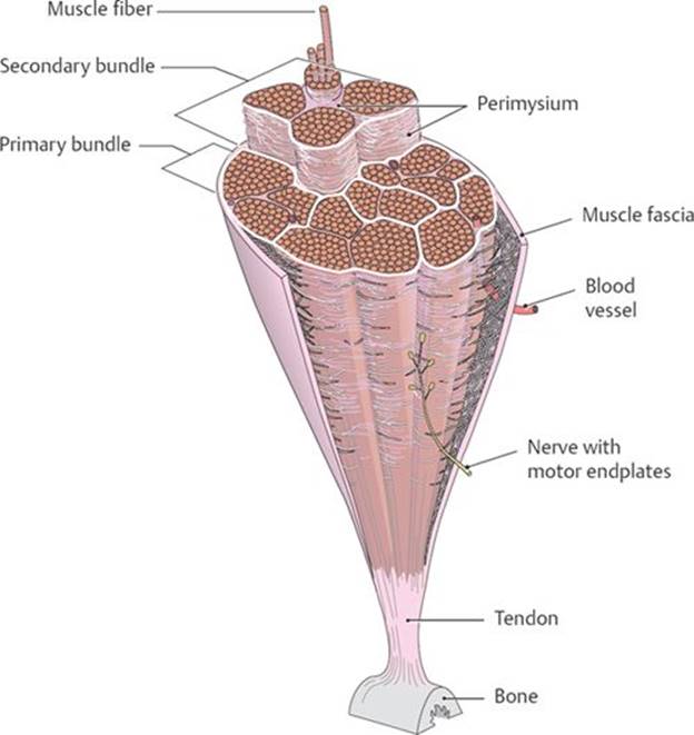
Fig. 1.10 Structure of a skeletal muscle
Cross section through a skeletal muscle.
1.7 The Circulatory System
– The heart and blood vessels, which make up the circulatory system (Figs. 1.11 and 1.12), transport blood to tissues of the body for the exchange of gases, waste products, and nutrients.
– The muscular heart provides the pumping action that maintains the flow of blood through the vessels.
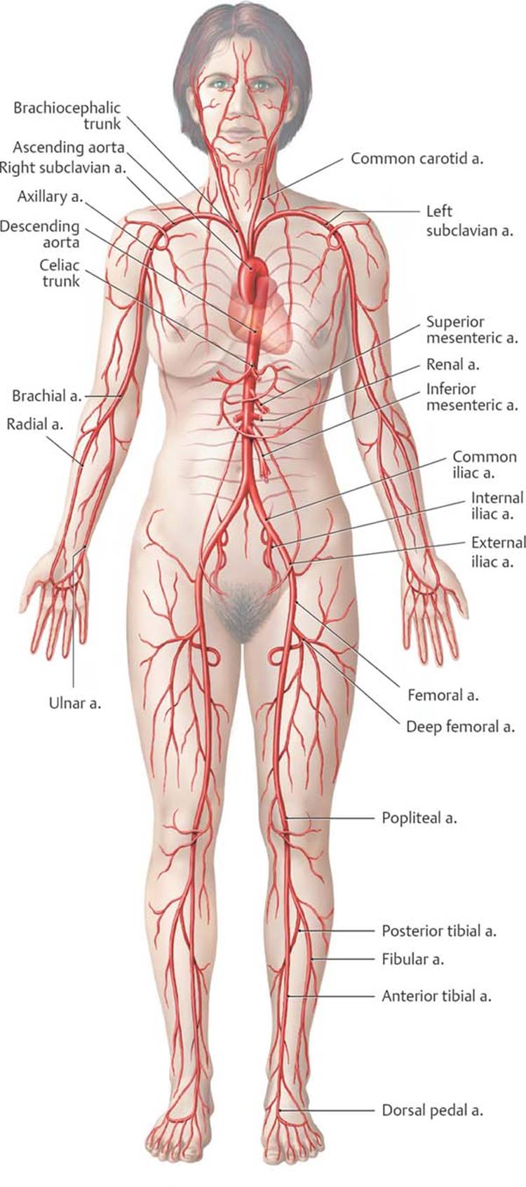
Fig. 1.11  Overview of the principal arteries in the systemic circulation
Anterior view.
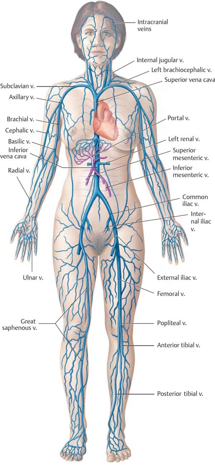
Fig. 1.12 Overview of the principal veins in the systemic circulation
Anterior view. The portal circulation of the liver is shown in purple.
– The blood vessels of the circulatory system (Fig. 1.13) are classified as follows:
• Arteries, which transport blood away from the heart and branch into many smaller arterioles
• Veins, which carry blood toward the heart and are formed by the convergence of many small venules
○ Many veins, particularly in the limbs, have multiple valves along their length to prevent backward flow due to gravity.
○ The veins are divided into superficial veins that travel in the superficial fascia and deep veins that accompany the arteries. Perforator veins connect the superficial and deep venous circulations.
○ Veins are more numerous and Veins are more numerous and more variable than arteries and often form venous plexuses (networks), which are named for the structure they surround (e.g., uterine venous plexus).
• Capillaries, which form networks that intervene between the arteries and veins at the terminal vascular beds, where gas, nutrient, and waste exchange occurs
• Sinusoids, which are wide, thin-walled vessels that replace capillaries in some organs, such as the liver

Fig. 1.13 Structure of blood vessels
Blood vessels in different regions of the systemic circulation, shown in cross section.
– The circulatory system has two circuits (Fig. 1.14):
1. The pulmonary circulation transports oxygen-poor blood from the right side of the heart to the lungs through pulmonary arteries. Oxygen-rich blood from the lungs flows back to the left side of the heart through pulmonary veins.
2. The systemic circulation distributes oxygen-rich blood from the left side of the heart to body tissues through the systemic arteries (the aorta and its branches). Oxygen-poor blood returns to the heart through the systemic veins (the superior and inferior venae cavae and their tributaries—sometimes referred to as the caval system— and the coronary sinus).
– A portal circulation is a route within the systemic circulation that diverts blood to a second capillary network before returning it to the systemic veins. The largest of these, the portal system in the liver, diverts blood from the gastrointestinal tract to the capillaries (sinusoids) in the liver before returning it to the systemic veins. A similar portal system is found in the pituitary gland.
– An anastomosis, a communication between arteries, allows blood to bypass its normal route and flow through an alternate, or collateral, route. Although blood volume through the anastomosis is usually minimal, it increases when the lumen of vessels along the normal route is obstructed.
– End arteries are vessels that lack anastomoses. Gradual narrowing of end arteries stimulates the formation of new vessels, but an abrupt obstruction of an end artery can cause necrosis (death) of the target tissue.

Fig. 1.14  Circulation
Schematic showing the pulmonary and systemic circulations. The portal circulation through the liver is part of the systemic circulation. Arteries are shown in red, veins in blue, and lymphatic vessels in green.
1.8 The Lymphatic System
The lymphatic system, which runs parallel to the circulatory system, consists of lymph, lymphatic vessels, and lymphoid organs.
– The lymphatic system performs the following functions:
• Drains excess extracellular fluid from body tissues and returns it to veins of the systemic circulation
• Mounts an immune response in the body
• Transports fat and large protein molecules that cannot be taken up by venous capillaries
– Lymphoid organs and tissues that are part of the body’s immune system include the thymus, bone marrow, spleen, lymph nodes, and tonsils. They also include bronchus-associated lymphatic tissue (BALT) in the airway and gut-associated lymphatic tissue (GALT), such as Peyer’s patches and the vermiform appendix, within the gastrointestinal tract (Fig. 1.15).
– Lymph, extracellular fluid that is extracted by lymph capillaries and transported by lymphatic vessels, is a clear, watery substance similar to blood plasma.
– The conducting vessels of the lymphatic system include
• blind-ended lymphatic capillaries that begin in the tissues and drain to lymphatic vessels;
• lymphatic vessels, which are interposed with lymph nodes along their length and drain to lymphatic trunks; and
• two major lymphatic trunks, the thoracic duct (left lymphatic trunk) and right lymphatic trunk, which drain into large veins of the neck.
– The thoracic duct (left lymphatic duct), which arises from the cisterna chyli (chyle cistern), a dilated lymphatic vessel in the abdomen, is the larger of the two major lymphatic trunks. It drains lymph from the right and left lower quadrants and left upper quadrant of the body. The smaller right lymphatic duct drains only the right upper quadrant of the body (Fig. 1.16).
– Lymph carried by the thoracic duct and right lymphatic duct returns to the systemic venous circulation at the left and right venous angles (junction of the internal jugular and subclavian veins), also known as the jugulosubclavian junction, in the neck (Fig. 1.17).

Fig. 1.15 Lymphatic system
The lymphatic system parallels the veins of the circulatory system and includes lymphatic vessels and lymphatic organs.
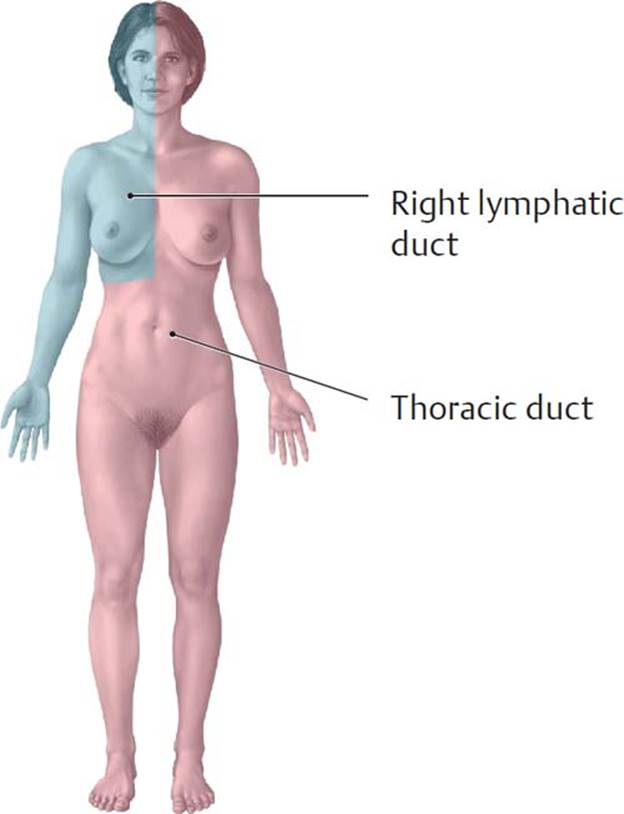
Fig. 1.16 Lymphatic drainage by body quadrants
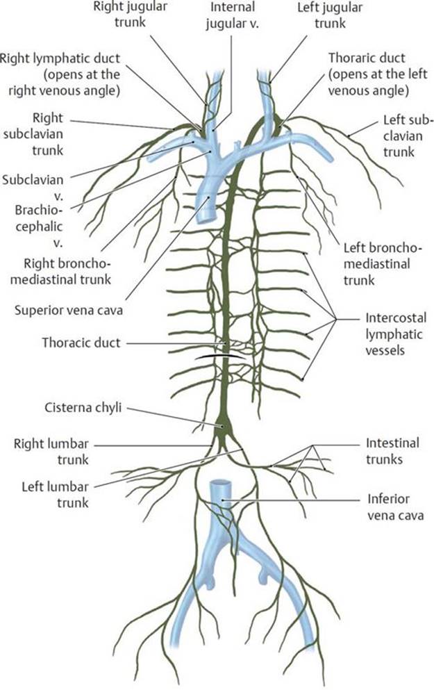
Fig. 1.17 Lymphatic pathways
Anterior view. The right lymphatic duct (~1 cm long) collects lymph from the right upper quadrant of the body and empties into the right venous angle at the junction of the right internal jugular vein with the right subclavian vein. Its major tributaries are
– the right jugular trunk (right half of the head and neck),
– the right subclavian trunk (right upper limb, right side of the chest and back wall), and
– the right bronchomediastinal trunk (organs of the right thoracic cavity).
The thoracic duct is approximately 40 cm long and transports lymph from the entire lower half of the body and left upper quadrant. It empties into the left venous angle between the left internal jugular vein and left subclavian vein. Its main tributaries are
– the left jugular trunk (left half of the head and neck),
– the left subclavian trunk (left upper limb, left side of the chest and back wall),
– the intestinal trunks (abdominal organs), and
– the right and left lumbar trunks (right and left lower limb; pelvic viscera; right and left pelvic, abdominal, and back wall).
The intercostal lymphatic vessels transport lymph from the left and right intercostal spaces to the lymphatic duct.
1.9 The Nervous System
The nervous system receives, transmits, and integrates information throughout the body through the conduction of nerve impulses.
– The nervous system has two major anatomic divisions (Fig. 1.18):
• A central nervous system (CNS) that consists of the brain and spinal cord
• A peripheral nervous system (PNS) that consists of 12 pairs of cranial nerves, 31 pairs of spinal nerves, and autonomic (visceral) nerves
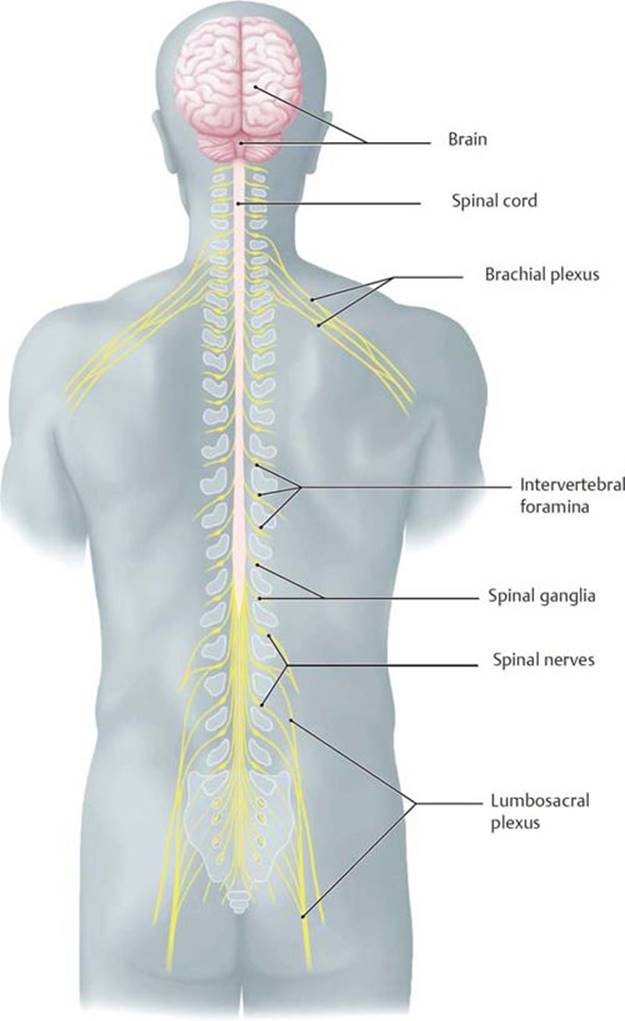
Fig. 1.18 Topography of the nervous system
Posterior view.
– The nervous system has two functional divisions:
• The somatic nervous system, which controls voluntary functions such as contraction of skeletal muscles
• The autonomic nervous system, which controls involuntary functions such as gland secretion
– There are two classes of cells within the nervous system:
• Nerve cells or neurons, the functional unit of the nervous system that are specialized for conducting nerve impulses
• Neuroglia, or glial cells, the nonneuronal cellular components of the nervous system that act as supporting cells and perform a variety of metabolic functions
– The neuron has a cell body (soma) with many short dendrites and a single long axon (Fig. 1.19).
• An aggregate of cell bodies is called a nucleus in the CNS and a ganglion in the PNS.
• Dendrites receive information and transmit impulses toward the cell body.
• Axons, or nerve fibers, transmit impulses away from the cell body. Bundles of axons in the CNS form tracts; bundles of axons in the PNS form nerves.
• A synapse is the site at which a neuron communicates with another neuron or with a receptor cell (typically, a cell of a muscle or gland).
• Many axons are surrounded by a lipid-rich insulating myelin sheath that increases the speed of impulse conduction. Myelin is formed by glial cells, which include Schwann cells in the PNS and oligodendrocytes in the CNS.
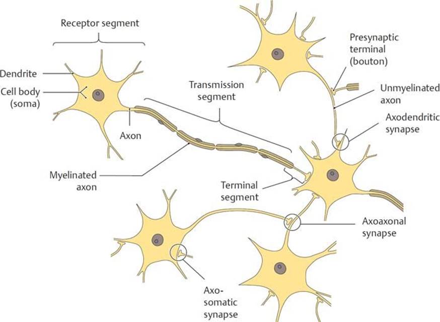
Fig. 1.19 Neurons
The nervous system is composed of neurons (nerve cells) and supporting neuroglial cells, which vastly outnumber neurons (10 to 1). Each neuron contains a cell body (soma) with one axon (projecting segment) and one or more dendrites (receptor segments). The release of neurotransmitters at synapses creates an excitatory or inhibitory postsynaptic potential at the target neuron. If this exceeds the depolarization threshold of the neuron, the axon “fires,” initiating the release of a neurotransmitter from its presynaptic terminal (bouton).
1.9a The Central Nervous System (Fig. 1.20)
– The brain and spinal cord of the central nervous system consist of
• gray matter that contains the cell bodies and dendrites of neurons;
• white matter that contains the axons of neurons, most of which are surrounded by a myelin sheath; and
• neuroglial cells that are abundant in both white and gray matter.
– The brain resides in the cranial cavity of the skull. Gray matter forms the cortex, or outer layer, of the brain and surrounds the inner areas of white matter. Axonal tracts of the white matter link regions of the brain with each other and with the spinal cord.
– The bony vertebral column encloses the spinal cord. Gray matter in the spinal cord is located centrally and is surrounded by the white matter. The gray matter forms an H-shaped area that consists of bilateral anterior horns, posterior horns, and, in the thoracic and upper lumbar region, lateral horns.
Referred pain
Referred pain is a sensation that originates from viscera but is perceived as if coming from an overlying or nearby somatic structure. It occurs because the somatic and visceral sensory fibers converge onto the same spinal cord segment. Diaphragmatic irritation from a splenic abscess, for example, is typically referred to the shoulder because both the diaphragm and the skin over the shoulder convey sensory information to C3–C5 segments of the spinal cord.
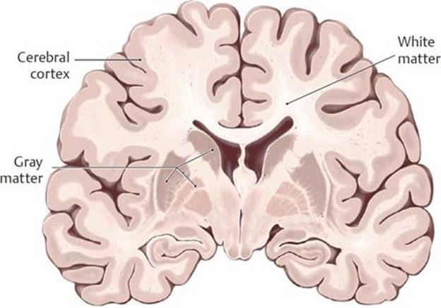
Fig. 1.20 Gray and white matter in the central nervous system
Nerve cell bodies appear gray in gross inspection, whereas nerve cell processes (axons) and their insulating myelin sheaths appear white.
A Coronal section through the brain.
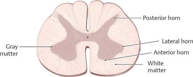
B Transverse section through the spinal cord.
1.9b The Peripheral Nervous System
– The anatomic components of the peripheral nervous system (Fig. 1.21) include the following:
• Twelve pairs of cranial nerves, designated by Roman numerals, that arise from the brain and primarily innervate structures of the head and neck. The vagus nerve (cranial nerve X) also innervates viscera of the thorax and abdomen.
• Thirty-one pairs of spinal nerves that arise from the spinal cord and exit the vertebral column through intervertebral foramina (openings between vertebrae). Spinal nerves are named for the section of the spinal cord from which they arise (e.g., T4 is the fourth segment of the thoracic part of the spinal cord).

Fig. 1.21 Spinal nerves and cranial nerves
Anterior view. Thirty-one pairs of spinal nerves arise from the spinal cord in the peripheral nervous system, compared with 12 pairs of cranial nerves that arise from the brain. The cranial nerve pairs are traditionally designated by Roman numerals.
– The functional components of the peripheral nervous system (Fig. 1.22) include the following:
• Sensory (afferent) nerves, which carry information regarding pain, temperature, and pressure to the central nervous system (CNS) from peripheral structures. Sensory nerves can contain somatic sensory fibers, which transmit the information from skin and skeletal muscles, and visceral sensory (visceral afferent) fibers, which transmit information from smooth muscle, cardiac muscle, and internal organs.
○ Sensations carried by visceral sensory fibers are vague and poorly localized (such as nausea) in contrast to sensations carried by somatic sensory fibers, which are sharp and localized (such as a paper cut).
• Motor (efferent) nerves, which transmit impulses from the CNS that elicit responses from peripheral target organs. They can contain somatic motor fibers, which innervate skeletal muscles, and visceral motor fibers, which innervate smooth muscle, cardiac muscle, and glands.
– Most nerves of the peripheral nervous system are mixed nerves that contain motor, sensory, and visceral fibers.
– Spinal nerves originate from the spinal cord and form by the merging of (Fig. 1.23)
• an anterior root carrying motor fibers whose cell bodies are located in the anterior horn of the spinal cord, and
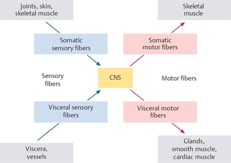
Fig. 1.22 Information flow in the nervous system
Fibers that carry information to the central nervous system (CNS) are called sensory or afferent fibers; fibers that carry signals away from the CNS are called motor or efferent fibers.
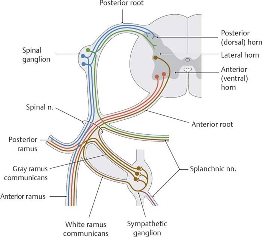
Fig. 1.23 Topographic and functional organization of a spinal cord segment
Somatic sensory (blue) and visceral sensory (green) fibers pass through the posterior root into the spinal cord and terminate in the posterior horn. Somatic motor fibers (red), originating in the anterior horn, and visceral motor fibers (brown), originating in the lateral horn, pass through the anterior root to the corresponding target organ. Somatic motor fibers synapse directly on their target organs (skeletal muscles), but the visceral motor fibers synapse with other autonomic fibers in discrete sympathetic ganglia or in ganglia embedded within the viscera.
• a posterior root carrying sensory fibers whose cell bodies are located in a spinal ganglion located outside the spinal cord.
– Spinal nerves emerge from the intervertebral foramen and split to form
• posterior rami that innervate structures of the back and
• anterior rami that form peripheral nerves and plexuses that innervate the rest of the body.
1.9c The Autonomic Nervous System
The autonomic nervous system, the visceral part of the peripheral nervous system, innervates blood vessels, glands, smooth muscle, and cardiac muscle.
– The autonomic nervous system consists of two divisions that often have antagonistic effects on the same organ (Fig. 1.24, Table 1.5):
• The sympathetic division allows the body to respond to stress (“fight or flight”).
• The parasympathetic division allows the body to maintain, or return to, a state of homeostasis (“rest and digest”).
TABLE 1.5  Effects of the Sympathetic and Parasympathetic Nervous Systems
|
Organ |
Sympathetic nervous system |
Parasympathetic nervous system |
|
Eye |
Pupillary dilation |
Pupillary constriction and increased curvature of the lens |
|
Salivary glands |
Decreased salivation (scant, viscous) |
Increased salivation (copious, watery) |
|
Heart |
Elevation of the heart rate |
Slowing of the heart rate |
|
Lungs |
Decreased bronchial secretions; bronchial dilation |
Increased bronchial secretions; bronchial constriction |
|
Gastrointestinal tract |
Decreased secretions and motor activity |
Increased secretions and motor activity |
|
Pancreas |
Decreased secretion from the endocrine part of the gland |
Increased secretion |
|
Male sex organs |
Ejaculation |
Erection |
|
Skin |
Vasoconstriction, sweat, secretion, piloerection |
No parasympathetic innervation |
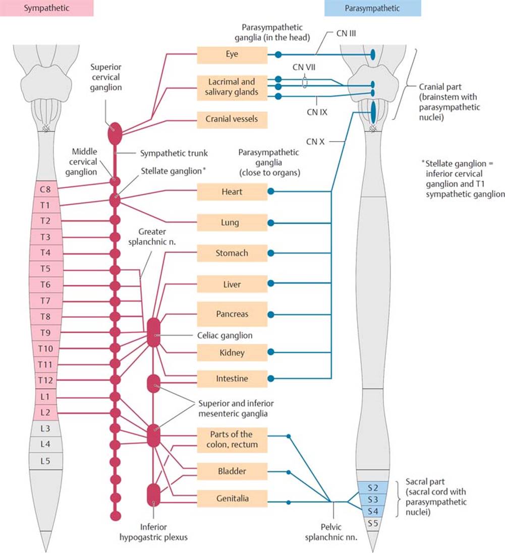
Fig. 1.24 Structure of the autonomic nervous system
– Nerves of the autonomic nervous system consist of a two-neuron path between the CNS and the target organ: a proximal preganglionic (presynaptic) neuron and distal postganglionic (postsynaptic) neuron that synapse in an intervening ganglion (Fig. 1.25).
• Sympathetic nerves arise from the thoracic and lumbar spinal cord (T1–L2) and exit the vertebral column with the corresponding spinal nerves.
○ Preganglionic sympathetic fibers Preganglionic sympathetic fibers leave the spinal nerve via white rami communicans to synapse on cell bodies of postganglionic fibers in paravertebral ganglia. These ganglia form a chain that runs along each side of the vertebral bodies known as the sympathetic trunk. Postganglionic fibers then rejoin the spinal nerve via gray rami communicans.
○ Some sympathetic preganglionic fibers form thoracic, lumbar, and sacral splanchnic nerves, autonomic nerves that bypass ganglia of the sympathetic trunk to synapse in prevertebral ganglia, such as the celiac ganglion. These nerves contribute to autonomic plexuses in the thorax, abdomen, and pelvis and innervate viscera in those regions.
• Parasympathetic nerves arise from the brain and the S2–S4 segments of the sacral spinal cord.
○ Preganglionic parasympathetic fibers Preganglionic parasympathetic fibers that arise from the brain travel with cranial nerves III, VII, IX, and X and synapse in parasympathetic ganglia of the head (or, in the case of the vagus nerve, ganglia near the target organ).
○ Pelvic splanchnic nerves are formed by the preganglionic parasympathetic fibers of the sacral spinal cord. They contribute to autonomic plexuses in the pelvis and abdomen and synapse in small ganglia located close to their target organ.
– The autonomic nervous system is considered to be a visceral motor system, although visceral sensory fibers accompany both sympathetic and parasympathetic nerves.
• Visceral sensory fibers carrying sensations of pain (visceral pain fibers) travel with sympathetic nerves.
• Visceral sensory fibers carrying sensations from physiological processes, such as distension of the bladder, travel with parasympathetic nerves.

Fig. 1.25 Autonomic nervous system circuitry
The autonomic nervous system innervates smooth muscle, cardiac muscle, and glands. It is divided into the sympathetic (red) and parasympathetic (blue) nervous systems, which often act in antagonistic ways to regulate blood flow, secretions, and organ function. Sensory (afferent) fibers are shown in green, motor (efferent) fibers are shown in purple.
1.10 Body Cavities and Internal Organ Systems
– The large organs of the endocrine, respiratory, digestive, urinary, and reproductive systems are housed in the thorax, abdomen, and pelvis.
– A thin serous (fluid-secreting) membrane lines the thoracic and abdominopelvic cavities of the trunk and their contents. A parietal layer forms the outer wall of the cavity and is continuous with a visceral layer that reflects from the outer wall to cover, or enclose, the viscera (see Figs. 12.2 and 12.3A). Thus the parietal pleura lines the outer wall of the pleural cavity, and the visceral pleura covers the surface of the lung within the cavity.
Review Questions: Introduction
1. Which of the following is associated with membranous bone formation?
A. Primary ossification center
B. Epiphyses
C. Direct ossification of mesenchymal templates
D. Long bones of the limbs
E. Diaphysis
2. A portal system is associated with
A. venous shunts that divert blood toward the heart
B. the pulmonary circulation
C. arterial–venous anastomoses
D. capillary beds in the liver
E. lymphatic capillaries
3. In the anatomic position the
A. eyes are directed forward
B. palms are directed forward
C. body is erect with arms at the sides
D. feet are directed forward
E. All of the above
4. Which of the following is true of splanchnic nerves?
A. They synapse in ganglia near their target organ.
B. They innervate viscera of the abdomen.
C. They may carry sympathetic fibers.
D. They may carry parasympathetic fibers.
E. All of the above
5. A 43-year-old high school teacher complained to her physician of abdominal bloating and pelvic pain. Radiographic studies showed a large tumor involving her right ovary. Although the patient was scheduled for surgery to remove the tumor, the physician was concerned about spread of the cancer along lymphatic channels. What is the lymphatic drainage pattern of pelvic viscera?
A. Ipsilateral drainage to the right and left lymphatic ducts
B. Contralateral drainage to the right and left lymphatic ducts
C. Bilateral drainage to the right and left lymphatic ducts
D. All pelvic viscera drain to the right lymphatic duct.
E. All pelvic viscera drain to the left lymphatic duct.
Answers and Explanations
1. C In the process of membranous ossification, embryonic mesenchymal templates are replaced by bone (Section 1.5).
A A primary ossification center, usually in the shaft of the long bones, is the site at which endochondral ossification begins.
B Epiphyses, located on either end of the long bones, are the secondary ossification centers for endochondral ossification.
D Long bones of the limbs undergo endochondral bone formation, in which a cartilaginous template forms from the embryonic mesenchyme before being replaced by bone.
E A diaphysis is the shaft of a long bone, which undergoes endochondral ossification.
2. A The body’s largest portal system diverts blood from capillary beds in the gastrointestinal tract to secondary capillary beds in the liver before returning it to systemic veins (Section 1.7).
B A portal system is a venous system within the systemic (general body) circulation, but not within the pulmonary (lung) circulation.
C A portal system diverts blood from one capillary network to another, unlike arterial–venous anastomoses, which divert blood away from capillary beds.
D Lymphatic capillaries are restricted to the lymphatic system and are not involved with portal venous systems.
3. E The anatomic position is the standard position of the body used in medical references. The body is upright, facing the observer, with arms at the side and the head, eyes, palms, and feet directed forward (Section 1.1).
A Eyes are directed forward, and other positions are correct as well (E).
B Palms are directed forward, and other positions are correct as well (E).
C Body is erect with arms at side, and other positions are correct as well (E).
D Feet are directed forward, and other positions are correct as well (E).
4. E All of the above (Section 1.9c).
A Splanchnic nerves synapse near their target organ either in prevertebral ganglia or in small ganglia on the viscera. B through D are correct also (E).
B Splanchnic nerves are autonomic nerves that innervate viscera in the thorax, abdomen, and pelvis. A, C, and D are correct also (E).
C Sympathetic fibers arise from the T1–L2 spinal cord to form thoracic, lumbar, and sacral splanchnic nerves. A, B, and D are correct also (E).
D Parasympathetic fibers arise from the S2–S4 spinal cord and form pelvic splanchnic nerves. A through C are correct also (E).
5. E The left lymphatic duct receives lymph from the entire body below the diaphgram, as well as from the left side of the thorax, head, and neck and the left upper limb (Section 1.8). The right lymphatic duct receives lymph only from the right side of the thorax, head, and neck and the right upper limb.
A Viscera from the right and left sides of the pelvis drain to the left lymphatic duct.
B All pelvic viscera drain to the left lymphatic duct.
C All pelvic viscera drain to the left lymphatic duct.
D Only the right upper quadrant of the body drains to the right lymphatic duct.