
The upper limb is characterized by its wide range of movement and fine motor ability. Coordinated movements at the pectoral girdle, as well as the shoulder, elbow, radioulnar, and wrist joints, position the hand for performing work as vital as eating and as complicated as playing a violin.
Schematics of upper limb muscles accompany the muscle tables (origin, attachment, innervation, and action) in the appropriate chapter sections. To see the muscles in situ, view the gallery of topographic images in Section 14.7at the end of the chapter.
14.1 The Pectoral Girdle
The pectoral girdle, formed by the clavicle and scapula, attaches the upper limb to the trunk (see Fig. 13.2A and B). The clavicle, acting as a strut, holds the scapula and humerus away from the trunk, allowing the free range of motion necessary for upper limb function.
14.1a Joints of the Pectoral Girdle
The joints of the pectoral girdle include articulations of the clavicle with the sternum and scapula, and a nonosseous joint that permits gliding movement between muscles of the trunk and scapula.
– The sternoclavicular joint is a strong but highly mobile synovial joint between the sternal end of the clavicle and the manubrium and 1st costal cartilage (Fig. 14.1).
• It is the only bony articulation between the upper limb and the trunk.
• An articular disk separates the articulating surfaces.
• Anterior and posterior sternoclavicular, costoclavicular, and interclavicular ligaments strengthen the joint.
• The joint allows the clavicle to elevate and rotate in conjunction with limb movements.
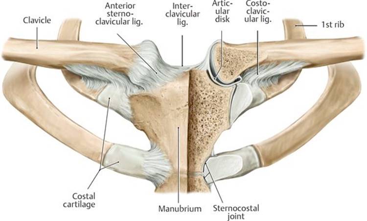
Fig. 14.1  Sternoclavicular joint
Anterior view with sternum coronally sectioned (left). Note: A fibrocartilaginous articular disk compensates for the mismatch of surfaces between the two saddle-shaped articular facets of the clavicle and the manubrium.
– The acromioclavicular joint is a plane type of synovial joint between the acromion of the scapula and acromial end of the clavicle (Fig. 14.2).
• An articular disk separates the articulating surfaces.
• An acromioclavicular ligament supports the joint superiorly.
• The coracoclavicular ligament, an extrinsic ligament (distant from the joint), strengthens the joint by anchoring the clavicle to the coracoid process. Its two parts are the conoid and trapezoid ligaments.
– The scapulothoracic joint is not a bony articulation but a functional relationship between the serratus anterior and subscapularis muscles that allows the scapula to glide and pivot on the chest wall (Fig. 14.3).
Injury to the long thoracic nerve
The superficial course of the long thoracic nerve (C5–C7) along the medial wall of the axilla puts it at risk of injury during regional surgeries such as axillary node dissection. Nerve damage results in the inability of the serratus anterior to laterally rotate the scapula, which is necessary to abduct the arm above the horizontal plane. The serratus anterior is also unable to support the scapula against the thoracic wall, which creates a “winged” scapula, especially noticeable when the subject presses the outstretched arm against a hard surface.
14.1b Muscles of the Pectoral Girdle
Muscles of the pectoral girdle attach the upper limb to the trunk and move and stabilize the pectoral girdle in response to movements of the glenohumeral joint at the shoulder (Table 14.1).

Fig. 14.2 Acromioclavicular joint
Anterior view. The acromioclavicular joint is a plane joint. Because the articulating surfaces are flat, they must be held in place by strong ligaments, greatly limiting the mobility of the joint.

Fig. 14.3 Scapulothoracic joint
Right side, superior view. In all movements of the shoulder girdle, the scapula glides on a curved surface of loose connective tissue between the serratus anterior and the subscapularis muscles. This surface can be considered a scapulothoracic joint.

A Subclavius and pectoralis minor, right, anterior view.

B Serratus anterior, right lateral view.
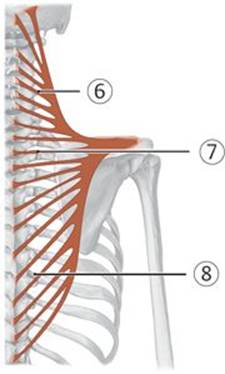
C Trapezius, right posterior view.
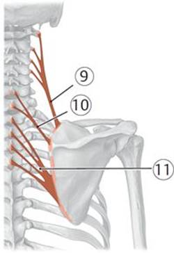
D Levator scapulae with rhomboids major and minor, right posterior view.
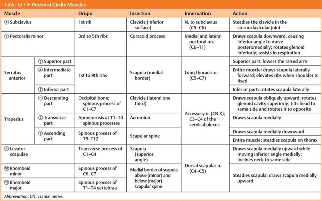
– Anterior muscles of the pectoral girdle, which lie on the anterior and lateral thoracic wall,
• include the subclavius, pectoralis minor, and serratus anterior and
• are anchored to the ribs, as well as to the bones of the pectoral girdle.
– Posterior muscles of the pectoral girdle, which are part of the superficial muscular layer of the back,
• include the trapezius, levator scapulae, rhomboid major, and rhomboid minor and
• arise from cervical and thoracic vertebrae and insert on the scapula.
– Movements of the pectoral girdle and the muscles that provide them are listed in Table 14.2.
TABLE 14.2  Movements of the Pectoral Girdle
|
Action |
Muscle |
|
Elevation |
Trapezius (descending part) Levator scapulae |
|
Depression |
Pectoralis minor Trapezius (ascending part) |
|
Protraction |
Pectoralis minor Serratus anterior |
|
Retraction |
Trapezius (transverse part) Rhomboid minor Rhomboid major |
|
Lateral rotation |
Serratus anterior (inferior part) Trapezius (descending part) |
|
Medial rotation |
Levator scapulae Rhomboid minor Rhomboid major |
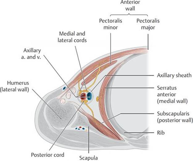
Fig. 14.4 Walls of the axilla
Right side, inferior view.
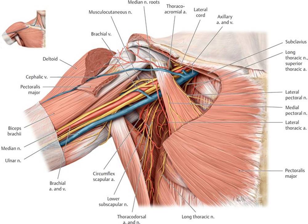
Fig. 14.5 Dissection of the axilla
Right shoulder, anterior view. Removed: Pectoralis major and clavipectoral fascia.
14.2 The Shoulder Region
The shoulder region includes the axilla, a passageway for neurovascular structures between the trunk and upper limb, and the glenohumeral joint, the largest joint of the upper limb. Muscles of the pectoral, scapular, and deltoid regions support the joint.
14.2a The Axilla
The axilla is a four-sided pyramidally shaped region between the upper parts of the arm and lateral thoracic wall (Fig. 14.4; see also Fig. 13.1A and C).
– Its boundaries are
• the cervicoaxillary canal, the narrow space between the clavicle and 1st rib, which forms the apex;
• the axillary fascia and axillary skin between the upper arm and the lateral thoracic wall, which form the base, or floor;
• the pectoralis major and pectoralis minor muscles, which form the anterior axillary wall (the lower edge of the pectoralis major forms a prominent ridge called the anterior axillary fold);
• the subscapularis, latissimus dorsi, and teres major muscles, which form the posterior axillary wall (the lower edge of the latissimus dorsi and teres major form a prominent ridge called the posterior axillary fold); and
• the lateral thoracic wall and the humerus of the arm, which form the medial and lateral walls, respectively.
– The contents of the axilla (Fig. 14.5), which are embedded in axillary fat, include
• the axillary artery and its branches,
• the axillary vein and its tributaries,
• axillary lymph nodes and vessels, and
• the cords and terminal nerves of the brachial plexus.
– An extension of the fascia of the neck forms a sleevelike axillary sheath that encloses the axillary vessels and the brachial plexus.
14.2b The Glenohumeral (Shoulder) Joint (Fig. 14.6A and B)
The glenohumeral joint is a ball-and-socket type of synovial joint between the shallow glenoid cavity of the scapula and the large head of the humerus.
– The glenoid labrum, a rim of fibrocartilage attached to the glenoid cavity, deepens the articular surface.
Glenohumeral dislocation
The glenohumeral joint is the most mobile but least stable joint of the body, and dislocations are frequent. Rotator cuff muscles provide the greatest stability, supporting the joint anteriorly, posteriorly, and superiorly, but inferior support is lacking. The coracoacromial arch, coracohumeral ligament, and capsular glenohumeral ligaments add further support. The majority of dislocations (90%) occur inferiorly, although most are labeled “anterior” dislocations based on the position of the displaced humeral head relative to the glenoid. These injuries can damage the axillary nerve and lead to a flattened shoulder profile. Posterior dislocations are rare and most often associated with seizures or electrocutions.
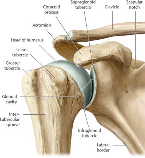
Fig. 14.6 Glenohumeral joint: Bony elements
Right shoulder, anterior view.
A Anterior view.

B Posterior view.

Fig. 14.7 Glenohumeral joint: Capsule and ligaments
Right shoulder, anterior view.

Fig. 14.8 Glenohumeral joint cavity
Right shoulder, anterior view.
– A fibrous capsule, lined by a synovial membrane, surrounds the joint (Fig. 14.7). Ligaments that support the joint capsule include
• anteriorly, three thickenings of the capsule, the superior, middle, and inferior glenohumeral ligaments, and
• superiorly, a coracohumeral ligament, that extends between the coracoid process of the scapula and lesser tuberosity of the humerus.
– A synovial membrane lines the joint space (synovial cavity) (Fig. 14.8).
• It forms a tubular sheath around the tendon of the biceps brachii as the tendon passes through the joint space.
• The synovial cavity communicates with a bursa under the subscapularis tendon (subtendinous bursa of the subscapularis).
– The coracoacromial ligament between the coracoid process and acromion prevents superior dislocation of the humerus.
– Three large bursae are associated with the glenohumeral joint (Fig. 14.9).
1. Anteriorly, the subtendinous bursa of the subscapularis, which lies between the tendon of the subscapularis and the neck of the scapula, communicates with the synovial cavity of the joint.
2. Superiorly, the subacromial bursa lies under the coracoacromial ligament and above the supraspinatus tendon and glenohumeral joint capsule.
3. Laterally, the subdeltoid bursa lies deep to the deltoid muscle and above the subscapularis tendon. It communicates with the subacromial bursa.
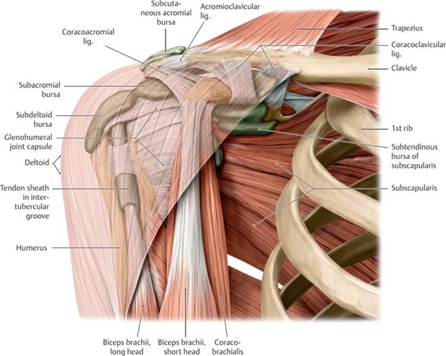
Fig. 14.9 Bursae of the shoulder region
Right shoulder, anterior view.

A Pectoralis major and coracobrachialis, right side, anterior view.

B Latissimus dorsi, right side, posterior view.
C Teres major, right side, posterior view.
D Deltoid, right side, right lateral view.
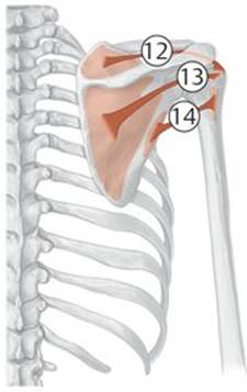
E Rotator cuff (supraspinatus, infraspinatus, and teres minor), right shoulder, posterior view.
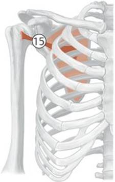
F Rotator cuff (subscapularis), right shoulder, anterior view.
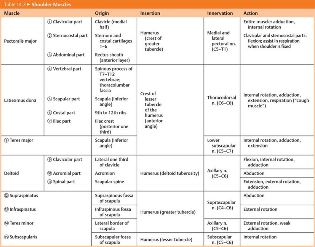
14.2c Muscles of the Shoulder Region
Muscles that cross the glenohumeral joint stabilize the head of the humerus in the glenoid cavity and assist in movements of the arm (Table 14.3)
– Two muscles of the trunk, the pectoralis major and latissimus dorsi, extend from the axial skeleton to the humerus and together provide strong adduction and medial rotation of the arm.
• The pectoralis major is a strong flexor of the arm.
• The latissimus dorsi is a strong extensor of the arm.
– Scapulohumeral muscles attach the humerus to the scapula and provide stability for the glenohumeral joint.
• The deltoid, which forms the rounded contour of the shoulder, abducts, flexes, and extends the arm.
• The teres major adducts and medially rotates the arm.
• The supraspinatus initiates abduction of the arm and assists the deltoid in the first 15 degrees of movement.
• The infraspinatus externally rotates the arm.
• The teres minor externally rotates the arm.
• The subscapularis on the anterior surface of the scapula internally rotates the arm.
– A musculotendinous rotator cuff around the glenohumeral joint includes four of the scapulohumeral muscles, which are the important dynamic stabilizers of the joint.
• The rotator cuff muscles include the supraspinatus, infraspinatus, teres minor, and subscapularis.
• Their tendons insert on, and reinforce, the fibrous capsule of the joint, forming a supportive cuff around the anterior, posterior, and superior aspects of the joint.
Rotator cuff tears
Rotator cuff tears can occur at any age but are most common in the older patient and usually involve the supraspinatus tendon. Degenerative changes and chronic inflammation from repetitive use (baseball pitchers are a good example) can cause the tendon to fray and rupture. When the subacromial and subdeltoid bursae tear in conjunction with the ruptured tendon, they become continuous with the cavity of the glenohumeral joint.
– A few muscles of the arm cross the glenohumeral joint and support its movements (see Table 14.5).
• The tendon of the long head of the biceps brachii passes through the intertubercular groove of the humerus and enters the joint cavity, where it is ensheathed by the synovial layer of the capsule. It prevents dislocation of the humerus during abduction and flexion.
• The short head of the biceps brachii and the coracobrachialis muscles cross the joint anteriorly and assist in flexion of the arm.
• The long head of the triceps brachii crosses the joint posteriorly and assists in adduction and extension.
– Movements at the glenohumeral joint and the muscles that provide them are listed in Table 14.4.
TABLE 14.4 Movements at the Glenohumeral Joint
|
Action |
Muscles |
|
Flexion |
Deltoid (clavicular part) Pectoralis major (clavicular and sternocostal parts) Coracobrachialis Biceps brachii (short head) |
|
Extension |
Deltoid (spinal part) Latissimus dorsi Teres major Triceps brachii (long head) |
|
Abduction |
Deltoid (acromial part) Supraspinatus |
|
Adduction |
Deltoid (clavicular and spinal parts) Pectoralis major Latissimus dorsi Teres major Triceps brachii (long head) |
|
Internal rotation |
Deltoid (clavicular part) Pectoralis major (clavicular part) Latissimus dorsi Teres major Subscapularis |
|
External rotation |
Deltoid (spinal part) Infraspinatus Teres minor |
14.2d Spaces of the Posterior Shoulder Region
Spaces formed between muscles of the shoulder and the scapula allow nerves and vessels to pass between the axilla and the posterior scapular and humeral regions (Fig. 14.10).
– The scapular notch, limited superiorly by the superior transverse scapular ligament, lies deep to the supraspinatus muscle. The suprascapular nerve passes below the ligament, and the suprascapular artery passes above it.
– The quadrangular space, bounded by the long head of the triceps, the humerus, and the teres major and teres minor muscles, transmits the posterior circumflex humeral artery and axillary nerve.
– The triangular space, bounded by the long head of the triceps and the teres major and teres minor muscles, transmits the circumflex scapular artery.
– The triceps hiatus, between the long and lateral heads of the triceps and below the teres major muscle, transmits the radial nerve and deep brachial artery.
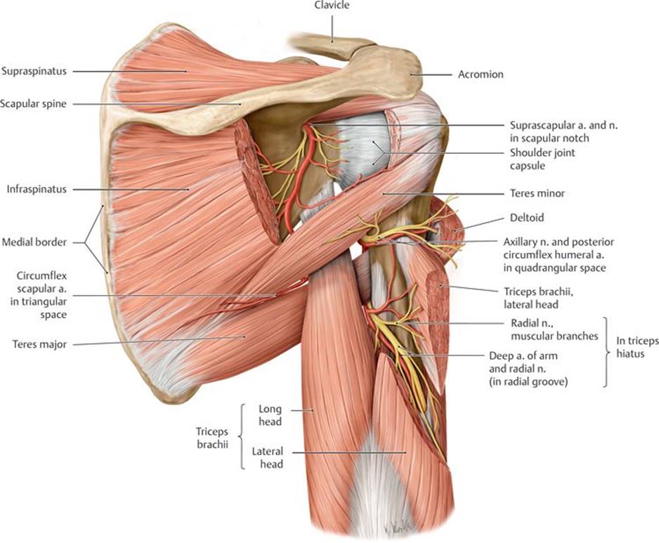
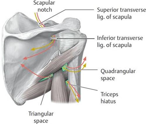
Fig. 14.10 Spaces of the posterior shoulder region
Right shoulder, posterior view. The course of arteries and nerves through the spaces are shown by red and yellow arrows in the schematic.
14.3 The Arm and Cubital Region
The arm (brachial region) extends from the shoulder to the elbow and contains the humerus and muscles of the arm. The cubital region contains the cubital fossa and the elbow joint.
14.3a Muscles of the Arm
Muscles of the arm move the shoulder and elbow joints. Brachial fascia that surrounds the arm divides these muscles into anterior and posterior compartments (Table 14.5).
– The anterior compartment contains
• muscles that flex the glenohumeral and elbow joints and supinate the radioulnar joint,
• the musculocutaneous nerve, and
• the brachial artery and vein.
– The posterior compartment contains
• muscles that extend the glenohumeral and elbow joints,
• the radial nerve, and
• the deep brachial artery and vein.
– The median and ulnar nerves descend along the medial side of the arm between the anterior and posterior compartments but do not innervate muscles of the arm.

A Biceps brachii, brachialis, and corachobrachialis, right arm, anterior view.

B Triceps brachii and anconeus, right arm, posterior view.

14.3b The Cubital Fossa
The cubital fossa is a shallow depression anterior to the elbow joint (Fig. 14.11A and B).
– Its boundaries are,
• medially, the pronator teres;
• laterally, the brachioradialis; and
• superiorly, a line that connects the medial and lateral epicondyles of the humerus.
– The cubital fossa contains
• the tendon of the biceps brachii,
• the brachial artery and vein,
• the proximal part of radial and ulnar arteries and veins, and
• the median and radial nerves and the cutaneous branch of the musculocutaneous nerve (lateral antebrachial cutaneous nerve).
– The bicipital aponeurosis, a fascial extension of the biceps brachii, forms the root of the fossa, and the medial cubital vein crosses the fossa superficially.
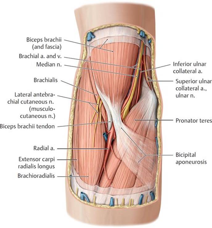
Fig. 14.11 Cubital region
Right elbow, anterior view.
A Superficial cubital fossa. Removed: Fasciae and epifascial neurovascular structures.

B Deep cubital fossa. Removed: Biceps brachii (distal muscle belly). Retracted: Brachioradialis.
14.3c The Elbow Joint
The elbow joint is made up of three separate synovial joints contained within a single joint capsule (Figs. 14.12A and B and 14.13).
– The hinge-type humeroulnar joint is an articulation between the trochlea of the humerus and the trochlear notch of the ulna.
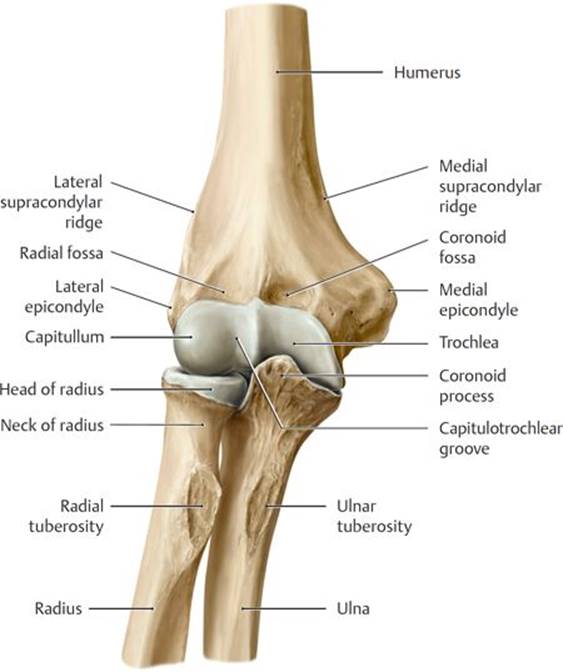
Fig. 14.12 Elbow (cubital) joint
Right elbow in extension. The elbow consists of three articulations: the humeroulnar, humeroradial, and proximal radioulnar.
A Anterior view.
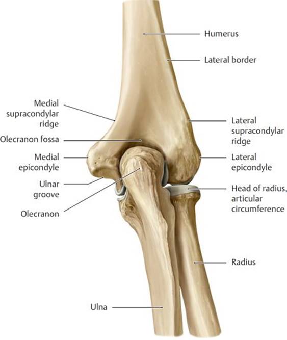
B Posterior view.
• The ulnar (medial) collateral ligament, which supports the joint medially, connects the coronoid process and olecranon with the medial epicondyle of the humerus.
– The hinge-type humeroradial joint is an articulation between the capitullum of the humerus and the head of the radius.
• The radial (lateral) collateral ligament, which supports the joint laterally, extends from the lateral epicondyle of the humerus to the annular ligament of the radius that encircles the radial neck.
– The proximal radioulnar joint, the articulation between the head of the radius and the radial notch of the ulna, is discussed further with the radioulnar joints of the forearm.
– Movements at the humeroulnar and humeroradial joints and the muscles that provide them are listed in Table 14.6.
TABLE 14.6 Movements at the Humeroulnar and Humeroradial Joints
|
Action |
Muscle |
|
Flexion |
Biceps brachii Brachialis Brachioradialis |
|
Extension |
Triceps brachii |
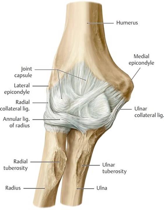
Fig. 14.13 Joint capsule of the elbow
Right elbow in extension, anterior view.
14.4 The Forearm
The forearm (antebrachial region) extends from the elbow to the wrist and contains the radius and ulna and the muscles of the forearm.
14.4a Radioulnar Joints
The radioulnar joints connect the bones of the forearm proximally at the elbow and distally at the wrist. Movement at these joints allows rotation of the distal radius around the ulna, causing supination (palm up) and pronation (palm down) of the hand (Figs. 14.14 and 14.15). These movements and the muscles of the arm and forearm that provide them are listed in Table 14.7.
TABLE 14.7 Movements at the Radioulnar Joints
|
Action |
Muscle |
|
Supination |
Supinator Biceps brachii |
|
Pronation |
Pronator teres Pronator quadratus |
– The proximal radioulnar joint is a synovial joint that allows rotation of the radial head within the cuff formed by the annular ligament and the radial notch of the ulna. This articulation is contained within the elbow joint capsule.
Subluxation of the radial head (nursemaid’s elbow)
In young children the immature radial head can be subluxated when the child’s arm is jerked upward. The movement tears the lax distal attachment of the annular ligament around the neck of the radius and allows the distal displacement of the radial head. The injured arm is held in a flexed and pronated position. Supination of the flexed elbow returns the joint to the correct orientation.
– The distal radioulnar joint has an L-shaped joint cavity with a triangular articular disk that separates the radio ulnar joint from the cavity of the wrist joint.
– An interosseous membrane connects the shafts of the radius and ulna and transfers energy absorbed by the distal radius to the proximal ulna.
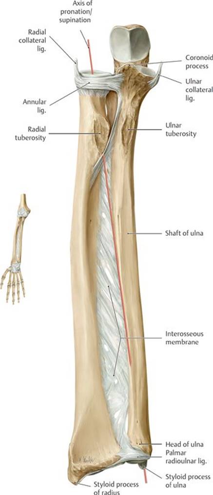
Fig. 14.14 Forearm in supination
Right forearm, anterior view.
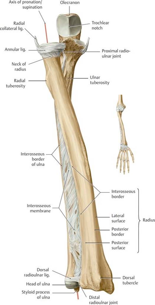
Fig. 14.15 Forearm in pronation
Right forearm, anterior view.
14.4b Muscles of the Forearm
Muscles of the forearm move joints of the elbow, wrist, and hand. Most forearm flexors and extensors have long tendons that cross the wrist and extend into the fingers. Intermuscular septa and the interosseous membrane create anterior and posterior muscular compartments.
– The anterior forearm compartment (Table 14.8) contains
• muscles that flex and pronate joints of the elbow, wrist, and hand;
• the median and ulnar nerves; and
• the ulnar and anterior interosseous arteries and veins.
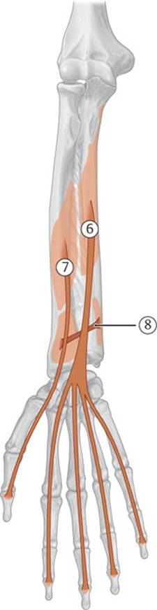
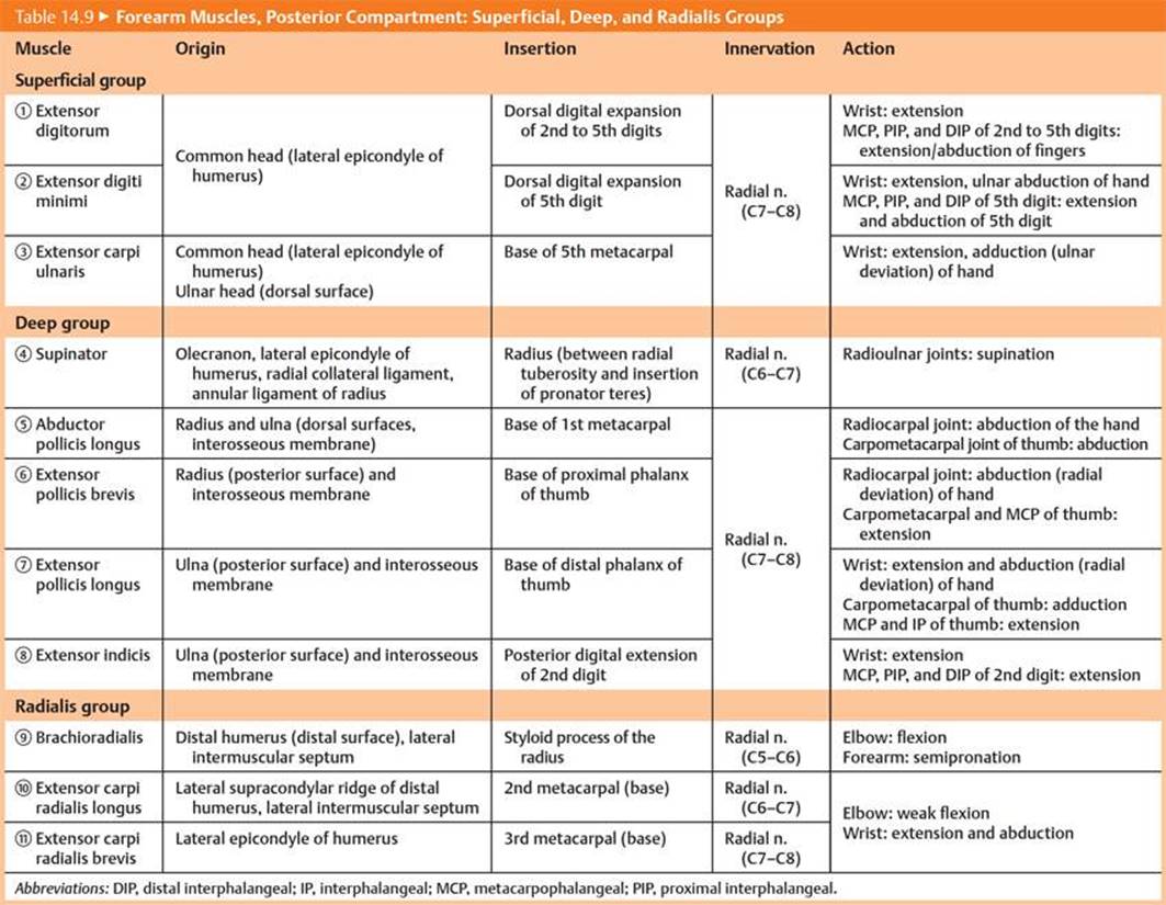
– The posterior forearm compartment (Table 14.9) contains
• muscles that extend joints of the elbow, wrist, and hand and supinate the radioulnar joint (one muscle, the brachioradialis, passes anterior to the elbow and therefore acts as a flexor, instead of an extensor of this joint);
• the radial nerve; and
• the radial and posterior interosseous arteries and veins.
Lateral epicondylitis
Repetitive use of the forearm extensors can inflame the attachment of the common extensor tendon at the lateral epicondyle (lateral epicondylitis). Pain is focused over the tendon insertion but radiates along the extensor forearm and is exacerbated by stretching of the extensor tendons by pronation and wrist flexion.
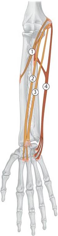
A Superficial muscles of anterior compartment of forearm.

B Intermediate muscle of anterior compartment of forearm.
C Deep muscles of anterior compartment of forearm.

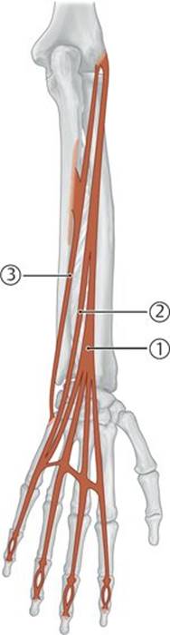
A Superficial muscles of posterior compartment of forearm.

B Deep muscles of posterior compartment of forearm.

C Radialis muscle group of posterior compartment of forearm.
14.5 The Wrist
The wrist, the narrow space between the forearm and hand, contains the carpal bones and the tendons of forearm muscles that move the wrist and fingers.
14.5a Joints of the Wrist
Joints of the wrist include the articulations between the distal radius and bones of the proximal row of carpals, articulations between adjacent carpal bones, and articulations between the distal carpal bones and the metacarpals of the hand (Fig. 14.16). Movements at the wrist joints and the muscles that provide them are listed in Table 14.10.
TABLE 14.10 Movements at the Wrist Joints
|
Action |
Muscle |
|
Flexion |
Flexor carpi radialis Flexor carpi ulnaris |
|
Extension |
Extensor carpi radialis longus Extensor carpi radialis brevis Extensor carpi ulnaris |
|
Abduction (radial deviation) |
Flexor carpi radialis Extensor carpi radialis longus Extensor carpi radialis brevis |
|
Adduction (ulnar deviation) |
Flexor carpi ulnaris Extensor carpi ulnaris |

Fig. 14.16 Joints of the wrist and hand
Right hand, posterior (dorsal) view.
– The radiocarpal joint is an articulation of the distal radius and articular disk of the distal radioulnar joint with the scaphoid and lunate.
• Palmar and dorsal radiocarpal ligaments and radial and ulnar collateral ligaments strengthen the joint.
– Intercarpal joints are the articulations between the carpal bones within each row; the midcarpal joint is the articulation between the bones of the proximal and distal carpal rows.
• Movements at these joints augment movements at the wrist joint.
– Carpometacarpal joints are the synovial articulations between the distal row of carpal bones and the metacarpals.
• Little or no movement occurs at the plane-type joints of the 2nd, 3rd, and 4th digits.
• The joint of the 5th digit between the metacarpus and the hamate is moderately mobile.
• The saddle-type joint between the metacarpus of the thumb and the trapezium in the distal carpal row allows movement in all directions, which is essential for thumb opposition (Fig. 14.17).
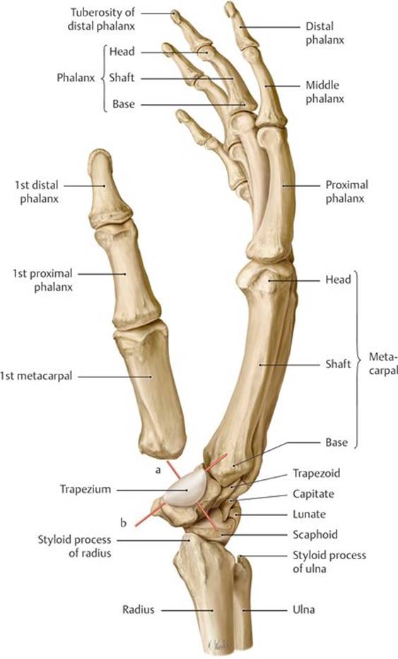
Fig. 14.17 Carpometacarpal joint of the thumb
Radial view. The 1st metacarpal bone has been moved slightly distally to expose the articular surface of the trapezium. Two cardinal axes of motion are shown here: (a) abduction/adduction and (b) flexion/extension.
14.5b Spaces of the Wrist
Neurovascular structures and the long tendons of the forearm muscles pass between the forearm and hand through narrow spaces that are usually defined by fascial thickenings.
– The carpal tunnel is a fascio-osseous space on the anterior wrist (Figs. 14.18A and B and 14.19).
• Carpal bones form the floor and sides; the flexor retinaculum forms the roof.
• The tendons of the flexor pollicis longus, flexor digitorum superficialis, and flexor digitorum profundus, and the median nerve pass through the tunnel.

Fig. 14.18 Carpal tunnel
Right hand, proximal view. The tight fit of sensitive neurovascular structures with closely apposed, frequently moving tendons in the carpal tunnel often causes problems (carpal tunnel syndrome) when any of the structures swell or degenerate.
A Cross section through the right wrist.
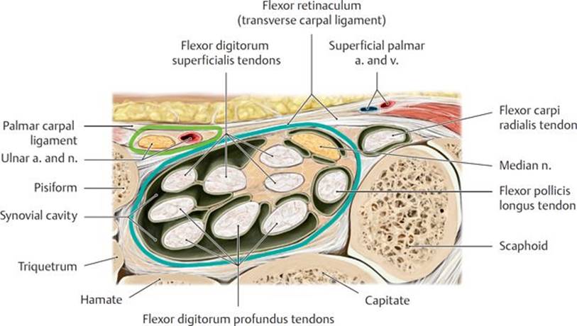
B Structures in the ulnar tunnel (green) and carpal tunnel (blue). Blowup of area enclosed by dash lines in A.
• A common flexor synovial tendon sheath encloses the flexor tendons as they pass through the carpal tunnel (see Fig. 14.32A).
• The palmar carpal ligament and flexor retinaculum prevent bowing of the flexor tendons as they cross the wrist.
Carpal tunnel syndrome
The carpal tunnel, defined by inflexible fibrous and osseous boundaries, can be compromised by the swelling of its contents, infiltration of fluid from inflammation or infection, protrusion of a dislocated carpal bone, or pressure from an external source. The median nerve is most sensitive to the increased pressure, and signs of carpal tunnel syndrome reflect the nerve’s distribution. These include tingling or numbness on the palmar surface of the lateral three and a half digits and weakness and eventual atrophy of the thenar muscles. The palmar cutaneous branch of the median nerve arises proximal to the canal and passes over the flexor retinaculum, so sensation of the palm remains intact.
– The ulnar tunnel (Guyon’s canal) is a narrow passageway on the medial side of the anterior wrist (see Figs. 14.18 and 14.19).
• The flexor retinaculum forms the floor, and the palmar carpal ligament forms the roof. The pisiform and hamate form the medial and lateral borders.
• The ulnar artery and nerve pass through the tunnel into the palm of the hand.
Ulnar nerve compression
Compression of the ulnar nerve at the wrist affects the innervation of most intrinsic hand muscles. When the patient attempts to form a fist, it results in a deformity known as a “claw hand”—the metacarpophalangeal joints are hyperextended due to the loss of the interossei muscles, and the interphalangeal joints are flexed.
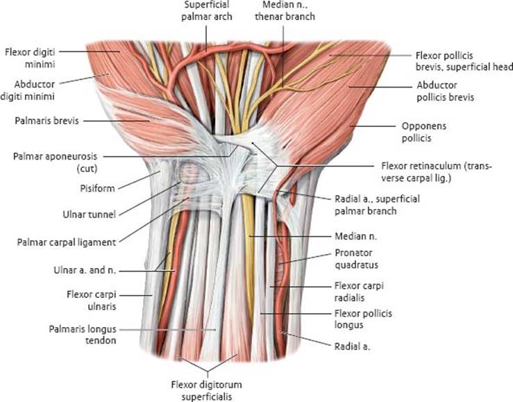
Fig. 14.19  Anterior carpal region
Right hand, anterior (palmar) view. Ulnar tunnel and deep palm. Carpal tunnel with flevor retinaculum transparent. Removed: Palmaris brevis, palmaris longus, palmar aponeurosis, and palmar carpal ligament.

Fig. 14.20 Anatomic snuffbox
Right hand, radial view. The three-sided “anatomic snuffbox” (shaded light yellow) is bounded by the tendons of insertion of the abductor pollicis longus and the extensor pollicis brevis and extensor pollicis longus.
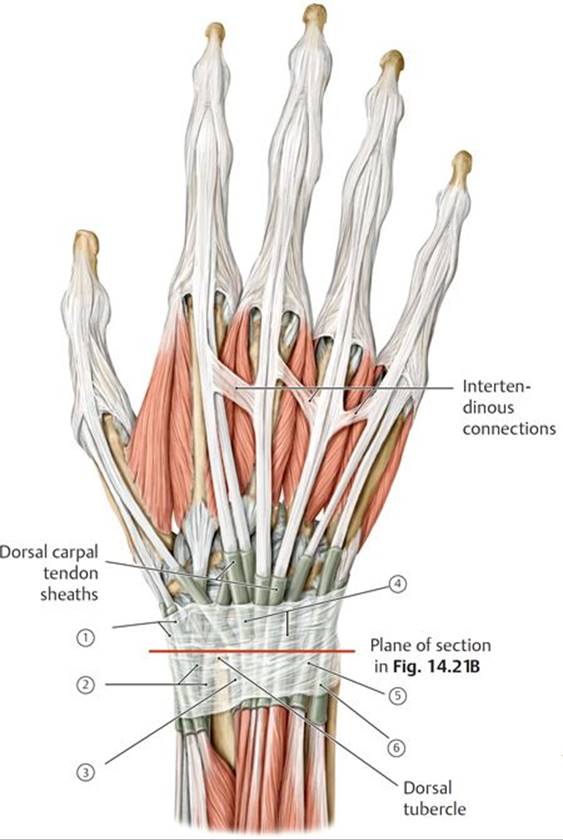
Fig. 14.21 Extensor retinaculum and dorsal compartments
Right hand.
A Posterior (dorsal) view.

B Proximal view of section indicated in A.
TABLE 14.11  Dorsal Compartments for Extensor Tendons
|
|
Abductor pollicis longus |
|
Extensor pollicis brevis |
|
|
|
Extensor carpi radialis longus |
|
Extensor carpi radialis brevis |
|
|
|
Extensor pollicis longus |
|
|
Extensor digitorum |
|
Extensor indicis |
|
|
|
Extensor digiti minimi |
|
|
Extensor carpi ulnaris |
– The anatomic snuffbox is a small triangular depression on the radial side of the dorsum of the wrist (Fig. 14.20).
• Tendons of the extensor pollicis longus, extensor pollicis brevis, and abductor pollicis longus form the borders. The scaphoid and trapezium form its floor.
• The radial artery passes through the snuffbox.
• The cephalic vein and superficial branch of the radial nerve cross the snuffbox superficially.
– Six small dorsal compartments (designated as 1st through 6th) form on the posterior surface of the wrist (Table 14.11; Fig. 14.21A and B).
• The extensor retinaculum forms their roof, and the dorsal surfaces of the distal radius and ulna form their floor.
• Extensor tendons of forearm muscles pass through the compartments onto the dorsum of the hand.
• Dorsal carpal synovial tendon sheaths enclose the extensor tendons as they pass through the dorsal compartments.
14.6 The Hand
The muscles and joints of the hand create a flexible tool that is adept at fine motor movements. The ability to grasp objects by positioning the thumb in opposition to the other fingers is a feature that is unique to humans and apes.
14.6a Joints of the Hand and Fingers
Joints of the hand and fingers are the articulations between the metacarpal bones of the palm and the proximal phalanges and between the proximal, middle, and distal phalanges of each digit (see Fig. 14.16). Movements at these joints and the muscles of the forearm and hand that provide them are listed in Tables 14.12 and 14.13.
TABLE 14.12 Movements at the Joints of the Fingers (Digits 2 through 5)
|
Action |
Muscle |
|
Flexion at MCP |
Lumbricals Interossei Flexor digiti minimi (only 5th digit) |
|
Flexion at DIP |
Flexor digitorum superficialis Lumbricals |
|
Flexion at PIP |
Flexor digitorum profundus |
|
Extension at MCP |
Extensor digitorum Extensor indicis (only 3rd digit) Extensor digiti minimi (only 5th digit) |
|
Extension at DIP and PIP |
Lumbricals Interossei |
|
Abduction at MCP |
Dorsal interossei Abductor digiti minimi (only 5th digit) |
|
Adduction |
Palmer interossei (digits 2, 4, and 5 only) |
|
Opposition |
Opponens digiti minimi (only 5th digit) |
|
Abbreviations: DIP, distal interphalangeal; MCP, metacarpophalangeal; PIP, proximal interphalangeal. |
|
TABLE 14.13 Movements at Joints of the Thumb
|
Action |
Muscle |
|
Flexion |
Flexor pollicis longus Flexor pollicis brevis |
|
Extension |
Extensor pollicis longus Extensor pollicis brevis |
|
Abduction |
Abductor pollicis longus Abductor pollicis brevis |
|
Adduction |
Adductor pollicis |
|
Opposition |
Opponens pollicis |
– Metacarpophalangeal (MCP) joints are condyloid synovial joints between the heads of the metacarpals and bases of the proximal phalanges.
• Movement in two planes, flexion-extension and abduction-adduction, occurs in digits 2 through 5.
• Only flexion and extension occur at the MCP joint of the thumb.
– Interphalangeal (IP) joints are hinge-type synovial joints between phalanges.
• Digits 2 through 4 have proximal interphalangeal (PIP) and distal interphalangeal (DIP) joints.
• The thumb has only a single IP joint.
• IP joints permit only flexion and extension.
– MCP and IP joints are surrounded by a fibrous capsule and supported by medial and lateral collateral ligaments.

Fig. 14.22 Surface anatomy of the dorsum of the hand
Right hand.
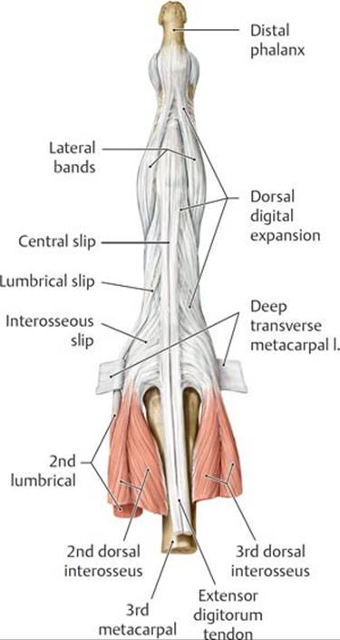
Fig. 14.23 Dorsal digital expansion
Right hand, middle finger, posterior view. The dorsal digital expansion permits the long digital flexors and the short muscles of the hand to act on all three finger joints.
14.6b The Dorsum of the Hand and Fingers
– The dorsum of the hand has the following surface anatomy (Fig. 14.22):
• The skin is thin and loose.
• A prominent superficial dorsal venous network gives rise to the cephalic and basilic veins.
• The heads of the metacarpals of the 2nd through 5th digits form distinct “knuckles” when the hand is flexed into a fist.
• The extensor tendons fan out from the wrist to the fingers.
– On the dorsum of the fingers, the long extensor tendons (of the posterior forearm compartment) flatten to form a dorsal digital expansion (extensor expansion, extensor hood), a triangular tendinous aponeurosis (Fig. 14.23). The dorsal digital expansion
• forms a hood that wraps around the sides of the distal metacarpus and proximal phalanx and holds the extensor tendon in place;
• inserts onto the middle and distal phalanges via a central slip and paired lateral bands; and
• is reinforced by the lumbricals and interossei muscles of the palm, which connect to the lateral bands and assist in extension of the interphalangeal joints of the fingers.
Extensor tendon rupture
Traumatic finger injuries can cause tendon ruptures that result in a variety of deformities. Rupture of the extensor tendon at the distal phalanx prohibits extension of the distal interphalangeal (DIP) joint. The unopposed action of the flexor digitorum profundus produces the characteristic “mallet finger” deformity in which the distal phalanx remains flexed.
14.6c The Palm of the Hand and Fingers
– The palm of the hand has the following surface anatomy (Fig. 14.24):
• The skin is thickened, firmly attached to the underlying fascia, and supplied with numerous sweat glands.
• A central concavity separates a thenar eminence at the base of the thumb from a hypothenar eminence at the base of the 5th digit.
• Longitudinal and transverse flexion creases form where the skin is tightly bound to the palmar fascia.
– Deep fascia over the central palm forms a tough, thickened palmar aponeurosis (Fig. 14.25), which
• firmly adheres to the skin of the palm,
• is continuous proximally with the flexor retinaculum and palmaris longus muscle, and
• is continuous distally with a transverse metacarpal ligament and the four digital fibrous sheaths of the fingers that surround the long flexor tendons and their synovial digital tendon sheaths.
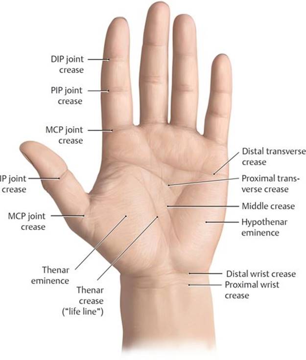
Fig. 14.24 Surface anatomy of the palm of the hand
Left hand. DIP, distal interphalangeal; IP, interphalangeal; MCP, meta-carpophalangeal; PIP, proximal interphalangeal.

Fig. 14.25 Palmar aponeurosis
Right hand, palmar surface.
* Also known as tranverse carpal ligament
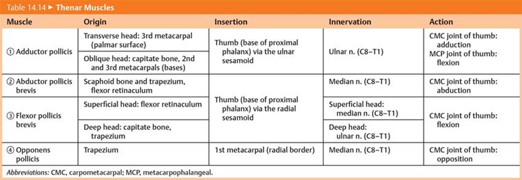
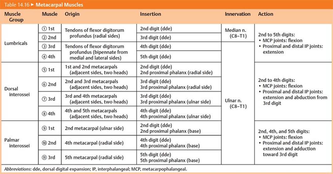
– The palmar aponeurosis and deep palmar fascia divide the palm into five muscular compartments (Tables 14.14, 14.15, and 14.16):
1. The thenar compartment, which contains thenar muscles that abduct, flex, and oppose the thumb.
2. The central compartment, which contains forearm flexor tendons that flex the fingers and lumbricals that flex and extend joints of the fingers.
3. The hypothenar compartment, which contains hypothenar muscles that flex, abduct, and oppose the 5th digit.
4. The adductor compartment, which contains the adductor pollicis muscle that adducts the thumb.
5. The interosseous compartment, which contains interossei muscles that abduct and adduct the fingers.
– Thenar and midpalmar spaces are potential spaces deep within the palm between the long flexor tendons and the fascia over the deep palmar muscles. The midpalmar space is continuous with the anterior forearm compartment through the carpal tunnel.
Dupytren’s disease
Dupytren’s disease is the progressive fibrosis and contracture of the longitudinal bands of the palmar fascia to the 4th and 5th digits, causing flexion of these fingers. It presents as painless nodular thickenings that progress to raised ridges on the palm. Surgical excision is usually required to release the bands.
– On the palmar surface of the fingers (see Fig. 14.33A),
• tendons of the flexor digitorum superficialis (FDS) split into two bands that insert on the middle phalanx,
• tendons of the flexor digitorum profundus pass between the bands of the FDS to insert on the distal phalanx, and
• synovial tendon sheaths surround the flexor tendons as they enter the fibrous tendon sheaths of the fingers (see Fig. 14.32A).
○ The digital synovial sheath of the 5th digit normally communicates with the common flexor sheath at the wrist.
○ The synovial sheath of the thumb extends into the wrist and may communicate with the sheath of the 5th digit and with the common synovial sheath.
○ The synovial sheaths of the 2nd, 3rd, and 4th digits usually remain independent from the common synovial sheath and other digital synovial sheaths.
– Muscles of the forearm and intrinsic muscles of the hand move the joints of the hand and fingers (see Tables 14.12 and 14.13).

Thenar and hypothenar muscles, right hand, anterior (palmar) view.

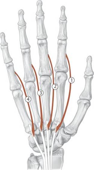
A Lumbricals, right hand, palmar view.
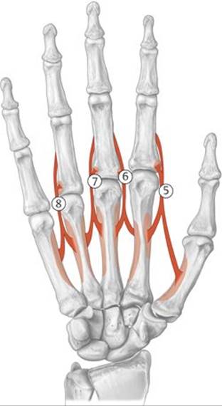
B Dorsal interossei, right hand, palmar view.

C Palmar interossei, right hand, palmar view.
Tenosynovitis
A puncture wound of the finger can initiate an infection in the digital synovial sheaths (tenosynovitis). Swelling of the tissue is painful and inhibits movement. Although synovial sheath connections vary, sheaths of the 2nd, 3rd, and 4th digits are usually independent and can confine the infection to that digit (unless the sheath ruptures). The sheath of the 5th digit, however, normally communicates with the common synovial sheath in the palm and wrist. Infection can track via this route through the carpal tunnel and into the anterior compartment. Likewise, infection in the thumb can track into the wrist and palm.
14.7 Topographic Views of Upper Limb Musculature
14.7a Shoulder and Arm
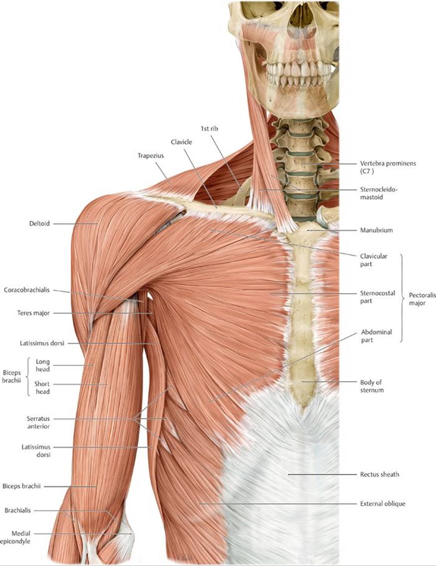
Fig. 14.26 Anterior shoulder muscles
Right side, anterior view. Muscle origins are shown in red, insertions in gray.
A Superficial dissection.
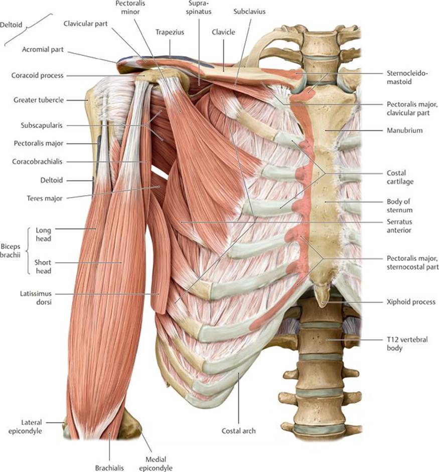
B Deep dissection. Removed: Sternocleidomastoid, trapezius, pectoralis major, deltoid, and external oblique muscles.

Fig. 14.27 Anterior dissection of arm
Right arm, anterior view. Muscle origins are shown in red, insertions in gray.
A Removed: Latissimus dorsi and serratus anterior.

B Removed: Subscapularis and supraspinatus muscles. Partially removed: Biceps brachii.

Fig. 14.28 Posterior shoulder muscles
Right side, posterior view.
A Superficial dissection.
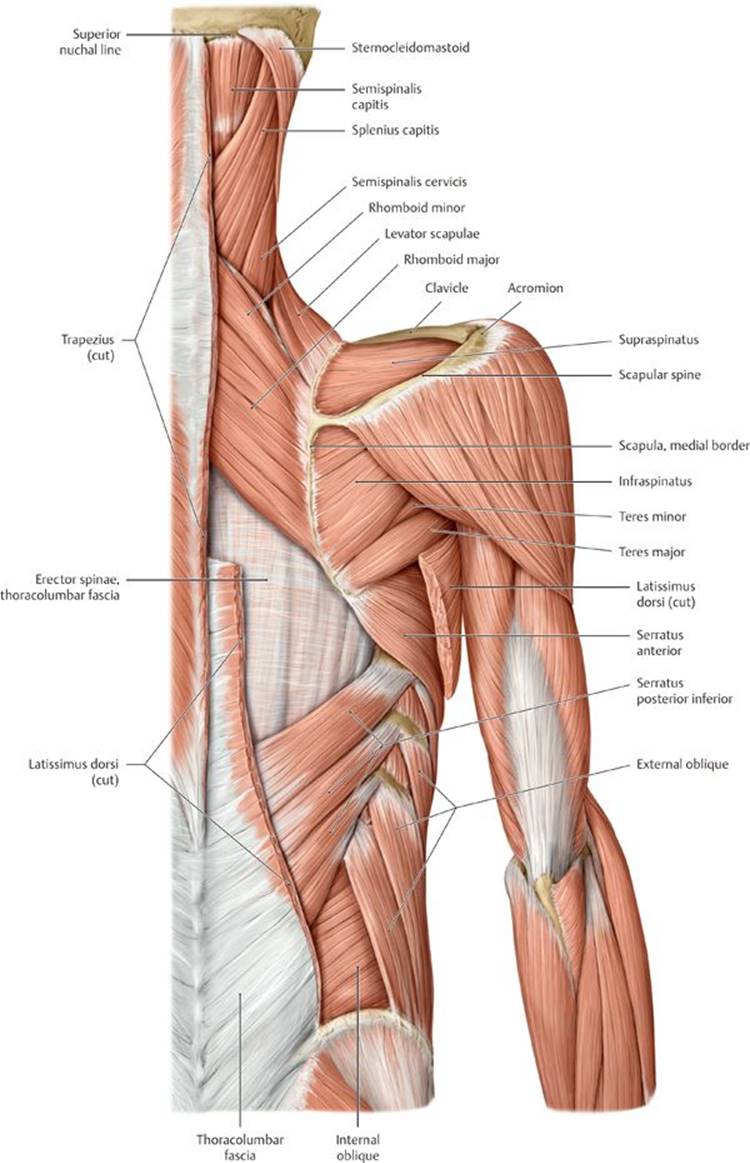
B Deep dissection. Partially removed: Trapezius and latissimus dorsi.

Fig. 14.29 Posterior dissection of arm
Right arm, posterior view. Muscle origins are shown in red, insertions in gray. Removed: Deltoid and forearm muscles.
14.7b Forearm and Wrist

Fig. 14.30 Anterior forearm muscles
Right forearm, anterior view. Muscle origins are shown in red, insertions in gray.
A Superficial flexors and radialis group.

B Removed: Radialis group (brachioradialis, extensor carpi radialis longus, and extensor carpi radialis brevis), flexor carpi radialis, flexor carpi ulnaris, abductor pollicis longus, palmaris longus, and biceps brachii.

C Removed: Pronator teres and flexor digitorum superficialis.

Fig. 14.31 Posterior forearm muscles
Right forearm, posterior view. Muscle origins are shown in red, insertions in gray.
A Superficial extensors and radialis group.

B Removed: Triceps brachii, anconeus, flexor carpi ulnaris, extensor carpi ulnaris, and extensor digitorum.
14.7c Hand

Fig. 14.32 Intrinsic muscles of the hand: Superficial and middle layers
Right hand, palmar surface.
A Superficial layer, with carpal and digital tendon sheaths exposed. Removed: Palmar aponeurosis, palmaris longus, antebrachial fascia, and palmaris brevis.
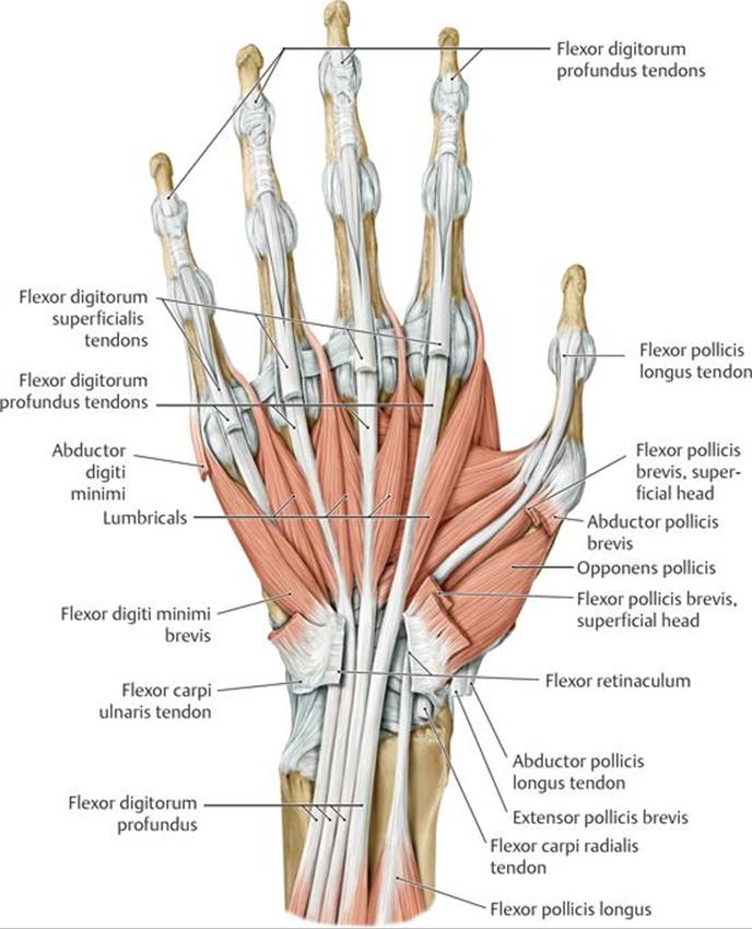
B Middle layer. Removed: Flexor digitorum superficialis, flexors carpi radialis and ulnaris, and pronator quadratus.
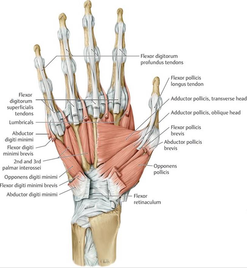
Fig. 14.33 Intrinsic muscles of the hand: Middle and deep layers Right hand, palmar surface.
A Middle layer. Cut: Flexor digitorum profundus, lumbricals, flexor pollicis longus, and flexor digiti minimi brevis.

B Deep layer. Cut: Opponens digiti minimi, opponens pollicis, flexor pollicis brevis, and adductor pollicis (transverse and oblique heads).
14.7d Compartments in Arm and Forearm

Fig. 14.34 Windowed dissection
Right limb, anterior view.
A Dissection of the arm.

B Dissection of the forearm.
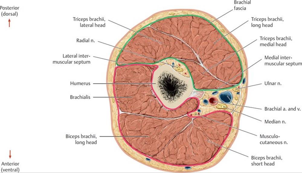
Fig. 14.35 Transverse sections
Right limb, proximal (superior) view. The anterior compartment is outlined in pink and the posterior compartment in green.
A Arm (plane of section in Fig. 14.34A).
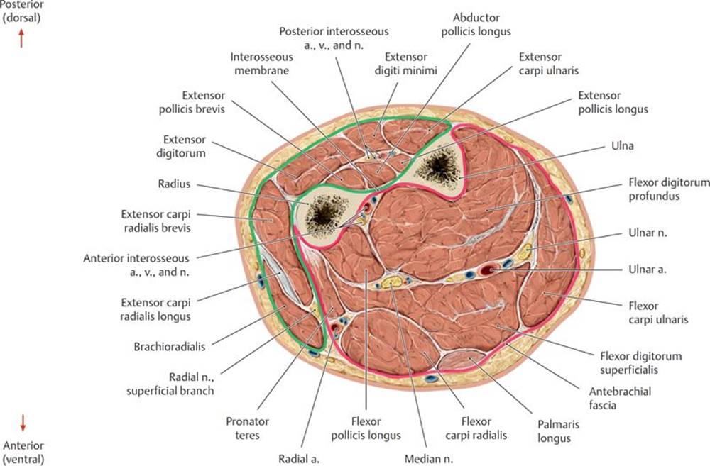
B Forearm (plane of section in Fig. 14.34B).


