
The back includes the vertebral column, the spinal cord and spinal nerves, and the overlying muscles and skin.
2.1a General Features
– The vertebral column
• encloses and protects the spinal cord,
• supports the head and trunk,
• provides an attachment for the limbs, and
• transfers the weight of the body to the lower limbs.
– The vertebral column, which extends from the skull to the coccyx, comprises 33 vertebrae and the intervening intervertebral disks, which are divided among five regions (Fig. 2.1):
• 7 cervical vertebrae
• 12 thoracic vertebrae
• 5 lumbar vertebrae
• 5 fused sacral vertebrae
• 4 (3–5) fused coccygeal vertebrae
Osteoporosis
The spine is the primary target for degenerative diseases of the skeleton, such as osteoporosis, in which the rate of reabsorption by osteoclasts exceeds that of bone formation by osteoblasts. The resulting loss of bone mass predisposes the individual to compression fractures of the spine.
– Within each region, individual vertebrae are identified by number, a designation often referred to as a vertebral level (such as T8 vertebral level).
– Vertebrae increase in size from the cervical to the lumbar regions and decrease in size from the top of the sacrum to the coccyx.
– The kyphotic curvatures of the thoracic and sacral regions of the vertebral column, known as primary curvatures, are curved posteriorly and are present in the fetus. The lordotic curvatures of the cervical and lumbar regions, curved anteriorly, are secondary curvatures that develop postnatally.

Fig. 2.1  Vertebral column
Left lateral view.
Abnormal curvatures of the vertebral column: kyphosis, lordosis, and scoliosis
Kyphosis (“hunchback”), an excessive anterior curvature of the thoracic spine, is often seen in elderly women. Although it may be congenital or posture related, it is usually secondary to degenerative changes (collapse) of the vertebral bodies. Lordosis (“swayback”), an excessive posterior curvature of the lumbar spine, frequently develops as a temporary side effect during pregnancy, but in nonpregnant individuals it may have pathologic or even weight-related causes. Scoliosis is a lateral curvature of the spine and may be congenital or neuromuscular, caused by diseases such as cerebral palsy and muscular dystrophy.
– A vertebral canal passes through the center of the vertebral column and encloses the spinal cord, the spinal meninges (membranes surrounding the spinal cord), and the roots of the spinal nerves, and associated vasculature (see Sections 2.3b, d).
– Intervertebral foramina, openings between vertebrae, allow the passage of spinal nerves.
– Spinal cord segments emit paired spinal nerves, and both the segment and the spinal nerve pair are identified by region and number (such as L4). Spinal cord segments do not necessarily lie adjacent to vertebra of the same number.
2.2 The Vertebral Column
2.2a Regional Characteristics of Vertebrae
Most vertebrae share a typical form (Fig. 2.2), although specific features vary by region.
– Most vertebrae have the following:
• An anterior vertebral body
• A posterior vertebral arch formed by paired pedicles and paired laminae (the pedicles attach to the vertebral body, and the paired laminae join to form a spinous process)
• Paired transverse processes that project laterally from the vertebral arch
• Superior and inferior articular processes that articulate with the vertebrae above and below
• A vertebral foramen encircled by the vertebral body and vertebral arch (the combined vertebral foramina of all vertebrae form the vertebral canal)
– Cervical vertebrae, the smallest of all of the vertebrae, support the head and form the posterior skeleton of the neck (Fig. 2.3). The seven cervical vertebrae are characterized as typical or atypical.

Fig. 2.2 Structural elements of a vertebra
Left posterosuperior view. With the exception of the atlas (C1) and axis (C2), all vertebrae consist of the same structural elements.
Spina bifida
Spina bifida is a congenital defect associated with the incomplete fusion of the vertebral arches (usually L5 and S1). In the mild form, spina bifida occulta, the defect is small and asymptomatic and is often marked only by a patch of hair, birthmark, or dimple over the affected vertebrae. Severe forms such as spina bifida cystica are caused by incomplete closure of the embryonic neural tube, resulting in incomplete development of the spinal cord, meninges, and overlying vertebrae. Severe cases are associated with neurologic deficits and herniation of the spinal cord and meninges through the vertebral arch defect.

Fig. 2.3  Cervical spine
Bones of the cervical spine, left lateral view.
• Typical cervical vertebrae (Fig. 2.4A)
○ C3–C6 have a small body, a large vertebral foramen, and often bifid (two-pronged) spinous processes.
• Atypical cervical vertebrae
○ C1, the atlas, lacks a vertebral body and spinous process (Fig. 2.4B). It has anterior and posterior vertebral arches that are connected on each side by lateral masses. C1 articulates with the occipital bone of the skull and C2.
○ C2, the axis, has a peglike dens projecting superiorly from its body that articulates with the anterior arch of C1 (Fig. 2.4C).
○ C7, the vertebra prominens, has a long, palpable spinous process.
• All cervical vertebrae have paired transverse foramina, openings formed by the anterior and posterior tubercles of each transverse process.
– Paired vertebral arteries ascend in the neck through the transverse foramina of C1–C6, pass through a groove on the posterior arch of C1, and enter the skull through a large opening, the foramen magnum.
– Thoracic vertebrae (Fig. 2.5A and B) have
• long spinous processes that project inferiorly,
• heart-shaped vertebral bodies,
• superior and inferior articular facets that are oriented in the coronal plane, and
• costal facets that articulate with the ribs.
– Lumbar vertebrae (Fig. 2.6A and B), the largest vertebrae, have
• large bodies,
• short, broad spinous processes, and
• an interarticular part (pars interarticularis), part of the lamina between the superior and inferior articular facets, that forms the neck of the “Scottie dog” seen on oblique views of lumbar spine radiographs. It is a common site of vertebral fractures (Fig. 2.7, Table 2.1).
Spondylolysis and spondylolisthesis
Spondylolysis is a fracture across the interarticular part of the lamina, usually at L5, and appears as a “collar” on the Scottie dog seen on lumbar radiographs. When the defect is bilateral, the vertebral body may separate from its vertebral arch and shift anteriorly relative to the vertebra below it, a condition known as spondylolisthesis. Mild cases can be asymptomatic, but more severe cases compress the spinal nerves and cause pain in the lower limbs and back.

Fig. 2.4 Cervical vertebrae
A Typical cervical vertebra (C4), superior view.
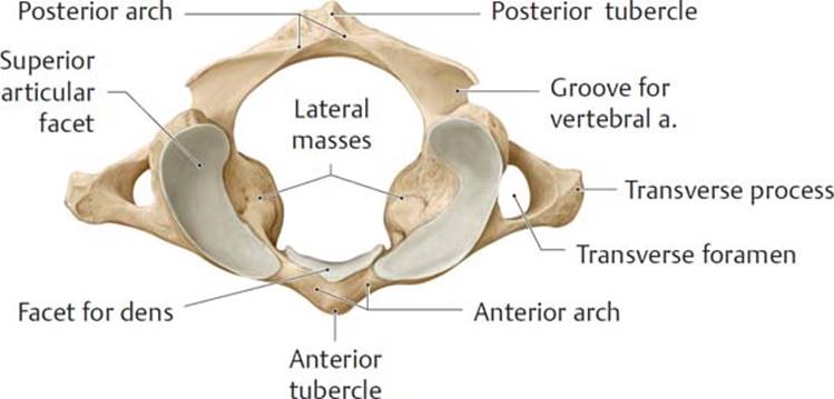
B Atlas (C1), superior view.

C Axis (C2), left lateral view.

Fig. 2.5 Thoracic spine
A Bones of the thoracic spine, left lateral view.

B Typical thoracic vertebra (T6), superior view.

Fig. 2.6 Lumbar spine
A Bones of the lumbar spine, left lateral view.
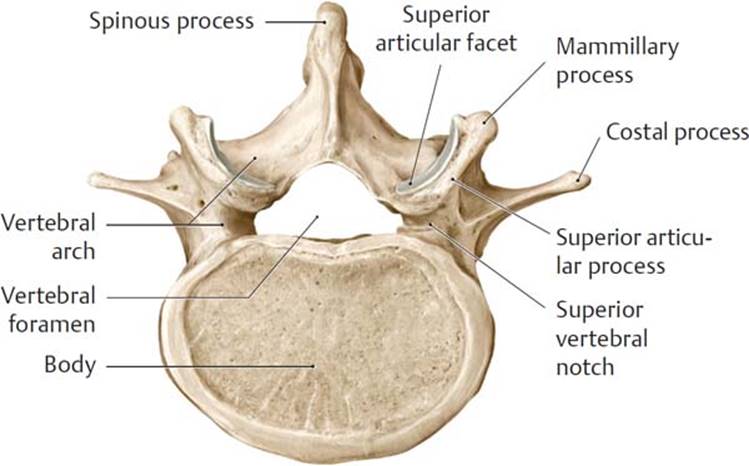
B Typical lumbar vertebra (L4), superior view.
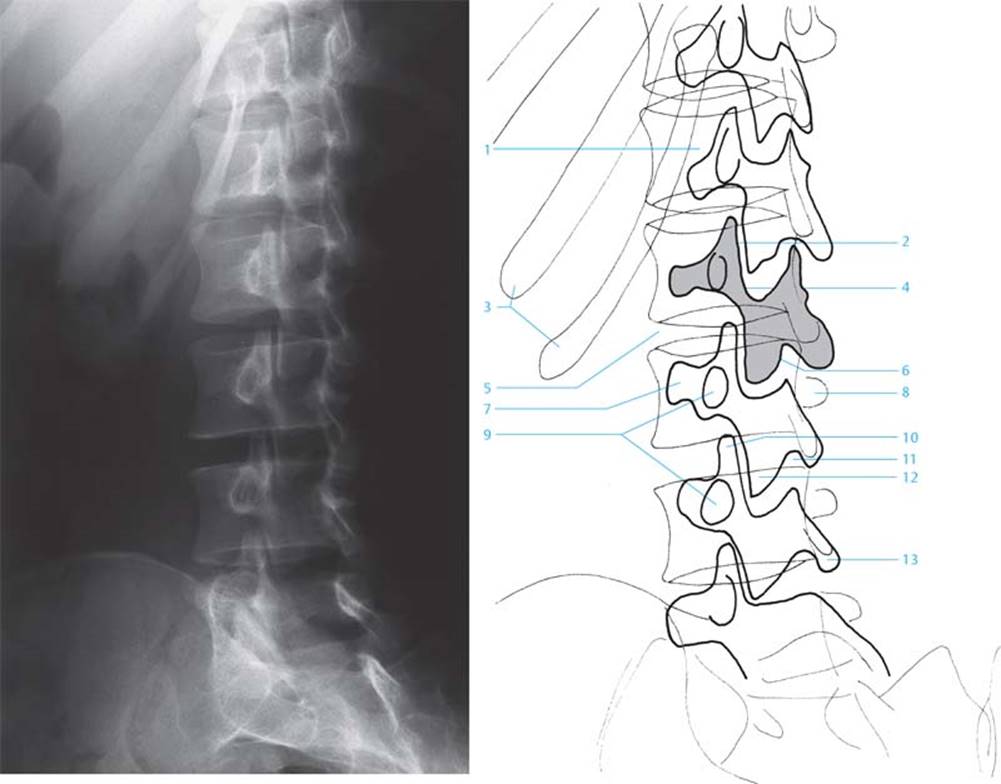
Fig. 2.7 Oblique view of the lumbar spine
1. Body of vertebra
2. Intervertebral disk space
3. Ribs
4. Interarticular part
5. Intervertebral disk space
6. Lamina
7. Ipsilateral transverse process
8. Contralateral transverse process
9. Pedicle
10. Superior articular process
11. Intervertebral foramen
12. Inferior articular process
13. Spinous process
– The five sacral vertebrae are fused into a single bone, the sacrum (Fig. 2.8A, B, and C), which forms the posterosuperior wall of the pelvis and articulates laterally with the hip bones. The sacrum contains
• the sacral canal, a continuation of the vertebral canal, which is open inferiorly at the sacral hiatus;
• the median sacral crest, the fused spinous processes of the sacral vertebrae;
• paired medial sacral crests that end inferiorly as the sacral cornua on either side of the sacral hiatus;
• four pairs of anterior and posterior sacral foramina for the passage of spinal nerve branches; and
• the promontory, formed by the anterior lip of the S1 vertebral body.
– The small coccygeal vertebrae, usually four (but this can vary from three to five), fuse into a single triangularly shaped bone, the coccyx, which articulates with the sacrum at the sacrococcygeal joint.

Fig. 2.8  Sacrum and coccyx
A Anterior view.

B Posterior view.

C Left lateral view.
2.2b Joints of the Vertebral Column
Joints of the vertebral column include articulations between adjacent vertebral bodies and articulations between adjacent vertebral arches. Joints also form between the vertebral column and the skull (Table 2.1). Individual vertebral joints allow small local movements, but the combination of these movements over multiple vertebral levels accounts for the considerable flexibility of the vertebral column.

TABLE 2.1  Joints of the Vertebral Column
|
Craniovertebral joints |
||
|
|
Atlanto-occipital joints |
Occiput–C1 |
|
|
Atlantoaxial joints |
C1–C2 |
|
Joints of the vertebral bodies |
||
|
|
Uncovertebral joints |
C3–C7 |
|
|
Intervertebral joints |
C2–S1 |
|
Joints of the vertebral bodies |
||
|
|
Zygapophyseal |
joints C1–S1 |
– Craniovertebral joints (Fig. 2.9A and B) are synovial joints between the skull and C1, and between C1 and C2:
• Paired atlanto-occipital joints between the occipital bone of the skull and the atlas (C1) allow flexion and extension of the head (as when nodding “yes”).
• Atlantoaxial joints, which include one median and two lateral articulations between the atlas and axis (C1 and C2), allow rotation of the head (as when saying “no”).
Injuries of the cervical spine
The laxity of the cervical spine makes it prone to hyperextension injuries, such as “whiplash,” the excessive and often violent backward movement of the head, resulting in fractures of the dens of the axis and traumatic spondylolisthesis (see Spondylolysis and Spondylolisthesis on page 22). Patient prognosis is largely dependent on the spinal level of the injuries.

Fig. 2.9 Craniovertebral joints
A Posterior view.
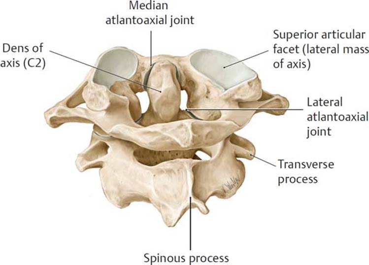
B Atlas and axis, posterosuperior view.
– Uncovertebral joints form between the uncinate processes (lateral lips on the superior edges of the vertebral bodies) of C3–C7 vertebrae and the vertebral bodies immediately superior to them.
• These joints, which are not present at birth, form during childhood, probably as a result of a fissure in the cartilage of the intervertebral disk that then assumes a jointlike character.
– Intervertebral joints form between intervertebral (IV) disks and the articular surfaces of vertebral bodies . There are no IV disks between C1 and C2.
• The IV disks act as shock absorbers and are composed of an outer fibrous ring, the anulus fibrosus, and a gelatinous core, the nucleus pulposus (Fig. 2.10).
• The height of the IV disk relative to the height of the vertebral body determines the degree of mobility of the joint; mobility is greatest in the cervical and lumbar regions.
• The differences between anterior and posterior heights of the cervical and lumbar disks contribute to the lordotic curvatures.

Fig. 2.10 Intervertebral disk
Fourth lumbar vertebra, superior view.
Herniation of intervertebral disks
As elasticity of the anulus fibrosus declines with age, compressive forces can cause the nucleus pulposus to protrude through weakened areas. If the fibrous ring of the anulus ruptures posteriorly, the herniated material may compress the contents of the dural sac, but posterolateral herniations that compress spinal nerves are most common, particularly at the L4–L5 or L5–S1 level. In the lumbar region, where spinal nerves exit the vertebral canal above the IV disk, the hernia is likely to compress the spinal nerve inferior to that level (e.g., a herniation of the L4–L5 disk will impact the L5 spinal nerve), and pain is felt along the corresponding dermatome.
– Zygapophyseal joints, also known as facet joints, are synovial joints that join the superior and inferior articular facets of adjacent vertebrae. The orientation of these joints differs between regions and influences the degree and direction of movement of the vertebral column.
• In the cervical region, the joints are mostly in the horizontal plane and allow movement in most directions.
• In the thorax, the joints largely lie in the coronal plane, limiting movement to lateral flexion.
• In the lumbar region, the joints are in the sagittal plane, facilitating flexion and extension.
Age-related changes in vertebrae
With advancing age, a decrease in bone density and aging of the IV disks can lead to an increase in compressive forces on the vertebral joints. Subsequent degenerative changes can include the depletion of articular cartilage and the formation of osteophytes (bony spurs). Osteophyte formation at the periphery of the vertebral bodies where they join the IV disks is known as spondylosis. Similar degenerative changes of the zygapophyseal joints indicate osteoarthritis, common in the cervical and lumbar spine but also manifested in the joints of the hand, hip, and knee.
2.2c Vertebral Ligaments
Vertebral ligaments support the joints of the vertebral column.
– Ligaments that support the cranial vertebral joints include (Fig. 2.11A and B)
• the atlanto-occipital membranes, which connect the occipital bone of the skull to the anterior and posterior arches of the atlas (C1);
• the alar ligaments, which secure the dens of C2 to the skull; and
• the cruciform ligament, formed by longitudinal fascicles (fibers) and a transverse ligament, which secures the dens against the anterior arch of the atlas.

Fig. 2.11 Dissection of the craniovertebral joint ligaments
Posterior view.
A Posterior longitudinal ligament. Removed: Spinal cord; vertebral canal windowed.

B Cruciform ligament of atlas (*). Removed: Tectorial membrane.
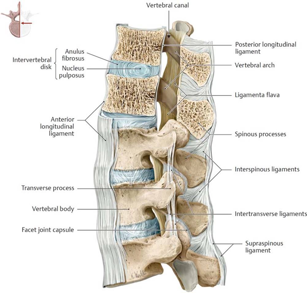
Fig. 2.12 Ligaments of the vertebral column: Thoracolumbar junction
Left lateral view of T11–L3, with T11–T12 sectioned in the midsagittal plane.
– Two longitudinal ligaments join all of the vertebral bodies (Figs. 2.12 and 2.13):
1. the anterior longitudinal ligament, a broad fibrous band extending from the occipital bone of the skull to the sacrum, attaches to the anterior and lateral surfaces of the vertebral bodies and IV disks and prevents hyperextension.
2. the posterior longitudinal ligament, a thin fibrous band extending from C2 to the sacrum along the anterior aspect of the vertebral canal, attaches primarily to the IV disks and offers weak resistance to hyperflexion. Superiorly, this ligament extends into the skull as the tectorial membrane (see Fig. 2.11A).
– Ligaments that join the vertebral arches (Fig. 2.14) include the following:
• the paired ligamenta flava, which join the laminae of adjacent vertebrae on the posterior wall of the vertebral canal. They limit flexion and provide postural support of the vertebral column (see also Fig. 2.12).
• the supraspinous ligament, which connects the posterior ridge of the spinous processes
• the nuchal ligament, a finlike expansion of the supraspinous ligament in the neck that extends from the occipital bone to the spinous process of C7
– Additional vertebral ligaments connect elements of the vertebral arches and spinous processes (see Fig. 2.12).
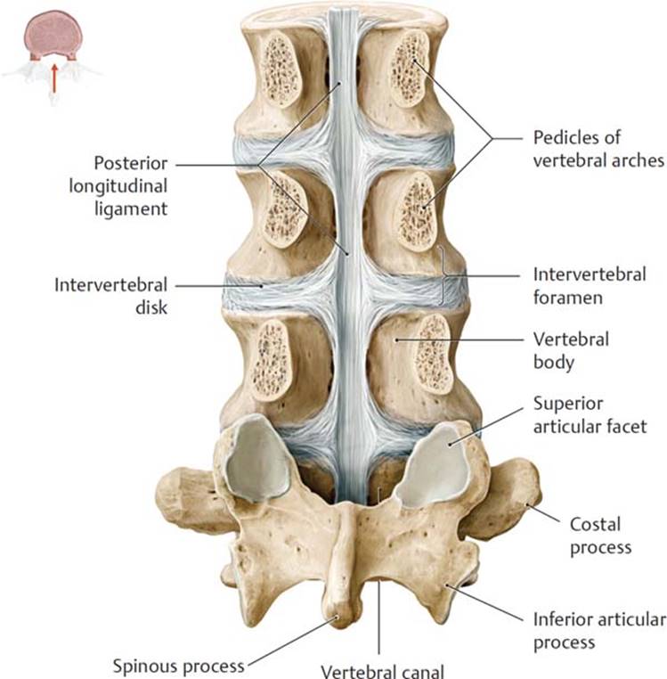
Fig. 2.13 Posterior longitudinal ligament
Posterior view of opened vertebral canal at level of L2–L5. Removed: L2–L4 vertebral arches at pedicular level.
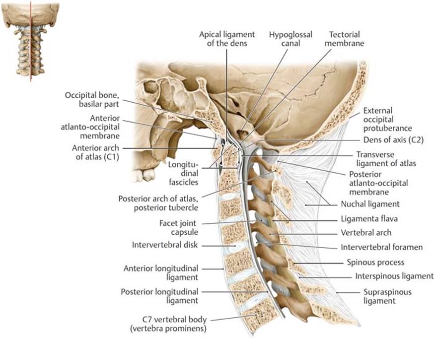
Fig. 2.14 Ligaments of the cervical spine
Midsagittal section, left lateral view. The nuchal ligament is the broadened, sagittally oriented part of the supraspinous ligament that extends from the vertebra prominens (C7) to the external occipital protuberance.
2.2d Neurovasculature of the Vertebral Column
– The following arteries supply the vertebrae, vertebral ligaments, meninges, and spinal cord (Fig. 2.15):
• The segmental arteries, paired branches of the descending aorta such as the posterior intercostal and lumbar arteries, that arise in the thoracic and lumbar regions
• Branches of the subclavian artery in the neck, including the vertebral and ascending cervical arteries
• The iliolumbar, medial, and lateral sacral arteries in the pelvis
– The vertebral venous (Batson) plexus surrounds the vertebral bodies and drains the spinal cord, meninges, and vertebrae (Figs. 2.16A and B).
• Anterior and posterior external plexuses surround the vertebrae, and anterior and posterior internal plexuses lie within the epidural space in the vertebral canal.
• Both the internal and external plexuses drain into intervertebral veins, which in turn drain to vertebral veins of the neck (see Fig. 2.20A; see also Fig. 17.20) and segmental veins (paired tributaries of the inferior vena cava and azygos system) in the thoracic, lumbar, and sacral regions.
• The veins of the vertebral venous plexus have few valves, which allows free venous communication between the skull, neck, thorax, abdomen, and pelvis.
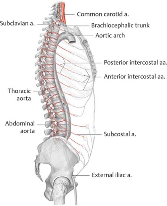
Fig. 2.15 Arteries of the trunk
Right lateral view.

Fig. 2.16 Vertebral venous plexus
The intervertebral and basivertebral veins connect the internal and external venous plexuses, which drain into the azygos system.
A Vertebral venous plexuses, superior view.
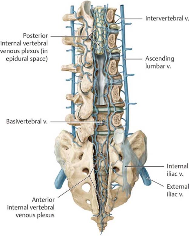
B Veins in the sacral and lumbar canals, posterior view with vertebral canal windowed.
Metastasis and the vertebral venous plexus
The vertebral venous plexus links the venous drainages of viscera in the thorax, abdomen, and pelvis and the venous sinuses of the brain. These communications have been identified as a likely route of metastases of carcinoma of the prostate (commonly), breast, and lung (less commonly) to the central nervous system and bone.
– Lymphatic drainage from the vertebrae and vertebral ligaments generally follows the arteries that supply each region and end in cervical, thoracic, lumbar, and sacral lymph nodes.
– Spinal nerves at each level innervate the vertebrae, vertebral joints, and spinal meninges through dorsal rami and meningeal branches of the anterior rami. A sympathetic branch also arises from the thoracic sympathetic ganglia.
2.3 The Spinal Cord and Spinal Nerves
The spinal cord is the part of the central nervous system that relays information between the brain and the body. The spinal cord, along with its spinal nerves, surrounding membranes (the meninges), and associated vasculature, is enclosed within the vertebral canal.

Fig. 2.17 Spinal cord in situ
Posterior view with vertebral canal windowed.
2.3a Spinal Cord
– The spinal cord, continuous with the medulla oblongata of the brain (see Fig. 18.8A) superiorly, exits the skull base through the foramen magnum of the occipital bone. It descends within the vertebral canal and terminates as the conus medullaris adjacent to the L1 vertebra (Fig. 2.17; see also Fig. 17.6).
– During development the longitudinal growth of the vertebral column exceeds that of the spinal cord. At birth the conus medullaris is at the level of the L3 vertebra, but in the adult it lies adjacent to the L1/L2 intervertebral disk.
– Because the adult spinal cord is considerably shorter than the vertebral column, occupying only the superior two thirds of the vertebral canal, most spinal cord segments do not lie adjacent to the vertebral level of the same number (Fig. 2.18).
– The spinal cord consists of 31 segments, each of which innervates a specific area of the trunk or limbs. Each spinal cord segment has paired spinal nerves that contain both sensory and motor neurons.

Fig. 2.18 Spinal cord segments and vertebral levels
The spinal cord is divided into four major regions: cervical, thoracic, lumbar, and sacral. Spinal cord segments are numbered by the exit points of their associated spinal nerves.
– Two swellings occur in the regions of the spinal cord that innervate the limbs:
• The cervical enlargement at C4–T1 is related to the brachial plexus, a plexus of nerves that innervate the upper limb.
• The lumbosacral enlargement at T11–S1 is related to the lumbar and sacral plexuses, nerve plexuses that innervate the abdominal wall and lower limb.
2.3b Meninges of the Spinal Cord
The spinal meninges are membranes that surround the spinal cord and nerve roots and that contain the cerebrospinal fluid (a fluid that cushions and nourishes the brain and spinal cord) (Figs. 2.19 and 2.20Aand B; see also Section 18.2b).
– The three layers of spinal meninges are continuous with the meninges that surround the brain:
1. Dura mater, a tough outer layer that forms the dural sac enclosing the spinal cord and extending along the nerve roots to the intervertebral foramina. The dural sac begins at the foramen magnum of the skull and ends at the level of S2.
2. Arachnoid mater, a delicate middle layer that is connected to the underlying membrane by arachnoid trabeculae (strands of connective tissue).
3. Pia mater, a thin layer that adheres to the surface of the spinal cord. Denticulate ligaments, transverse extensions of the pia mater, attach to the dura mater and suspend the spinal cord within the dural sac.

Fig. 2.19 Spinal cord and its meningeal layers
Posterior view. The dura mater is opened, and the arachnoid mater is sectioned.
– The filum terminale, a thin cord of pia mater, extends from the conus medullaris to the apex of the dural sac. There it is surrounded by spinal dura mater and extends to the end of the vertebral canal, where it anchors both membranes to the coccyx.
– Three spaces separate the layers of meninges.
• The epidural space lies between the bony wall of the vertebral canal and the dura mater. It contains fat and the vertebral venous plexus.
• The subdural spac e, a potential space between the dura and arachnoid layers, contains a thin film of lubricating fluid.
• The subarachnoid space lies deep to the arachnoid layer and contains the cerebrospinal fluid. The lumbar cistern is an enlargement of the subarachnoid space within the dural sac inferior to the conus medullaris.
Lumbar puncture, spinal anesthesia, and epidural anesthesia
A lumbar puncture, used to extract cerebrospinal fluid from the spinal subarachnoid space, is administered by inserting a needle between the spinous process of L3 and L4 (sometimes between L4 and L5). The needle pierces the ligamentum flavum and wall of the dural sac before entering the lumbar cistern. The injection of a local anesthetic for spinal anesthesia is also administered in this manner. A similar approach may be used for epidural anesthesia, to anesthetize emerging spinal nerves, but the anesthetic is injected into the epidural space without entering the dural sac. A caudal approach through the sacral hiatus also allows access to the epidural space.
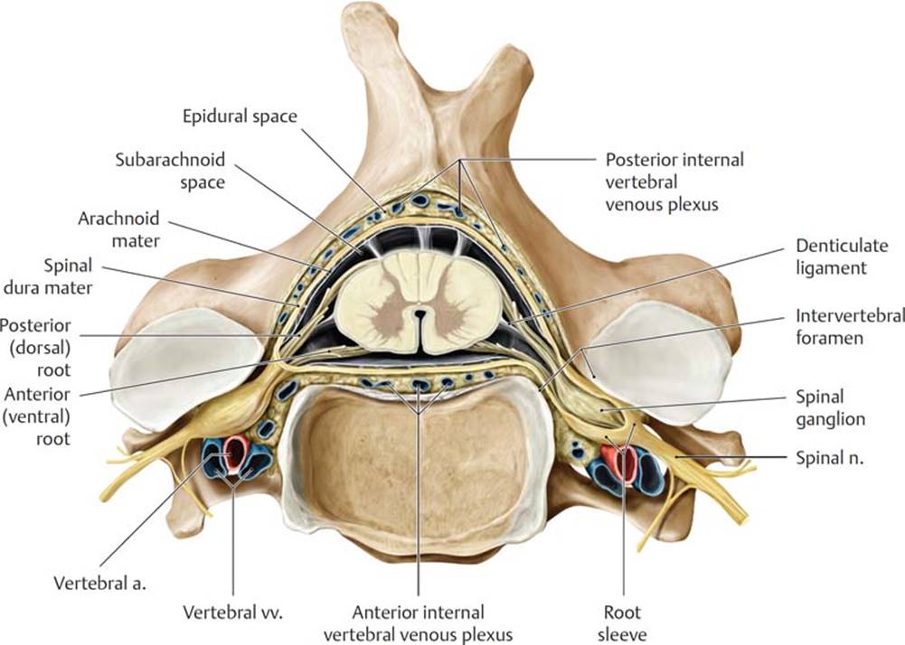
Fig. 2.20 Spinal cord in situ: Transverse section
Superior view.
A Spinal cord at level of C4 vertebra.
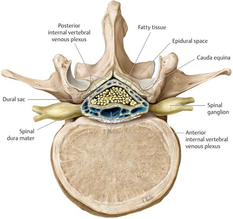
B Cauda equina at level of L2 vertebra.
2.3c Blood Supply to the Spinal Cord (Fig. 2.21A and B)
– Longitudinal spinal arteries supply the superior part of the spinal cord.
• A single anterior spinal artery arises from the two vertebral arteries (branches of the subclavian arteries) and supplies the anterior two thirds of the spinal cord.
• Paired posterior spinal arteries arise from the vertebral arteries (or one of their branches, the posterior cerebellar artery) and supply the posterior third of the spinal cord.
– Anterior and posterior segmental medullary arteries are large, irregularly spaced vessels that communicate with the spinal arteries.
• They arise from branches of the subclavian artery and segmental arteries in the thoracic and lumbar region.
• The medullary arteries enter the vertebral canal through the intervertebral foramina and are found mainly at the cervical and lumbar enlargements.

Fig. 2.21 Arteries of the spinal cord
The unpaired anterior and paired posterior spinal arteries typically arise from the vertebral arteries. As they descend within the vertebral canal, the spinal arteries are reinforced by anterior and posterior segmental medullary arteries. Depending on the spinal level, these reinforcing branches may arise from the vertebral, ascending or deep cervical, posterior intercostal, lumbar, or lateral sacral arteries.
A Spinal and segmental medullary arteries.

B Arterial supply system.
– The great anterior segmental medullary artery (of Adam-kiewicz), a single large, usually left-sided vessel, can provide an important contribution to the circulation of the lower two thirds of the spinal cord.
• It arises as a branch of a lower thoracic or lumbar segmental artery.
• It enters the vertebral canal through an intervertebral foramen in the lower thorax or upper lumbar region.
– The anterior and posterior radicular arteries are small arteries that supply the roots of the spinal nerves and the superficial gray matter of the spinal cord. They do not communicate with the spinal arteries.
– Veins of the spinal cord, which are more numerous than the arteries, have the same distribution, anastomose freely with one another, and drain into the internal vertebral plexus (Fig. 2.22).

Fig. 2.22 Veins of the spinal cord
The interior of the spinal cord drains via venous plexuses into an anterior and a posterior spinal vein. The radicular and spinal veins connect the veins of the spinal cord with the internal vertebral venous plexus. The intervertebral and basivertebral veins connect the internal and external vertebral venous plexuses, which drain into the azygos system.
2.3d Spinal Nerves
Spinal nerves transmit information between peripheral body tissues and the spinal cord. A single pair of spinal nerves arises from each spinal cord segment.
– There are 31 pairs of spinal nerves: 8 cervical, 12 thoracic, 5 lumbar, 5 sacral, and 1 coccygeal.
– Spinal nerves are mixed (sensory and motor) nerves, which arise bilaterally from each spinal cord segment (Fig. 2.23). They are formed by the merging of
• anterior roots containing motor (efferent) fibers and
• posterior roots containing sensory (afferent) fibers.
– Spinal nerves are somatic nerves but most also carry visceral nerve fibers.
• Spinal nerves T1–L2 carry sympathetic fibers that synapse in the ganglia of the sympathetic trunk or in preaortic ganglia.
• Spinal nerves S2–S4 carry parasympathetic fibers that contribute to the visceral nerve plexuses of the pelvis.
– Each spinal nerve divides into an anterior ramus and a posterior ramus. Anterior rami innervate the anterolateral trunk wall and the limbs; posterior rami innervate the skin and muscles of the back and posterior scalp.
– Spinal nerves from each spinal cord segment pass through the intervertebral foramina at the corresponding vertebral level.
• Cervical nerves C1–C7 exit the vertebral canal superior to the vertebra of the same number (e.g., C4 spinal nerve exits between C3 and C4 vertebrae).
• C8 spinal nerve exits below the C7 vertebra (between the C7 and T1 vertebrae).
• Spinal nerves T1–Co1 exit the canal inferior to the corresponding vertebra.
– Because the spinal cord is shorter than the vertebral column, nerve roots from the lower spinal cord (L2–Co1) must descend below the conus medullaris within the lumbar cistern of the dural sac before exiting through the respective intervertebral foramina. This loose group of nerve roots is called the cauda equina (see Figs. 2.18 and 2.20B).
– Anterior rami of thoracic spinal nerves become intercostal nerves, which run in the spaces between ribs of the thoracic wall. Anterior rami of the cervical, lumbar, and sacral regions (with contributions from T1) form plexuses (Fig. 2.24A and B):
• Cervical plexus (C1–C4)
• Brachial plexus (C5–T1)
• Lumbar plexus (L1–L4)
• Sacral plexus (L4–S3)
– The cutaneous (sensory) branches of the pair of spinal nerves derived from a spinal cord segment supply specific areas of skin called dermatomes (Fig. 2.25). The muscles that are innervated by the motor branches of each pair of spinal nerves are called myotomes.

Fig. 2.23 Spinal cord segment
Anterior view. The spinal cord consists of 31 segments innervating a specific area in the trunk or limbs, Afferent (sensory) posterior rootlets and efferent (motor) anterior rootlets form the posterior and anterior roots, respectively. The two roots fuse to form a mixed spinal nerve, which then divides into various branches.
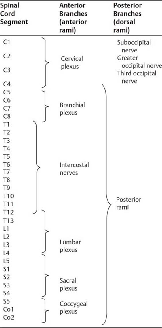
Fig. 2.24 Nerves of the trunk wall
A Anterior and posterior branches of the spinal nerves.
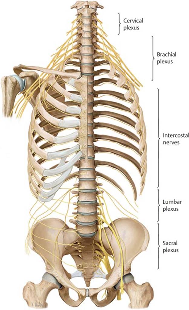
B Nerves of the trunk wall, anterior view. Removed: Anterior part of the left half of the thoracic cage.

Fig. 2.25 Dermatomes of the head, trunk, and limbs
Each spinal cord segment innervates a particular skin area (dermatome).
2.4 Muscles of the Back and Suboccipital Region (Table 2.2)
– Extrinsic muscles, the most superficial muscles that overlie the back, stabilize and move the upper limb. (See Chapter 14 for a discussion of the muscles of the upper limb.)
• Extrinsic muscles include the trapezius, latissimus dorsi, levator scapulae, and rhomboid major and minor.
– Intrinsic muscles, which attach to vertebrae or ribs, move and support the vertebral column.
• They are arranged in superficial, intermediate, and deep layers (Figs. 2.26 and 2.27A and B).
• The superficial layer includes the splenius muscle group that covers the deeper neck muscles laterally and posteriorly (Fig. 2.26). These muscles extend and rotate the head and neck. They extend superolaterally from the spinous processes of cervical and upper thoracic vertebrae to the occipital bone and transverse processes of C1 and C2
• The intermidiate layer includes the erector spinae muscle group that extends from the midline of the back to the angle of the ribs laterally. These large muscles are the main extensors and stabilizers of the thoracic and lumbar vertebral column. They include
○ the iliocostalis, the most lateral column that arises from the thoracolumbar fascia, the sacrum, iliac crest, and ribs and extends superolaterally to the ribs and to cervical and lumbar vertebrae.
○ the longissimus, the middle column that arises from the sacrum, iliac crest, spinous processes of lumbar vertebrae, and transverse processes of thoracic and cervical vertebrae. It inserts superiorly on the temporal bone of the skull, to cervical, thoraric and lumbar vertebrae and to the ribs.
○ the spinalis, the most medical column that extends between the spinous processes of cervical and thoracic vertebrae
TABLE 2.2 Muscles of the Back and Suboccipital Region
|
Muscle Group |
Innervation |
Action |
|
Intrinsic muscles of the back |
||
|
Superficial layer Splenius capitis Splenius cervicis |
Posterior rami of cervical spinal nerves |
Extend, rotate, and laterally flex the head and cervical spine |
|
Intermediate layer (erector spinae) Spinalis Longissimus Iliocostalis |
Posterior rami of spinal nerves |
Extend and laterally flex the spine |
|
Deep layer Transversospinalis group Rotatores (brevis and longus) Multifidus Semispinalis Deep segmental group Interspinales Intertransversarii Levatores costarum |
Posterior rami of spinal nerves |
Extend, rotate, and laterally flex the head and spine |
|
Muscles of the suboccipital region |
||
|
Rectus capitus posterior major Rectus capitus posterior minor Obliquus capitus superior Obliquus capitus inferior |
Suboccipital n. (C1) |
Extend and rotate the head |
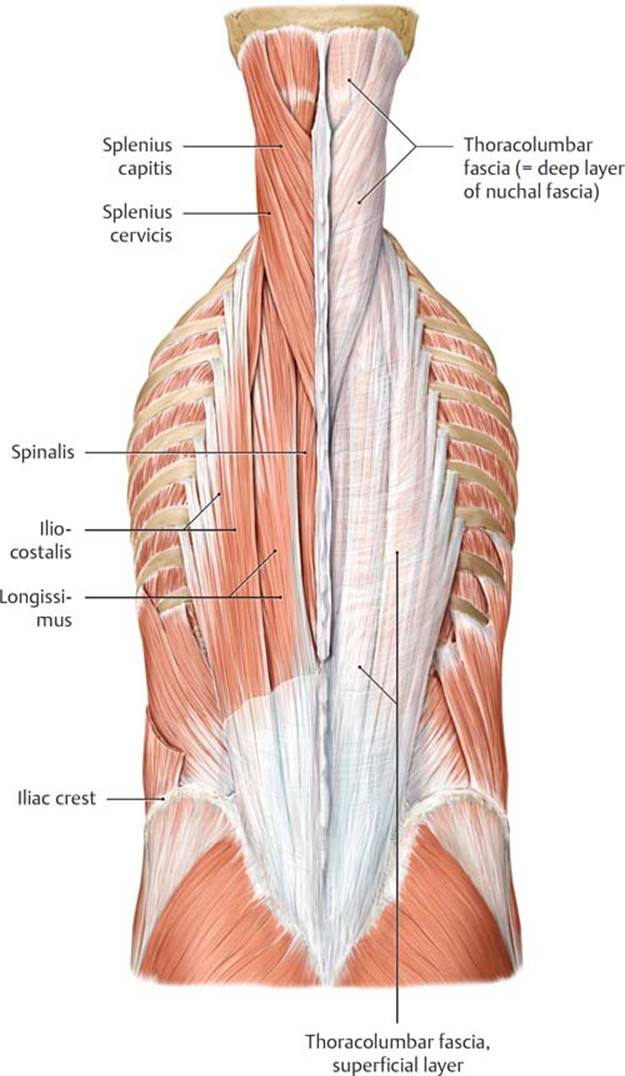
Fig. 2.26 Superficial and intermediate muscles of the back
Posterior view. Removed: Thoracolumbar fascia (left).
• The deep layer includes short muscles at multiple vertebral levels that produce small movements along the entire vertebral column (Fig. 2.27A and B). They are divided into transversospinalis muscle group and a deep segmental muscle group. The transversospinalis muscles extend between the transverse and spinous processes of the vertebrae, They include:
○ the semispinalis muscles, the most superficial this group
○ the multifidis, most prominent in the lumbar region
○ the rotatores, the deepest muscles of the transversospinalis group, best developed in the thoraric region
• The deep segmental muscles are minor muscles of the back. They include the interspinales and intertransversarii that connect adjacent vertebrae, and the levatores costarum that connect vertebrae to ribs.

Fig. 2.27 Deep intrinsic back muscles
Posterior view.
A Transversospinalis muscles: Rotatores, multifidus, and semispinalis.
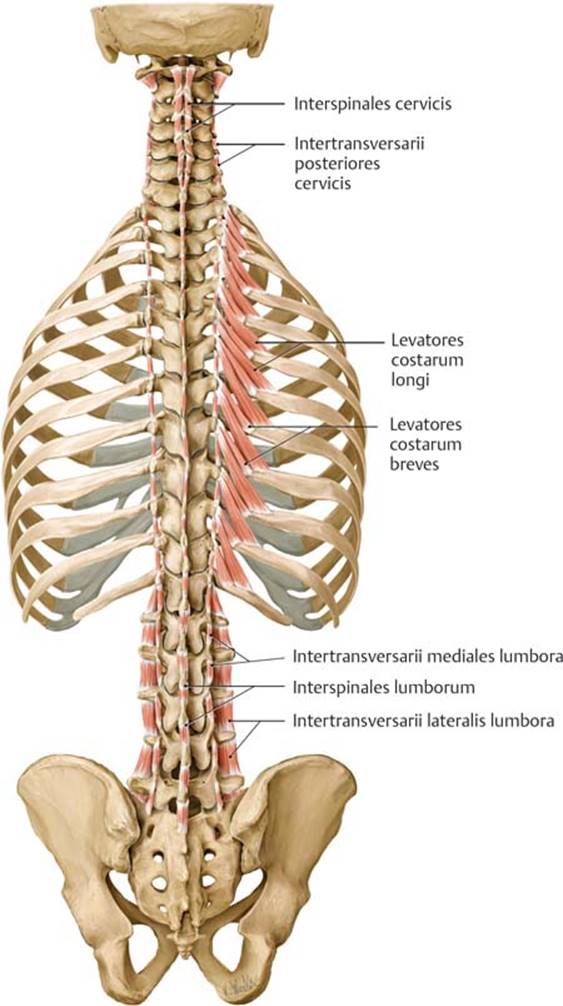
B Deep segmental muscles: Interspinales, intertransversarii, and levatores costarum.
• A deep fascia that encloses the intrinsic muscles runs laterally from the posterior midline to the cervical and lumbar transverse processes and to the ribs. This thoracolumbar fascia continues into the neck as the deep layer of the nuchal fascia, the posterior extension of the cervical fascia (see Section 21.1).
– Muscles of the posterior neck occupy the small suboccipital compartment (Fig. 2.28) that is inferior to the base of the skull and deep to the trapezius and intrinsic back muscles that extend into the neck. The suboccipital muscles arise from C1 or C2 and extend upward to insert on the occipital bone or transverse process of C1. All assist in the positioning of the head and are innervated by the suboccipital nerve, the posterior ramus of C1. They include the rectus capitis posterior major, rectus capitis posterior minor, obliquus capitis inferior, and obliquus capitis superior.
– Posterior intercostal and lumbar arteries (branches of the descending aorta and subclavian artery) supply the skin and muscles of the back. The veins of the back accompany the arteries and are tributaries of the azygos system.
– Posterior rami of intercostal and lumbar nerves supply the skin and intrinsic muscles of the back (Figs. 2.29 and 2.30).

Fig. 2.28 Short nuchal and craniovertebral joint muscles
Suboccipital muscles, posterior view.
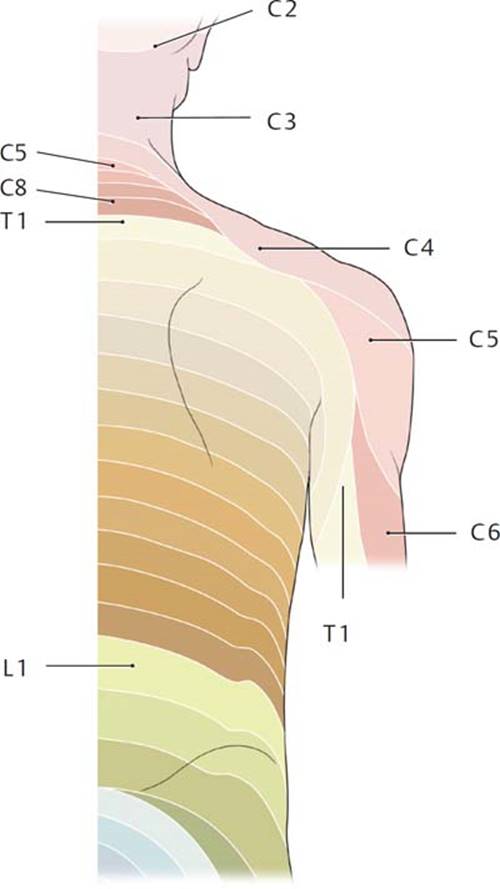
Fig. 2.29 Cutaneous innervation of the back
Dermatomes: Segmental (radicular) cutaneous innervation of the back. Note: The posterior ramus of C1 is purely motor; there is consequently no C1 dermatome.

Fig. 2.30 Nerves of the back
Cross section through the vertebral column and spinal cord with surrounding musculature, superior view.


