
That Takes a Lot of Nerve
NERVOUS TISSUE
Nervous tissue sends electrical signals from one place to another in the body. These signals can either bring information to the central nervous system from the body for processing or send information out to the body. You would trigger nervous tissue reaction if you touched a hot stove, for example. Your neurons would tell your brain that you are touching something hot (bringing information to the central nervous system). Your brain would tell your body to move your hand off the stove (sending information out to the body).
Neurons
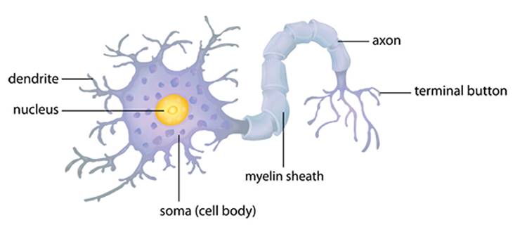
Neurons are the signaling cells of the nervous system and come in a myriad of shapes and sizes. Generally, neurons have structures called neurites extending from the cell body, which make the cell look somewhat spiky and create the characteristic appearance of a neuron. What these neurites are named is based on the direction the signals travel. For instance, if the electrical signal travels toward the cell body, the neurite is called a dendrite. If the signal moves away from the cell body, it is an axon. Typically neurons will have many smaller-diameter dendrites and one longer, thicker axon.
Neuroglia
While neurons are the signaling cells of the nervous system, they only make up 20 percent of the nervous system. The bulk is made up of the supporting cells of the nervous system that are collectively referred to as neuroglia.
Why are neuroglia sometimes called “nerve glue”?
The word “neuroglia” comes from two Greek words that together mean “nerve glue.” Neuroglial cells don’t actually glue anything to anything. When they were first discovered, scientists thought they served a binding purpose, thus their name.
In much the same way as the movie stars on the screen are only a small fraction of the people involved in making a movie, the behind-the-scenes supporting members for the nervous system are the neuroglial cells (they are also called glial cells or glia). Glial cells are smaller than neurons and do not have neurites. They are not involved in actively sending or receiving signals. Rather, they support neurons by maintaining their surroundings and modulating the uptake of neurotransmitters. Glial cells also help neurons recover from injury and perform other supporting functions.
Anatomy of a Word
neurotransmitters
Neurotransmitters are the chemicals that transmit signals between neurons. Neurons have small spaces between them, called synapses, and neurotransmitters help signals pass through synapses by converting the electrical impulse of the neuron into a chemical messenger.
Myelinating Cells
One group of neuroglia insulates the axons of neurons. These cells wrap their plasma membranes around the axon 40 or 50 times, much like an electrician wrapping electrical tape around a bare wire. The term myelin refers to the area of the membrane that is tightly wrapped around the axon (to myelinate something is to wrap around it). Myelin insulates the axon, and in doing so creates a much more rapid conduction velocity of the electrical signal. For the neurons in areas of the body such as in your arms and legs, these neurons will be myelinated by Schwann cells, named after Theodor Schwann, the physiologist who discovered them. They are specific types of glial cells that exist in the peripheral nervous system and are responsible for wrapping peripheral nerves.
In the central nervous system, however, oligodendrocytes myelinate axons. Much like an octopus with many arms, these cells extend several cellular structures to axons and thus one oligodendrocyte can myelinate several different axons.
Astrocytes
Astrocytes, so-called because of their star shape, are another type of neuroglial cell. They protect the nervous system from infection by covering blood vessels with their structures and regulating the movement of materials out of the blood stream and into the nervous system. As part of the blood-brain barrier, they screen for pathogens and assist in the transportation of nutrients as well as the processing of waste.
Microglia
Microglia cells are the vacuum cleaners of the central nervous system. Known as phagocytic cells, they patrol the nervous system looking for pathogens and debris and removing them through a process called phagocytosis, or engulfing the foreign object into their cell bodies, effectively eliminating these detrimental materials before they can do any damage.
Ependymal Cells
The last type of neuroglial cell present in the central nervous system is the ependymal cell. These cells produce cerebrospinal fluid, which flows around the spinal cord and the brain and functions as a shock absorber against blows to the head and spine. Additionally, as cerebrospinal fluid is produced and as it is eliminated it creates a current that assists the blood-brain barrier; pathogens have to swim across this river of flowing cerebrospinal fluid before gaining access to the central nervous system.
Together these various cells form the tissues and organs of the central and peripheral nervous systems in the human body.