The wall of the thorax and the wall of the abdomen are one, topographically and developmentally, the essential difference being the presence of ribs in the part primarily concerned with respiration.
The skin varies in texture, tending to be thin in front and thick behind. Distribution of hair varies with sex, age and race. The tension lines run almost horizontally around the body wall, except over and above the breast (Fig. 1.2, p. 2).
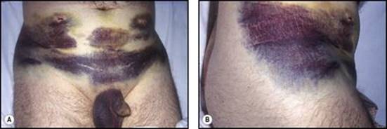
In the subcutaneous tissue over the dilatable part of the body wall, namely the anterior abdominal wall and lower part of the thoracic wall in front of the midaxillary lines, the fibrous septa of the subcutaneous tissue are condensed beneath the fat into a thin but strong membranous layer of superficial fascia, the fascia of Scarpa. This fascia allows the fatty layer of superficial fascia, the fascia of Camper, to slide freely over the underlying thoracic wall, rectus sheath and external oblique aponeurosis. It fades out over the upper thoracic wall and along the midaxillary lines. The fascia of Scarpa is continued over the penis and scrotum as the superficial fascia of the penis (Buck's fascia) and the superficial perineal fascia (Colles' fascia; see pp. 318 and 319). Below, over the thighs, it is attached to the fascia lata along the flexure skin crease of the hip, extending laterally from the pubic tubercle just below the inguinal ligament (Fig. 4.1).
|
|
|
Figure 4.1 Bilateral damage to the inferior epigastric artery—during the introduction of instruments through both iliac fossae for minimal access surgery—has resulted in blood leaking at the port sites and tracking down in a plane deep to Scarpa's fascia: A to the penis and scrotum and B to the upper thigh as far as the level of the flexure skin crease of the hip. |
Blood supply
The intercostal, subcostal and lumbar arteries pass forward in the neurovascular plane (see p. 11) to supply the flanks; the internal thoracic and the superior and inferior epigastric arteries supply the ventral midline tissues. From all these arteries cutaneous branches pass to the superficial fat and skin. The venous return from the subcutaneous tissue does not follow the arteries. The blood is collected by an anastomosing network of veins that radiate away from the umbilicus. Below this level they pass to the great saphenous vein in the groin; above the umbilicus they run up to the lateral thoracic vein and so to the axillary vein. From the umbilicus a few paraumbilical veins accompany the ligamentum teres and drain to the left branch of the portal vein; they may distend in portal obstruction, giving rise, if the distension spreads to the subcutaneous veins, to a pattern of dilated veins around the umbilicus, the caput Medusae. A longitudinal channel, the thoracoepigastric vein uniting the lateral thoracic vein with the superficial epigastric vein above the inguinal ligament, provides a communication between superior and inferior venae cavae and often becomes prominent in cases of obstruction of the inferior vena cava.
Lymph drainage
Lymphatic channels from the subcutaneous tissue and skin follow the veins to axillary and superficial inguinal nodes. From above the level of the umbilicus, lymph from the front of the body goes to the anterior (pectoral) group and from the back of the body to the posterior (scapular) group of axillary nodes. From the umbilicus downwards lymph from the anterior aspect of the abdominal wall and perineum goes to the medial group and from the lateral and posterior aspects of the abdominal wall to the lateral group of superficial inguinal nodes.
Nerve supply
Above the second rib and the manubriosternal joint the skin is supplied by supraclavicular branches of the cervical plexus (C4; see Fig. 1.7, p. 12). Below this level a midline and paramedian strip of skin is supplied by the anterior cutaneous branches of the spinal nerves from T2 to L1; the skin in the upper epigastric region is supplied by T7, in the umbilical region by T10 and suprapubic skin by L1. A broad lateral strip is supplied by the lateral cutaneous branches of the spinal nerves from T2 or 3 to L1; these branches emerge in the midaxillary line. The lateral cutaneous branches of T12 and the iliohypogastric nerve descend over the iliac crest to also supply the skin of the buttock. The ilioinguinal nerve has no lateral cutaneous branch; it is the collateral branch of the iliohypogastric, both coming from L1 nerve. A posterior strip of skin is innervated by the posterior rami of spinal nerves, by their medial branches in the upper thoracic and their lateral branches in the lower thoracic and lumbosacral parts (see Fig. 1.6, p. 12).