
Anterior aspect of the neck (Figs 3.1—3.8) 22
Larynx 22
Anterior triangle (Fig. 3.8) 24
Submandibular region (Figs 3.9, 3.10) 24
Large vessels of the neck (Fig 3.11) 25
Lateral aspect of the neck (Figs 3.12–3.19) 26
Posterior triangle (Fig. 3.16) 26
Cutaneous innervation of the head and neck (Fig. 3.18) 28
Lymph nodes of the head and neck (Fig. 3.19) 29
Anterior aspect of the neck (Figs 3.1—3.8)
The anterior aspect of the neck is bounded by the body and angles of the mandible superiorly, and the superior border and sternal notch of the manubrium and the clavicles inferiorly (Fig. 3.2). Laterally it is continuous with the lateral and posterior surface without a specific line of demarcation. The clavicles are subcutaneous throughout their length; they articulate medially at the sternoclavicular and laterally at the acromioclavicular (Fig. 3.13, p. 26) joints.
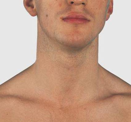
3.1 Anterior aspect of the neck
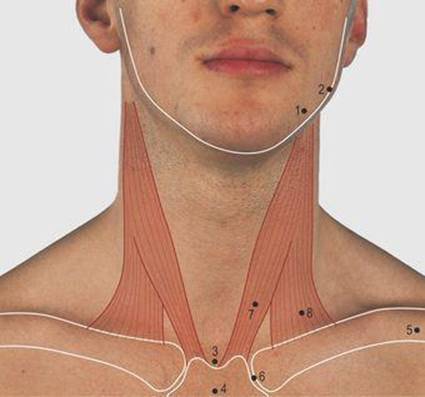
3.2 Anterior aspect of the neck: bones and muscles
1 Body of mandible
2 Angle of mandible
3 Sternal notch
4 Manubrium
5 Clavicle
6 Sternoclavicular joint
7 Sternocleidomastoid muscle – sternal head
8 Sternocleidomastoid muscle – clavicular head
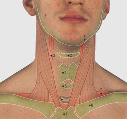
3.3 Anterior aspect of neck: palpable structures
1 Body of mandible
2 Hyoid bone
3 Bifurcation of common carotid artery
4 Thyroid cartilage
5 Cricoid cartilage
6 Base between third and fourth tracheal rings
7 Manubrium sterni
8 Clavicle
9 Subclavian artery
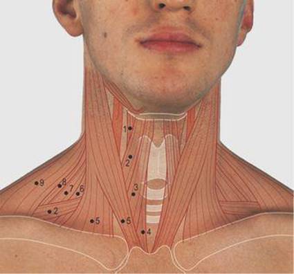
3.4 Anterior aspect of neck: muscles
1 Thyrohyoid
2 Omohyoid
3 Sternohyoid
4 Sternothyroid
5 Sternal and clavicular heads of sternocleidomastoid
6 Scalenus anterior
7 Scalenus medius
8 Levator scapuli
9 Trapezius
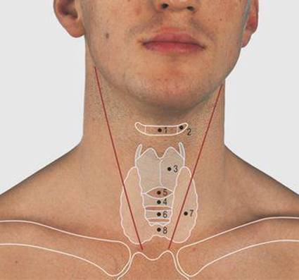
3.5 Larynx and thyroid gland
1 Body of hyoid bone
2 Greater horn of hyoid bone
3 Thyroid cartilage
4 Cricoid cartilage
5 Cricothyroid membrane
6 First tracheal ring
7 Lateral lobe of thyroid gland
8 Isthmus of thyroid gland
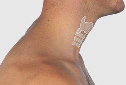
3.6 Laryngeal prominence in the male
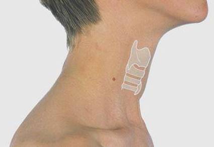
3.7 Laryngeal prominence in the female
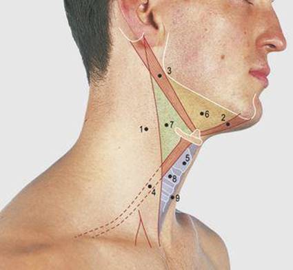
3.8 Anterior triangle of the neck
1 Sternocleidomastoid muscle
2 Anterior belly of digastric muscle
3 Posterior belly of digastric muscle
4 Omohyoid muscle
5 Thyroid cartilage
6 Digastric triangle
7 Carotid triangle
8 Cricoid cartilage
9 Muscular triangle
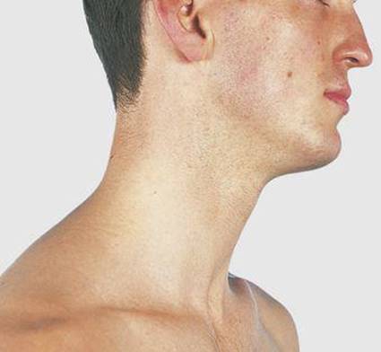
3.9 Submandibular region
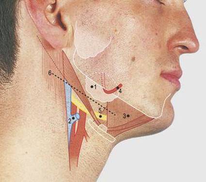
3.10 Submandibular region: soft tissues
1 Submandibular gland
2 Common facial vein
3 Mylohyoid muscle
4 Facial artery
5 Hypoglossal nerve
6 Incision for the surgical approach to the submandibulargland
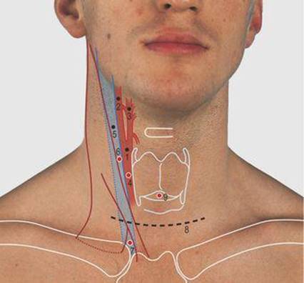
3.11 Carotid arteries, internal jugular vein and applied anatomy
1 Common carotid artery
2 Internal carotid artery
3 External carotid artery
4 Point of access to common carotid artery
5 Internal jugular vein
6 Point of access to internal jugular vein above sternocleidomastoid muscle
7 Point of access to internal jugular vein between the heads of the sternocleidomastoid muscle
8 Collar incision
9 Cricothyroid puncture site
Each sternocleidomastoid muscle is attached to the mastoid process and superior nuchal line of the temporal and occipital bones superiorly, and has sternal and clavicular heads inferiorly. The anterior borders of the two muscles form a prominent V shape when contracting together, such as in protruding the chin or raising the head from the lying position. When acting independently, each rotates and laterally flexes the head on the neck, as when looking under a table.
Larynx
The larynx lies in the midline covered only by skin, platysma muscle and superficial and deep fascia. It can be seen to rise during swallowing. Superiorly, the U-shaped hyoid bone is palpable; a central body is bounded laterally by two greater horns (Fig. 3.5). The bone can be gripped between finger and thumb and if gently pressed backwards and rocked from side to side, the tips of the greater horns can be felt grating against the bodies of the cervical vertebrae, confirming that only thin prevertebral muscles and the wall of the pharynx separate the larynx from the vertebral column. The hyoid bone is at the level of the third cervical vertebra. Suprahyoid muscles pass to the pharynx, the mandible and into the tongue.
The thyroid cartilage forms a midline prominence which is more obvious in the adult male; the angle between the sides of the cartilage (alae) is approximately 90° in the adult male (the Adam's apple, Fig. 3.6) and 120° in the female (Fig. 3.7). The vocal cords are attached to the back of this prominence, and muscles attached to the oblique line, on the outer surface of the cartilage, to the hyoid bone superiorly and the back of the manubrium inferiorly. The inferior constrictor muscle of the pharynx is attached to both the thyroid and cricoid cartilages. The latter forms the lower border of the larynx and is the only complete ring of cartilage in the respiratory passage; the adjacent tracheal cartilages are deficient posteriorly. The cricoid and thyroid cartilages are attached anteriorly by the cricothyroid membrane. The lower border of the cricoid is at the level of the sixth cervical vertebra.
Pressure of the cricoid cartilage onto the vertebral column compresses the pharynx, this being a useful manoeuvre to prevent regurgitation of stomach contents into the oropharynx and then into the airway during anaesthesia.
The concept of speech and its use in language is a cortical phenomenon, but its production (phonation) depends on an energy source, a point of vibration and a resonating chamber. The energy is produced by the force of expired air from the lungs and the vibration primarily by the vocal cords. The resonating chamber is the column of air above the vocal cords, including the larynx, pharynx, nasal cavities and mouth.
The primary vowel tones are produced by the sudden opening of the vocal cords during expiration. These glottal sounds are further modified by narrowings in the vocal tract at a higher level, articulating consonants and other sounds – e.g. ‘g’ and ‘k’ by the pharynx and palate; ‘d’, ‘s’, ‘n’, ‘r’ and ‘th’ by the tongue and teeth; ‘f’ and ‘v’ by the lower lip and upper teeth; and ‘p’, ‘b’, ‘o’ and ‘oo’ by the lips.
Each lateral lobe of the thyroid gland lies adjacent to the thyroid cartilage below the oblique line, overlapped by the strap muscles (omohyoid, sternohyoid and sternothyroid). The lateral lobes are not easily palpable as they are partly covered by the sternocleidomastoid muscles (Figs 3.4, 3.5). However, generalised enlargement of the gland (goitre), or a local nodule, is more easily palpable and it is then possible to see the gland moving with the larynx on swallowing. The gland is most easily examined by standing behind a sitting subject. The isthmus of the thyroid gland unites the lateral lobes across the midline anterior to the second and third tracheal cartilages. The first cartilage and isthmus can usually be felt in the midline below the cricoid cartilage. A goitre may be due to iodine deficiency, hormonal imbalance or neoplastic changes within the gland. Surgical access is through a transverse cervical collar incision (Fig. 3.11, p. 25) and care must be taken not to damage the trachea, the recurrent laryngeal nerves or the parathyroid glands.
Anterior triangle (Fig. 3.8)
The sternocleidomastoid muscle divides the anterior and lateral aspects of the neck; the region in front of it on each side is termed the anterior triangle. Two muscles, attached to the hyoid bone (not visible in the living), further divide the anterior triangle into three triangles. The anterior belly of the digastric muscle is attached to the posterior surface of the mandible near the midline and the posterior belly to the mastoid process. The two bellies are joined by an intermediate tendon which is slung to the hyoid bone, and, together with the lower border of the mandible, they form the digastric triangle. The omohyoid muscle passes from the hyoid bone deep to the sternocleidomastoid muscle and, with the anterior border of this muscle and the posterior belly of the digastric muscle, forms the carotid triangle. The muscular triangle is bounded by the omohyoid, the anterior border of sternocleidomastoid, and the midline, from the hyoid bone to the sternum.
A fourth triangle, the submental, crosses the midline; its apex is at the symphysis menti (i.e. the midline bony union of the primitive two halves of the mandible), its lateral margin is the anterior belly of both digastric muscles and its base the body of the hyoid bone.
Submandibular region (Figs 3.9, 3.10)
The digastric triangle contains the submandibular gland (Fig. 3.10). The gland lies on and around the posterior border of the mylohyoid muscle, partly overlapped by the body of the mandible just in front of its angle. The facial artery passes over the superior surface of the gland then around the inferior border of the mandible.
The submandibular gland is most easily palpated bimanually, between a finger of one hand placed in the floor of the mouth and the fingers of the second hand placed in the neck over the superficial aspect of the gland. A number of lymph nodes are related to the superficial surface of the gland.
The surgical approach to the submandibular gland is made 2.5 cm below and parallel to the lower border of the mandible in order to avoid the mandibular branch of the facial nerve, which may dip below the mandible as it passes to the lower facial muscles. This incision divides skin, platysma muscle and the superficial and deep fascia, to reach the capsule of the gland and the prominent nodes around it. The same incision can be used to biopsy these nodes or nodes in the carotid triangle.
Large vessels of the neck
The larynx is bounded on each side by a cylindrical sheath of fascia (carotid sheath) containing the common carotid artery and the internal jugular vein, and behind these the vagus (10th cranial) nerve; these vessels are covered anteriorly for much of their course by the sternocleidomastoid muscle. The common carotid artery can be palpated above and through the muscle by pressing it posteriorly onto the transverse processes of the cervical vertebrae. It divides at the level of the upper border of the thyroid cartilage into the internal and external carotid arteries. The internal carotid artery passes cranially, with the internal jugular vein, up to and through the base of the skull to supply the brain. The external carotid artery gives branches to the thyroid gland, tongue, face, scalp, pharynx, palate, jaws and nose.
The internal jugular vein lies lateral to the common and internal carotid arteries. In the normal individual, the venous blood pressure is approximately 11cm above that of the right atrium. By positioning a subject with the upper half of the body raised at an angle of approximately 30° and the head rotated to the opposite side, the characteristic pulse wave of the internal jugular vein can be observed. A needle may be introduced into the common carotid artery for radiological procedures. Access to the internal jugular vein is obtained either superior to the sternocleidomastoid muscle or between its two inferior heads.
A common operation in this region is partial or total removal of the thyroid gland for benign or malignant enlargement. In the collar incision, the skin, platysma muscle and superficial and deep fascia are divided along the line shown (Fig. 3.11). The pretracheal fascia between the strap muscles is divided vertically, or transversely with the strap muscles, to reach the gland; the choice depends on the size and required access to the gland. In diseases of the larynx or in a patient with respiratory difficulty, a temporary or permanent opening may be required into the trachea (tracheostomy). The skin incision may be the central third of the collar incision or a vertical midline approach. The tracheal opening is usually over the second and third tracheal rings and the isthmus of the thyroid gland may also require division. Incision or needle puncture of the cricothyroid membrane enters the larynx below the vocal cords and provides an alternative means of accessing the airway; this is of particular value in children, where the upper trachea is less accessible.
An incision along the anterior border of the sternocleidomastoid muscle through the skin, platysma and superficial and deep fascia, brings the dissection onto the carotid sheath for surgery on the carotid arteries. The upper end of the incision provides access to the submandibular group of lymph nodes (Fig. 3.19, p. 29). Medial dissection anterior or posterior to the carotid sheath provides access to the pharynx superiorly and the oesophagus inferiorly.
Lateral aspect of the neck (Figs 3.12-3.19)
Laterally, the neck is bounded superiorly by the body, the angle and the ramus of the mandible and the temporomandibular joint (Fig. 2.17, p. 15). Behind this are the external acoustic meatus, the mastoid process and the superior nuchal line (Fig. 3.12). The tip of the transverse process of the atlas can be felt by gentle pressure midway between the angle of the mandible and the mastoid process.

3.12 Lateral aspect of the neek
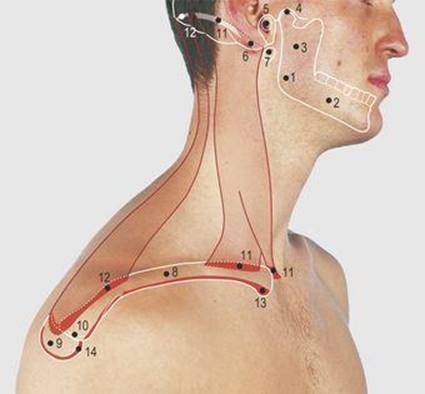
3.13 Lateral aspect of neck: bones and attachments of shoulder girdle muscle
1 Angle of mandible
2 Body of mandible
3 Ramus of mandible
4 Temporomandibular joint
5 External acoustic meatus
6 Mastoid process
7 Tip of transverse process of atlas vertebra
8 Clavicle
9 Acromion
10 Acromioclavicular joint
11 Sternocleidomastoid
12 Trapezius
13 Pectoralis major
14 Deltoid

3.14 Lateral aspect of the neck: palpable structures
1 Mastoid process
2 Angle of mandible
3 Hyoid bone
4 Laryngeal prominence of thyroid cartilage
5 Cricoid cartilage
6 Tracheal ring
7 Manubrium sterni
8 Manubriosternal joint (angle of Louis)
9 Second costal cartilage
10 Clavicle
11 Acromion
12 Spine of scapula
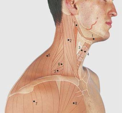
3.15 Lateral aspect of neck: muscles
1 Posterior and anterior bellies of digastric
2 Sternocleidomastoid
3 Omohyoid
4 Sternohyoid
5 Trapezius
6 Scalenus anterior
7 Deltoid
8 Pectoralis major

3.16 Posterior triangle of the neck
1 Sternocleidomastoid muscle
2 Trapezius muscle
3 Superior nuchal line
4 Clavicle
5 Scalenus anterior muscle
6 Apex of lung
7 Needle insertion point for brachial plexus anaesthesia
8 Needle insertion point for scalene regional anaesthesia
9 Supraclavicular incision to approach subclavian artery, neck of first rib and adjacent structures
10 Posterior triangle of neck
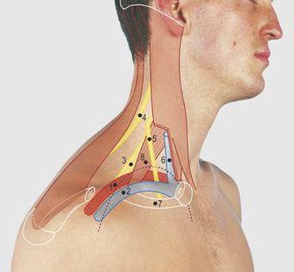
3.17 Subclavian vessels and brachial plexus
1 Subclavian artery
2 Subclavian vein
3 Brachial plexus (upper trunk)
4 Accessory (11th cranial) nerve
5 Phrenic nerve
6 Internal jugular vein
7 First rib
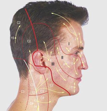
3.18 Cutaneous innervation of the head and neck
1 Supratrochlear
2 Supraorbital
3 Infratrochlear
4 External nasal
5 Infraorbital
6 Lacrimal
7 Zygomaticotemporal
8 Zygomaticofacial
9 Auriculotemporal
10 Buccal
11 Mental
12 Great auricular
13 Transverse cervical
14 Supraclavicular
15 Greater occipital
16 Lesser occipital
I, II, III Ophthalmic, maxillary and mandibular divisions of the trigeminal nerve

3.19 Lymph nodes of the head and neck
1 Submental
2 Submandibular
3 Parotid, superficial and deep
4 Posterior auricular
5 Occipital
6 Jugulodigastric
7 Jugulo-omohyoid
8 Supraclavicular
9 Deep cervical lymph chain around the internal jugular vein
10 Superficial cervical lymph chains around external and anterior jugular veins
Inferiorly, the region is bounded by the clavicle and the acromion which forms the tip of the shoulder. The two bones are united by the acromioclavicular joint.
Posterior triangle (Fig. 3.16)
The posterolateral border of the sternocleidomastoid muscle, with the middle third of the clavicle and the anterior border of the trapezius muscle, form the borders of the posterior triangle of the neck. In a thin subject with the head turned and flexed to the opposite side, the omohyoid can be seen crossing the triangle. The lateral border of scalenus anterior may also be visible. It appears to a variable extent in the anteroinferior angle of the posterior triangle, depending on the size of the clavicular head of the sternocleidomastoid. The apex of the lung rises 3 cm above the medial third of the clavicle. The subclavian artery arches over the lung and the apical pleura behind the scalenus anterior muscle, before passing deep to the clavicle to become the axillary artery, at the outer border of the first rib, and pass through the cervico-axillary canal into the arm (Fig. 3.17). The subclavian artery can be palpated behind the middle of the clavicle by downward pressure onto the first rib.
The subclavian vein lies anterior to the scalenus anterior muscle and is slightly inferior to its artery, being covered by the clavicle.
The vein is frequently used for vascular access. It is usually approached inferiorly but a needle can be inserted into it from above. The point of the needle passes downwards, close to the posterior surface of the clavicle to avoid puncturing the apex of the lung.
The trunks of the brachial plexus may be palpable as they pass obliquely laterally across the anteroinferior angle of the posterior triangle; the lower trunk is, however, deeply placed posterior to the subclavian artery. The trunks divide posterior to the clavicle.
Local anaesthetic can be inserted around the plexus to anaesthetise the arm. A needle is inserted along the lateral border and deep to the scalenus anterior muscle, beneath the prevertebral fascia. More lateral placement of the needle may puncture the apex of the lung.
The accessory (11th cranial) nerve passes anterior to the lateral mass of the atlas and descends through the sternocleidomastoid muscle, then emerging near the middle of the muscle's posterolateral border, it crosses the posterior triangle to reach the trapezius muscle; it supplies both these muscles.
A supraclavicular incision is used to biopsy supraclavicular lymph nodes and to approach the subclavian artery, brachial plexus and cervical sympathetic chain. The last structure is deeply placed across the neck of the first rib. The incision divides the skin, platysma muscle, superficial and deep fascia and the omohyoid muscle. A pad of fat surrounds the omohyoid muscle and encloses the supraclavicular and transverse cervical vessels. The scalenus anterior muscle is divided in line with the incision to reach the subclavian artery and brachial plexus. Care must be taken to preserve the phrenic nerve as it crosses the scalenus anterior muscle and the apex of the lung behind the muscle.
Cutaneous innervation of the head and neck (Fig. 3.18)
The trigeminal (5th cranial) nerve supplies the facial area, in front of the thick line shown in Figure 3.18, through its named branches. The remaining cutaneous supply is from the cervical nerves; the dorsal roots innervate the skin posterior to the dotted line. The greater occipital nerve is derived from the second cervical nerve; the first provides motor innervation of the suboccipital muscles but has no cutaneous contribution from its posterior primary rami. The ventral roots, forming the cervical plexus, innervate the skin anterior to the dotted line, through the named branches and the cervical dermatomes as shown.
Lymph nodes of the head and neck (Fig. 3.19)
The lymphatic drainage of the head and neck is through deep and superficial lymphatic rings, around the base of the skull, and the deep and superficial cervical lymph chains. The deep ring around the oro- and nasopharynx includes the palatine tonsil. The superficial ring comprises the occipital, posterior auricular, superficial and deep parotid, submandibular and submental nodes. The deep cervical lymph chain lies around the internal jugular vein, prominent nodes being in the carotid triangle, around the digastric and omohyoid muscles, and in the posterior triangle above the clavicle. Superficial lymph chains lie along the external and anterior jugular veins.
Enlarged cervical lymph nodes are the commonest neck lumps seen in clinical practice. They are usually associated with tonsillar or upper respiratory tract infection but may also be the site of metastases from malignancies of the nasal cavities, thyroid gland, lungs and breast. Occasionally, malignancy of the upper abdomen can present with enlargement of a left supraclavicular node.
The order of palpation of cervical lymph nodes is not critical, provided all groups are included. They may be examined from the front, or from behind as for the thyroid gland. In the former, simultaneous examination of the two sides can be undertaken with the pulps of the second, third and fourth fingers of both hands. Examined in turn are the occipital, posterior auricular, superficial and deep parotid, submandibular and submental lymph nodes. This is followed by palpation downwards, along the anterior border of the sternocleidomastoid muscle to the clavicle, palpating the internal jugular lymph chain.
An enlarged node can be missed deep to the lower end of the sternocleidomastoid muscle if not carefully searched for by gently squeezing the finger and thumb deep to the lower end of the muscle. Passing backwards from this area, the supraclavicular nodes are examined and then, by ascending along the posterior border of the sternocleidomastoid muscle, the external jugular lymph chain. Additional nodes may be present anterior to the larynx and along the anterior jugular vein.