
Anterior abdominal wall (Figs 5.1–5.8) 42
Surface markings of the alimentary tract (Fig. 5.6) 43
Surface markings of non-alimentary tract viscera (Figs 5.8, 5.11) 45
Abdominal incisions (Fig 5.9) 45
Posterior abdominal wall (Figs 5.10–5.12) 46
Cutaneous innervations of the trunk (Figs 5.13, 5.14) 47
Abdominal examination 48
Inguinal region, perineum, scrotum and penis (Figs 5.15–5.18) 49
Scrotum and penis 49
Female perineum (Figs 5.19, 5.20) 50
Anterior abdominal wall (Figs 5.1-5.8)
The anterior abdominal wall is bounded superiorly by the lower costal margin and inferiorly by the symphysis pubis, the pubic crest and tubercle, the inguinal ligament, the anterior superior iliac spine and the iliac crest, from medial to lateral (Fig. 5.2). The wall comprises three flat sheet-like muscles. They fuse medially to form a fibrous sheath for the vertically running rectus abdominis muscles, lying on each side of the midline. The sheaths of the two sides meet in the midline as a fibrous raphe, known as the linea alba. The three transverse lines of attachment of the rectus abdominis muscle to the anterior sheath can be seen in a muscular subject above the umbilicus; they are termed tendinous intersections (Fig. 5.1). The superior end of the rectus abdominis muscle crosses the anterior rib cage to be attached to the fifth, sixth and seventh costal cartilages from lateral to medial. The lateral line of formation of the rectus sheath is known as the linea semilunaris.

5.1 Anterior abdominal wall

5.2 Anterior abdominal wall: bones
1 Costal margin
2 Xiphisternum
3 Fifth, sixth and seventh costal cartilages
4 Symphysis pubis
5 Body of pubis
6 Pubic tubercle
7 Inguinal ligament
8 Anterior superior iliac spine
9 Iliac crest
10 Iliac tubercle
11 Linea alba
12 Linea semilunaris
13 Rectus abdominis muscle
14 Tendinous intersections

5.3 Anterior abdominal wall: palpable structures
1 Body of sternum
2 Xiphisternum
3 Costal margin
4 Tubercle of ilium
5 Anterior superior iliac spine
6 Upper border of two pubic bones, united by midline symphysis pubis
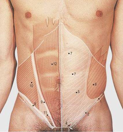
5.4 Anterior abdominal wall: muscles
1 Anterior superior iliac spine
2 Inguinal ligament
3 Pubic tubercle
4 Superficial inguinal ring
5 External oblique aponeurosis
6 External oblique muscle fibres
7 Tendinous intersections of rectus abdominis
8, 9 Cut edges of external oblique
10 Internal oblique muscle fibres
11 Conjoint tendon
12 Rectus abdominis muscle

5.5 Regions of the abdomen
1 Subcostal plane
2 Transtubercular plane
3 Transpyloric plane
4 Epigastrium
5 Umbilical
6 Suprapubic
7 Hypochondrium
8 Lumbar
9 Iliac

5.6 Surface markings of the alimentary tract
1 Oesophagus
2 Stomach
3 Pyloric antrum
4 Duodenum
5 Duodenojejunal flexure
6 Terminal ileum
7 Caecum
8 Appendix (in pelvic position)
9 Ascending colon
10 Transverse colon
11 Descending colon
12 Sigmoid colon
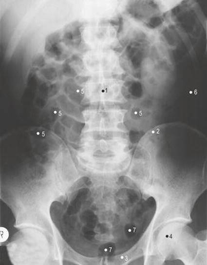
5.7 Abdomen: plain abdominal radiograph
1 Third lumbar vertebra
2 Ilium
3 Superior ramus of pubis
4 Head of femur
5 Gas shadows in small gut
6 Gas shadows in left colon
7 Gas shadows in rectum
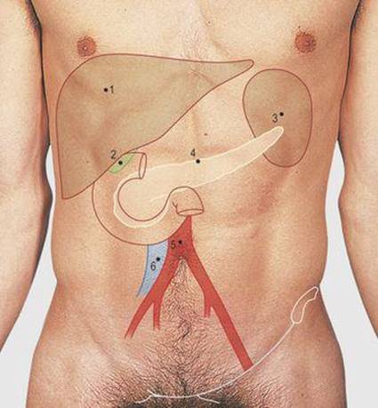
5.8 Surface marking of non-alimentary tract abdominal viscera (The structures labelled 3–6 lie on the posterior abdominal wall)
1 Liver
2 Gall bladder
3 Spleen
4 Pancreas
5 Aortic bifurcation
6 Formation of inferior vena cava
The most superficial of the three sheet-like abdominal muscles is the external oblique; it is attached superiorly by slips to the lateral aspect of the lower eight ribs and interdigitates with the serratus anterior over the middle four ribs. Medially, the muscle forms part of the rectus sheath and inferiorly it is attached to the symphysis pubis, pubic tubercle, anterior superior iliac spine and the anterior half of the iliac crest. The muscle is extensively aponeurotic medial to, and below, a line descending vertically downwards from the ninth costal cartilage to below the level of the umbilicus. The line then curves laterally towards the anterior superior iliac spine. Between the spine and the pubic tubercle, the aponeurotic edge is recurved on itself to form the inguinal ligament. The muscle has a free posterior border but this is not usually visible or palpable in a living subject. The muscles of the anterior abdominal wall are supplied by the lower five intercostal, the subcostal and the first lumbar nerves.
The anterior abdominal wall is divided for descriptive purposes into nine regions by two horizontal lines (subcostal and transtubercular) and two vertical lines through the midpoint of each clavicle (Fig. 5.5 – these lines cross the costal margin at the tip of the ninth costal cartilage and the midinguinal point). The subcostal plane passes through the lower border of the third lumbar vertebra; the transtubercular plane passes through the iliac tubercle and the fifth lumbar spine. The three central regions formed by these lines are the epigastrium, umbilical and suprapubic, from above downwards. The three regions on each side are the hypochondrium, lumbar and iliac, from above downwards. Another plane sometimes referred to in clinical practice is the horizontal, transduodenal (transpyloric) plane, passing through the first part of the duodenum. It is at the level of the lower border of the first lumbar vertebra; ventrally, the transpyloric plane lies midway between the xiphisternal joint and the umbilicus. The abdominal cavity extends upwards behind the lower ribs under the diaphragm and downwards into the pelvis to the pelvic diaphragm (formed by the levator ani muscles).
Surface markings of the alimentary tract (Fig. 5.6)
The oesophagus passes through the diaphragm at the level of the 10th thoracic vertebra, just to the left of the midline. The duodenum starts variably to the right of the midline, sometimes in, but frequently far below, the transpyloric plane. The stomach lies between these two points and is of variable size, partly covered by the rib cage and filling the epigastrium. The duodenum curves in C fashion, convex to the right, to end at the duodenojejunal flexure, this being sited near the midline and approximately in the subcostal plane. The root of the small gut mesentery passes obliquely over the posterior abdominal wall from the flexure to the right iliac fossa, and the central abdomen and upper pelvis are filled with loops of small gut.
The base of the appendix is sited in the right iliac fossa. The surface marking is approximately at the junction of the middle and lateral thirds of a line joining the umbilicus to the anterior superior iliac spine. Initially, inflammation of the appendix produces referred pain around the umbilicus (i.e. the 10th thoracic dermatome) but, as it progresses and involves the parietal peritoneum, local tenderness becomes more marked in the right iliac fossa. The tip of the appendix is usually positioned laterally, inferior to, or behind the caecum, but it may pass downwards into the pelvis or be related to the terminal ileum. The caecum, ascending colon and hepatic flexure of the colon on the right side, and the splenic flexure descending colon on the left, are largely retroperitoneal structures whereas the transverse and sigmoid colons have a mesentery and vary in their position, although they are usually in the regions shown (Fig. 5.6).
Pain produced by inflammation of part of the gut may initially be referred, e.g. the small gut and appendix to the umbilical region, and the large gut to the suprapubic region. However, once the inflammation affects the adjacent parietal peritoneum, the pain and associated tenderness become focal to the area of disease.
Surface markings of non-alimentary tract viscera (Figs 5.8, 5.11)
The liver lies in the right hypochondrium above the costal margin. Its upper border, bulging into the diaphragm, is at the level of the fourth intercostal space. The left lobe passes deep to the xiphisternum across the epigastrium. The gall bladder projects just below the liver at the point where the midclavicular line crosses the costal margin, i.e. the ninth costal cartilage. The spleen is situated posteriorly along the line of the left ninth to 11th ribs. The kidneys lie on the posterior abdominal wall (Fig. 5.11). The pancreas passes from the concavity of the duodenum, slightly obliquely upwards across the midline, over the posterior abdominal wall; its tail passes into the hilum of the spleen.
The abdominal aorta lies on the posterior abdominal wall near the midline and to the left of the inferior vena cava. It divides at the level of the umbilicus, at the level of the body of the fourth lumbar vertebra. The common and external iliac arteries pass from the umbilicus to the midinguinal point (midway between the anterior superior iliac spine and the symphysis pubis) to continue as the femoral artery. The inferior vena cava is formed at the level of the body of the fifth lumbar vertebra, its common iliac tributaries lying to the right and posterior to their respective arteries. The external iliac and femoral veins lie medial to their respective arteries. Note that the umbilicus varies at the level of the disc between the third and fourth lumbar vertebrae in young muscular adults, but descends over the body of the fourth lumbar vertebra in middle age.
Abdominal incisions
Most abdominal viscera can be reached through a midline incision (Fig. 5.9). This may be in the upper or lower abdomen, or along its full length, depending on the target organ. The incision divides the skin, the two layers of superficial fascia and the fibres of the linea alba, this being wide enough to divide in the midline without entering either rectus sheath. The transversalis fascia and peritoneum are also divided in the line of the skin incision.

5.9 Abdominal incisions: anterior
1 Midline
2 Paramedian
3 Subcostal
4 Roof top
5 Transverse
6 Renal
7 Appendix
8 Suprapubic
9 Point for insertion of peritoneal catheter
10 Point for insertion of laparoscope
11 Access point for liver biopsy
The liver and gall bladder may be conveniently approached through a right subcostal incision. The rectus muscle and anterior and posterior rectus sheath are divided in line with the incision, as are the three sheet-like abdominal muscles. A left subcostal incision can be used to approach the spleen and the left and right subcostal incisions can be combined across the midline (rooftop incision) to give a wide bilateral access to the kidneys and suprarenal glands.
The bladder, and the uterus and its adnexia, can be approached through a transverse suprapubic (Pfannenstiel) incision. The skin, two layers of superficial fascia and the anterior rectus sheath are divided in line with the incision. The rectus abdominis muscles are not adherent to the sheath at this level and can be retracted laterally to expose the transversalis fascia and peritoneum; these structures are divided in line with the skin incision. It is possible to approach the inferior aspect of the bladder and the prostate gland in a plane posterior to the symphysis pubis using the suprapubic approach, retracting the peritoneum but not dividing it, i.e. staying outside the peritoneal cavity.
The appendix is approached through an oblique incision about 3cm above and medial to the anterior superior iliac spine, dividing the skin and two layers of superficial fascia. The external oblique muscle and aponeurotic fibres run in the same plane and are separated in this line. The internal oblique and transverse abdominus muscles, running in a different plane, are also split in line with their fibres from the centre of the incision so that, when repaired at the end of the procedure, the lines of division overlap each other in a gridiron fashion. A wider approach to the appendix can be obtained by dividing the three abdominal wall muscles in line with a longer oblique skin incision, and a similar left iliac incision can be used to approach the sigmoid colon.
A transverse incision through the rectus muscle in the transpyloric plane is used to approach the gastroduodenal junction in babies with abnormalities of this region. Although the kidney can be reached from anteriorly, its posterior position favours a more posterior approach. A wide exposure can be obtained through an incision along the line of the 12th rib, the abdominal wall muscles being divided and the kidney approached retroperitoneally (Fig. 5.11).

5.10 Posterior abdominal wall
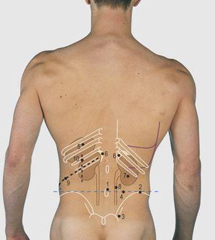
5.11 Posterior abdominal wall: bones and soft tissues
1 Fourth lumbar spine
2 Iliac crest
3 Posterior superior iliac spine
4 Intercristal plane
5 Spleen
6 Suprarenal glands
7 Right kidney
8 Right ureter
9 Nephrectomy incision
10 Lumbotomy incision (this provides a limited exposure to the kidney and suprarenal gland)
A needle may be inserted into the liver usually through the right seventh intercostal space in the anterior axillary line. The needle passes through the lateral recess of the pleural cavity and the diaphragm but does not pass through the lung; the needle is inserted with the subject holding a breath in expiration. The procedure may be used to obtain a liver biopsy or to introduce radio-opaque material into an obstructed dilated biliary system. Other commonly undertaken biopsies are of the kidney, through the site of its posterior surface marking (Fig. 5.11).
A needle may be passed into the peritoneal cavity, in a site such as the iliac fossa, to identify and sample any abnormal peritoneal fluid. Peritoneal tubes for renal dialysis can be inserted through the linea alba 2cm below the umbilicus; the dialysate is introduced and then siphoned off after a number of hours.
Posterior abdominal wall (Figs 5.10-5.12)
In the posterior lower trunk, the lower ribs, iliac crest, posterior superior and inferior iliac spines, lumbar spines and the posterior aspect of the sacrum are palpable (Fig. 5.11). The intercristal line, joining the iliac crests, passes between the spines of the third and fourth lumbar vertebrae; often it crosses the fourth spine. Compare this with Figure 4.9 (p. 35) to relate the position of the abdominal viscera to the lungs.
The latissimus dorsi (Fig. 7.16, p. 70) takes its origin over this region and deep to it are the powerful erector spinae muscles. The spleen lies posterolateral on the left chest, deep to the ninth to 11th ribs; it is susceptible to damage by trauma of this region. The kidneys lie approximately in the transpyloric plane, the right being slightly superior. Their long axes are slightly angled, the inferior pole lying away from the vertical plane; they may be needled through these sites to obtain a tissue biopsy or drain an obstructed renal pelvis.
A needle can be introduced into the aorta for the injection of radio-opaque material, demonstrating the aorta and its lower limb branches. The point of insertion is on the left side, at the level of the first lumbar vertebra, 7cm from the midline and angled 45° medially; when the needle hits the vertebral column it is gradually angled clear of the bone and then advanced forward into the aorta. Lumbar puncture is considered on p. 61.
Cutaneous innervations of the trunk (Figs 5.13, 5.14)
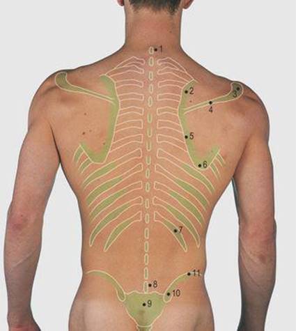
5.12 Posterior abdominal wall: palpable structures
1 Spine of 7th cervical vertebra (vertebra prominens)
2 Upper medial border of scapula
3 Acromion
4 Spine of scapula
5 Lower medial border of scapula
6 Inferior angle of scapula
7 Twelfth rib
8 Spine of 5th lumbar vertebra
9 Sacrum
10 Posterior superior iliac spine
11 Iliac crest
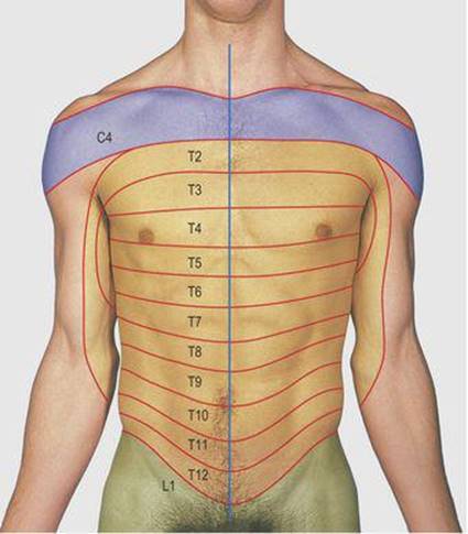
5.13 Cutaneous innervation of the trunk: anterior
The numbers denote the cutaneous innervation from the anterior primary rami of the cervical, thoracic and lumbar nerves. There is extensive overlap of consecutive dermatomes both anteriorly and posteriorly.

5.14 Cutaneous innervation of the trunk: posterior
The numbers denote the cutaneous innervation from the cervical, thoracic, lumbar and sacral nerves. The dotted lines enclose the areas innervated by the posterior primary rami.
Abdominal examination
The abdomen is examined with the subject lying flat on his/her back on a couch with a single pillow supporting the head and with the legs extended and uncrossed. This is the recumbent (supine) position. The abdomen is fully exposed, the breasts being covered in the female and the pubic region in both sexes until specific examination of these areas is required. The size, shape and symmetry of the abdomen are noted and the position and form of the umbilicus. At rest, respiratory movement is predominantly diaphragmatic and the abdominal wall reflects this activity. The abdominal muscles tense during coughing and on raising the head off the couch. In a thin person, central aortic pulsation and occasionally gut peristalsis may be visible. These movements are best observed by kneeling so that the examiner's eyes are at the level of the anterior abdominal wall. Not uncommonly, abnormalities such as scars of previous operations and inguinal hernias, which are protrusions of abdominal viscera through the superficial inguinal ring, may be observed. The latter are accentuated by coughing, this being referred to as an expansile cough impulse.
Palpation should be with clean, warm hands, with the observer standing on the right of the subject and the right hand carrying out most of the manoeuvres, even in a left-handed person. The hand first gently rests on the four corner regions of the abdomen in a circular order; this gentle palpation assesses muscle tone in the abdominal wall and any differences between areas. A tense abdominal wall can be felt by asking the subject to cough or raise his/her head off the pillow. In normal subjects examination produces no pain but abdominal disease may be accompanied by tenderness and this will produce protective voluntary tensing (guarding) or involuntary muscle tension (rigidity) of the abdominal wall.
Following this initial assessment of abdominal tone, each region is examined in turn, palpating for abdominal viscera and abdominal masses. It is customary to start with the right hypochondrium and then to proceed in an orderly fashion so that no region is omitted. A possible scheme is to pass on to the epigastrium, followed by the left hypochondrium, umbilical, left iliac, suprapubic and right iliac regions, leaving the lumbar regions until last, as these require bimanual examination. This order is sometimes related to local findings, any tender area being best left until last, but all regions must always be palpated. Palpation of posterior abdominal wall viscera and masses is by gentle depression of the relaxed anterior abdominal wall, compressing viscera onto the vertebral bodies and muscles of the posterior abdominal wall. The shape of any palpable organ is defined by gentle finger palpation around the area.
In the right hypochondrium, the liver edge may just be palpable in a thin individual. The liver descends on inspiration and this fact is made use of for identification. The flat of the right hand is placed across the right side of the abdomen with the index finger parallel to the costal margin. The right hand is depressed into the abdomen during expiration and pressure is maintained during inspiration. The distending abdomen lifts the hand but any descending firm liver edge will be felt by the lateral border of the index finger. In enormous enlargement, the liver can extend to the right iliac fossa, so the hand is depressed first in this area and then ascends one to three fingers' breadth at a time with each respiratory cycle, repeating the same manoeuvre until the costal margin is reached, or the edge is located.
An enlarged gall bladder moves with the liver and in line with the tip of the ninth costal cartilage, i.e. where the midclavicular line crosses the costal margin. The epigastrium contains much of the stomach, which is not usually palpable, and the left lobe of the liver which, if enlarged, is located as described above. In a thin person, the pulsation of the aorta may be felt through the stomach and pancreas. The left hypochondrium contains the rest of the stomach and the spleen may enlarge into this area. Initially splenic enlargement is below the costal margin in the left anterior axillary line but a very large spleen crosses the umbilical region and reaches the right iliac fossa. The mode of examination for the notch on the anterior edge of the spleen is the same as that for the liver, working cranially along this line of enlargement.
The semisolid contents of the colon mean that it is often palpable in the normal subject. The transverse colon may be felt across the umbilical region and the sigmoid colon is felt in the left iliac fossa. The caecum is often palpable in the right iliac fossa. The ascending and descending colons pass through the right and left lumbar regions respectively. Other normal organs which may be palpable are the aorta in the umbilical region, and a full urinary bladder or pregnant uterus rising out of the pelvis into the suprapubic region. Palpation of the umbilicus with the tip of the finger may reveal a circular defect in the linear alba. This is usually insignificant, but larger defects may transmit viscera producing an umbilical hernia with a cough impulse.
The lower pole of a normal right kidney may be palpated and this is felt by backward pressure of the flat of the right hand in the right lumbar region, combined with forward pressure from the left hand placed behind the abdomen, opposite the right hand. The kidney descends on inspiration and can be felt between the two hands in this bimanual palpation. For bimanual examination of the left lumbar region, the right hand is applied across the anterior abdomen in the usual fashion, and the left hand is either crossed behind the back of the subject with the examiner kneeling or placed behind the left lumbar region with the examiner leaning over the subject.
Percussion is used to define the lower borders of the liver and spleen, being undertaken along the defined lines of their enlargement. The enlarged urinary bladder and uterus are dull to percussion as are abdominal masses and fluid within the peritoneal cavity. The latter is known as ascites and produces dullness in the flanks in the supine position. If the subject then rolls onto one side, the upper dull area will become resonant due to shifting of the fluid by gravity, a feature known as shifting dullness.
Bowel sounds can be heard with a stethoscope over all the abdomen. After a meal they are loudest and most frequent, but at other times it may be necessary to wait for a minute to hear them. Turbulence in the abdominal aorta or iliac vessels may produce sounds detectable with a stethoscope.
Inguinal region, perineum, scrotum and penis (Figs 5.15-5.18)
The aponeurotic lower border of the external oblique muscle (Fig. 5.4, p. 43) between the anterior superior iliac spine and the pubic tubercle is rolled back on itself to form a firm edge, the inguinal ligament. Just above and medial to the pubic tubercle there is an oval defect in the aponeurosis of the external oblique known as the superficial inguinal ring. In the male it forms the external opening of the canal in the abdominal wall through which the testis passes en route to the scrotum. The ring admits the tip of a little finger; it may be difficult to palpate in an obese subject and, alternatively, the tip of the finger can invaginate the skin of the scrotum upwards, behind the subcutaneous fat into the ring. The inguinal canal starts at a round opening in the transversalis fascia, the deep inguinal ring. This is not palpable but is situated about 1cm above the middle of the inguinal ligament.

5.15 Inguinal region

5.16 Inguinal region: bones and soft tissues
1 Anterior superior iliac spine
2 Inguinal ligament
3 Pubic tubercle
4 Symphysis pubis
5 Superficial inguinal ring
6 Deep inguinal ring
7 Femoral artery
8 Femoral vein
9 Femoral canal
10 Femoral nerve
11 Inguinal hernia incision
12 Femoral hernia incision

5.17 Testis and spermatic cord
1 Testis
2 Superior pole of epididymis
3 Spermatic cord
4 Vas deferens
5 Superficial inguinal ring
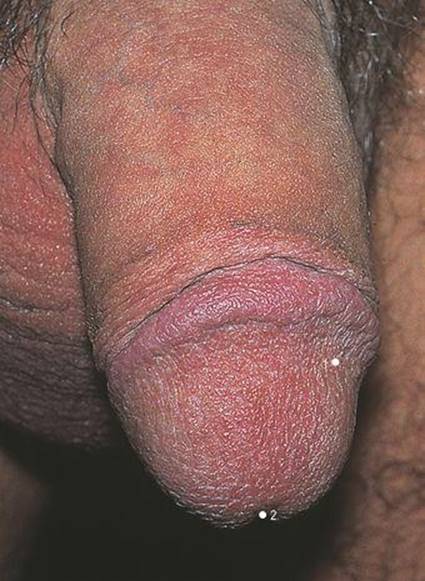
5.18 Penis
Compare this circumcised penis with that of Fig. 5.15, where the foreskin is intact
1 Glans penis
2 Urethral opening
The femoral artery is found deep to the ligament at the midinguinal point (midway between the anterior superior iliac spine and the symphysis pubis). The inferior epigastric artery ascends medially from this point, medial to the deep ring. The femoral vein lies medial to the artery, within a common sheath. An areolar channel medial to the vein, allowing its expansion, forms the medial compartment of the sheath and contains fat, a few deep inguinal lymph nodes and the ascending lymphatic channels; it is termed the femoral canal (Fig. 8.8, p.102). The sheath merges with the adventitia of the vessels at the level of the saphenous opening, where the saphenous vein pierces the deep fascia to drain into the femoral vein.
Testicular descent draws a sac of peritoneum with the testicular vessels into the scrotum and the lower end of this sac surrounds the organ.
The communication with the peritoneal cavity usually closes off, but if it remains patent, peritoneal contents may pass into the sac along the length of the inguinal canal and into the scrotum; this condition is known as an indirect inguinal hernia. Gut may also bulge through a weak posterior wall of the inguinal canal medial to the inferior epigastric artery and protrude through the superficial inguinal ring; this is termed a direct inguinal hernia. A large inguinal hernia can be easily felt, particularly if the subject stands up and coughs. For small hernias, the tip of the little finger is introduced into the superficial inguinal ring as described above and will detect a cough impulse. Although the inguinal canal is rudimentary in the female, both forms of inguinal hernia may occur. Occasionally a peritoneal sac can be protruded into the femoral canal and emerge through the saphenous opening. This is known as a femoral hernia. The incisions shown in Figure. 5.16 are used to approach inguinal and femoral hernias, removing the abnormal peritoneal sac and repairing the defect in the abdominal wall.
Scrotum and penis
The normal testis can be gently palpated through the walls of the scrotum, together with the upper and lower poles of the epididymis (Figure. 5.15). The vas (ductus) deferens ascends from the lower pole, posterior to the testis and can be palpated up to the superficial inguinal ring, rolling the cord-like structure between finger and thumb. The lower end of the peritoneal sac, drawn into the scrotum during testicular descent and surrounding the testis, normally contains a film of lubricant fluid within it.
If excessive fluid is produced, which may be related to a disease process, the condition is known as a hydrocele. The body of the penis ends in the expanded glans penis containing the external opening of the urethra. The skin over the glans, the prepuce (foreskin), is the tissue removed in circumcision. It is usually retractable after the age of 3 or 4 but may contract circumferentially over the end of the glans or, if then retracted,forms a constriction proximal to the glans; this condition is known as a paraphimosis. Examination of the scrotum, its contents and the penis should be a routine part of every full examination of a male patient, confirming a normal testis, epididymis and vas deferens bilaterally, and a normal penis.
Female perineum (Figs 5.19, 5.20)
The opening of the vagina (the introitus) is surrounded by two labial (majora and minora) folds on each side. The labia majora unite anterosuperiorly around the clitoris, the embryological equivalent of the penis. The urethra opens between the clitoris and the vagina. The anus is situated in the midline, anterior to the coccyx and in line with the ischial tuberosities. The pelvic contents can be palpated with a gloved finger passed per rectum or vaginam (PR or PV). In digital examination of the rectum, the patient is usually lying on her left side on the edge of a couch, the examiner standing behind her back. On insertion of the right index finger, the examiner assesses the tone of the anal sphincter. Posteriorly is found the curve of the sacrum. The coccyx can be felt between the inserted finger and the thumb resting superficially, and laterally the ischial spines and tuberosities. In the male, the posterior aspect of the prostate gland can be palpated anteriorly and at a higher level, through the anterior rectal wall, the contents of the rectovesical pouch. The contents usually are loops of sigmoid colon and small gut, and sometimes the tip of the appendix.

5.19 Female perineum
1 Labium majora
2 Labium minora
3 Clitoris
4 Urethral opening
5 Vaginal opening
6 Anus
7 Inferior pubic ramus
8 Acetabulum
9 Ischial tuberosity
10 Sacrotuberous ligament
11 Posterior inferior iliac spine
12 Sacrum
13 Coccyx
14 Sacrospinous ligament
15 Gluteus maximus
16 Hamstring muscles
17 Adductor magnus
18 Gracilis
19 Adductor longus
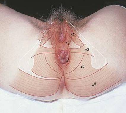
5.20 Muscles of female perineum
1 Ischiocavernosus
2 Bulbospongiosus
3 Transversus perinei superficialis
4 Sphincter ani externus
5 Levator ani
6 Gluteus maximus
Vaginal examination allows palpation of the cervix and, by pressing the left hand over the suprapubic region, bimanual examination usually enables the size and position of the uterus to be assessed. Ovaries lying on the broad ligament may also be palpable and the contents of the recto-uterine pouch (of Douglas) may be noted. Although such examinations are not necessarily very acceptable to clinician or patient, they can elicit important information about pelvic viscera. A PR is an essential part of every full clinical examination and a PV when obstetric or gynaecological conditions are being considered.