
Anterior aspect of the shoulder and upper arm (Figs 7.1-7.12) 64
Actions of the biceps muscle 67
Posterior aspect of the shoulder and upper arm (Figs 7.13-7.20) 69
Movements of the scapula and shoulder joint 71
Cubital fossa (Figs 7.21-7.26) 73
Anterior aspect of the forearm (Figs 7.27-7.33) 75
Posterior aspect of the elbow and forearm (Figs 7.34-7.39) 77
Movements of the elbow and radioulnar joints (Figs 7.40-7.41) 79
Anterior aspect of the wrist and hand (Figs 7.42-7.49) 80
Anatomical snuff box (Figs 7.50-7.54) 84
Dorsal aspect of the wrist and hand (Figs 7.55-7.58) 85
Movements of the wrist and hand (Figs 7.59-7.62) 87
Thenar and hypothenar eminences (Figs 7.63-7.65) 88
Thenar eminence 88
Hypothenar eminence 89
Finger movements (Figs 7.66-7.71) 89
Interosseous muscles 89
Lumbrical muscles 91
Movements of the hand (Figs 7.72-7.77) 91
Innervation of the upper limb (Figs 7.78-7.81) 92
Arteries of the upper limb (Fig. 7.82) 95
Superfi cial structures of the upper limb (Figs 7.83, 7.84) 96
Anterior aspect of the shoulder and upper arm (Figs 7.1-7.12)
The shoulder girdle is the means by which the humerus of the upper limb is attached to the axial skeleton. It consists of the scapula and the clavicle (Fig. 7.2) and is supported by powerful proximal muscles.
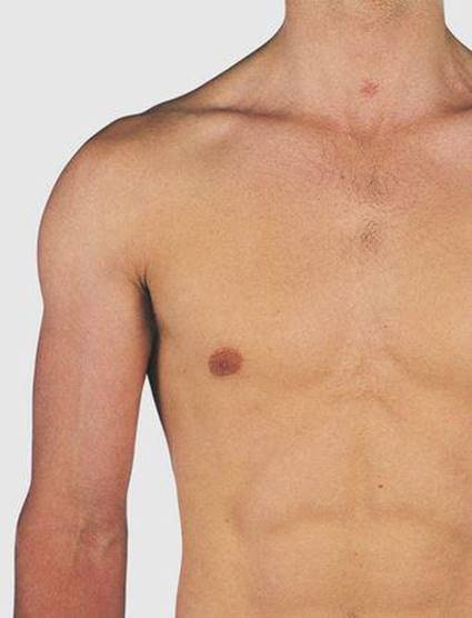
7.1 Anterior aspect of the shoulder and upper arm

7.2 Anterior aspect of the shoulder and upper arm: bones
1 Clavicle
2 Sternoclavicular joint
3 Costoclavicular ligament
4 Coracoid process
5 Coracoclavicular ligaments
6 Acromion
7 Humerus
8 Greater tuberosity
9 Lesser tuberosity
10 Bicipital groove

7.3 Anterior aspect of shoulder and upper arm: palpable structures
1 Clavicle
2 Acromion
3 Corocoid process
4 Greater tuberosity
5 Tendon of long head of biceps
6 Lesser tuberosity
7 Head of humerus
8 Lateral epicondyle
9 Medial epicondyle
10 Brachial artery

7.4 Anterior aspect of the shoulder
1 Clavicle
2 Infraglenoid tubercle
3 Shoulder joint
4 Head of humerus
5 Greater tuberosity
6 Shaft of humerus

7.5 Anterior aspect of the shoulder and upper arm: muscle attachments
1 Pectoralis minor
2 and 11 Coracobrachialis
3 Short head of biceps
4 Long head of biceps
5 Long head of triceps
6 Supraspinatus
7 Subscapularis
8 Pectoralis major
9 Latissimus dorsi
10 Teres major
12 Deltoid
13 Brachialis
14 Sternocostal and
15 Clavicular heads of pectoralis major

7.6 Anterior aspect of the shoulder and upper arm: muscles
1 Pectoralis major
2 Pectoralis minor
3 Deltoid
4 Long head of biceps
5 Short head of biceps
6 Brachialis
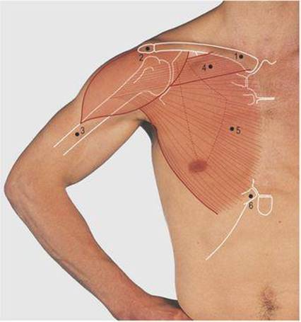
7.7 Action of the pectoralis major muscle
1 Clavicle
2 Acromion
3 Humerus
4 Pectoralis major clavicular head
5 Pectoralis major sternocostal head
6 Seventh costal cartilage

7.8 Shoulder joint: anterior aspect
1 Costoclavicular ligament
2 Conoid ligament
3 Trapezoid ligament
4 Coracohumeral ligament
5 Short head of biceps
6 Coracobrachialis
7 Pectoralis minor
8 Long head of triceps
9 Subscapularis
10 Anterior capsule of shoulder joint with opening of subscapular bursa
11 Long head of biceps
12 Latissimus dorsi
13 Pectoralis major

7.9 Anterior aspect of the shoulder and upper arm: vessels and nerves
1 Subclavian artery
2 Axillary artery
3 Brachial artery
4 Axillary vein
5 Site of access to subclavian vein
6 Brachial plexus: trunks separated for clarity
7 Median nerve
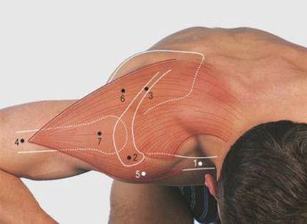
7.10 Attachments and action of the deltoid muscle
1 Clavicle
2 Acromion
3 Spine of the scapula
4 Humerus
5 Anterior fibres
6 Posterior fibres
7 Lateral fibres
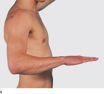


7.11 Actions of the biceps muscle
A, pronation;
B, supination;
C, forced flexion and supination
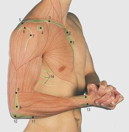
7.12 Lateral aspect of upper limb: palpable structures
1 Body of sternum
2 Manubrium sterni
3 Clavicle
4 Corocoid process
5 Acromion
6 Greater tuberosity
7 Lesser tuberosity
8 Lateral supracondylar ridge
9 Lateral epicondyle
10 Olecranon
11 Subcutaneous border of ulna
12 Radial tuberosity
13 Head of ulna
14 Lateral aspect of ribs
Compression forces from the limb are transmitted through the humerus, scapula and clavicle to the axial skeleton. The clavicle articulates medially with the sternum and first rib at the sternoclavicular joint; the costoclavicular ligament is a powerful shock absorber. The clavicle articulates laterally with the scapula at the acromioclavicular joint and through strong coracoclavicular ligaments. The anterior surface of the clavicle, the acromion and the spine of the scapula are subcutaneous and palpable.
The lateral third of the clavicle, the acromion and the spine give lateral attachment to the trapezius muscle (Fig. 7.16, p. 70), which raises, laterally rotates and draws the scapula medially. It is supplied by the accessory (11th cranial) nerve. The deltoid muscle takes its medial attachments from the lateral quarter of the clavicle, the acromion and the spine of the scapula, and forms the smooth contour of the shoulder. Laterally, it is attached to the deltoid tubercle on the lateral aspect of the humerus. It is the prime abductor of the arm, its anterior fibres contributing to flexion and the posterior fibres to extension of the limb; it is supplied by the axillary nerve. The upper end of the humerus can be palpated through the fibres of the relaxed deltoid.
The pectoralis major muscle has two medial heads (Fig. 7.6). The clavicular is attached to the medial two-thirds of the clavicle and the sternocostal to the anterior surface of the sternum, the upper five to seven costal cartilages and the upper part of the external oblique aponeurosis. Laterally, the two heads converge onto a narrow tendon which passes deep to the deltoid muscle to be attached to the lateral lip of the bicipital groove on the humerus. The superior head overlaps the inferior and together they form the bulk of the anterior axillary fold (Fig. 4.14, p. 38). The muscle as a whole is a powerful abductor and medial rotator of the arm; the clavicular head, with the anterior fibres of deltoid, flexes the arm. In contrast, the sternocostal head is a powerful extensor from the flexed position, acting with latissimus dorsi (Fig. 7.16, p. 70), e.g. pulling the body up on an overhead bar and the follow-through of a tennis serve. It can be demonstrated clinically by pressing hands on hips (Fig. 7.7). The muscle is supplied by the medial and the lateral pectoral nerves from the brachial plexus.
The cephalic vein lies in the deltopectoral groove medial to the anterior fibres of the deltoid muscle. It passes deeply through the clavipectoral fascia (a layer between the pectoralis minor and the clavicle) to enter the axillary vein.
The axillary artery and vein can be exposed through an incision through skin and superficial fascia, separating the two heads of the pectoralis major muscle and dividing the clavipectoral fascia. The axillary vein becomes the subclavian vein at the outer border of the first rib behind the middle of the clavicle and is closely related to the posterior surface of this bone. A needle inserted below the clavicle and aimed superomedially adjacent to the posterior aspect of the bone will enter the vein; this provides an important point of access (Fig. 7.9).
The brachial artery lies in the groove between the biceps and brachialis muscles along the length of the arm: it can be palpated by lateral pressure onto the humerus. The median nerve crosses anterior to the brachial artery in the mid-upper arm and comes to lie on its medial side in the cubital fossa. The shoulder joint may be approached through an anterior incision along the anterior border of the deltoid muscle, dividing its attachment to the clavicle and the tendon of the subscapularis muscle, to expose the capsule and anterior surface of the joint.
Actions of the biceps muscle
The tendon of the long head of the biceps muscle is palpable between the lesser and the greater tuberosities of the humerus. The coracoid process of the scapula gives attachment to the coracobrachialis, the short head of the biceps and the pectoralis minor muscles. It can be palpated deeply 1 cm below the clavicle under the medial fibres of the deltoid muscle.
The biceps muscle forms a prominence on the anterior aspect of the arm. Its short head is attached to the coracoid process and the long head passes between the greater and lesser humeral tuberosities, within the bicipital groove, and is attached to the superior aspect of the glenoid fossa on the scapula. Inferiorly the muscle is attached to the bicipital tuberosity on the radius; it has no attachments to the humerus. The muscle flexes the elbow and is a powerful supinator of the forearm: combining these actions produces the greatest prominence of the muscle belly (Fig. 7.11C). The muscle is supplied by the musculocutaneous nerve, which also supplies the coracobrachialis and brachialis muscles. The former muscle is not easily palpable in the lateral aspect of the axilla but the latter forms, with the biceps, the muscle bulk of the anterior aspect of the upper arm.
Posterior aspect of the shoulder and upper arm (Figs 7.13-7.20)
The acromion and spine of the scapula are subcutaneous. The deltoid muscle forms the smooth prominence of the shoulder overlapping the upper end of the humerus; its posterior fibres aid in extension of the shoulder (Fig. 7.16).

7.13 Posterior aspect of the shoulder and upper arm
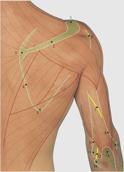
7.14 Posterior aspect of shoulder and upper arm: palpable structures
1 Acromion
2 Spine of scapula
3 Medial angle
4 Medial border
5 Inferior angle of scapula
6 Greater tuberosity
7 Radial nerve deep to fibres of triceps
8 Lateral supracondylar ridge
9 Lateral epicondyle
10 Medial supracondylar ridge
11 Medial epicondyle
12 Ulnar nerve
13 Olecranon process
14 Head of ulna
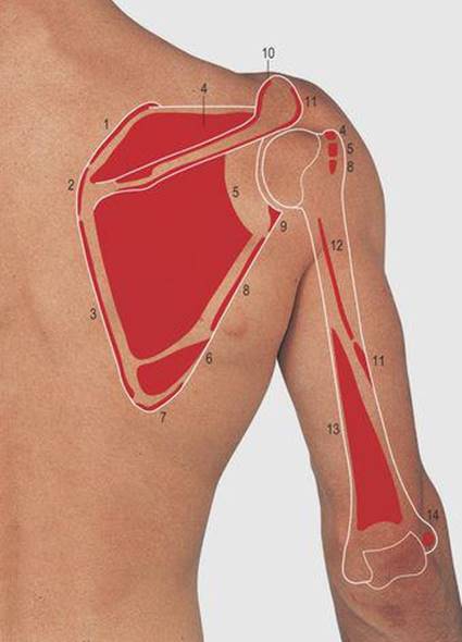
7.15 Posterior aspect of the shoulder and upper arm: muscle attachments
1 Levator scapulae
2 Rhomboideus minor
3 Rhomboideus major
4 Supraspinatus
5 Infraspinatus
6 Teres major
7 Latissimus dorsi
8 Teres minor
9 Long head of triceps
10 Trapezius
11 Deltoid
12 Lateral head of triceps
13 Medial head of triceps
14 Anconeus
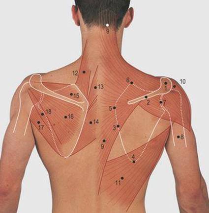
7.16 Posterior aspect of the shoulder and upper arm: bones and muscles of the shoulder girdle
Right, superficial muscles; left, deep muscles
1 Acromion
2 Spine of scapula
3 Medial border of scapula
4 Inferior angle of scapula
5 Medial angle of scapula
6 Superior angle of scapula
7 Glenoid fossa
8 Humerus
9 Trapezius
10 Deltoid
11 Latissimus dorsi
12 Levator scapulae
13 Rhomboideus minor
14 Rhomboideus major
15 Supraspinatus
16 Infraspinatus
17 Teres major
18 Teres minor
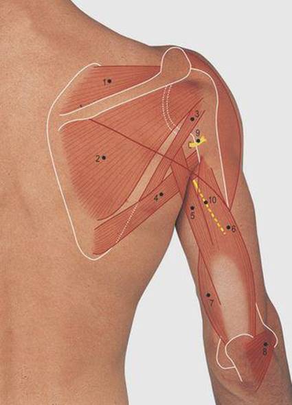
7.17 Posterior aspect of the shoulder and upper arm: structures deep to the deltoid muscle
1 Supraspinatus
2 Infraspinatus
3 Teres minor
4 Teres major
5 Long head of triceps
6 Lateral head of triceps
7 Medial head of triceps
8 Anconeus
9 Axillary nerve
10 Radial nerve
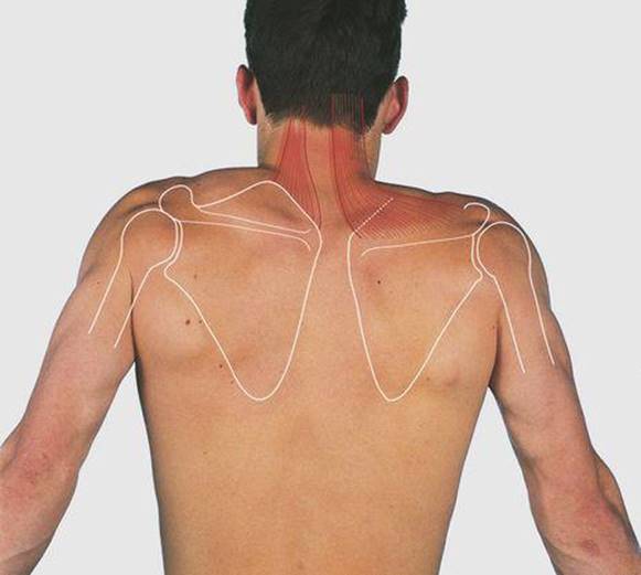
7.18 Scapula elevation

7.19 Scapula retraction
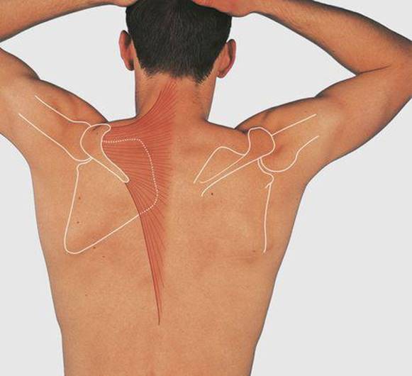
7.20 Lateral rotation of the scapula
The trapezius forms a wide triangular muscle sheet with its base medially attached to the medial half of the superior nuchal line of the occipital bone, the ligamentum nuchae and the spines and interspinous ligaments of the lower cervical and all the thoracic vertebrae. Laterally the muscle is attached to the lateral third of the clavicle and to the length of the acromion and the spine of the scapula. The wide medial attachment of the muscle produces a wide range of scapular movements, raising, laterally rotating and drawing the bone medially (Fig. 7.16 and 7.18-7.20). The muscle is supplied by the accessory (11th cranial) nerve.
The latissimus dorsi muscle also has a wide medial attachment, to the lumbar spines, lumbar fascia and posterior half of the iliac crest. Its fibres pass upwards and laterally to the floor of the bicipital groove on the humerus. The muscle is also attached to the inferior angle of the scapula, producing medial rotation, and helps to prevent this angle from jutting out from the chest wall during shoulder movements. The latissimus dorsi is a powerful adductor of the arm; its narrow tendon wraps around the teres major muscle in the posterior axillary wall. It is supplied by the thoracodorsal branch of the posterior cord of the brachial plexus, the nerve descending over the medial wall of the axilla to enter the muscle in the axilla.
The lower part of the posterior axillary fold is formed mainly of the teres major muscle, passing from the dorsum of the inferior angle of the scapula to the medial lip of the bicipital groove on the humerus. The lateral border of the scapula can be felt through the bulk of the teres major muscle. The subscapularis muscle contributes to the posterior axillary wall superiorly. Together, the supraspinatus, infraspinatus, teres minor and subscapularis are termed the ‘rotator cuff’ muscles, being closely applied to the shoulder joint and maintaining its stability.
The teres major and the subscapularis muscles pass anterior to the joint, the latter to the lesser tuberosity, and are medial rotators of the shoulder joint. The infraspinatus and teres minor muscles pass posterior to the joint to the greater tuberosity and are lateral rotators. The former muscle is attached medially to the posterior aspect of the scapula below its spine and is palpable, the latter to its lateral border.
The supraspinatus muscle is attached to the posterior aspect of the scapula above the spine and is palpable deep to the trapezius. It passes laterally under the acromion to the greater tuberosity of the humerus. It also stabilises the shoulder joint and is important in initiating abduction, as the deltoid muscle has insufficient mechanical advantage to initiate this movement from the adducted position.
If the supraspinatus muscle is inactive the arm has to be swung or flicked by the hip laterally, away from the body, to enable the deltoid to take over abduction. While not primarily a rotator, supraspinatus does form one of the ‘cuff’ muscles.
The supraspinatus tendon is compressed between the greater tuberosity of the humerus and the acromion in mid-abduction; the arm has to be laterally rotated for full abduction to take place. This can be demonstrated by holding the medial and lateral epicondyles of the humerus through the full range of abduction. The supraspinatus and infraspinatus muscles are supplied by the suprascapular nerve from the upper trunk, and the teres major and suprascapularis muscles by the subscapular nerves from the posterior cord of the brachial plexus. All the intrinsic scapular muscles derive their nerve supply from the fifth and sixth cervical spinal nerve roots. The rotator muscles and supraspinatus stabilise the shoulder joint anteriorly, posteriorly and superiorly.
The muscle bulk of the posterior aspect of the upper arm is produced by the triceps muscle with a smaller contribution from the anconeus near the elbow (Fig. 7.14). The triceps is attached to the posterior shaft of the humerus above (lateral head) and below (medial head) the radial groove and, by its long head, below the glenoid fossa on the scapula. The muscle is attached distally to the palpable olecranon process of the ulna and is a powerful extensor of the elbow; it is supplied by the radial nerve.
Falls onto the outstretched hand can result in fractures of the clavicle; this usually occurs at the junction of the middle and lateral thirds. The shoulder joint is least supported inferiorly, and dislocation in this direction is usually due to a blow to the upper lateral aspect of the abducted humerus. The axillary nerve is at risk. Sensation should be assessed over the distal attachment of the deltoid, since this examination, of the upper lateral cutaneous branch of the axillary nerve, is less painful than testing the action of deltoid in the immediate post-traumatic period. Fractures of the humerus are usually by direct violence. At the upper end this is through the surgical neck; the axillary nerve is again at risk as it lies adjacent to this site. Fractures of the humeral shaft are usually spiral, and the radial nerve, lying in its groove on the posterior aspect of the bone, is at risk; nerve injury produces wrist drop. The triceps is spared in this injury, as it is supplied from above this level.
Movements of the scapula and shoulder joint
Movements of the upper limb away from the trunk involve movements both of the scapula and of the shoulder joint. This can be confirmed by watching a subject from behind and then trying to hold the scapula still during shoulder movements (Figs 7.18-7.20). Powerful muscles raise, lower, draw medially and laterally, and rotate the bone medially (medial movement of the inferior angle and downward facing of the glenoid fossa) and laterally. During these movements there is also some compensatory movement in the joints at either end of the clavicle.
The scapula is elevated, as in shrugging the shoulders, by the upper fibres of the trapezius and the levator scapulae muscles. The latter muscle lies deep to the trapezius and is attached to the medial border above the spine of the scapula and superiorly to the cervical transverse processes. It forms some of the muscle bulk of the posterolateral aspect of the neck. The scapula is depressed by the action of gravity and the serratus anterior and pectoralis minor muscles. The scapula is moved forwards on the chest wall, as in pushing and punching, by the serratus anterior and pectoralis minor muscles. The attachments of the latissimus dorsi to the inferior angle hold it onto the chest wall, but if the serratus anterior muscle is paralysed the inferior angle juts out during these movements, a condition known as winging of the scapula. Retraction of the scapula, as in bracing the shoulders, is by trapezius and, deep to this, the rhomboid muscles, passing between the medial aspect of the scapula and the thoracic spines. Lateral rotation of the scapula is produced by serratus anterior and trapezius, and medial rotation by levator scapulae, pectoralis minor and the rhomboid muscles.
Flexion of the shoulder joint is by the clavicular head of the pectoralis major, the anterior fibres of the deltoid and coracobrachialis muscles. Extension is by the posterior fibres of deltoid and, from the flexed position, by the sternocostal head of pectoralis major and latissimus dorsi muscles. Abduction is initiated by the supraspinatus and continued by the deltoid muscles. Medial rotation is by pectoralis major, the anterior fibres of deltoid and latissimus dorsi, teres major and subscapularis muscles. Lateral rotation is by the posterior fibres of deltoid, teres minor and the infraspinatus muscles.
Cubital fossa (Figs 7.21-7.26)
The cubital fossa is bounded superiorly by a line through the palpable medial and lateral epicondyles of the humerus, laterally by the brachioradialis muscle and medially by the pronator teres muscle (Figs 7.22 and 7.25). The brachialis muscle has a wide attachment to the anterior aspect of the humerus and its tendon passes to the coronoid process of the ulna in the floor of the cubital fossa, making it difficult to palpate the bones on the anterior aspect of the elbow joint. The prominent biceps tendon passes to the bicipital tuberosity, on the medial side of the upper end of the radius, through the middle of the fossa.

7.21 Cubital fossa and the anterior aspect of the forearm
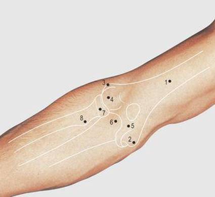
7.22 Cubital fossa: bones
1 Humerus
2 Medial epicondyle of humerus
3 Lateral epicondyle of humerus
4 Capitulum
5 Trochlea
6 Coronoid process of ulna
7 Head of radius
8 Bicipital tuberosity
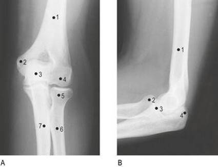
7.23 Elbow joint
A anterior
1 Shaft of humerus
2 Medial epicondyle
3 Trochlea, overlain by upper end of ulna
4 Capitulum
5 Head of radius
6 Shaft of radius
7 Shaft of ulna
B lateral
1 Shaft of humerus
2 Head of radius
3 Coronoid process of ulna
4 Olecranon process of ulna
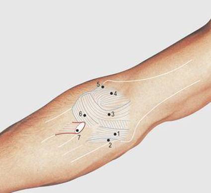
7.24 Elbow joint: anterior aspect
1 Anterior band of medial ligament
2 Oblique band of medial ligament
3 and 4 Anterior capsule
5 Lateral ligament
6 Annular ligament
7 Tendon of biceps
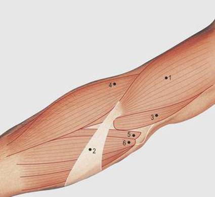
7.25 Cubital fossa: soft tissues
1 Biceps
2 Bicipital aponeurosis
3 Brachialis
4 Brachioradialis
5 Pronator teres
6 Common flexor origin

7.26 Cubital fossa: vessels
1 Cephalic vein
2 Basilic vein
3 Median cubital vein
4 Brachial artery
5 Ulnar artery
6 Radial artery
The firm thin bicipital aponeurosis passes from the medial side of the tendon in the roof of the fossa to the subcutaneous surface of the ulna (Fig. 7.25). The aponeurosis makes it more difficult to feel the brachial artery which has the median nerve lying on its medial side. To feel the artery, the elbow is fully extended and the vessel compressed back onto the joint; it passes deeply into the apex of the cubital fossa where it divides into the radial and ulnar arteries.
The brachial artery may be used to obtain an arterial blood sample or to insert catheters for cardiac and other arterial investigations. The median nerve leaves the fossa between the humeral and ulnar heads of pronator teres, and deep to the superficial muscles of the forearm. The cephalic, basilic and median cubital veins in the roof of the cubital fossa are commonly used for obtaining venous samples (Fig. 7.26).
Anterior aspect of the forearm (Figs 7.27-7.33)
The pronator teres muscle is attached proximally just above the common flexor origin, on the anterior surface of the medial epicondyle. The common flexor origin gives attachment to the flexor carpi radialis, palmaris longus, flexor digitorum superficialis and flexor carpi ulnaris (Figs 7.30-7.33). The pronator teres gains an additional head from the coronoid process of the ulna, and the flexor carpi ulnaris one from the posterior subcutaneous border of the bone; the former muscle is attached distally to the lateral aspect of the mid-shaft of the radius, and it pronates the forearm.

7.27 Anterior aspect of the forearm

7.28 Anterior aspect of the forearm: bones
1 Humerus
2 Ulna
3 Radius
4 Scaphoid
5 Lunate
6 Triquetral

7.29 Anterior aspect of forearm: palpable structures
1 Lateral supracondylar ridge
2 Lateral epicondyle
3 Medial supracondylar ridge
4 Medial epicondyle
5 Brachial artery
6 Ulnar styloid process
7 Radial styloid process
8 Radial artery
9 Ulnar artery

7.30 Anterior aspect of the forearm: muscle attachments
1 Brachioradialis
2 Extensor carpi radialis longus
3 Common extensor origin
4 and 13 Pronator teres
5 Common flexor origin
6 and 12 Flexor digitorum superficialis
7 Deep head of pronator teres
8 Brachialis
9 Flexor digitorum profundus
10 Biceps
11 Supinator
14 Flexor pollicis longus
15 Pronator quadratus
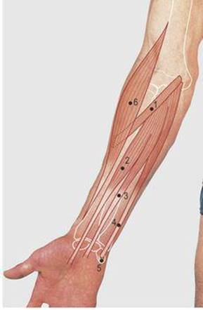
7.31 Anterior aspect of the forearm: superficial muscles
1 Pronator teres
2 Flexor carpi radialis
3 Palmaris longus
4 Flexor carpi ulnaris
5 Pisiform bone
6 Brachioradialis

7.32 Flexor digitorum superficialis (intermediate level)
1 Humero-ulnar head
2 Radial head
3 Tendons to middle and ring fingers lying anterior to those to the index and little fingers
4 Ulnar head of supinator
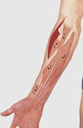
7.33 Anterior aspect of the forearm: deep muscles
1 Tendon of biceps
2 Supinator, humeral head
3 Flexor pollicis longus
4 Flexor digitorum profundus
5 Pronator quadratus
The tendons of the other common flexor origin muscles pass to the hand (Fig. 7.49, p. 84). These muscles are also weak flexors of the elbow: flexor carpi ulnaris is supplied by the ulnar nerve; the remainder by the median. The flexor pollicis longus, flexor digitorum profundus and, distally, the pronator quadratus muscles are deeply placed and impalpable in the forearm; all three are supplied by the anterior interosseus branch of the median nerve, the flexor digitorum profundus receiving an additional supply from the ulnar nerve (Fig. 7.32).
The brachoradialis muscle is attached proximally to the upper two-thirds of the lateral supracondylar ridge of the humerus and distally to the lateral aspect of the distal end of the radius; it pronates or supinates the forearm into the midprone position (flexion of the elbow joint is most powerful in this position). The muscle overlies the radial nerve and its posterior interosseous branch in the cubital fossa and is supplied by the former. The radial nerve also supplies the extensor carpi radialis longus muscle which is attached to the lower third of the lateral supracondylar ridge of the humerus; the muscle is deeply placed in the forearm but its tendon is palpable at the wrist (Fig. 7.58, p. 87).
Posterior aspect of the elbow and forearm (Figs 7.34-7.39)
The bones forming the elbow joint are more easily palpable posteriorly. The medial and lateral epicondyles can be grasped between finger and thumb and they are useful indicators of the position of the humerus in rotation of the shoulder joint (Fig. 7.35). The posterior border of the ulna is subcutaneous throughout its length. The triceps form the main posterior muscle bulk of the upper arm and is attached to the palpable olecranon process. The head of the radius, ensheathed in the annular ligament, is palpable through the muscles attached to the common extensor origin on the posterior aspect of the lateral epicondyle. These muscles are the extensor carpi radialis brevis, extensor digitorum, extensor digiti minimi and extensor carpi ulnaris; they are weak extensors of the elbow joint and are supplied by the posterior interosseous nerve (Fig. 7.39).

7.34 Posterior aspect of the elbow and forearm
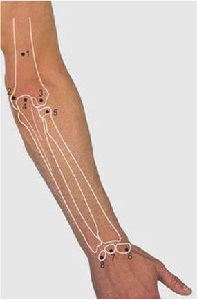
7.35 Posterior aspect of the elbow and forearm: bones
1 Humerus
2 Medial epicondyle of humerus
3 Lateral epicondyle of humerus
4 Olecranon process of ulna
5 Head of radius
6 Scaphoid
7 Lunate
8 Triquetral
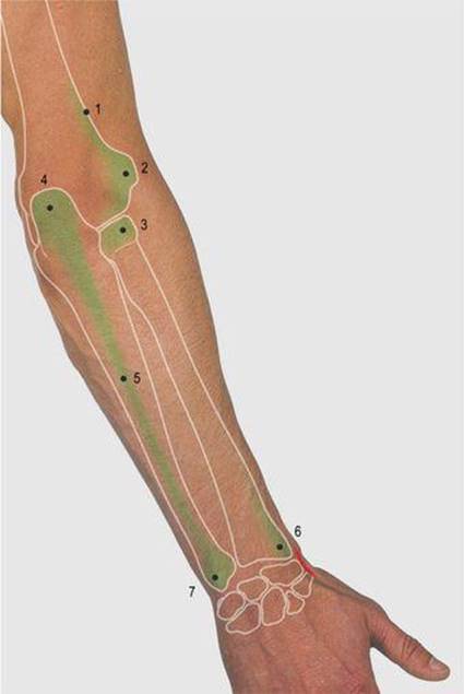
7.36 Posterior aspect of elbow and forearm: palpable structures
1 Lateral supracondylar ridge
2 Lateral epicondyle
3 Head of radius
4 Olecranon process
5 Subcutaneous border of ulna
6 Radial styloid process
7 Ulnar styloid process
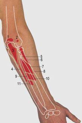
7.37 Posterior aspect of the elbow and forearm: muscle attachments
1 Triceps
2 and 3 Anconeus
4 Posterior border of ulna giving attachment to extensor carpi ulnaris, flexor carpi ulnaris and flexor digitorum profundus
5 Biceps
6 Supinator
7 Abductor pollicis longus
8 Pronator teres
9 Extensor pollicis longus
10 Extensor pollicis brevis
11 Extensor indicis

7.38 Posterior aspect of the elbow and forearm: superficial muscles
1 Brachioradialis
2 Extensor carpi radialis longus
3 Anconeus
4 Extensor carpi radialis brevis
5 Extensor digitorum
6 Extensor carpi ulnaris
7 Extensor digiti minimi
8 Abductor pollicis longus
9 Extensor pollicis brevis
10 Extensor pollicis longus
11 Ulnar nerve
12 Flexor carpi ulnaris

7.39 Posterior aspect of the elbow and forearm: deep muscles
1 Supinator, humeroulnar head
2 Abductor pollicis longus
3 Extensor pollicis brevis
4 Extensor pollicis longus
5 Extensor carpi radialis longus tendon
6 Extensor carpi radialis brevis tendon
7 Extensor indicis
8 Flexor carpi ulnaris
The supinator is also attached to the common extensor origin at a deeper level and receives a head from the lateral aspect of the upper end of the ulna. The muscle wraps around the upper end of the radius to be attached to the anterior surface. It and the other deep impalpable muscles on the posterior aspect of the forearm are supplied by the posterior interosseous nerve; the tendons of these muscles are palpable at the wrist (Fig. 7.58, p. 87). The ulnar nerve lies posterior to the medial epicondyle and can be rolled over the bone at this point, before passing between the humeral and ulnar heads of the flexor carpi ulnaris. If knocked at this point it produces a pins and needles sensation in the forearm.
Movements of the elbow and radioulnar joints (Figs 7.40-7.41)
The elbow is flexed by the biceps and brachialis muscles, assisted by brachioradialis and the muscles attached to the common flexor origin. Extension is by the triceps muscle, with a weak contribution from the muscles of common extensor origin.

7.40 Forearm in full supination

7.41 Forearm in full pronation
In the anatomical position, the palm faces forwards, the position of the forearm being in full supination (Fig. 7.40). The lower end of the radius may be rotated medially, anterior to the ulna through 180°, to bring the thumb from lateral to medial and the dorsum of the hand anteriorly; this is the position of full pronation (Fig. 7.41). These, and intermediate positions, place the hand in the most appropriate position for a required movement. The biceps and supinator muscles supinate and the pronator teres and pronator quadratus pronate. This last muscle is a deep small muscle joining the anterior aspect of the distal quarter of the radius and ulna near the wrist. The brachioradialis brings the forearm into the midprone position.
In the anatomical position, the styloid processes of the radius and ulna are palpable on their respective sides of the wrist joint; the radial styloid is 1cm distal to the ulna. In full pronation, the radial styloid is still palpable but medially placed. The bone most easily palpable on the anterolateral aspect of the wrist is now the head of the ulna. The distal end of the ulna undergoes some medial-to-lateral displacement during pronation, this being more marked when the elbow is flexed. However, there is no rotation of the ulna and the styloid process is no longer palpable.
The bone across the supracondylar region of the humerus is very thin in children, and prone to fracture. The distal fragment in this injury is displaced posteriorly and a knuckle of brachial artery may be trapped between the broken bone ends. Radial artery pulsation must be carefully monitored as, if an occluded brachial artery is left untreated, forearm muscle ischaemia can give rise to a contracture (Volkmann's), with long-term disability.
Forearm fractures in children characteristically break along one side of the bone, which bends rather than snaps. This is like a broken sapling branch of a tree, and is thus called a greenstick fracture. In old age, the bones become soft and crumble more easily. A characteristic upper limb fracture in the elderly is through the distal end of the radius, a centimetre proximal to its articular surface (Colles’ fracture), sustained by a fall onto the outstretched hand.
Anterior aspect of the wrist and hand (Figs 7.42-7.49)
The bones of the hand and their relation to observable skin creases are shown in Figure 7.43. When flexing the wrist against resistance, three tendons (flexor carpi radialis, palmaris longus and flexor carpi ulnaris) stand out. The radial artery is easily palpable on the lower end of the radius lateral to flexor carpi radialis. This is the usual site to assess the pulse when examining a patient and to establish the rate, rhythm, volume and character of the cardiac output and firmness of the arterial wall. A needle may be inserted into the artery at this point. The flexor carpi radialis is attached distally to the base of the second and third metacarpal bones. The median nerve lies deep to the palmaris longus, which provides some protection to it in cuts across the wrist. The muscle is attached to the flexor retinaculum and the apex of the palmar aponeurosis; it is occasionally absent. The flexor carpi ulnaris muscle is attached to the pisiform bone and thence onto the hamate and the base of the fifth metarcarpal bones (Fig. 7.46). The ulnar artery and nerve lie lateral to the tendon but are partly covered by the palmaris brevis muscle and the medial superficial part of the flexor retiniculum; they are not easily palpable.
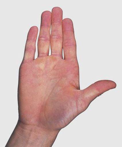
7.42 Anterior aspect of the wrist and hand

7.43 Anterior aspect of the wrist and hand: bones
Note the relation of bones to the skin creases
1 Ulna
2 Radius
3 Ulnar styloid process
4 Radial styloid process
5 Pisiform
6 Triquetral
7 Lunate
8 Scaphoid
9 Scaphoid tubercle
10 Hamate
11 Hook of hamate
12 Capitate
13 Trapezoid
14 Trapezium
15 Ridge of trapezium
16 Metacarpals 1–5
17 Phalanges

7.44 Wrist joint and hand
A anterior
1 Terminal phalanx
2 Middle phalanges
3 Proximal phalanges
4 Metacarpals
5 Hamate
6 Capitate
7 Overlapping trapezoid and trapezium
8 Scaphoid
9 Lunate
10 Triquetral, overlapped by pisiform
11 Styloid process of radius
12 Shaft of radius
13 Styloid process of ulna
14 Shaft of ulna
B anterior view showing epiphyses
1 Epiphysis of proximal phalanx
2 Epiphysis of first metacarpal
3 Distal epiphysis of radius
4 Distal epiphysis of ulna
C lateral view
1 First metacarpal
2 Lunate
3 Overlapping shafts of ulna and radius

7.45 Anterior aspect of the wrist and hand: muscle attachments
1 and 3 Opponens digiti minimi
2 Pisometacarpal ligament
4 Flexor digiti minimi
5 Abductor digiti minimi
6 Flexor carpi ulnaris
7 Abductor pollicis brevis
8 Opponens pollicis
9 Flexor pollicis brevis
10 Abductor pollicis longus
11 and 17 Palmar interossei
12, 16 Transverse and 20 oblique heads and abductor pollici
13 Flexor pollicis brevis and abductor pollicis brevis
14 Flexor pollicis longus
15 Flexor carpi radialis
18 Flexor digitorum superficialis
19 Flexor digitorum profundus
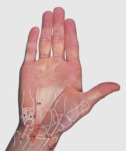
7.46 Anterior aspect of the wrist and hand: superficial tendons
1 Flexor carpi ulnaris
2 Pisohamate ligament
3 Pisometacarpal ligament
4 Palmaris longus
5 Palmar aponeurosis
6 Flexor carpi radialis
7 Radial artery
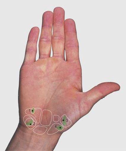
7.47 Flexor retinaculum: palpable attachments
1 Hook of hamate
2 Pisiform
3 Ridge of trapezium
4 Scaphoid tubercle
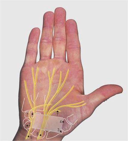
7.48 Flexor retinaculum
1 Hook of hamate
2 Pisiform bone
3 Scaphoid tubercle
4 Ridge of trapezium
5 Ulnar nerve
6 Median nerve
7 Recurrent branch of median nerve
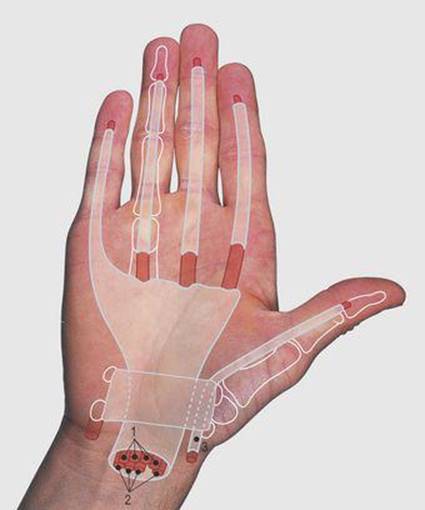
7.49 Anterior aspect of the wrist and hand: digital flexor tendons and sheaths
1 Flexor digitorum superficialis
2 Flexor digitorum profundus
3 Flexor pollicis longus
The flexor retinaculum is a 2.5–3 cm rectangular fibrous band between the pisiform and the hook of the hamate medially and the tubercle of the scaphoid and ridge of the trapezium laterally (Fig. 7.47 and Fig. 7.48). The pisiform is easily palpable and the other three bones are felt by deeper palpation. The tendons of the flexor digitorum superficialis and profundus and the flexor pollicis longus are deeply placed anterior to the wrist joint and pass with the median nerve deep to the flexor retinaculum.
The tunnel formed by the retinaculum and the carpus has limited space and an increase in pressure can compress the median nerve, producing pain and numbness in the lateral aspect of the hand, and wasting of the thenar muscles (the carpal tunnel syndrome). The flexor retinaculum may be divided through an incision across the wrist joint, or endoscopically, to relieve pressure in this condition.
The deep flexor tendons are enclosed in synovial sheaths over the wrist and into the fingers as shown in Figure 7.49. These have clinical significance as infection can spread along a sheath.
The deep fascia over the palm is thickened centrally to form the palmar aponeurosis; this renders the long flexor tendons impalpable at this site. The flexor retinaculum and attachment of the sheaths prevent bowing of the tendons across the wrist during flexion.
The flexor digitorum superficialis and profundus tendons are surrounded by synovial and fibrous sheaths in the fingers and although movement can be felt, individual tendons cannot be palpated. The flexor digitorum profundus is attached to the base of the distal phalanx and the flexor digitorum superficialis tendon divides into two slips which pass on either side of the profundus tendon to be attached to the middle phalanx. The flexor pollicis longus tendon has its own synovial and fibrous sheaths and is attached to the distal phalanx of the thumb.
The skin of the palm is hairless and thickened. The configuration of dermal papillae serves to anchor the epidermis to the dermis, providing a friction surface for gripping and other manual tasks; fibrofatty compartments over the palm produce a firm cushioned area of contact. The palm has a large number of sweat glands which serve to cool the surface during activity and maintain the suppleness of the skin.
The skin over the dorsum of the hand is more mobile and elastic than that of the palm, allowing for stretching during full finger and wrist flexion. The hand, particularly at the fingertips, is densely innervated, these sensory nerve endings facilitating delicate tactile discrimination, such as in reading Braille and feeling in the dark and around corners.
The nail is attached to the surrounding skin by the cuticle (eponychium). Infection may enter a break in this layer, resulting in inflammation of the surrounding tissues and the formation of pus under the nail, a condition known as a paronychia.
Anatomical snuff box (Figs 7.50-7.54)
The anatomical snuff box is a depression on the lateral aspect of the wrist which is accentuated when the thumb is extended. The bones of its floor are the radial styloid, the wrist joint, the scaphoid, trapezium and the base of the first metacarpal (Fig. 7.51 and 7.52).

7.50 Anatomical snuff box

7.51 Anatomical snuff box: bones
1 Radial styloid
2 Scaphoid
3 Trapezium
4 First metacarpal

7.52 Anatomical snuff box: palpable structures
1 Radial styloid process
2 Scaphoid
3 Trapezium
4 Base of first metacarpal
5 Radial artery
6 Radial nerve
7 Extensor pollicis longus
8 Extensor pollicis brevis and abductor pollicis longus
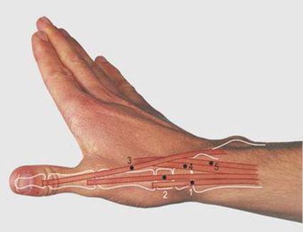
7.53 Anatomical snuff box: tendons
1 Abductor pollicis longus
2 Extensor pollicis brevis
3 Extensor pollicis longus
4 Extensor carpi radialis longus
5 Extensor carpi radialis brevis
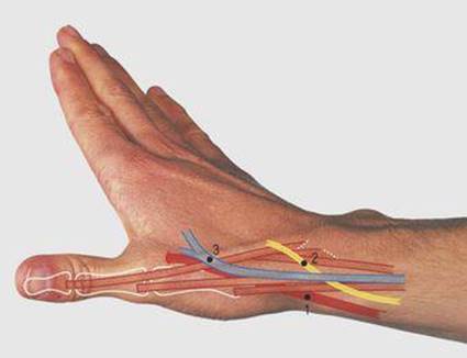
7.54 Anatomical snuff box: radial artery and nerve, and cephalic vein
1 Radial artery
2 Radial nerve
3 Cephalic vein
Tenderness at this site occurs with injuries of the scaphoid bone and synovitis of the adjacent tendon sheaths.
The snuff box is bounded anteriorly by the tendons of abductor pollicis longus and extensor pollicis brevis and posteriorly by extensor pollicis longus (Fig. 7.53). These three muscles are deeply placed in the forearm and are supplied by the posterior interosseous nerve.
The abductor pollicis longus is attached distally to the first metacarpal and the extensor pollicis brevis to the proximal phalanx of the thumb. The extensor pollicis longus is attached to the distal phalanx of the thumb. The fossa is crossed by the tendons of the extensor carpi radialis longus and brevis as these pass, respectively, to the bases of the second and third metacarpals. The superficial branch of the radial nerve crosses the brachioradialis tendon and can be palpated at this point. It then crosses the snuff box to divide into its digital branches (Fig. 7.54). The radial artery crosses the lateral aspect of the wrist before passing between the first and second metacarpals into the palm. Its pulsation can be felt in the floor of the snuff box. The cephalic vein crosses the fossa superficially.
Dorsal aspect of the wrist and hand (Figs 7.55-7.58)
The distal end of the radius is palpable posteriorly, particularly the dorsal tubercle, around which hooks the tendon of extensor pollicis longus. The triquetral, lunate and scaphoid bones, from medial to lateral, form the distal articulation of the wrist joint (Fig. 7.56). These, other carpal bones and the bases of the medial four metacarpals are not easily palpated. The remainder of the metacarpals and phalanges can be palpated throughout their length posteriorly. A finger placed in the depression over the middle of the wrist joint lies between the radius proximally, while distally the finger is related to the capitate on full extension and the lunate on full flexion. The second to fourth metacarpophalangeal joints are easily palpable posteriorly. The first metacarpal is far more mobile than the second to fourth and its carpometacarpal joint is readily palpable. The interphalangeal joints are palpable around their circumference.

7.55 Dorsal aspect of the supine wrist and hand

7.56 Dorsal aspect of the wrist and hand: bones
1 Ulna
2 Ulna styloid
3 Radius
4 Radial styloid
5 Dorsal tubercle of radius
6 Triquetral
7 Lunate
8 Scaphoid
9 Hamate
10 Capitate
11 Trapezoid
12 Trapezium
13 Carpometacarpal joint of thumb
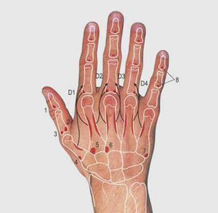
7.57 Dorsal aspect of the wrist and hand: muscle attachments
1 Extensor pollicis longus
2 Adductor pollicis
3 Extensor pollicis brevis
4 Abductor pollicis longus
5 Extensor carpi radialis longus
6 Extensor carpi radialis brevis
7 Extensor carpi ulnaris
8 Extensor digitorum
D1–4 dorsal interossei: the arrows indicate the secondary attachment to the extensor digital expansion

7.58 Dorsal aspect of the wrist and hand: tendons
1 Extensor carpi ulnaris
2 Extensor digitorum
3 Extensor indicis
4 Extensor digiti minimi
5 Extensor carpi radialis brevis
6 Extensor carpi radialis longus
7 Extensor retinaculum
8 Extensor digital expansion
The digital extensor tendons are closely related to the posterior aspect of the radius and retain their relations during pronation. They are held down to the bone by a fibrous sheet, the extensor retinaculum. This passes obliquely from the lateral side of the distal radius to the medial side of the carpus (Fig. 7.58); septa from it pass between the extensor tendons forming osseofascial tunnels. Synovial sheaths cover the tendons beneath and distal to the retinaculum. Each extensor digitorum tendon forms a triangle (the dorsal expansion) over the metacarpophalangeal joint. The corresponding interossei and lumbrical muscles are attached to the base of this expansion. At the apex of the triangle the long tendon reforms and then divides into three slips over the proximal interphalangeal joint. The middle slip is attached to the base of the middle phalanx and the outer slips reunite before being attached to the base of the terminal phalanx.
Two further tendons may be visible on the dorsum of the wrist. The extensor digiti minimi lies medial to the long tendon of the little finger and is inserted into its dorsal tendinous expansion. The extensor indicis is a deep muscle of the forearm supplied by the posterior interosseous nerve; its tendon lies medial to the long tendon of the index finger and it is also inserted into the dorsal expansion. Both extensor digiti minimi and extensor indicis tendons can be removed and used for grafting with minimal loss of function in the hand. The veins over the dorsum of the hand are usually prominent and provide useful sites for venous access, as does the cephalic vein over the anatomical snuff box.
Note that Figures 7.50-7.54 are photographed with the forearm and hand in pronation with the head of the ulna prominent. In contrast, Figures 7.55-7.58 are photographed from behind with the forearm and hand in supination and the styloid process of the ulna can be seen.
Movements of the wrist and hand (Figs 7.59-7.62)
Movements at the wrist (radiocarpal) joint are combined with and augmented by movement at the midcarpal (between the proximal and distal rows of the carpus) joint. Flexion occurs mainly at the midcarpal joint and is produced by the flexor carpi radialis and flexor carpi ulnaris assisted by the long digital flexor muscles (Fig. 7.59). Extension is mainly at the wrist joint and is produced by the extensor carpi radialis longus and brevis and the extensor carpi ulnaris, assisted by the long digital extensor muscles (Fig. 7.60). Abduction is mainly at the midcarpal joint and produced by flexor carpi radialis and extensor carpi radialis longus and brevis (Fig. 7.61). Adduction is mainly at the wrist joint and produced by the flexor carpi ulnaris and extensor carpi ulnaris (Fig. 7.62).

7.59 Wrist flexion
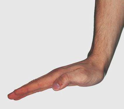
7.60 Wrist extension
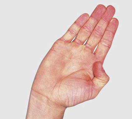
7.61 Wrist abduction

7.62 Wrist adduction
Thenar and hypothenar eminences (Figs 7.63-7.65)
Thenar eminence
The muscle bulk of the lateral aspect of the palm is known as the thenar eminence and it is formed by the short muscles of the thumb. The abductor, flexor and opponens pollicis muscles are attached to the scaphoid tubercle, the ridge of the trapezium and the adjacent flexor retinaculum. The abductor pollicis brevis and flexor pollicis brevis both pass to the radial side of the proximal phalanx of the thumb and the opponens pollicis to the whole length of the radial margin of the first metacarpal bone. The three muscles are supplied by the median nerve, by a recurrent branch after the nerve exits from the carpal tunnel. The adductor pollicis is deeply placed in the palm and is attached medially by two heads to the capitate and second and third metacarpals. Distally it passes laterally to the proximal phalanx of the thumb. It is supplied by the ulnar nerve. The muscle bulk in the first intermetacarpal space includes adductor pollicis, the first dorsal and palmar interossei, and the first lumbrical.

7.63 Thenar and hypothenar eminences
1 Abductor pollicis brevis
2 Flexor pollicis brevis
3 Opponens pollicis
4 Adductor pollicis oblique head
5 Adductor pollicis transverse head
6 Abductor digiti minimi
7 Flexor digiti minimi
8 Opponens digiti minimi
9 Flexor retinaculum
10 Palmar aponeurosis
11 Flexor fibrous sheaths

7.64 Flexion and opposition of the thumb and little finger
(Movement: 2/3 thumb, 1/3 little finger)
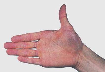
7.65 Extension of the thumb
There is considerable mobility at the saddle-shaped carpometacarpal joint of the thumb. Movements are described in relation to the plane of the nail bed rather than the coronal plane of the body. Flexion is combined with medial rotation and is produced by flexor pollicis longus and brevis and opponens pollicis. Extension is combined with lateral rotation and brought about by extensor pollicis longus and brevis and abductor pollicis longus. The abductor pollicis brevis produces abduction (away from the palm, in the plane of the nail, i.e. 90° to the plane of the palm), the adductor pollicis adduction (return from the abducted position), and the opponens pollicis opposition of the thumb to the index and other digits.
Hypothenar eminence
The hypothenar eminence is formed by the abductor digiti minimi, flexor digiti minimi and opponens digiti minimi, arising from the flexor retinaculum and adjacent medial bones, and passing distally to the ulnar side of the proximal phalanx and the ulnar margin of the fifth metacarpal bone. The little finger is less mobile than the thumb, but slight rotation is present. The muscles are supplied by the ulnar nerve. The thenar and hypothenar muscles are within their own osseofascial compartment of deep fascia.
Finger movements (Figs 7.66-7.71)
Interosseous muscles
Movements of the carpometacarpal joint of the thumb and all metacarpophalangeal joints are flexion, extension, abduction, adduction, circumduction and some rotation. For digits 2–5 (index to minimus), flexion and extension are in the sagittal plane, while abduction and adduction are in the coronal plane (i.e. the plane of the palm). Movements of the thumb are at 90° to the other digits, flexion and extension in the coronal plane (i.e. the plane of the palm), abduction and adduction in the sagittal plane; a combination of abduction, flexion and medial rotation produces opposition of the thumb. In Figure 7.64, the little finger is flexed and laterally rotated in opposition to the thumb. Abduction and adduction of digits 2–5 (away from and towards the midline of the middle finger) are brought about by the palmar and dorsal interosseous muscles arising from the sides of the length of the metacarpals and forming the muscle bulk palpable between these bones; this is particularly marked between the first and second metacarpals. The four smaller palmar interosseous muscles are attached to the palmar surfaces of the first, second, fourth and fifth metacarpal bones.

7.66 Palmar interossei
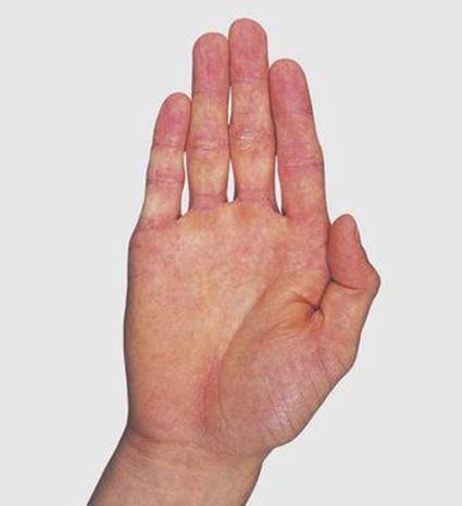
7.67 Finger adduction and extension
Thumb in adducted position: note partial flexion of its metacarpophalangeal and solitary interphalangeal joints

7.68 Dorsal interossei

7.69 Finger abduction and extension with thumb extension

7.70 Finger flexion

7.71 Action of the lumbrical muscles
The four dorsal interosseous muscles are larger and more powerful, arising by two heads from the adjacent sides of the metacarpal bones. The distal tendons of all the interossei gain attachment to the corresponding proximal phalanx and extensor expansion. These attachments are such that the palmar muscles adduct (PAD) and the dorsal muscles abduct (DAB). The interossei also flex the proximal phalanx at the metacarpophalangeal joint and, through their attachment to the extensor expansion, extend the proximal and distal interphalangeal joints.
Lumbrical muscles
The lumbricals are four slender muscles arising from the lateral side of each flexor digitorum profundus tendon in the palm; they are attached to the lateral side of the dorsal expansion of the same finger. Their action is to flex the metacarpophalangeal joint and extend the proximal and distal interphalangeal joints, aided by the interosseous muscles.
All the interosseous and the medial two lumbrical muscles are supplied by the ulnar nerve and the lateral two lumbricals by the median nerve. Further flexion of the metacarpal and interphalangeal joints of the finger is by the long flexor tendons; the flexor digitorum profundus alone acts on the distal interphalangeal joint.
Extension of the fingers is mainly by the long digital extensor muscles aided by extensor indicis and extensor digiti minimi. The collateral slips of the extensor expansion over the proximal interphalangeal joint reunite beyond the expansion. The reformed tendon is attached to the base of the distal phalanx and extends the distal interphalangeal joint.
Movements of the hand (Figs 7.72-7.77)

7.72 Precision grip

7.73 Pinch grip

7.74 Writing

7.75 Power grip

7.76 Hook grip

7.77 Unscrewing the top of a jar
Innervation of the upper limb (Figs 7.78-7.81)
The innervation of a limb (arm or leg) follows three distinctive patterns of clinical relevance in terms of their segmental origin and distribution. They reflect their different modes of development: these are (a) cutaneous, (b) to the muscles and joints, and (c) autonomic. Relevant terms are: axis of the limb; preaxial border (lateral border, radial border, bearing the thumb and cephalic vein); postaxial border (medial border, ulnar border, bearing the little finger and basilic vein); apex of the limb (middle three digits); dermatome (skin area supplied by a single spinal nerve) and myotome (muscle volume supplied by a single spinal nerve).

7.78 Cutaneous nerves of the upper limb: anterior
1 Supraclavicular
2 Upper lateral cutaneous nerve of arm
3 Intercostobrachial
4 Anterior cutaneous nerve of arm (branch of 5)
5 Medial cutaneous nerve of arm
6 Medial cutaneous nerve of forearm
7 Lateral cutaneous nerve of forearm
8 Palmar branch of ulnar
9 Palmar branch of median
10 Superficial terminal branch of radial
11 Ulnar digital
12 Median digital
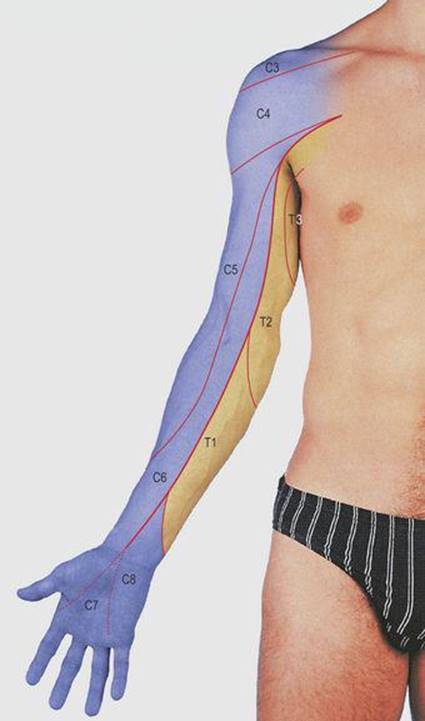
7.79 Cutaneous dermatomes of the upper limb: anterior
The numbers denote the nerve roots. The dark line is the anterior axial line
C, cervical; T, thoracic

7.80 Cutaneous nerves of the upper limb: posterior
1 Supraclavicular
2 Upper lateral cutaneous nerve of arm
3 Posterior cutaneous nerve of arm
4 Intercostobrachial
5 Lower lateral cutaneous nerve of arm
6 Medial cutaneous nerve of arm
7 Posterior cutaneous nerve of forearm
8 Lateral cutaneous nerve of forearm
9 Medial cutaneous nerve of forearm
10 Radial
11 Dorsal branch of ulnar
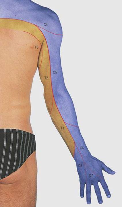
7.81 Cutaneous dermatomes of the upper limb: posterior
The numbers denote the nerve roots. The dark line is the posterior axial line
C, cervical; T, thoracic
The highest (spinal nerve level) dermatome is at the preaxial root of the limb, then successive dermatomes proceed down the preaxial border to the apex, and then return along the postaxial border to reach the trunk at the limb root. Areas supplied by successive dermatomes have considerable overlap in their innervation. Areas supplied by discontinuous (non-successive dermatomes) have minimal or no overlap; these are the dorsal and ventral axial lines (Figs. 7.79, 7.81).
In contrast, the highest myotomes (spinal nerve level) are sited proximally, the intermediate where their name implies, and the lowest are distal (the intrinsic muscles of the hand or foot).
The skin of the upper limb is supplied by the C5 to T1 dermatomes (but with additions from C4 and T2, 3) through the brachial plexus. The fourth cervical root supplies the skin over the tip of the shoulder and the third thoracic that over the axilla. The sixth root supplies the skin over the thumb and the eighth that of the little finger. The distribution of the seventh root to the central fingers is variable. When testing for sensory disturbance, the greatest difference can be demonstrated across axial lines because of the considerable overlap between the innervation of adjacent dermatomes elsewhere.
In brachial plexus injuries of the upper trunk (C5, 6 – birth injuries and severe downward traction on the shoulder), the deltoid, teres minor, supraspinatus, infraspinatus, biceps and brachialis and sometimes other muscles are paralysed. The limb hangs limply, medially rotated and fully pronated, in the ‘tip’ position. There is sensory diminution on the radial side of the arm and forearm. Lower trunk injuries (C8, T1) can also occur at birth, from grasping overhead to break a fall or from pressure from a cervical rib; they produce paralysis of the small muscles of the hand and the long digital flexors, resulting in the characteristic clawed hand, and sensory diminution on the ulnar side of the arm and forearm.
Axillary nerve injuries are usually associated with dislocation of the shoulder joint or during its reduction, and fractures of the surgical neck of the humerus; they result in paralysis of the deltoid muscle and anaesthesia of the skin over the deltoid tuberosity. The radial nerve is most easily damaged in fractures of the humerus as it crosses the radial groove. As the triceps muscle is supplied from above this level, the disability is paralysis of the wrist extensor muscles (wrist drop). Sensory loss is distal to the snuff box and often minimal.
Median nerve injuries are commonest in the carpal tunnel, resulting in paralysis of the thenar muscles (except adductor pollicis) and the lateral two lumbricals. There is sensory palmar loss over the thumb, index, middle and half the ring finger. More proximal injuries result in additional paralysis of the forearm flexors and absence of pronation.
The ulnar nerve is subject to injury as it crosses the posterior aspect of the medial epicondyle of the humerus. There is paralysis of all the remaining small muscles of the hand, and also flexor carpi ulnaris and part of flexor digitorum profundus; sensory loss is over the ulnar side of the hand. When the long digital flexor muscles can contract unopposed by lumbricals and interossei, as with injuries of the ulnar or median nerves at the wrist, there is clawing of the hand.
The biceps reflex is lost with lesions of the fifth and sixth roots, the brachioradialis with the sixth, and the triceps with the sixth and seventh cervical root injuries.
The preganglionic autonomic supply to the arm is from T2 to T6.
Arteries of the upper limb (Fig. 7.82)

7.82 Arteries of the upper limb
1 Subclavian
2 Axillary
3 Brachial
4 Radial
5 Ulnar
6 Common interosseous
7 Superficial palmar arch
8 Deep palmar arch
Superficial structures of the upper limb (Figs. 7.83, 7.84)

7.83 Anterior aspect of upper limb: superficial structures
1 Sternal and
2 Clavicular heads of pectoralis major
3 Deltoid
4 Biceps
5 Triceps
6 Brachialis
7 Bicipital aponeurosis
8 Pronator teres
9 Brachioradialis
10 Flexor carpi radialis
11 Palmaris longus
12 Flexor carpi ulnaris

7.84 Posterior aspect of upper limb: superficial structures
1 Deltoid
2 Triceps
3 Tendon of triceps
4 Anconeus
5 Flexor carpi ulnaris
6 Brachioradialis and extensor carpi radialis longus
7 Extensor digitorum
8 Extensor carpi ulnaris
9 Abductor pollicis longus