
Somatic Motor Pathways
The somatic motor pathways of the brain and spinal cord are divided into pyramidal and extrapyramidal systems. Both these systems control the motor activities of body through lower motor neurons. The pyramidal system has a direct route to the lower motor neurons, while the extrapyramidal system has an indirect, tortuous route to these neurons. The lesions of somatic motor pathways lead to paralysis.
Clinical Correlation
Paralysis
The term paralysis denotes abolition of function, either motor or sensory, but in common clinical practice, this term is used to denote only loss of motor function.
Types of Paralysis
• Hemiplegia is the paralysis of one half of the body and usually involves upper and lower limbs. If the paralysis is partial, the term hemiparesis is used.
• Monoplegia is the paralysis of one limb only, i.e. the paralysis is restricted to a single limb.
• Diplegia is the bilateral paralysis of the corresponding parts, viz. both upper limbs or both lower limbs (paraplegia), or bilateral facial paralysis (facial diplegia).
• Paraplegia is the paralysis of two upper or two lower limbs but generally, the term paraplegia is used for the paralysis of the two lower limbs.
• Quadriplegia is the paralysis of all the four limbs.
Pyramidal System
The pyramidal system is the main voluntary motor pathway. It consists of two neurons, the upper and lower motor neurons. The upper motor neurons arise in the cerebral cortex and descend to relay in the motor nuclei of the cranial nerves (corticonuclear fibres) and anterior horn cells of the spinal cord (corticospinal fibres).
The fibres arising from cranial nerve nuclei and anterior horn cells (lower motor neurons) pass through cranial and spinal nerves to supply the skeletal muscles (the final common pathway).
Conventionally the term pyramidal tract refers specifically to a group of corticospinal fibres (corticospinal tract) which occupies the pyramid of the medulla oblongata. However, clinically it includes both corticospinal and corticonuclear fibres.
Corticospinal (pyramidal) tract (Fig. 17.1)
The fibres of corticospinal tract arise from pyramidal cells of the cerebral cortex (areas 4, 6 and 8). These fibres descend and converge in the corona radiata, to pass through the internal capsule where they occupy the genu and anterior two-third of its posterior limb. Then they descend through the midbrain occupying the middle three-fifth of the crus cerebri. On entering the pons (basilar part), the tract is dispersed into many smaller longitudinal bundles by pontine nuclei and transverse pontocerebellar fibres. These bundles regroup in the upper part of medulla, near its ventral aspect to produce a pyramid-shaped swelling known as pyramid (hence the name ‘pyramidal tract’). In the lower part of the medulla, the majority of these fibres (about 75%) cross to the opposite side and descend in the spinal cord occupying the posterior part of lateral white column as the crossed pyramidal tract or lateral corti-cospinal tract. The fibres of this tract establish connections with the anterior horn cells of anterior grey column at various levels of spinal cord. The majority of uncrossed fibres (20%) descend into the anterior white column of spinal cord forming what is called uncrossed pyramidal tract or anterior corticospinal tract. The remaining (5%) fibres continue with the lateral corticospinal tract of the same side. On reaching the appropriate level of spinal cord these fibres along with the fibres of anterior corticospinal tract also cross to the opposite side and establish connections with the anterior horn cells like that of lateral corti-cospinal tract. In this way, all the corticospinal fibres ultimately connect the cerebral cortex of one side with the anterior horn cells in the opposite half of the spinal cord. Therefore, the lesions of corticospinal tract above the pyramidal decussation will give rise to paralysis on the opposite side.

FIG. 17.1 Course and termination of the corticospinal and corticonuclear tracts. The inset on the right side shows an abbreviated form of motor pathway. (UMN = upper motor neuron, INN = internuncial neuron, LMN = lower motor neuron.)
N.B. According to Barr, ML (1972) those fibres of corti-cospinal tract, which do not take part in the pyramidal decussation terminate in the ipsilateral grey matter and account in part for the bilateral cortical control of the muscles of the neck and the trunk.
Functions of the corticospinal tract (pyramidal tract)
The corticospinal tract forms a pathway that confers the speed and agility to the voluntary movements by contraction of individual or small group of muscles, particularly those moving the hands, fingers, feet and toes. Thus the integrity of corticospinal tract is essential for performing the rapid skilled voluntary movements.
Corticonuclear tract (Fig. 7.16)
The corticonuclear fibres arise and course in company with the fibres of corticospinal tract. In the internal capsule they occupy the genu. In the midbrain they occupy a small part of crus cerebri, immediately medial to the cor-ticospinal fibres. At various levels of brainstem, most of corticonuclear fibres cross to the opposite side to synapse with the cells of cranial nerve nuclei, either directly or through interneurons. The cranial nerve nuclei that supply striated muscles are functionally equivalent to the anterior horn cells of spinal cord. Some of them terminate on the ipsilateral cranial nerve nuclei also.
Points to Note
• The pyramidal tract contains about one million fibres in the human.
• The majority of corticospinal fibres terminate on interneurons/ internuncial neurons which in turn carry the impulses to anterior horn cells. Only 9–10% synapse directly with anterior horn cells.
• Fibres of lateral corticospinal tract extend to the lowest segments of the cord, while that of anterior corticospinal tract extend only up to the midthoracic level.
• The longest fibres of corticospinal tract, viz. those to lower segments of cord lie most superficially, while shortest fibres lie most medially.
• The fibres of corticospinal tract in addition to motor cortex, also arise from sensory cortex (one-third from premotor area and remaining one-third from primary sensory area and superior parietal lobule). The fibres arising from sensory cortex (parietal lobe) end in nucleus gracilis, nucleus cuneatus and substantia gelatinosa. They do not control motor activity but regulate the input of sensory impulses to the brain.
• The representation of the musculature of the body differs at different levels. (In the primary motor cortex the body is represented upside down, in the internal capsule the motor fibres to head lie anteriorly and those for leg lie posteriorly, in the midbrain the fibres for the face lie medially while those for leg lie laterally.)
Arterial supply of areas of brain and spinal cord occupied by pyramidal tract
In view of the frequent involvement of the pyramidal tract in cerebrovascular accidents, the arterial supply of the areas of the brain and the spinal cord occupying this tract is listed in detail in Table 17.1.
Table 17.1
Arterial supply of the different parts of brain and spinal cord containing pyramidal tract
|
Parts |
Arterial supply |
|
Motor cortex |
|
|
• Leg area |
Anterior cerebral artery |
|
• Face, trunk and arm areas |
Middle cerebral artery arm areas |
|
Internal capsule |
Branches of middle cerebral artery |
|
Midbrain (cms cerebri) |
Posterior cerebral artery |
|
Pons |
Pontine branches of basilar artery |
|
Medulla |
Medullary branches of vertebral artery |
|
Spinal cord |
Segmental branches of anterior spinal artery |
N.B. The pyramidal tract is most frequently involved in cerebrovascular accident where it passes through the internal capsule.
Clinical Correlation
Effects of the lesions of corticospinal tracts
The lesions of corticospinal tracts/upper motor neuron lesions result in hemiplegia with or without involvement of cranial nerves. The classical signs are as follows:
• Spastic paralysis, due to involvement of upper motor neurons (UMN). Normally the lower motor neurons (LMNs) are under control of UMNs. Once the UMNs are damaged, they have no control on LMNs. Consequently, LMNs become hyperactive causing hypertonia or spasticity of muscles and exaggerated tendon reflexes.
– Babinski's sign is present, i.e. great toe becomes dorsiflexed and other toes fan outward when the skin along the lateral aspect of the sole of the foot is scratched with a blunt object.
– Superficial abdominal reflexes are absent, i.e. abdominal muscles fail to contract when the skin of the abdomen is scratched,
– Cremasteric reflex is absent, i.e. cremasteric muscle fails to contract when the skin on the medial side of the upper part of thigh is stroked.
– Loss of performance of fine skilled voluntary movements.
The signs and symptoms of hemiplegia differ according to the site of lesion (Table 17.2).
Table 17.2
Clinical features of hemiplegia

N.B. The lesions of corticospinal tract above the pyramids produce contralateral paralysis, whereas lesions below the pyramids cause ipsilateral paralysis.
N.B. The most important characteristic feature of unilateral brainstem lesion (haemorrhage, tumour) is ‘alternating hemiplegia’ which is characterised by:
(a) ipsilateral cranial nerve palsy (at the level of lesion), and
(b) contralateral hemiplegia, i.e. loss of motor power and sensations in the limbs (below the level of lesion).
Extrapyramidal System
Phylogenetically, it is an older system than the pyramidal system. It consists of all the motor tracts of the brain and spinal cord which do not pass through the medullary pyramids. The extrapyramidal system works hand in hand with the pyramidal system to perform voluntary movements (Flowcharts 17.1 and 17.2).

FLOWCHART 17.1 Indirect motor pathways through which the corpus striatum influences the spinal cord.
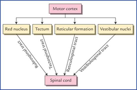
FLOWCHART 17.2 Indirect motor pathways through which the cerebral cortex influences the spinal cord. These are generally described as extrapyramidal tracts.
Components of extrapyramidal system
The extrapyramidal system includes subcortical centres such as corpus striatum, globus pallidus, tectum, red nucleus, reticular formation, vestibular nuclei and neocerebellum.
The corpus striatum influences descending pathways principally by its cortical connections. The other subcorti-cal centres influence the lower motor neurons in the spinal cord directly through rubrospinal, reticulospinal, tectospinal, vestibulospinal, and olivospinal tracts (Flowchart 17.2).
Functions of extrapyramidal system
• Postural adjustments of the body to maintain balance.
• Gross synergistic voluntary movements in group of muscles affecting proximal joints of the limbs.
• Movements performed unconsciously, like swinging of arms during walking.
• Regulatory influence over the reflex activities.
The differences between the pyramidal and extrapyrami-dal systems are given in Table 17.3.
Table 17.3
Differences between the pyramidal and extrapyramidal systems
|
Pyramidal system |
Extrapyramidal system |
|
|
Phylogeny |
Phylogenetically recent in acquisition, present only in mammals and achieving its greatest development in man |
Phylogenetically older than pyramidal system |
|
Function |
Responsible for non-postural, precise movements of small muscles involved in skilful activity |
Responsible for gross postural (stereotyped) movements involving large groups of muscles |
|
Pathways |
Connected directly to the lower motor neurons. Therefore impulses reach the LMNs, through a direct route |
Connected indirectly (polysynaptic pathway) to lower motor neurons. Therefore, impulses reach the LMNs through a circuitous route |
|
Effects of lesion |
No increase in muscle tone |
Muscle tone increased (spasticity) |
|
Cortical fibres |
Arise predominantly in primary motor area (Brodmann's area 4) |
Arise predominantly in premotor area (Brodmann's area 6) |
|
Subcortical centres and basal ganglia |
Play no role in pyramidal system |
Play a key role in extrapyramidal system |
N.B. Naturally occurring lesions in man rarely, if ever involve pyramidal pathway without simultaneous involvement of extrapyramidal pathways therefore the division of motor pathways into pyramidal and extrapyramidal systems is of little or no clinical relevance.
Points to Note
• The extrapyramidal fibres originating from cerebral cortex, in contrast to pyramidal fibres, make synapse with subcortical centres before establishing contact with the lower motor neurons at the segmental level.
• The cortical sites of origin of extrapyramidal and pyramidal fibres overlap extensively.
Clinical Correlation
Effects of lesions of extrapyramidal tracts
• Paralysis with little or no muscular atrophy (except that due to disuse)
• Spasticity or hypertonicity of the muscles
• Exaggerated deep reflexes, viz. knee jerk, ankle jerk, etc.
• Clasp-knife rigidity.
Upper and lower motor neuron lesions
Any lesion that destroys any part of the pyramidal tract is termed upper motor neuron (UMN) lesion.
The classical example of upper motor neuron paralysis is hemiplegia.
In UMN lesion, the muscles are not paralyzed because UMNs do not supply muscles directly. Actually what happens is that the control of UMNs on LMNs which supply the muscles is lost. As a result LMNs become hyperactive. Consequently, the muscles of the limbs may become spastic and involuntary movements may occur which are uncontrollable and often lead to severe rigidity or spasm. Reflexes are exaggerated. There is no loss of muscle tone and no wasting of the affected muscles. A little wasting may occur due to disuse atrophy.
Similarly, any lesion of anterior horn cells or cranial nerve nuclei or motor fibres of peripheral nerves is described as lower motor neuron (LMN) lesion. The classical examples of LMN paralysis are poliomyelitisdue to involvement of anterior horn cells of spinal cord, and Bell's palsy due to involvement of facial nerve.
In LMN lesion, the affected muscles are paralyzed. They become limp and flaccid. There is wasting and normal reflexes are lost.
Both types of lesions manifest in the form of paralysis of muscles and loss of motor power. The important differences of upper and lower motor neuron lesions are given in Table 17.4.
Table 17.4
Differences in clinical features of upper and lower motor neuron lesions
|
Features |
Upper motor neuron lesion |
Lower motor neuron lesion |
|
Muscle tone |
Increased (hypertonia or spasticity) |
Decreased (hypotonia or flaccidity) |
|
Muscle wasting |
Absent (except due to disuse atrophy) |
Present (atrophy) |
|
Extent of paralysis |
Widespread |
Localized |
|
Babinski's sign* |
Present |
Absent |
|
Tendon reflexes, viz. knee and ankle jerks |
Exaggerated |
Diminished or lost |
|
Muscle clonus † |
Present |
Absent |
*When outer border of plantar surface of the foot is scratched with a blunt object in patient with a pyramidal (UMN) lesion it results in slow dorsiflexion of great toe accompanied by fanning of lateral toes. In normal person it results in plantar flexion of toes.
†A quick and continued stretch of a tendon, e.g. by sudden downward pulling of patella in patient with upper motor neuron lesion results in appearance of repetitive, rhythmical contraction and relaxation of quadriceps instead of single contraction as in normal individuals.
General Somatic Sensory Pathways
The general somatic sensory pathways are concerned with sensory modalities of pain, touch, temperature, vibration, tactile discrimination, pressure and proprioception. They transmit these sensations from somatic receptors in skin, muscles, tendons, joints) to the central nervous system.
The general somatic sensory pathways are divided into spi-nothalamocerebral, spinocerebellar, and trigeminothalamic pathways. The sensations carried by the first group of pathways are only perceived consciously.
Spinothalamocerebral Pathways
Spinothalamocerebral pathways consist of a series of three sensory neurons: primary, secondary and tertiary.
The cell bodies of primary (first order) sensory neurons lie in the dorsal root ganglia of spinal nerves and sensory ganglia of cranial nerves, the cell bodies of secondary (second order) sensory neurons lie in the grey matter of the spinal cord and sensory nuclei within the brainstem, and cell bodies of tertiary (third order) sensory neurons lie in the thalamus.
The primary sensory neurons bring sensations from receptors to the secondary neurons which transmit it to the tertiary neurons; which finally convey it to the primary somatic sensory cortex of the cerebral hemisphere.
Points to Note
• The second order sensory neurons cross to the opposite side, hence sensations from left side of body pass to the right cerebral hemisphere and those from right side of the body to the left cerebral hemisphere.
• All types of sensory modalities are consciously perceived at the level of cerebral cortex except pain and probably temperature which may be consciously perceived in the thalamus.
• Interneurons also called internuncial neurons may be interposed between the main sensory neurons.
• The cell bodies of all the first order sensory neurons lie outside the CNS except those in the mesencephalic nucleus of trigeminal nerve which lie within the CNS (brainstem).
Various types of sensations carried by different somatic sensory pathways are summarized in Flowchart 17.3.

FLOWCHART 17.3 Sensations carried by different somatic sensory pathways.
Spinothalamic pathways (pathways for simple touch, pain and temperature)
Lateral spinothalamic tract (Fig. 17.2)
The lateral spinothalamic tract carries pain and temperature sensations from the opposite side of the body.

FIG. 17.2 Lateral and ventral spinothalamic tracts.
The first order sensory neurons carrying these sensations enter the spinal cord through the lateral division of the dorsal root of the spinal nerve. In the cord these fibres ascend or descend for one or two segments as dorsolateral tract of Lissauer at the tip of posterior horn and then relay into the posterior horn cells in the region of substantia gelatinosa.
The axons of second order sensory neurons arise from posterior horn cells, cross over to the opposite side in the anterior commissure in front of the spinal canal and reach the opposite lateral white column, where they turn upwards forming the lateral spinothalamic tract. The fibres of this tract terminate in the cells of ventral posterolateral (VPL) nucleus of the thalamus.
The axons of third order sensory neurons arise from the cells of VPL nucleus of thalamus and project to the primary sensory cortex of the cerebral hemisphere (area 3, 1, 2).
Clinical Correlation
• The damage of lateral spinothalamic tract causes loss of pain and temperature sensation on the opposite side of the body one or two segments below the level of lesion. The patient will not, therefore, respond to pin-prick or recognize hot or cold objects placed in contact with the skin.
• In the spinothalamic tract the pain fibres are lateral to the temperature fibres. In the cervical region the pain fibres become very superficial in the lateral white column, hence cordotomy can be performed safely at this level to relieve the pain in the opposite half of the body.
Ventral spinothalamic tract (Fig. 17.2)
The ventral (anterior) spinothalamic tract carries light touch, pressure, tickle, and itch sensations from the opposite side of the body.
The first order sensory neurons carrying these sensations enter the spinal cord through the lateral division of dorsal root of the spinal nerve. In the cord these fibres may ascend or descend for 8 to 10 segments in dorsolateral tract of Lissauer before terminating into the posterior horn cells in the region of substantia gelatinosa.
The axons of second order sensory neurons arise from posterior horn cells, cross over to the opposite side in the anterior white commissure in front of the spinal canal to reach the opposite anterior white column, where they turn upwards to form the ventral spinothalamic tract. As it ascends, it merges with the lateral spinothalamic tract in the brainstem and relay into the ventral posterolateral (VPL) nucleus of the thalamus.
The axons of third order sensory neurons arise from cells of VPL nucleus of thalamus and project to the primary sensory area of cerebral cortex (areas 3, 1, 2).
Clinical Correlation
The damage of anterior spinothalamic tract causes loss of light touch and pressure on the opposite side of the body below the level of the lesion. The patient will not feel the light touch of a cotton wisp or pressure of a blunt object placed against the skin.
N.B. The discriminative touch will still be present, as it is carried by fasciculus gracilis and fasciculus cuneatus.
Points to Note
• The lateral spinothalamic tract carries pain and temperature sensations from whole of the opposite half of body except from the region of the head and neck from where these sensations are carried by Vth, IXth and Xth cranial nerves.
• The transmission of pain and temperature in the posterior horns may be altered (inhibition or facilitation) by nerve impulses coming from the sensory motor strip of the cerebral cortex, especially that from the somesthetic area.
• Throughout the course of spinothalamic tracts the fibres lie in a somatotopic arrangement with sacral fibres being the later almost and fibres from lumbar, thoracic, and cervical segments medially in that order.
Dorsal column medial lemniscal pathways (pathways for conscious proprioception)
Fasciculus gracilis, fasciculus cuneatus and medial lemniscus
The fasciculus gracilis and fasciculus cuneatus carry the sensations of proprioception, two-point discrimination and vibration (Fig. 17.3).

FIG. 17.3. Dorsal column medial lemniscal pathways carrying conscious proprioceptive sensations. (FG = fasciculus gracilis, FC = fasciculus cuneatus.)
The first order sensory neurons carrying these sensations enter the spinal cord through the medial division of the posterior spinal nerve root and continue in the posterior white column as fasciculus gracilis and fasciculus cuneatus to the medulla, where they relay in the nucleus gracilis and nucleus cuneatus respectively.
The axons of second order sensory neurons arise from cells of the nucleus gracilis and nucleus cuneatus, curve as internal arcuate fibres, cross to the opposite side, decussate with those of opposite side in lower medulla (called sensory decussation) and then turn upwards forming the medial lemniscus. The fibres of medial lemniscus ascend up successively through medulla and pons to relay into the ventral posterolateral (VPL) nucleus of the thalamus.
The axons of third order sensory neurons arise from cells of VPL nucleus of thalamus and project to the primary sensory area of the cerebral cortex (area 3, 1, 2).
Clinical Correlation
The lesions of posterior white columns (i.e. fasciculus gracilis and fasciculus cuneatus) cause loss of sensations of proprioception, two-point discrimination and vibration on the same side of body below the level of lesion, whereas the lesions of medial lemnis-cus produce contralateral loss of these sensations.
Due to loss of proprioception, the patient fails to get information from the muscles and joints to consciousness, hence he is not able to know the position and movements of the ipsilateral limb. For example if you dorsiflex his big-toe, he cannot tell whether the toe is pointing upwards or downwards if his eyes are closed.
The loss of vibration sense can be easily tested by applying a vibrating tuning fork to the bony prominences, such as lateral malleolus of fibula or styloid process of radius.
The loss of tactile discrimination is tested by putting the compass on the skin and gradually separating the two points of the compass until the patient can appreciate them as two separate points, not as one.
Points to Note
• The characteristics of fine touch or discriminative touch are that, one can recognize the location of stimulated points with precision and is also aware that the two points are touched simultaneously, even though they are close together.
• The touch and pressure sensations carried by the ventral spinotha-lamic tract are described as crude touch in contrast to the same sensations carried by the dorsal column pathways which are described as fine touch because the localization and two-point discrimination of sensations carried by ventral spinothalamic tract is poor.
• Fibres conveying sensations from the lower half of the body below the sixth thoracic segment, form the fasciculus gracilis placed medially in the posterior white column while the fibres from the upper half of the body form the fasciculus cuneatus placed laterally.
• The dorsal column pathways should ideally be called dorsal column-medial lemniscal system or pathways as it includes dorsal white columns of the spinal cord and the medial lemniscus of the brainstem.
Spinocerebellar Pathways (Pathways for Unconscious Proprioceptive Sensations)
Here it is important to mention that these sensory pathways consist of only two sensory neurons. The fibres of second order sensory neurons in spite of going to the thala-mus, go to the cerebellar cortex through posterior and anterior spinocerebellar, and cuneocerebellar tracts.
The cell bodies of primary sensory neurons lie in the dorsal root ganglia, and those of secondary sensory neurons in the thoracic nucleus (nucleus dorsalis).
Dorsal (posterior) spinocerebellar tract (Fig. 17.4)
The central processes of first order sensory neurons carrying the proprioceptive sensations enter the cord through posterior roots of the spinal nerves and terminate in the cells of nucleus dorsalis (Clarke's column)which is situated on the medial side of the base of the posterior horn.

FIG. 17.4 Spinocerebellar pathways (i.e. posterior and anterior spinocerebellar and cuneocerebellar tracts). Note, posterior spinocere-bellar and cuneocerebellar tracts enter the cerebellum through inferior cerebellar peduncle.
The axons of second order sensory neurons arise from the cells of Clarke's column, and enter the dorsolateral part of the lateral white column on the same side and ascend to form the dorsal spinocerebellar tract.
The fibres of this tract enter the cerebellum through the inferior cerebellar peduncle and project (end) in the ipsilat-eral cerebellar cortex.
Ventral (anterior) spinocerebellar tract (Fig. 17.4)
The central processes of first order sensory neurons carrying the proprioceptive sensations enter the cord through the posterior roots of the spinal nerves and terminate in the nucleus dorsalis at the base of the posterior horn. Most of axons of second order sensory neurons arising from nucleus dorsalis cross to the opposite side and reach the ventrolateral part of the lateral white column and ascend anterior to the fibres of dorsal spinocerebellar tract as the ventral spinocerebellar tract, however some of them ascend as anterior spinocerebellar tract in the lateral white column of the same side.
The fibres ascend through the medulla oblongata and pons up to the midbrain then bends downwards to enter the cerebellum through the superior cerebellar peduncle and terminate in the ipsilateral cerebellar cortex.
N.B. The posterior spinocerebellar tract consists of only uncrossed fibres white the anterior spinocerebellar tract consists of both crossed (mainly) and uncrossed fibres.
The fibres of dorsal spinocerebellar tract enter the cerebellum through the inferior cerebellar peduncle and those of anterior spinocerebellar tract through superior cerebellar peduncle.
Cuneocerebellar tract (posterior external arcuate fibres) (Fig. 17.4)
As the Clarke's column is located only from C8 to L3 spinal segments. The axons of first order sensory neurons carrying proprioceptive sensations from upper limb and entering the spinal cord above C8 segment, ascend in the ipsilateral dorsal funiculus to terminate by synapsing with the cells of accessory cuneate nucleus of the medulla. The axons of second order sensory neurons arising from cells of accessory cuneate nucleus enter the ipsilateral cerebellum through the inferior cerebellar peduncle.
N.B. The cuneocerebellar tract is the counterpart of the posterior spinocerebellar tract for the upper limb.
Points to Note
• The dorsal and ventral spinocerebellar tracts carry proprioceptive sensations from lower limb and trunk while the cuneocerebellar tract carries the proprioceptive sensations from the upper limb.
• It is believed that most of the crossed fibres in the anterior spino-cerebellar tract cross again in the cerebellar white matter to reach the ipsilateral cerebellar cortex.
• Recent investigations have shown that some exteroceptive sensations (touch and pressure) may reach the cerebellum through these pathways.
Clinical Correlation
The information carried by dorsal and ventral spino-cerebellar, and cuneocerebellar tracts enable the cerebellum to perform unconscious coordination. The lesions of these tracts result in hypotonia of muscles and ataxia.
Trigeminothalamic Pathways
The trigeminothalamic pathways carry general and pro-prioceptive sensations from face and oral cavity to the somatosensory cortex.
Ventral trigeminothalamic tract (Fig. 17.5)
The ventral trigeminothalamic tract carries pain and temperature sensations from the face and the oral cavity.

FIG. 17.5 Trigeminothalamic pathways. (CNV=trigeminal nerve.)
The cell bodies of first order sensory neurons are located in trigeminal ganglion. The axons of these neurons descend in the spinal tract of trigeminal nerve and synapse with the cell bodies of second order sensory neurons located in the spinal nucleus of the trigeminal nerve.
The axons of second order sensory neurons cross over to the opposite side ascend up to form the ventral trigemi-nothalamic tract and relay in the contralateral ventral posteromedial (VPM) nucleus of the thalamus.
The axons of third order sensory neurons arise from nerve cells of VPM nucleus of thalamus and project through the posterior limb of internal capsule to the face area of the somatosensory area (Brodmann's area 3, 1, and 2) of the cerebral cortex.
Dorsal trigeminothalamic tract (Fig. 17.5)
The dorsal trigeminothalamic tract carries sensations of tactile discrimination and pressure from face and oral cavity.
The first order sensory neurons are located in the trigeminal ganglion. The axons of these neurons synapse with the cell bodies of second order sensory neurons located in the principal sensory nucleus of the trigeminal nerve.
The axons of second order sensory neurons ascend up to form the dorsal trigeminothalamic tract and relay in the ipsilateral VPM nucleus of the thalamus.
The axons of third order sensory neurons arise from nerve cells VPM nucleus of thalamus and project through the posterior limb of the internal capsule to the face area of mostly ipsilateral of the somatosensory cortex (Brodmann's area 3, 1 and 2).
Clinical Problems
1. Explain, why the Babinski's sign is present in adults only if there is a lesion of corticospinal (pyramidal) tract (upper motor neuron lesions) but in infants and children up to the age of one it is normally present.
2. Explain, why light touch is very sensitive but not highly discriminative (poorly localized).
3. The lesions of the posterior white column (fasciculus gracilis and fasciculus cuneatus) cause the loss of proprioception, fine touch, and vibration on the same side of the body whereas, the lesions of medial lemniscus cause the loss of these sensations on the opposite side of the body. Explain why it is so.
Clinical Problem Solving
1. When the skin along the lateral aspect of the sole of a foot is scratched with a blunt object the normal response is plantar flexion of all the toes. This is because, normally corticospinal tracts produce plantar flexion of toes in response to sensory stimulation of the skin of the sole of the foot. When corticospinal tracts are damaged (UMN lesion), the influence of other descending tracts become apparent in the form of withdrawal reflex, characterised by dorsiflexion of the great toe and fanning of the other toes (Babinski's sign).
The Babinski's sign is normally present in children during the first year of their life, because the corti-cospinal tract is not myelinated until the end of the first year of life (the myelination of corticospinal tract starts 10 to 14 days after the birth and completed by the end of first year).
N.B. The corticospinal tracts become functional only after their constituent fibres are myelinated.
2. The first order sensory neurons of anterior spinothalamic tract carrying (light touch), after entering the spinal cord, ascend or descend for 8 to 10 segments before synapsing with the second order sensory neurons. While ascending or descending the first order sensory neurons give collateral branches that synapse with second order sensory neurons of several segments of the cord. Thus collateral branches from number of first order sensory neurons carrying information of light touch from sensory receptors in different patches of skin converge on a single second order neuron and enhance its afferent conduction. As a result, the light touch requires less peripheral stimulation to produce action potentials in the ascending pathway (anterior spinothalamic tract) with consequent increased light touch sensitivity. However it results in less discriminative information because sensory receptors from more than one points of skin have input on to the same second order neuron, which cannot distinguish one small area of skin from another within the zone where its sensory receptors are located.
3. See Clinical correlation on page 205.