
The peripheral nerve lesions are common in clinical practice and can be caused by a wide variety of diseases like trauma, neoplasms, infection, metabolic diseases (diabetes) and chemical toxins such as lead.
Therefore, it is of paramount importance for a physician to know the basic structure of peripheral nerves. Further, he also needs to know the structure and function of nerve fibres, and the process of their myelination, so that he could understand the mode of conduction of nerve impulses, and appreciate the process of nerve degeneration and regeneration.
Nerve Fibres
An axon of a nerve cell is termed nerve fibre. The bundles of nerve fibres found in the central nervous system (CNS) are referred to as nerve tracts while the bundles of nerve fibres found in the peripheral nervous system are called peripheral nerves. Two types of nerve fibres are present in the nervous system, viz. myelinated and non-myelinated.
Myelinated and Non-myelinated Nerve Fibres
In the peripheral nervous system, all axons (nerve fibres) are enveloped by the specialised Schwann cells which provide both structural and metabolic support to them.
In general, small diameter axons, for example those of the autonomic nervous system (ANS) and small pain fibres, are simply enveloped by the cytoplasm of Schwann cells; these nerve fibres are said to be non-myelinated. The large diameter fibres are wrapped by a variable number of concentric layers of Schwann cell plasma membrane forming the so-called myelin sheath, and such nerve fibres are said to be myelinated. Within the CNS, the myelination is similar to that in the peripheral nervous system except that the myelin sheath is formed by cells called oligodendrocytes.
Myelination (formation of myelin)
The myelination is the process by which nerve fibres acquire myelin sheaths which enhance the conduction of nerve impulses.
The process of myelination begins before birth in the late fetal period but is not complete until a year or more later after the birth.
Myelination of the peripheral nerve fibres (Fig. 3.1)
The myelination begins near the origin of the axon and ends just before its terminal branches.

FIG. 3.1 Myelination. (A) Stages in the formation of myelin sheath. The axon invaginates the cytoplasm of the Schwann cell and becomes suspended by a mesaxon (1 and 2). The mesaxon elongates and bound around the axon spirally (3, 4 and 5).
The axon invaginates the side of a Schwann cell, as a result the plasma membrane of Schwann cell forms a mesaxon, which suspends the axon within the Schwann cell. The layer of plasma membrane immediately around the axon is continuous with the remainder of the plasma membrane through a double layered mesaxon (Fig. 3.1A).
The Schwann cell now rotates around the axon so that mesaxon becomes wrapped repeatedly around the axon forming spirals around it. As the process continues, the cytoplasm is extruded from the spirals into the Schwann cell body. On maturity, the inner layers of plasma membrane fuse with each other so that axon becomes surrounded by several layers of modified membrane which together constitute the myelin sheath. Thus, myelin sheath consists of many regular layers of plasma membrane material, which is predominantly white lipid protein, giving the myelinated axons a whitish appearance. It insulates the axon from extracellular environment thus preventing ion fluxes across the plasma membrane of the nerve fibre/axon.
The thickness of myelin sheath depends on the number of spirals of Schwann cell membrane. In electron micrographs of cross-sections of myelinated nerve fibres, the myelin is seen to be laminated consisting of major and minor dense lines. The darker major dense line (about 2.5 nm thick) consists of two inner protein layers of the plasma membrane that are fused together. The lighter minor dense line (about 10 nm thick) is formed by the approximation of the outer surfaces of adjacent plasma membranes and is made up of lipid.
Each Schwann cell extends for a short distance along the nerve fibre and at its termination its role is supplemented by an another Schwann cell with which it interdigitates closely.
In the CNS, oligodendrocytes responsible for the process of myelination, follows the similar pattern as of Schwann cell in the PNS; a single oligodendrocyte, however, forms the myelin sheath around several axons.
A myelinated nerve fibre, therefore, consists of an axon, a myelin sheath and a neurilemmal/Schwann sheath. The myelin sheath is segmented, the segments being separated at regular intervals by nodes of Ranvier. The areas between the nodes are called internodes.
Functions of the myelin sheath
• Provides support to the nerve fibres.
• Aids in conduction of the nerve impulses.
• Insulates an axon from the extracellular environment.
• Responsible for the colour of the white matter of the brain and spinal cord.
The non-myelinated fibres are also surrounded by Schwann cells (Fig. 3.2). Several axons become longitudinally invagi-nated into the cytoplasm of a Schwann cell so that each fibre is embedded in a groove in the Schwann cell cytoplasm. The Schwann cell plasma membrane fuses along the opening of the groove, thus effectively sealing the nerve fibre within an extracellular compartment. As many as 15 or more axons may share a single Schwann cell.
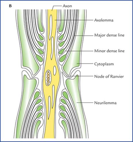
FIG. 3.1 (B) The longitudinal section of the myelinated nerve showing fine structure of the node of Ranvier.

FIG. 3.2 Relationship of several non-myelinated axons to a Schwann cell. (A) Axons lying near the plasma membrane of a Schwann cell. (B) The axons are longitudinally invaginated into the cytoplasm of Schwann cell.
Each axon is surrounded by a single layer of plasma membrane of schwann cell, hence it is unmyelinated. There are no nodes of Ranvier. Consequently the action potential travels along the whole length of axolemma without the accelerating factor of node-to-node (saltatory) conduction. This accounts for slow rate of conduction of nerve impulse in the unmyelinated fibres.
Conduction of Action Potential along an Axon
Like all the cells, the resting (unstimulated) neuron maintains an ionic gradient across its plasma membrane, thereby creating an electrical potential called resting membrane potential. Thus, in resting neuron its plasma membrane remains polarized. The excitability (a fundamental property of neurons) involves a change in membrane permeability in response to appropriate stimuli so that the ionic gradient across the plasma membrane is reversed and the plasma membrane becomes depolarized. A wave of depolarization known as action potential then spreads along the plasma membrane. This is followed by the process of repolarization in which membrane rapidly re-establishes its resting potential.
The speed of conduction of the action potential, along an axon depends on the myelination of the axon (Fig. 3.3). The action potentials are conducted more rapidly in myelinated than in non-myelinated axons. In non-myelinated fibres, the action potential passes continuously along the axolemma, progressively exciting neighbouring areas of membrane. In myelinated fibres, the myelin sheath serves as an insulator. Consequently a myelinated nerve fibre can be stimulated only at the nodes of Ranvier, where the axon is naked and the ions can pass freely through the plasma membrane between the extracellular fluid and the axo-plasm. Therefore, in these fibres the action potential jumps from one node to the next. The action potential at one node sets up a current in the surrounding tissue fluid, which quickly produces depolarization at the next node. The action potential conduction in a myelinated fibre is like a grasshopper jumping, whereas action potential conduction in a non-myelinated fibre is like a grasshopper walking. The action potential will naturally move more rapidly by jumping.

FIG. 3.3 The conduction of action potential along an axon. (A) In myelinated axon the action potential is conducted from one node of Ranvier to another (saltatory conduction). (B) In non-myelinated axon the action potential is conducted along the entire length of the axon.
This leaping of the action potential from one node of Ranvier to another in the myelinated nerve fibres is called saltatory conduction (L. saltare = to leap).
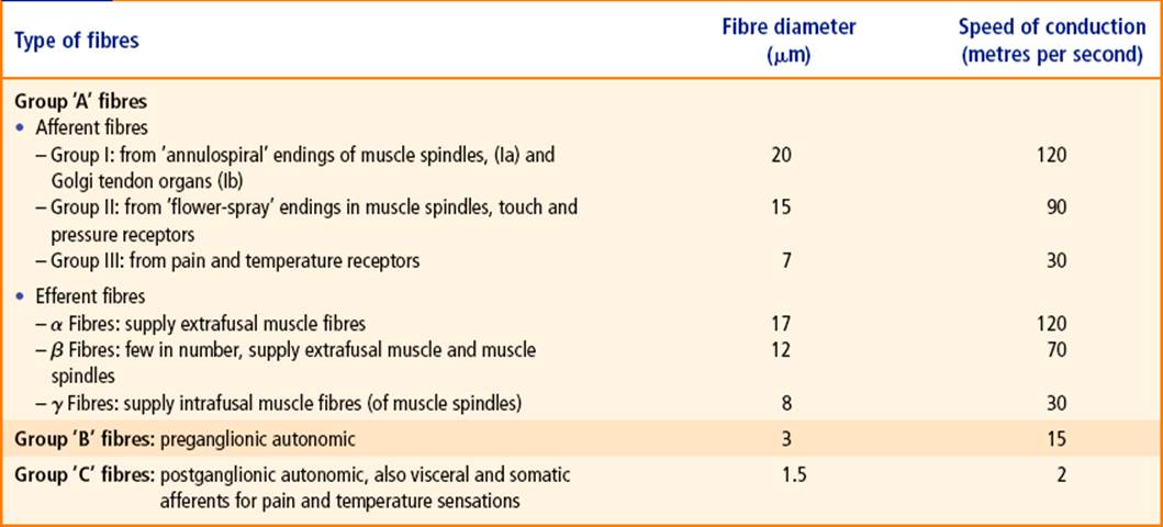
In addition to myelination, the diameter of axons affects the speed of conduction of action potential. The conduction of action potential is faster along large diameter axons than small diameter axons because large diameter axons provide less resistance to action potential propagation. In the large motor fibres (alpha fibres), the rate of conduction may be as high as 70-120 meters per second.
The smaller sensory fibres have slower conduction rate (Table 3.1).
Tabel 3.1
Classification of peripheral nerve fibres
Clinical Correlation
The process of myelination begins before birth (late in the fetal development), and continues rapidly until the end of first year after birth and continues more slowly thereafter. Thus, the development of myelin sheath is associated with the infant's continuing development of rapid and better coordinated responses. For example, the fibres of corticospinal tract which control the reflex emptying of urinary bladder get myelinated and begin to function at 3-6 years of age. Therefore reflex emptying of bladder and enuresis (bed-wetting) is normal in infants.
Classification of Peripheral Nerve Fibres
According to the axonal diameter (including myelin sheath if present) and speed of conduction, the peripheral nerve fibres are classified into three main groups: A, B and C.
• Type A fibres are large diameter, myelinated axons and therefore conduct action potentials at a great speed (15 – 120 m/sec). Motor neurons supplying skeletal muscles and most sensory neurons have type A fibres. Consequently, rapid response to external stimuli is possible as there is rapid input of sensory information to CNS on one hand and rapid output of action potential to skeletal muscle on the other hand.
N.B. All types of fibres in group A and B are myelinated whereas group C fibres are non-myelinated.
• Type B fibres are medium-diameter, myelinated axons and conduct action potentials at a slow speed (3-15 m/sec).
• Type C fibres are small-diameter non-myelinated axons that conduct action potentials at a very slow speed (2 m/sec or less).
The type B and C fibres are primarily found in the ANS, which supplies internal organs such as stomach, intestine. The responses necessary to maintain internal homeostasis such as digestion need not be as rapid as to external environment.
The group A fibres are further classified into somatic sensory (I, II, III) and motor (a, (3, 7) subgroups. Table 3.1 shows the types of nerve fibres and their maximum diameters and conduction rates.
Peripheral Nerves
The peripheral nerves comprise 12 pairs of cranial and 31 pairs of spinal nerves.
Most of these nerves are composed of both motor and sensory fibres, and therefore called mixed nerves. Some of the cranial nerves, however are composed of either sensory nerve fibres only (sensory nerves) or motor nerve fibres only (motor nerves).
The impulses enter or leave the CNS via the cranial and spinal nerves.
According to the area of innervation the nerve fibres within the spinal nerves may be classified into the following types (Fig. 3.4):
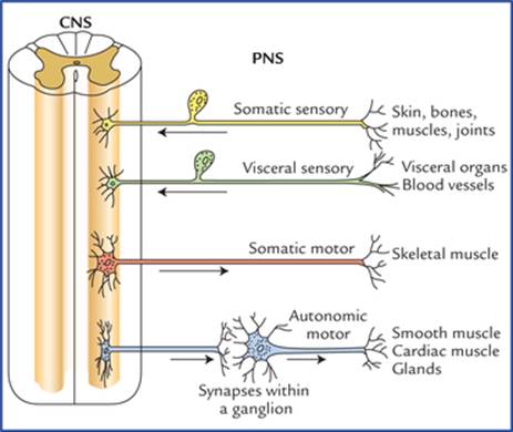
FIG. 3.4 Classification of the nerve fibres according to the area of innervation. Note the relationship of the sensory and motor fibres of the peripheral nervous system (PNS) with the central nervous system (CNS).
1. Somatic sensory fibres: convey nerve impulses from skin, bones, muscles and joints to the CNS.
2. Somatic motor fibres: carry nerve impulses from CNS to the skeletal muscles.
3. Visceral sensory fibres: convey nerve impulses from visceral organs and blood vessels to the CNS.
4. Visceral motor fibres (also called autonomic motor fibres): carry impulses from CNS to the cardiac muscle, glands, and smooth muscles within the visceral organs.
The types of nerve fibres present within the cranial nerves are described in Chapter 9.
Cranial nerves
There are 12 pairs of cranial nerves which arise from brain and leave the cranial cavity by passing through the foramina in the skull. Three of these nerves (olfactory I, optic II, and vestibulocochlear VIII) are composed entirely of sensory (afferent) nerve fibres bringing sensations to the brain; five of them (oculomotor III, trochlear IV, abducent VI, accessory XI, and hypoglossal XII) are composed entirely of motor (efferent) fibres, while the remainder (trigeminal V, facial VII, glossopharyngeal IX, and vagus X) possess both sensory (afferent) and motor (efferent) fibres (for details, seeChapter 9).
Spinal nerves
There are 31 pairs of spinal nerves which arise from the spinal cord and pass through intervertebral foramina in the vertebral column. The spinal nerves are named according to the regions of the vertebral column with which they are associated. There are 8 cervical, 12 thoracic, 5 lumbar, 5 sacral, and 1 coccygeal nerves.
Typical spinal nerve
A typical spinal nerve arises from spinal cord by two roots: an anterior root and a posterior root (Fig. 3.5). The anterior root consists of bundles of nerve fibres which carry nerve impulses away from the spinal cord; these fibres are called motor (efferent) fibres. Their cells of origin lie in the anterior horn of the spinal cord.

FIG. 3.5 A typical spinal (segmental) nerve.
The posterior root consists of bundles of nerve fibres which carry impulses to the spinal cord, these are called sensory (afferent) fibres. The cell bodies of these nerve fibres are located outside the spinal cord in a swelling on the posterior root called posterior root ganglion. The posterior root ganglion is located in the intervertebral foramen.
The spinal nerve roots pass from the spinal cord to the intervertebral foramen, where they unite to form a spinal nerve. Thus, the spinal nerve is made up of both motor and sensory fibres.
After emerging from the intervertebral foramen, each spinal nerve first gives a recurrent meningeal branch which re-enters the vertebral canal to innervate the meninges, then it divides into a large anterior ramus and a smaller posterior ramus, each containing both motor and sensory fibres. The posterior ramus passes posteriorly around the vertebral column to supply the muscles and skin of the back. The anterior ramus runs anteriorly to supply the muscles and skin over the anterolateral part of the body wall.
The anterior ramus in its initial part is connected to the sympathetic ganglion by grey and white rami communicates.
Plexus formation
Except for thoracic nerves from T3 to T11, the anterior primary rami of all the spinal nerves join together and/or branch to form a network of nerves known as nerve plexus. There are three major plexuses:
• Cervical plexus (Fig. 3.6), made up of C1 through C4 spinal nerves, innervates the muscles of neck and diaphragm.

FIG. 3.6 Cervical plexus.
• Brachial plexus (Fig. 3.7), made up of C5 through T1 spinal nerves, innervates muscles of the upper limb.

FIG. 3.7 Brachial plexus. (DS = dorsal scapular, SS = suprascapular, NS = nerve to subclavius, LP = lateral pectoral nerve, S = subscapular nerve, T = thoracodorsal nerve, MP = medial pectoral nerve, MCA = medial cutaneous nerve of arm, MCF = medial cutaneous nerve of forearm.)
• Lumbosacral plexus (Fig. 3.8), made up of L1 through S5 spinal nerves, innervates muscles of the lower limb. It is divided into two portions:

FIG. 3.8 The lumbosacral plexus.
– Lumbar portion (lumbar plexus) L1 through L4, supplies mostly muscles of the thigh.
– Sacral portion (sacral plexus) L5 through S5, supplies mostly muscles of leg and foot.
The branches arising from plexuses are also termed peripheral nerves.
The formation of nerve plexus allows individual nerve fibres to pass from one peripheral nerve to another. A nerve plexus thus permits a redistribution of nerve fibres within the different peripheral nerves.
The detailed description of these plexuses and peripheral nerves derived from them is beyond the scope of this book.
Structure of the Peripheral Nerve (Fig. 3.9)
Each peripheral nerve trunk consists of a number of nerve fibre bundles or fasciculi.

FIG. 3.9 The structure of a peripheral nerve showing three protective coverings.
There are three protective coverings of connective tissue in each nerve trunk:
1. Endoneurium: It is a loose delicate connective tissue that surrounds the individual nerve fibres. In fact it lies between the nerve fibres within a nerve bundle.
2. Perineurium: It is a smooth sheath, made up of a condensed layer of collagenous connective tissue that surrounds the bundle of nerve fibres.
3. Epineurium: It is a dense connective tissue sheath which surrounds and encloses the bundles of nerve fibres forming the nerve trunk (i.e. it surrounds the entire nerve). It contains tiny blood and lymph vessels.
The fibres within a peripheral nerve trunk derive considerable mechanical strength from these three layers of connective tissue.
Clinical Correlation
• The connective tissue of epineurium, perineurium and endoneurium is in continuity with each other. The blood capillaries and lymphatics ramify in this connective tissue. The major arterial occlusion in the limbs may cause severe pain due to ischaemic neuritis.
• The injuries of peripheral nerve are quite common and can occur due to compression, traction, trauma, injection, cuts, etc.
The nerve injuries are of three types:
1. Neurotmesis: In this both axon and its myelin sheath is damaged.
2. Axonotmesis: In this axon is damaged but its myelin sheath is preserved.
3. Neuropraxia: In this both axon and its myelin sheath are preserved.
Recovery can occur in cases of neuropraxia and axonotmesis but functional loss is inevitable in case of neurotmesis.
Degeneration and Regeneration of the Peripheral Nerves after Injury
Degeneration of the nerve fibre (Fig. 3.10)
When a nerve fibre is cut, the axon is no longer in continuity with its trophic centre*—the nerve cell body. The immediate reaction in the neuron is that of degeneration. The series of degenerative changes will take place: (a) in the nerve fibre—(i) the distal segment that is separated from the cell body, (ii) a portion of axon proximal to the injury, and (b) possibly in the cell body from which the axon arises.

FIG. 3.10 Degeneration of a nerve fibre (Wallerian degeneration).
Changes in the nerve fibre
• The distal segment immediately undergoes degeneration from the site of lesion to its termination. This process of anterograde degeneration is called Wallerian degeneration.
• The degeneration also extends proximally from the site of lesion, for a short distance as far as the first node of Ranvier.
In the process of degeneration, parts of the axon distal and proximal to cut, disintegrates and their myelin sheaths break up into lipid droplets.
Changes in the nerve cell body
• The cell body swells and nucleus becomes eccentric.
• The Nissl bodies disintegrate and become fine and granular, and dispersed throughout the cytoplasm, a process known as chromatolysis.
The amount of swelling of the cell body and chromatolysis is greatest when the injury to the axon is close to the cell body. The changes that occur in the cell body following an injury to its axon are referred to as retrograde degeneration.
Regeneration of nerve fibre (Fig. 3.11)
Regeneration of nerve fibre usually begins two weeks after the injury. The various regenerative steps are as follows:

FIG. 3.11 Regeneration of a divided nerve.
• The macrophages migrate at the site of lesion and remove the debris by phagocytoses.
• The Schwann cells then proliferate and fill the endoneural tube to form a solid cellular cord (column of cells). The endoneural sheath and the contained cord of Schwann cells is known as band fibre.
A small gap that exists between the proximal and distal stumps is also filled by the proliferating Schwann cells. The macrophages probably secrete the substances the nerve growth factors that cause proliferation of Schwann cells.
The proximal axon now gives rise to multiple sprouts with bulbous tips that enter the proximal end of endoneural tube of distal segment.
The course of sprouts is guided by the cord of Schwann cells. Several sprouts from different axons may enter into one endoneural tube but only one of the sprouts from each axon persists, the remainders degenerate. The persisting sprout now grows distally to reinnervate a motor or sensory end organ.
If it stops growing or wanders in some new direction, the normal function will not return.
Factors necessary for satisfactory regeneration
• Endoneural sheath should be intact, viz. in crush nerve injuries, the axon is divided and its blood supply is interfered but the endoneural sheaths remain intact.
• Distance between proximal and distal stumps should not be more than few millimetres.
• Infection should be absent at the site of wound.
• Presence of nerve growth factors.
• Proper physiotherapy: Inadequate physiotherapy to the paralyzed muscles will cause their atrophy before it is reinnervated by the regenerating axons.
N.B. The axon grows at the rate of 3.5 to 4.5 mm per day, and if the cut ends of the peripheral nerves are sutured the regeneration is facilitated.
Clinical Correlation
• If a mixed nerve (containing sensory, motor and autonomic fibres) is cut, then during the process of regeneration, its autonomic, motor and sensory fibres may travel to an inappropriate peripheral end organs as the growing axon of one type may enter the endoneural tube of the other type. This results in inappropriate responses. The classical example is Frey's syndrome, a clinical condition in which when the patient eats, the ipsilateral cheek becomes red, hot and painful, followed by beads of perspiration. Further, there is hyperaesthesia in front and above the ear. This syndrome occurs following auriculotemporal nerve injury. What happens actually is that when the auriculotemporal nerve (a mixed nerve) is cut, the axons conveying secretory impulses to salivary glands grow into the endoneural tubes of axons supplying cutaneous receptors for pain, touch and temperature; and sympathetic axons supplying sweat glands and blood vessels. Consequently, a stimulus intended for salivary secretion during eating evokes cutaneous hyperaesthesia, sweating and flushing.
• If the distance between the proximal and distal stumps of the completely severed nerve is greater, or the gap becomes filled with the proliferating fibrous tissue or by adjacent muscular tissue that bulges into the gap. The growing axonal sprouts enter into the surrounding connective tissue and form a tangled mass called neuroma.
Ganglia (Swelling or Knot)
The collection of nerve cell bodies outside the CNS is called ganglion. They are of two types: (a) sensory ganglia, and (b) autonomic ganglia.
Sensory Ganglia
The sensory ganglia located on the dorsal roots of spinal nerves and on the trunks of some cranial nerves (trigemi-nal, facial, glossopharyngeal and vagus) have the same structure. Each ganglion has a connective tissue capsule surrounding the cell bodies of primary sensory neurons. These neurons are of unipolar (pseudounipolar) type having oval or rounded cell body. A single process leaves the cell body and after a short convoluted course bifurcates at a T-junction into peripheral and central processes. The peripheral process terminates in the peripheral receptors and the central process enters the CNS.
The peripheral process conducting impulses towards the cell body is functionally a dendrite but has the structural and physiological characteristics of an axon.
The impulses pass directly from the peripheral to the central process bypassing the cell body. Each nerve cell body is closely surrounded by a layer of flattened cells called capsular cells or satellite cells. The satellite cells are similar in structure to Schwann cells and separate the nerve cell bodies from the capillaries. They help to provide nutrition, and structural support to the nerve cells of ganglia.
Autonomic Ganglia
The autonomic ganglia are of two types: sympathetic and parasympathetic.
The sympathetic ganglia are located in the sympathetic chains present one on each side into the paravertebral region (paravertebral ganglia). They are also located in front of the vertebral column in the abdominal cavity— prevertebral ganglia (e.g. coeliac, superior mesenteric and inferior mesenteric ganglia).
The parasympathetic ganglia are located close to or in the walls of the viscera.
Each autonomic ganglion consists of a collection of mul-tipolar neurons, and is covered by a layer of connective tissue called capsule.The autonomic ganglia are the sites where preganglionic nerve fibres synapse on postganglionic neurons. They are relay stations in the visceromotor (auto-nomic motor) pathway. The preganglionic fibres are myelinated while the postganglionic fibres are non-myelinated.
Clinical Correlation
• Peripheral neuropathy (neuritis)
It is the most common disorder of the peripheral nervous system. It consists of degenerative changes in the peripheral nerves which produce sensory loss and motor weakness. The distal portions of the nerves are affected first, hence symptoms appears first in hands and feet (glove and stocking paraesthesia). There are multiple causes of peripheral of neuropathy, viz. nutritional deficiencies such as deficiency of vit. B1, B6and B12, toxins of various kinds such as drugs, alcohol, and metabolic disorders such as diabetes.
• Entrapment syndromes
They occur due to compression of peripheral nerves, e.g. compression of median nerve in the carpal tunnel (carpal tunnel syndrome).
• Radiculopathy
It occurs due to compression of nerve roots due to variety of disorders, e.g. spondylosis, disc prolapse, etc.
Clinical Problems
1. In patients with multiple sclerosis and diabetes mellitus, there is an impaired control of skeletal and smooth muscles. Why?
2. By what route the tetanus toxin travel from the site of wound to the central nervous system?
3. The injured nerve fibres in the peripheral nervous system are able to regenerate but in the central nervous system they fail to do so. Why?
4. A man on returning home after attending a party on Saturday night, slept on a hard chair the whole night with his right hand suspended over the edge of the chair back. Next day when he woke up, he found that he was not able to extend his right hand, but by end of the day his hand became normal without treatment. What is the likely diagnosis? Mention its anatomical basis.
5. If muscles are paralyzed following a nerve injury due to crush or traction, the recovery is rapid and nearly complete, but if the paralysis occurs due to complete section of a nerve trunk, the recovery is not possible at all. Mention the reason for this.
Clinical Problem Solving
1. In diseases like multiple sclerosis (skleros = hardening) and diabetes mellitus the myelin sheath is gradually destroyed. Consequently, the transmission of nerve impulse is slowed leading to an impaired control of the skeletal and smooth muscles.
2. The tetanus toxin travels from the site of wound to the CNS through the spaces in the endoneurium of nerve fibres.
3. The regeneration of nerve fibres is possible in the peripheral nervous system due to following factors:
(a) Presence of endoneural tubes.
(b) Presence of large number of Schwann cells.
(c) Presence of nerves growth factors (seepage 26).
In the CNS also there is an attempt at regeneration of the axons as evidenced by sprouting of the axons, but the process ceases after 2 weeks due to the following factors:
(a) Absence of endoneural tubes.
(b) Failure of oligodendrocytes to serve in the same manner as Schwann cells. The oligodendrocytes in number and are at a short distance away from the axon consequently when myelin sheath degenerates, no column of cells are formed to guide the growing axon sprouts.
(c) Absence of nerve growth factors in the CNS.
(d) Laying down of scar tissue by the active astrocytes.
4. This is typical case of Saturday night paralysis due to neurapraxia of radial nerve. The term neurapraxia is applied to transient nerve block due to pressure on the nerve. The paralysis is incomplete and the recovery is rapid and complete. Also seepage 25.
5. This is because in nerve lesion due to crush injury or traction, the axons are damaged but the surrounding connective tissue remains intact, a condition called axonotmesis. Consequently, the nerve regenerates and function returns to normal. However, if there is complete section of the nerve, both axons and surrounding connective tissue sheaths are damaged, a condition called neurotmesis. Consequently, the nerve fails to regenerate.
*Trophic = having to do with the nutrition.