
This chapter explores breath anatomy from a yogic perspective, using the cell as a starting point. This most basic unit of life can teach us an enormous amount about yoga. In fact, we can derive the most essential yogic concepts from observing the cell’s form and function. Furthermore, when we understand the basics of a single cell, we can understand the basics of anything made out of cells, such as the human body.
YOGA LESSONS FROM A CELL
Cells are the fundamental building blocks of life, from single-celled plants to multitrillion-celled animals. The human body, which is made up of roughly 100 trillion cells, begins as two newly created cells.
A cell consists of three parts: the cell membrane, the nucleus, and the cytoplasm. The membrane separates a cell’s internal environment, which consists of the cytoplasm and nucleus, from its external environment, which contains the nutrients that the cell requires.
After nutrients have penetrated the membrane, they are metabolized and turned into energy that fuels a cell’s life functions. An unavoidable by-product of all metabolic activity is waste, which must get back out through the same membrane. Any impairment to a cell’s ability to let nutrients in or let waste out results in death by starvation or toxicity. The yogic concepts that relate to this functional activity of the cell are prana and apana. The concepts that relate to the structural properties of the membrane that support that function are sthira and sukha.
Prana and Apana
The Sanskrit term prana is derived from pra-, a prefix meaning before, and an, a verb meaning to breathe, to blow, and to live. Prana refers to what nourishes a living thing, but it has also come to mean the action that brings the nourishment in. Within this chapter, the term will refer to the functional life processes of a single entity. When capitalized, Prana is a more universal term that can be used to designate the manifestation of all creative life force.
All living systems require a balance of forces, and the yogic concept that complements prana is apana, which is derived from apa, meaning away, off, or down. Apana refers to the waste that’s being eliminated as well as the action of elimination. These two fundamental yogic terms—prana and apana—encompass the essential functions of life on every level, from cell to organism.
Sthira and Sukha
If prana and apana are expressions of function, what of the structural conditions that have to exist in a cell in order for nutrition to enter and waste to exit? This is the function of the membrane—a structure that must be just permeable enough to allow material to pass in and out (see figure 1.1, page 2). If the membrane is too permeable, the cell loses integrity, causing it to either explode from pressures within or implode from pressures without.
In a cell, as in all living things, the principle that balances permeability is stability. The yogic terms that reflect these polarities are sthira and sukha. In Sanskrit, sthira can mean firm, hard, solid, compact, strong, unfluctuating, durable, lasting, or permanent. Sukha is composed of two roots: su meaning good and kha meaning space. It means easy, pleasant, agreeable, gentle, and mild. It also refers to a state of well-being, free of obstacles.

All successful living things must balance containment and permeability, rigidity and plasticity, persistence and adaptability, and space and boundaries. This is how life avoids destruction through starvation or toxicity and through implosion or explosion.
Successful man-made structures also exhibit a balance of sthira and sukha. For example, a suspension bridge is flexible enough to survive wind and earthquakes, but stable enough to support its load-bearing surfaces. This image also invokes the principles of tension and compression, which are discussed in chapter 2.
Sukha also means having a good axle hole, implying a space at the center that allows function. Like a wheel, a person needs to have good space at his or her center, or functional connections become impossible.
Human Pathways of Prana and Apana: Nutrition In, Waste Out
The body’s pathways for nutrients and waste are not as simple as those of a cell, but not so complex that we can’t easily describe them in terms of prana and apana.
Figure 1.2 shows a simplified version of our nutritional and waste pathways. It shows how the human system is open at the top and at the bottom. We take in prana—solid and liquid nourishment—at the top of the system. These solids and liquids enter the alimentary canal, move through the digestive process, and, after a lot of twists and turns, move down and out as waste matter. This is the only way waste can go, because the exits are at the bottom. It is clear that the force of apana, when acting on solid and liquid waste, must move down to get out.
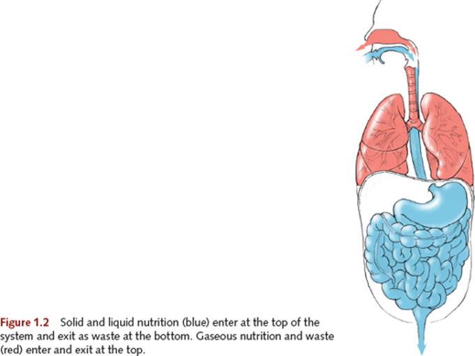
Prana also enters our bodies in gaseous form: the breath. Like solids and liquids, it enters at the top, where it remains above the diaphragm in the lungs (see figure 1.3), exchanging gases with the capillaries at the alveoli. The waste gas in the lungs needs to be expelled, but it gets out the same way it came in. The force of apana, when acting on respiratory waste gas, must move up to get out. Apana must be able to operate freely both upward and downward, depending on what type of waste it acts upon.

The ability to reverse apana’s downward action is a basic and useful skill acquired through yoga practice, but not something most people are able to do without training. People are accustomed to pushing down to operate their apana. Many have learned that whenever something needs to be eliminated from the body, the body must squeeze in and push down. That is why, when most beginning students are asked to exhale completely, they activate their breathing muscles as if they are urinating or defecating.
Sukha and Dukha
Prana and apana must have a healthy reciprocal relationship in the body; thus, the body’s pathways must be clear of obstructing forces. In yogic terms, our breathing bodies must be in a state of sukha, translated literally as good space. Bad space is referred to as dukha, which is derived from dus, meaning bad, difficult, or hard, and kha, meaning space. It is generally translated as suffering, uneasy, uncomfortable, unpleasant, and difficult.
This model points to the fundamental methodology of all classical yoga practice, which seeks to uncover and resolve blockages or obstructions (kleshas1) to improve function. Essentially, when we make more good space our pranic forces flow freely and restore normal, healthy function.
The modern master of yoga therapy, T.K.V. Desikachar, has often said that yoga therapy is 90 percent waste removal.
Because exhalation is an action of removing waste from the system, another practical way of applying this insight is that if we take care of the exhalation, the inhalation takes care of itself. If we get rid of the unwanted, we make room for what is needed.
Being Born to Breath and Gravity
When a fetus is in utero, the mother does the breathing. Her lungs deliver oxygen to the uterus and placenta. From there it travels to the umbilical cord, which takes about half the oxygenated blood to the inferior vena cava while the other half enters the liver. The two sides of the heart are connected, bypassing the lungs, which remain dormant until the child is born. Needless to say, human fetal circulation is very different from ex-utero circulation.
Being born means being severed from the umbilical cord—the lifeline that has sustained the fetus for nine months. Suddenly, and for the first time, the infant needs to engage in actions that ensure continued survival. The very first of these actions declares physical and physiological independence. It is the first breath, and it is the most important and forceful inhalation a human will ever take.
The initial inflation of the lungs triggers enormous changes to the entire circulatory system, which has previously been geared toward receiving oxygenated blood from the placenta. That first breath causes a massive surge of blood into the lungs, the right and left sides of the heart to separate into two pumps, and the specialized vessels of fetal circulation to shut down, seal off, and become ligaments that support the abdominal organs.
That first inhalation must be so forceful because it needs to overcome the initial surface tension of the previously inactive lung tissue. The force required to overcome that tension is three or four times greater than that of a normal inhalation.2
Another radical reversal that occurs at the moment of birth is the sudden experience of body weight in space. Inside the womb, the fetus is in a cushioned, supportive, fluid-filled environment. Suddenly, the child’s entire universe expands—the limbs and head can move freely, and the baby must be supported in gravity.
Because adults swaddle babies and move them around from place to place, stability and mobility may not seem to be so much of an issue early in life. In fact, infants begin to develop their posture immediately after taking their first breath, as soon as they begin to nurse. The complex, coordinated action of simultaneously breathing, sucking, and swallowing eventually provides them with the tonic strength to accomplish their first postural skill—supporting the weight of the head. This is no small feat for the infant, considering that an infant’s head constitutes one fourth of its overall body length, compared to one eighth for an adult.
Head support involves the coordinated action of many muscles and, as with all weight-bearing skills, a balancing act between mobilization and stabilization. Postural development continues from the head downward until after about a year, when babies begin walking, culminating in the completion of the lumbar curve at about 10 years of age (see chapter 2).
Having a healthy life on Earth requires an integrated relationship between breath and posture, prana and apana, and sthira and sukha. If something goes wrong with one of these functions, by definition it will go wrong with the others. In this light, yoga practice can be viewed as a way of integrating the body’s systems so we spend more time in a state of sukha than in dukha.
To summarize, from the moment of birth, humans are confronted by breath and gravity, two forces that were not present in utero. To thrive, we need to reconcile those forces as long as we draw breath on this planet.
BREATHING DEFINED: MOVEMENT IN TWO CAVITIES
Breathing is traditionally defined in medical texts as the process of taking air into and expelling it from the lungs. This process—the passage of air into and out of the lungs—is movement; specifically, it is movement in the body’s cavities, which I will refer to as shape change. So, for the purposes of this exploration, here’s our definition:
Breathing is the shape change of the body’s cavities.
The simplified illustration of the human body in figure 1.4 shows that the torso consists of two cavities, thoracic and abdominal. These cavities share some properties, and they have important distinctions as well. Both contain vital organs: The thoracic cavity contains the heart and lungs, and the abdominal cavity contains the stomach, liver, gall bladder, spleen, pancreas, small and large intestines, kidneys, and bladder.

Both cavities open at one end to the external environment—the thoracic at the top, and the abdominal at the bottom. The cavities open to each other3 by means of an important shared, dividing structure, the diaphragm. Another important shared property is that both cavities are bound posteriorly by the spine. The two cavities also share the quality of mobility—they change shape. This shape-changing ability is most relevant to breathing; without this movement, the body cannot breathe at all.
Although both the abdominal and thoracic cavities change shape, an important structural difference exists in how they do so.
The Water Balloon and the Accordion
The abdominal cavity changes shape like a flexible, fluid-filled structure such as a water balloon. When you squeeze one end of a water balloon, the other end bulges (figure 1.5).

That is because water is noncompressible. Your hand’s action only moves the fixed volume of water from one region of the flexible container to another. The same principle applies when the movements of breathing compress the abdominal cavity; a squeeze in one region produces a bulge in another. In the context of breathing, the abdominal cavity changes shape but not volume. In the context of life processes other than breathing, the abdominal cavity does change volume. When you drink a large volume of liquid or eat a big meal, the overall volume of the abdominal cavity increases as a result of expanded abdominal organs (stomach, intestines, and bladder). Any volume increase in the abdominal cavity produces a corresponding decrease in the volume of the thoracic cavity. That is why it is more difficult to breathe after a big meal, before a big bowel movement, or when pregnant.
In contrast to the abdominal cavity, the thoracic cavity changes both shape and volume; it behaves as a flexible gas-filled container, similar to an accordion bellows. When you squeeze an accordion, you create a reduction in the volume of the bellows and air is forced out. When you pull the bellows open, its volume increases and air is pulled in (figure 1.6). This occurs because the accordion is compressible and expandable, as is air. The same is true of the thoracic cavity, which, unlike the abdominal cavity and its contents, can change its shape and volume in breathing.
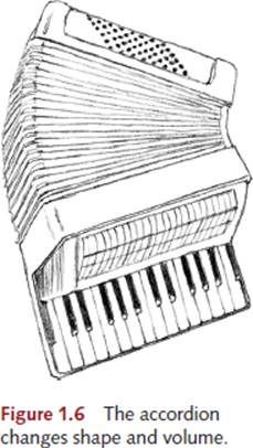
Let’s now imagine the thoracic and abdominal cavities as an accordion stacked on top of a water balloon. This image gives a sense of the relationship of the two cavities in breathing; movement in one will necessarily result in movement in the other. Recall that during an inhalation (the shape change permitting air to be pushed into the lungs by the planet’s atmospheric pressure), the thoracic cavity expands its volume. This pushes downward on the abdominal cavity, which changes shape as a result of the pressure from above.
By defining breathing as shape change, it becomes very easy to understand what constitutes effective or obstructed breath—it is simply the ability or inability of the structures that define and surround the body’s cavities to change shape.
The Universe Breathes Us
Volume and pressure are inversely related; when volume increases, pressure decreases, and when volume decreases, pressure increases. Because air always flows toward areas of lower pressure, increasing the volume inside the thoracic cavity will decrease pressure and cause air to flow into it. This is an inhalation.
It is important to note that in spite of how it feels when you inhale, you do not actually pull air into the body. On the contrary, air is pushed into the body by the atmospheric pressure (14.7 pounds per square inch, or 1.03 kg/cm2) that always surrounds you. This means that the actual force that gets air into the lungs is outside of the body. The energy expended in breathing produces a shape change that lowers the pressure in the chest cavity and permits the air to be pushed into the body by the weight of the planet’s atmosphere. In other words, you create the space, and the universe fills it.
During relaxed, quiet breathing such as while sleeping, an exhalation is a passive reversal of this process. The thoracic cavity and lung tissue—which have been stretched open during the inhalation—spring back to their initial volume, pushing the air out and returning them to their previous shapes. This is referred to as a passive recoil. Any reduction in the elasticity of these tissues results in a reduction of the body’s ability to exhale passively, leading to a host of respiratory problems such as emphysema and pulmonary fibrosis, which greatly compromise the elasticity of the lung tissue.
In breathing patterns that involve active exhaling, such as blowing out candles, speaking, singing, and performing various yoga exercises, the musculature surrounding the two cavities contracts in such a way that the abdominal cavity is pushed upward into the thoracic cavity or the thoracic cavity is pushed downward onto the abdominal cavity, or any combination of the two.
Three-Dimensional Shape Changes of Breathing
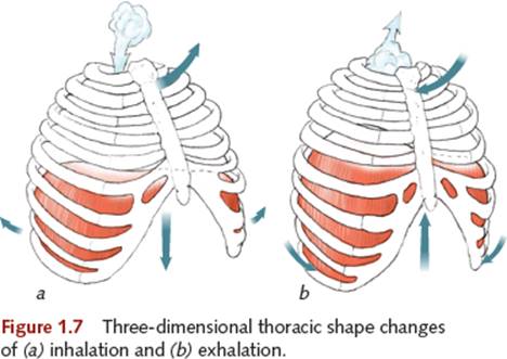
Because the lungs occupy a three-dimensional space in the thoracic cavity, when this space changes shape to cause air movement, it changes shape three-dimensionally. Specifically, an inhalation involves the chest cavity increasing its volume from top to bottom, from side to side, and from front to back, and an exhalation involves a reduction of volume in those three dimensions (see figure 1.7).
Because thoracic shape change is inextricably linked to abdominal shape change, you can also say that the abdominal cavity also changes shape (not volume) in three dimensions—it can be squeezed from top to bottom, from side to side, or from front to back (see figure 1.8). In a living, breathing body, thoracic shape change cannot occur without abdominal shape change. That is why the condition of the abdominal region has such an influence on the quality of our breathing and why the quality of our breathing has a powerful effect on the health of our abdominal organs.

EXPANDED DEFINITION OF BREATHING
Based on the information we have so far, here’s an expanded definition of breathing:
Breathing, the process of taking air into and expelling it from the lungs, is caused by a three-dimensional shape change in the thoracic and abdominal cavities.
Defining breathing in this manner explains not only what it is but also how it is done. As a thought experiment, try this: Substitute the term shape change for the word breathing whenever discussing the breath. For example, “I just had a really good breath” really means “I just had a really good shape change.” More important, “I’m having difficulty breathing” really means “I’m having trouble changing the shape of my cavities.” This concept has profound therapeutic implications, because it tells us where to start looking for the root causes of breath and postural issues, and it can eventually lead us to examine the supporting, shape-changing structure that occupies the back of the body’s two primary cavities—the spine, which is discussed in chapter 2.
A key observation that has been made in yogic teachings is that spinal movements are an intrinsic component of the shape-changing activity of the cavities (breathing). This is why such a huge component of yoga practice involves coordinating the movements of the spine with the process of inhaling and exhaling.
There’s a reason why students are instructed to inhale during spinal extension and exhale during spinal flexion. Fundamentally, the spinal shape change of extension is an inhale and the spinal shape change of spinal flexion is an exhale.
THE DIAPHRAGM’S ROLE IN BREATHING
A single muscle, the diaphragm, is capable of producing—on its own—all of the three-dimensional movements of breath. This is why just about every anatomy book describes the diaphragm as the principal muscle of breathing. Let’s add the diaphragm to our shape-change definition of breathing to begin our exploration of this remarkable muscle:
The diaphragm is the principal muscle that causes three-dimensional shape change in the thoracic and abdominal cavities.
To understand how the diaphragm causes this shape change, it is important to examine its shape and location in the body, where it is attached and what is attached to it, its action, and its relationship to the other muscles of breathing.
Shape and Location
The deeply domed shape of the diaphragm has evoked many images. Two of the most common are a jellyfish and a parachute (figure 1.9). It is important to note that the diaphragm’s shape is created by the organs it encloses and supports. Deprived of its relationship with those organs, its dome would collapse, much like a stocking cap without a head in it. It is also evident that the diaphragm has an asymmetrical double-dome shape; the right dome rises higher than the left. The liver pushes up from below the right dome, and the heart pushes down from above the left dome (see figure 1.10 on page 9).
The diaphragm divides the torso into the thoracic and abdominal cavities. It is the floor of the thoracic cavity and the roof of the abdominal cavity. Its structure extends through a wide section of the body. The uppermost part reaches the space between the third and fourth ribs, and its lowest fibers attach to the front of the third and second lumbar vertebrae; nipple to navel is one way to describe it.

Muscular Attachments of the Diaphragm
Muscles attach at origin and insertion points. The determination of origin or insertion is dependent on two factors: structure and function.
· Structurally, the end of the muscle closest to the core of the body—the proximal end—is usually referred to as the origin. The distal end, the one that attaches more peripherally, is usually referred to as the insertion.
· Functionally, the end of the muscle that is more stable on contraction is referred to as the origin, and the more mobile end the insertion.
Although this seems to make sense—proximal structures are generally more stable than distal ones—this is only true some of the time, as is explored further in chapter 4. For example, a reversal of functional origins and insertions occurs when you have a mobile core and stable extremities while moving the body through space.
The muscle that moves space through the body—the diaphragm—possesses an unmistakably three-dimensional form and function, which makes its origin and insertion anything but cut and dried. To avoid confusion as we begin to examine the attachments of its muscular fibers, we simply refer to the diaphragm’s lower attachments and upper attachments.
Lower Attachments
The lower edges of the diaphragm’s fibers attach at four distinct regions. Traditional texts list only three regions: sternal, costal, and lumbar (see figure 1.10).
· 1. Sternal—The back of the xiphoid process at the bottom of the sternum
· 2. Costal—The inner costal cartilage surfaces of ribs 6 through 10
· 3. Arcuate—The arcuate ligament4 that runs from rib 10’s cartilage to the lumbar spine, attaching along the way to the floating ribs (11 and 12) and the transverse process and body of L1
· 4. Lumbar—The crura (Latin for legs) at the front of the lumbar spine, L3 on right and L2 on left

Upper Attachments
All the muscular fibers of the diaphragm rise upward in the body from their lower attachments. They eventually arrive at the flattened, horizontal top of the muscle, the central tendon, into which they blend. In essence, the diaphragm connects to itself—its own center, which is fibrous noncontractile tissue. The central tendon’s vertical movements within the body are limited by its strong connection to the heart’s fibrous pericardium, to which it is inextricably linked.
Traditional texts refer to the lower attachments as the muscle’s origin, and the central tendon as the insertion. The following text offers our reevaluation of that assumption.
Challenging Traditional Labeling of Origin and Insertion
As we will see later in this chapter, there is much confusion among breathing teachers about the action of the diaphragm. Why is there so much confusion, and where did it begin? A major factor may be that the structural origin and insertion of the diaphragm have historically been mislabeled in anatomy texts. This has resulted in a functional confusion about which end of the muscle is stable and which is mobile when the diaphragm’s fibers contract.
Assumptions About Structure In terms of structure, traditional anatomy texts present the origin of the diaphragm as its lower attachments, and the central tendon is labeled as its insertion. Upon closer scrutiny, this categorization breaks down.
Let’s see how true this is for the location of your diaphragm’s lower attachments (see figure 1.10 on page 9). If you place your fingertips at the base of your sternum, you can usually touch the tip of your xiphoid process. You can then sweep your fingers around the edges of your costal cartilage, and from there around your back to the region of the floating ribs, and then to the top of your lumbar spine.
At every point of contact you just traced on your body, your fingertips were as little as 1/4 inch (0.6 cm) and no more than one 1 inch (2.5 cm) away from the sternal, costal, arcuate, or lumbar attachments of your diaphragm. Your fingers were on the surface of your body, not near its core, and neither were the attachments you just traced.
Now, let’s see if you can trace your diaphragm’s upper attachments. Can you get your fingertips close to your central tendon? Not really, because it is at the core of the body. In fact, your heart is anchored to it. Describing this structure as central is apt, which is why using a term that is usually reserved for distal structures (insertion) is all the more confusing.
Lower Fibers The lower muscular fibers of the diaphragm attach to flexible cartilage and ligament. The bottom of the xiphoid process is mostly cartilage. The costal cartilage is springy and flexible and has many joints that attach it to the ribs, which are among the more than 100 joints that make up the rib cage articulations. The arcuate ligament is a long, ropy band that attaches to the tips of the floating ribs. The front surface of the lumbar spine is covered with the anterior longitudinal ligament, which is anchored to the anterior surfaces of the cartilaginous intervertebral discs as well as the anterior surfaces of the lumbar vertebrae.
Assuming that the rib cage is allowed to move freely, we can make a strong case that these lower attachments of the diaphragm have considerable potential for movement. Even the crura have this potential in situations involving lumbar motion and the action of the psoas muscles, which share common attachments in the upper lumbar region.
Upper Fibers The center of the diaphragm and the heart have never been apart. The tissue that will become the central tendon actually originates outside of the thoracic cavity in our embryonic development. At this early stage, it is called the transverse septum, and it lies adjacent to the primordial heart tissue. With the inward folding of the embryo’s structure in the fourth week in utero, the heart and transverse septum move together into the thoracic cavity. Once the transverse septum is in this location, the muscular tissue of the diaphragm grows toward it from the interior surface of the abdominal wall. Thus, the association of the central tendon with the heart is the original manifestation of the diaphragm, and further justifies labeling it as its origin.
Because of its firm anchorage to the heart, the tough, fibrous tissue of the central tendon has limited ability to move vertically within the thoracic cavity (between 1/2 to 1 inch). Therefore, the upper muscular attachments of the diaphragm closest to the central tendon have little movement potential. However, the muscular domes that rise up on either side of the central tendon do have the ability to strongly push downward on the abdominal viscera, and this (not the downward movement of the central tendon itself) mostly accounts for the bulging of the upper abdomen commonly referred to as a belly breath.
Conclusions For all the reasons just mentioned, we have concluded that traditional texts reverse the structural labeling of origin and insertion of the diaphragm by describing distal structures (lower attachments) as origin and proximal structures (upper attachments) as insertion. This structural confusion leads to a functional confusion because of the assumption that muscular insertions are mobile and muscular origins are stable. We will explore this shortly.
Organic Relations
Studying the diaphragm’s origin and insertion allows us to understand what structures it is attached to. But unlike other muscles, the diaphragm has a lot of structures attached to it. This is what is meant by the term organic relations.
As the prime mover of the thoracic and abdominal cavities, the diaphragm is a place of anchorage for the connective tissue that surrounds the thoracic and abdominal organs. The names of these important structures are easily remembered as the three Ps:
· • Pleura, which surrounds the lungs
· • Pericardium, which surrounds the heart
· • Peritoneum, which surrounds the abdominal organs
It should be clear that the shape-changing activity of these cavities has a profound effect on the movements of the organs they contain. The diaphragm is a fundamental source of these movements, but the viscera are also a source of resistance and stabilization for the diaphragm. This reciprocal relationship illuminates why the coordinated movements of breath and body promoted by yoga practice can lead to such dramatic improvements in the overall health and functioning of all the body’s systems.
Action of the Diaphragm
It is important to remember that the muscular fibers of the diaphragm are oriented primarily along the vertical (up–down) axis of the body (see figure 1.11).
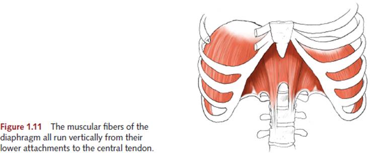
As with all muscles, the contracting fibers of the diaphragm pull their two ends (the central tendon and the base of the rib cage) toward each other. This action is the fundamental cause of the three-dimensional thoracoabdominal shape changes of breathing.
Because the diaphragm has multidimensional action, the type of movement it produces depends on which region of its attachment is stable and which is mobile.
To illustrate this with a more visible movement, the psoas major muscle creates hip flexion either by moving the leg toward the front of the spine, as in standing on one leg and flexing the opposite hip, or by moving the front of the spine toward the leg, as in sit-ups with the legs braced. In both cases, the psoas major is contracting and flexing the hip joint. What differs is which end of the muscle is stable and which is mobile. Needless to say, a stable torso and moving leg look very different from a moving torso and a stable leg.
Variety of Diaphragmatic Breaths
Just as you can think of the psoas major as either a leg mover or a trunk mover, you can think of the diaphragm as either a belly bulger or a rib cage lifter (see figure 1.12). The muscular action of the diaphragm is most often associated with a bulging movement in the upper abdomen, which is commonly referred to as a belly breath or abdominal breath, and confusingly referred to as a diaphragmatic breath. This is only one type of diaphragmatic breath—one in which the base of the rib cage (lower attachments) is stable and the domes (upper attachments) are mobile (see figure 1.13a).
If we reverse these conditions by stabilizing the upper domes while relaxing the rib cage, a diaphragmatic contraction causes an expansion of the rib cage (see figure 1.13b). This is called a chest breath, which many believe to be caused by the action of muscles other than the diaphragm. This mistaken idea creates a false dichotomy between diaphragmatic and so-called “non-diaphragmatic” breathing.

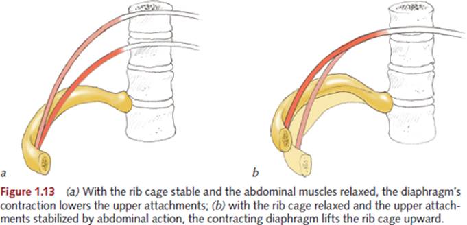
The unfortunate result of this error is that many people receiving breath training who exhibit chest rather than belly movement are told that they are not using the diaphragm, which is entirely wrong. Except in cases of paralysis, the diaphragm is always used for breathing. The real issue is whether or not the diaphragm is able to work efficiently, meaning how well it can coordinate with all the other muscles that can affect shape change. Yoga practice can help with precisely this coordination.
If it were possible to release all of the muscular action surrounding our cavities, the diaphragm’s action would cause both the chest and abdomen to move simultaneously. This rarely occurs because the need to stabilize the body’s mass in gravity causes many of the respiratory stabilizing muscles—which are also postural muscles—to remain active through all phases of breathing, even while supine. From this perspective, our postural habits are synonymous with our breathing habits.
Engine of Three-Dimensional Shape Change
The specific patterns we encounter in yoga asana or breathing practice (pranayama) result from the action of accessory muscles—muscles other than the diaphragm—that can change the shape of the cavities. They have the same relationship to the diaphragm that the steering mechanism of a car has to its engine.
The engine is the prime mover of a car. All mechanical and electrical movements that are associated with a car’s operation are generated by the engine. Similarly, three-dimensional, thoracoabdominal shape changes of breathing are primarily generated by the diaphragm.
When you drive, the only direct control you exert over the function of the engine is the speed of its spinning. Pushing the gas pedal makes the engine spin faster, and releasing the pedal makes it spin slower. When breathing, the only direct, volitional control you have over your diaphragm is its timing. Within limits you can control when it fires, but when it ceases contracting, a passive recoil creates the exhalation, just as your car’s gas pedal springs upward to decelerate upon release of your foot.
Steering Shape Change
Everyone knows you don’t steer a car with its engine. To channel the power of the engine in a particular direction, you need the transmission, brakes, steering, and suspension. In the same way, you don’t steer your breathing with your diaphragm. To control the power of the breath and guide it into specific patterns, you need the assistance of accessory muscles.
From the standpoint of this engine analogy, the notion that improving breath function by training the diaphragm is flawed. After all, you don’t become a better driver by learning how to work only the gas pedal. Most of the skills you acquire in driver training have to do with coordinating the acceleration of the car with steering, braking, and awareness of your surroundings. Likewise, breath training is really accessory muscle training. Only when all the musculature of the body is coordinated and integrated with the action of the diaphragm can breathing be efficient and effective.
The notion that diaphragmatic action is limited to abdominal bulging (belly breathing) is as inaccurate as asserting that an engine is only capable of moving the car forward and that some separate source of power governs reverse movement. This automotive error results from not understanding the relationship of the car’s engine to its transmission; the breathing error results from not understanding the diaphragm’s relationship to rib cage movement and to the accessory muscles.
A related error equates belly movement with proper breathing and chest movement with improper breathing. This is just as silly as stating that a car is best served by only going forward at all times. Driving a car with no reverse gear will eventually leave you stuck somewhere.
Accessory Muscles of Respiration
Although there is universal agreement that the diaphragm is the principle muscle of breathing, there are varied and sometimes conflicting ways of categorizing the other muscles that participate in breathing. By restating our definition of breathing, we can define as accessory muscles any muscle other than the diaphragm that can cause a shape change in the cavities. It is irrelevant whether shape change leads to inhalation (an increase of thoracic volume) or exhalation (a decrease in thoracic volume), because muscles that control both can be active during any phase of breathing.
Let’s use this perspective to analyze the differences and similarities between a few types of breathing.
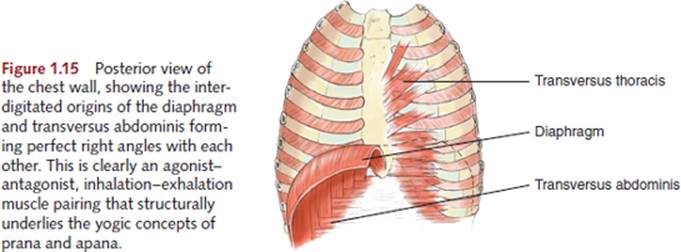
In a belly breath, the costal attachments of the diaphragm are stabilized by muscles that pull the rib cage downward: the internal intercostals, the transversus thoracis, and others (see figures 1.15 and 1.16 on the following page). These muscles are generally classified as exhaling muscles, but here they actively participate in shaping an inhalation.
In a chest breath, the upper attachments of the diaphragm are stabilized by the lower abdominal muscles, also regarded as exhaling muscles, but in this case, they clearly act to produce a pattern of inhaling. It should be noted that in both the chest and belly breaths, one region of accessory muscles had to be relaxed while the other was active. In the belly breath, the abdominal wall released, and in the chest breath, the so-called rib cage depressors had to let go.
In the cleansing technique of kapalabhati (kapala meaning skull and bhati meaning light or shine), in which strong, voluntary exhalations are the focus, the base of the rib cage needs to be lifted and held open in order to allow the lower abdominal region to freely, rhythmically change shape. Here the “inhaling” muscles of the external intercostals remain active during exhalation.
Abdominal and Thoracic Accessory Muscles
The abdominal cavity and its musculature can be imagined as a water balloon surrounded on all sides by elastic fibers running in all directions (figure 1.14).
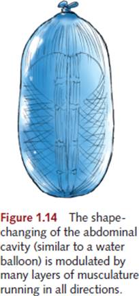
In concert with diaphragmatic contractions, the shortening and lengthening of these fibers produce the infinitely variable shape changes associated with respiration. As the tone of the diaphragm increases during inhalation, the tone of some abdominal muscles must decrease to allow the diaphragm to move. If you contract all your abdominal muscles at once and try to inhale, you’ll notice that it’s quite difficult because you’ve limited the ability of your abdomen to change shape.
The abdominal muscle group does not affect breathing only by limiting or permitting shape change in the abdominal cavity. Because these muscles also attach directly to the rib cage, they directly affect its ability to change shape.
The abdominal muscles that have the most direct effect on breathing are the ones that attach at the same place as the diaphragm, the transversus abdominis. This deepest layer of the abdominal wall arises from the costal cartilage at the base of the rib cage’s inner surface. The fibers of the transversus abdominis are interdigitated (interwoven) at right angles with those of the diaphragm, whose fibers ascend vertically, whereas those of the transversus abdominis run horizontally (see figure 1.15). This makes the transversus abdominis the direct antagonist to the diaphragm’s action of expanding the rib cage. The same layer of horizontal fibers extends this action upward into the posterior thoracic wall as the tranversus thoracis, a depressor of the sternum.
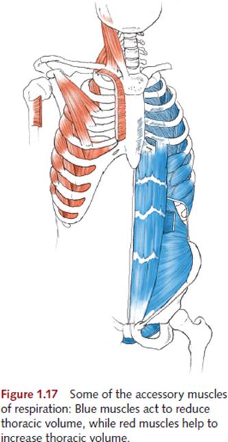
The other layers of the abdominal wall have similar counterparts in the thoracic cavity. The external obliques turn into the external intercostals, and the internal obliques turn into the internal intercostals (see figure 1.16). Of all these thoracoabdominal layers of muscle, only the external intercostals are capable of increasing thoracic volume. All the others produce a reduction of thoracic volume, either by depressing the rib cage or pushing upward on the upper attachments of the diaphragm.

Other Accessory Muscles
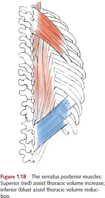
Chest, neck, and back muscles can increase the volume of the rib cage (see figures 1.17 and 1.18), but they are far more inefficient at doing this than the diaphragm and external intercostals. This inefficiency is the result of the fact that the location and attachment of these muscles do not provide good leverage on the rib cage, and the usual role of these muscles is not respiration. They are primarily head, neck, shoulder girdle, and arm mobilizers—actions that require them to be stable proximally (toward the core of the body) and mobile distally (toward the periphery of the body). For these muscles to expand the rib cage, this relationship must be reversed; the distal insertions must be stabilized by yet more muscles so the proximal origins can be mobilized. That makes these the least efficient of the accessory muscles, and considering the degree of muscular tension that accessory breathing entails, the net payoff in oxygenation makes it a poor energetic investment. That is why improved breathing is observable as decreased tension in the accessory mechanism, which happens when the diaphragm, with its enormously efficient shape-changing ability, operates as unencumbered as possible.
THE OTHER TWO DIAPHRAGMS
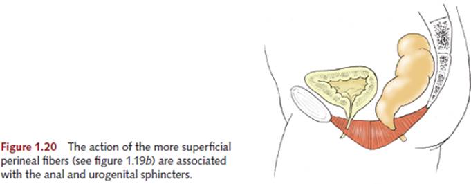
Along with the respiratory diaphragm, breathing involves the coordinated action of the pelvic and vocal diaphragms. Of particular interest to yoga practitioners is the action of mula bandha, or root lock (mulameaning firmly fixed or root, and bandha meaning binding, bonding, or tying), which is a lifting action produced in the pelvic floor muscles (shown in figure 1.19) that also includes the lower fibers of the deep abdominal layers. Mula bandha is an action that moves apana upward and stabilizes the upper attachments of the diaphragm. Inhaling while this bandha is active requires a release of the attachments of the upper abdominal wall, which permits the diaphragm to lift the base of the rib cage upward. This lifting action is referred to as uddiyana bandha, or flying upward lock.
It is important to note that the more superficial muscular fibers of the perineum need not be involved in mula bandha, because they are not efficient lifters of the pelvic floor. They also contain the anal and urethral sphincters, which are associated with the downward movement of apana (elimination of solid and liquid waste), as shown in figure 1.20.

Vocal Diaphragm
The gateway to the respiratory passages is the glottis, shown in figure 1.21, which is not a structure but a space between the vocal folds (cords).

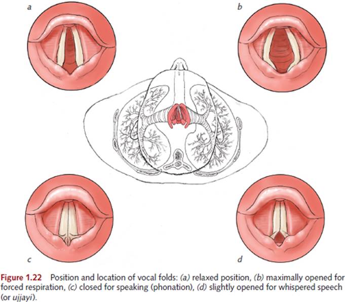
Yoga practitioners are accustomed to regulating this space in various ways based on what they are doing with their breath, voice, and posture. When at rest, the muscles that control the vocal cords can be relaxed so that the glottis is being neither restricted nor enlarged (see figure 1.22a). This occurs in sleep and in the more restful, restorative practices in yoga.
When doing breathing exercises that involve deep, rapid movements of breath, such as kapalabhati or bhastrika (bhastra meaning bellows) the muscles that pull the vocal folds apart (abduction) contract to create a larger passage for the air movements (see figure 1.22b).
When chanting, singing, or speaking, the vocal folds are drawn together (adduction), which causes them to vibrate as the exhaled air is forced across them. This vibration is termed phonation (see figure 1.22c).
When the exercises call for long, deep, slow breaths, the glottis can be partially closed, with only a small opening at the back of the cords (see figure 1.22d). This is the same action that creates whispered speech; in yoga it’s known as ujjayi, the victorious breath (ud meaning to flow out and jaya meaning victory or triumph). This action also creates more postural support in the body, as we will explore in the next section.
The Bandhas
All three diaphragms (pelvic, respiratory, and vocal) come together with ujjayi in yoga movements that are coordinated with inhaling and exhaling. In addition to giving more length and texture to the breath, the valve of ujjayi creates a kind of back pressure throughout the abdominal and thoracic cavities. This pressure can protect the spine during the long, slow flexion and extension movements that occur in the breath-synchronized flowing practice of vinyasa (arrangement or placement), such as during sun salutations. In yogic terms, these coordinated actions of the diaphragms (bandhas) create more sthira (stability) in the body, protecting it from injury by redistributing mechanical stress.
Figure 1.23 shows a mechanical analysis of the body entering into a forward bend from two perspectives. In figure 1.23a, we see the torso moving without breath support. Because the breathing musculature surrounding the cavities is not engaged, there is no single center of gravity to the shape, and a partial center of gravity (B) is acting upon the long arm of a lever (C), of which the fulcrum point (A) is at the vulnerable disc of the lumbosacral junction. The weight of the torso is being controlled by the posterior musculature, which compressively acts on the short end of the lever (D). The body instinctively resents this extremely poor leverage, and that’s why we tend to hold our breath in situations like this to avoid damaging our spinal structures.

Figure 1.23b on page 19 pictures the same movement employing the glottal valve of ujjayi (E), which automatically engages the breathing musculature. This creates support along the entire anterior surface of the spine because it rests on the stabilized body cavities. The body now has a single center of gravity, which is being supported safely by the pelvis and legs. This is what is commonly referred to as frontal support.
An additional effect of moving and supporting the body through this kind of resistance is the creation of heat in the system, which can be used in many beneficial ways. These practices are referred to as brhmana (brh meaning increase or expand), which implies heat, expansion, and the development of power and strength as well as the ability to withstand stress. Brhmana is also associated with inhaling, nourishment, prana, and the chest region.
When relaxing the body in the more released, horizontal, or restorative practices, it is important to disengage the bandhas and glottal constrictions that are associated with vertical postural support. This relaxing side of yoga embodies the quality of langhana (meaning fasting or hunger), which is associated with coolness, condensation, relaxation, and release, as well as the development of sensitivity and inward focus. Langhana is also associated with exhaling, elimination, apana, and the abdominal region.
Because the ultimate goal of yoga breath training is to free up the system from habitual, dysfunctional restrictions, the first thing we need to do is free ourselves from the idea that there is a single correct way to breathe. As useful as the bandhas are when supporting the center of gravity and moving the spine through space, we need to release the brhmana forces of sthira in the system when pursuing the langhana, relaxation, and release of sukha.
INTRINSIC EQUILIBRIUM: PRESSURE ZONES
Intrinsic equilibrium refers to several important mechanisms that combine to make the human torso a self-supporting structure, which has an inherent tendency to seek upward movement.
The most important of these mechanisms is in the visceral component of the torso—the pressure differential between the lower abdomen (highest pressure), the upper abdomen (middle pressure), and the thoracic space (lowest pressure). Since energy always migrates from a region of higher pressure toward a region of lower pressure, this means that the lower and upper abdominal contents are constantly migrating upward toward thoracic space.5
The bony components of the torso—the spine, rib cage, and pelvis—all share a common characteristic: They are knit together under mechanical tension, like coiled springs restrained by elastic bands. When the sternum is divided for thoracic surgery, the two halves spring open and need to be pushed back together in order to be closed up again. At the front of the pelvis, the two pubic rami are joined at the pubic symphysis, a pressurized joint that softens and opens in childbirth and hopefully reknits afterward.
The spine’s intervertebral discs are constantly pushing the vertebral bodies apart—an action that is resisted by the ligamentous and bony structures of the spine’s posterior column. This combined push–pull of forces makes the spinal column as a whole a very springy structure that always seeks to return to neutral.
Note that all of these features of the body operate independently of muscle contraction—in fact, it is the unconscious, habitual activity of our postural and breathing musculature that obstructs the effect of intrinsic equilibrium. So, establishing an upright relationship to gravity, in the deepest sense, is less about exerting the correct muscular effort than it is about discovering and releasing the habitual muscular effort that is obstructing the natural tendency of the body to be supported all on its own.
This view of the body’s anatomical support mechanisms is completely in harmony with the perspective on yoga practice offered by Patañjali. We achieve yoga by identifying and removing the kleshas (afflictions) from our system.
CONCLUSION
When translated, the term pranayama is commonly broken into the two roots prana, meaning life or breath energy, and yama, meaning restraint or control. Because the breath is only partially under our voluntary control, this translation gives a very limited view of breath practice.
A fuller understanding of the term is available when the second long “aa” (pranaaayama) is recognized. This means that the second root is ayama.
In Sanskrit, the prefix a negates the term it precedes. This means that pranayama refers to a process that unrestrains the breath. It also honors the aspects of the breath that are not under our voluntary control.
This is why Patañjali’s definition of kriya yoga (see page x in the introduction) so beautifully links with the idea that the breath is our best, most intimately available teacher of the deepest principles of yoga.
In this light, it is clear that the practice of unrestraining the breath can be seen as synonymous with the identification and release of the bodily tensions that obstruct the expression of our system’s intrinsic equilibrium.
1 Klestr means that which causes pain or suffering.
2 The initial inflation of the lungs is assisted by the presence of surfactant, a substance that lowers the surface tension of the stiff, newborn lung tissue. Because surfactant is produced very late in intrauterine life, babies who are born prematurely (before 28 weeks of gestation) have a hard time breathing.
3 The three openings (hiati) in the diaphragm are for the arterial supply to the lower body (aortic hiatus), the venous return from the lower body to the heart (inferior vena cava) and the esophagus (esophageal hiatus). Hiatus is the Latin past participle of hiare—to stand open or yawn.
4 Traditional texts label each arc of the arcuate ligament individually. It is much clearer to think of it as a single, long ligament that attaches to the tips of the bony surfaces mentioned. In dissection, when the arcuate ligament is deprived of these attachments, it clearly stretches out into a single, straight ligament.
5 When a lobe of the lung is removed (lobectomy), the diaphragm and abdominal organs are drawn upward and fill the extra space.