
Objectives
The student understands the primary disturbances, compensatory responses, decompensatory processes, and possible therapeutic interventions that pertain to various abnormal cardiovascular situations:
● Defines circulatory shock.
● dentifies the primary disturbances that can account for cardiogenic, hypovolemic, anaphylactic, septic, and neurogenic shock states.
● Lists the compensatory processes that may arise during various types of circulatory shock.
● dentifies the decompensatory processes that may arise during shock and describes how these lead to irreversible shock states.
● ndicates how coronary artery disease may lead to abnormal cardiac function.
● Defines the term angina pectoris and describes the mechanisms that promote its development.
. Indicates the mechanisms by which various therapeutic interventions may alleviate angina and myocardial ischemia in association with coronary artery disease.
- Defines the term cardiomyopathy and differentiates between dilated and hypertrophied cardiomyopathies.
- Defines the term heart failure and differentiates between acute and chronic heart failure and between systolic and diastolic failure.
● dentifies the short-term and long-term compensatory processes that accompany chronic systolic heart failure.
● Describes the advantages and disadvantages of the fluid accumulation that accompanies systolic heart failure.
● Defines pulmonary and systemic arterial hypertension.
● dentifies the various factors that may contribute to the development of systemic hypertension.
● Describes the role of the kidney in establishing and/or maintaining systemic hypertension.
Cardiovascular disease represents the number one cause of deaths in most industrialized countries. How and when a person’s cardiovascular system breaks down depends upon an increasing number of known risk factors. These include family history and genetic predisposition to various CV system malfunctions, poor diet and/or obesity, inactivity, environmental pollutants, and the acute or chronic effects of various infective agents. In this last chapter, a variety of specific pathologies that may be evoked by these risk factors are introduced. We have already described specific pathologies associated with abnormal cardiac electrical activities and valve function in Chapter 5, so will now focus on the pathologies associated with system-wide abnormalities. It is not intended as an in-depth coverage of cardiovascular diseases but rather as an introductory presentation of how the physiological processes described previously are evoked and/or altered during various abnormal cardiovascular states. In each case, there is generally a primary disturbance associated with malfunction of some component of the cardiovascular system that threatens the maintenance of normal arterial pressure. These primary disturbances evoke appropriate compensatory reflex responses to restore arterial pressure. Often, however, pathological situations also lead to inappropriate “decompensatory processes,” which tend to accelerate the deterioration of cardiovascular function. Therapeutic interventions may be required and are often designed to limit or reverse these decompensatory processes. Students are again encouraged to review the summary of cardiovascular variables and their determinants in Appendix C because a thorough knowledge of this material will greatly help understand the physiological consequences of these abnormalities.
Circulatory shock
A state of circulatory “shock” exists whenever there is a generalized, severe reduction in blood supply to the body tissues and the metabolic needs of the tissues are not met. Even with all cardiovascular compensatory mechanisms activated, arterial pressure is usually (though not always) low in shock. In severe shock states of any etiology, inadequate brain blood flow leads to loss of consciousness often with sudden onset (called syncope). The approach to understanding the causes and selecting an appropriate treatment depends on determination of the underlying primary disturbance. Recall that arterial pressure is determined by cardiac output and total peripheral resistance, so any loss in blood pressure is a result of a decrease in either one or both of these variables.
Primary Disturbances
In general, the shock state is precipitated by 1 of 3 cardiovascular crises: (1) severely depressed myocardial functional ability, (2) grossly inadequate cardiac filling due to low mean circulatory filling pressure, or (3) profound systemic vasodilation either due to the abnormal presence of powerful vasodilators or due to the absence of neurogenic tone normally supplied by the sympathetic nervous system. The consequences of these primary disturbances are represented in the 5 categories of shock summarized in Figure 11-1.
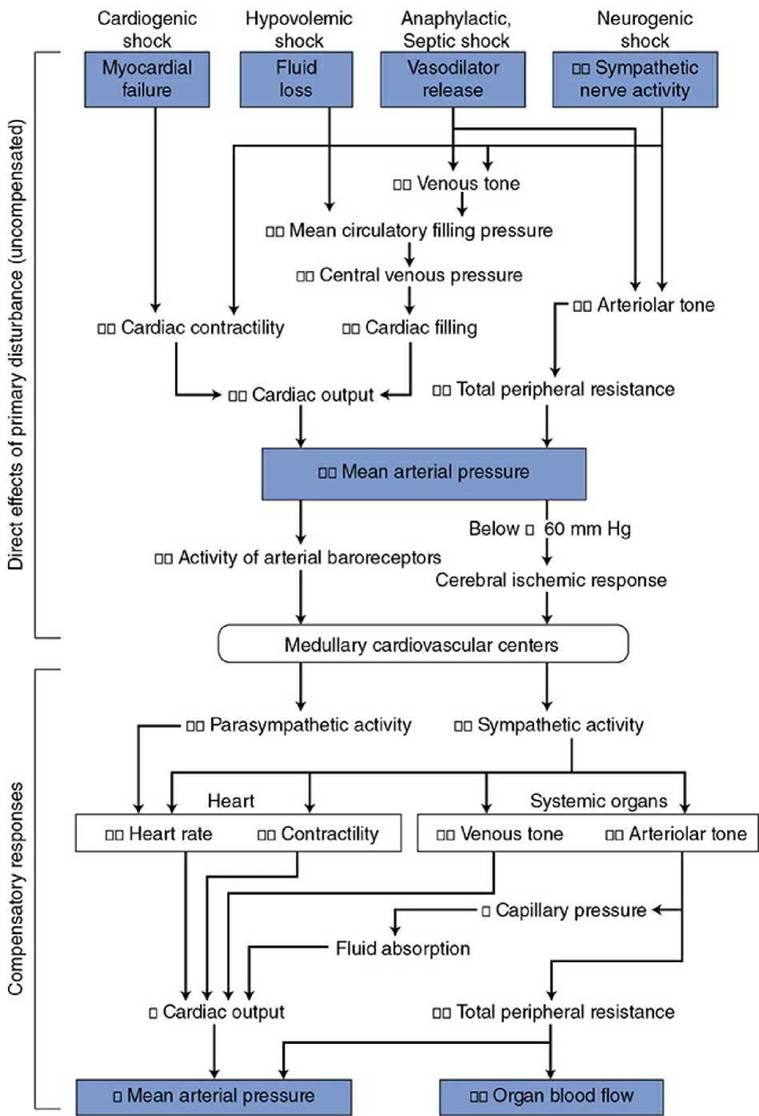
Figure 11-1. Cardiovascular alterations in shock.
1. Cardiogenic shock occurs whenever cardiac pumping ability is compromised (e.g., as a result of severe arrhythmias, abrupt valve malfunction, coronary occlusions, or myocardial infarction). The direct consequence of any of these abnormalities is a significant fall in cardiac output.
2. Hypovolemic shock accompanies significant hemorrhage (usually greater than 20% of blood volume), or fluid depletion from lack of adequate fluid intake, severe burns, chronic diarrhea, or prolonged vomiting. These situations induce shock by depleting body fluids and thus circulating blood volume. The direct consequence of hypovolemia is inadequate cardiac filling and reduced stroke volume.
Note: There are some situations that may result in reduced cardiac filling that are not related to hypovolemia. For example, cardiac tamponade associated with fluid accumulation in the pericardial sac (from pericardial infection, coronary vessel rupture, chest wounds, etc.) prevents adequate diastolic filling. Another example of reduced cardiac filling unrelated to hypovolemia is the occurrence of a pulmonary embolus (a clot mobilized from systemic veins lodging in a pulmonary vessel). In addition to potential damage to lung tissue, difficulty breathing, and severe discomfort, the disruption of left ventricular cardiac filling may result in a severe systemic shock state.
3. Anaphylactic shock occurs as a result of a severe allergic reaction to an antigen to which the patient has developed sensitivity (e.g., insect bites, antibiotics, and certain foods). This immunological event, also called an “immediate hypersensitivity reaction,” is mediated by several substances (such as histamine, prostaglandins, leukotrienes, and bradykinin) that, by multiple mechanisms, results in substantial arteriolar vasodilation, increases in microvascular permeability, and loss of peripheral venous tone. These combine to reduce both total peripheral resistance and cardiac output.
4. Septic shock is also caused by profound vasodilation but specifically from substances released into the circulating blood by infective agents. One of the most common is endotoxin, a lipopolysaccharide released from bacteria. This substance induces the formation of a nitric oxide synthase (called inducible nitric oxide synthase to distinguish it from the normally present constitutive nitric oxide synthase) in endothelial cells, vascular smooth muscle, and macrophages that then produce large amounts of the potent vasodilator, nitric oxide. The term distributive shock is sometimes used to describe both the anaphylactic and septic shock states.
5. Neurogenic shock is produced by loss of vascular tone due to inhibition of the normal tonic activity of the sympathetic vasoconstrictor nerves and often occurs with deep general anesthesia or in reflex response to deep pain associated with traumatic injuries. It may also be accompanied by an increase in vagal activity, which significantly slows the cardiac beating rate. This type of shock is often referred to a vasovagal syncope. The transient syncope evoked by strong emotions is a mild form of neurogenic shock and is usually quickly reversible.
As shown in the top half of Figure 11-1, the common primary disturbances in all forms of shock are decreased cardiac output and/or total peripheral resistance leading to decreased mean arterial pressure. Generally, the reduction in arterial pressure is substantial, and so therefore, is the influence on the cardiovascular centers from reduced arterial baroreceptor discharge rate. In addition, in the case of hypovolemic, anaphylactic, and septic shock, diminished activity of the cardiopulmonary baroreceptors due to a decrease in central venous pressure and/or volume acts on the medullary cardiovascular centers to stimulate sympathetic output. 1 If arterial pressure falls below approximately 60 mm Hg, brain blood flow begins to fall and this elicits the cerebral ischemic response. As indicated in Chapter 9, the cerebral ischemic response causes intense activation of the sympathetic nerves.
Compensatory Mechanisms
■ In general, the various forms of shock evoke the compensatory responses in the autonomic nervous system that we would expect from a fall in blood pressure. 2 These increases in sympathetic activity and decreases in parasympathetic activity are indicated in the bottom half of Figure 11-1. Cardiac and peripheral vascular compensatory responses to shock, however, may be much more intense than those that accompany more ordinary cardiovascular disturbances. Many of the commonly recognized symptoms of shock (e.g., pallor, cold clammy skin, rapid heart rate, muscle weakness, and venous constriction) are a result of greatly increased sympathetic nerve activity. When the immediate compensatory processes are inadequate, the individual may also show signs of abnormally low arterial pressure and reduced cerebral perfusion, such as dizziness, confusion, or loss of consciousness.
Additional compensatory processes initiated during the shock state may include the following:
1. Rapid and shallow breathing occurs, which promotes venous return to the heart by action of the respiratory pump.
2. Increased renin release from the kidney as a result of sympathetic stimulation promotes the formation of the hormone, angiotensin II, which is a potent vasoconstrictor and participates in the increase in total peripheral resistance even in mild shock states.
3. Increased circulating levels of vasopressin (also known as antidiuretic hormone) from the posterior pituitary gland contribute to the increase in total peripheral resistance. This hormone is released in response to decreased firing of the cardiopulmonary and arterial baroreceptors.
4. Increased circulating levels of epinephrine from the adrenal medulla in response to sympathetic stimulation contribute to systemic vasoconstriction.
5. Reduced capillary hydrostatic pressure resulting from intense arteriolar constriction reduces capillary hydrostatic pressure and promotes fluid movement from the interstitial space into the vascular space.
6. Increased glycogenolysis in the liver induced by epinephrine and norepinephrine results in a release of glucose and a rise in blood (and interstitial) glucose levels and, more importantly, a rise in extracellular osmolarity by as many as 20 mOsm. This will induce a shift of fluid from the intracellular space into the extracellular (including intravascular) space.
The latter 2 processes result in a sort of “autotransfusion” that can move as much as a liter of fluid into the vascular space in the first hour after the onset of the shock episode. This fluid shift accounts for the reduction in hematocrit that is commonly observed in hemorrhagic shock. The extent of fluid shift may be limited by a reduction in colloid osmotic pressure.
In addition to the immediate compensatory responses shown in Figure 11-1, fluid retention mechanisms are evoked by hypovolemic states that affect the situation in the long term. The production and release of the antidiuretic hormone (vasopressin) from the posterior pituitary promote water retention by the kidneys. Furthermore, activation of the renin- angiotensin-aldosterone pathway promotes renal sodium retention (via aldosterone) and the thirst sensation and drinking behavior (via angiotensin II). These processes contribute to the replenishment of extracellular fluid volume within a few days of the shock episode.
Decompensatory Processes
Often the strong compensatory responses during shock evoked by the intense sympathetic activation are capable of preventing drastic reductions in arterial pressure. However, because the compensatory mechanisms involve overwhelming arteriolar vasoconstriction, perfusion of tissues other than the heart and the brain may be inadequate despite nearly normal arterial pressure. For example, blood flow through vital organs such as the liver, gastrointestinal tract, and kidneys may be reduced nearly to zero by intense sympathetic activation. The possibility of permanent renal, hepatic, or GI tract ischemic damage is a very real concern even in seemingly mild shock situations. Patients who have apparently recovered from a state of shock may die several days later because of renal failure, uremia, or sepsis due to bacterial penetration of the weakened mucosal barrier in the GI tract.
The immediate danger with shock is that it may enter the progressive stage, wherein the general cardiovascular situation progressively degenerates, or, worse yet, enter the irreversible stage, where no intervention can halt the ultimate collapse of cardiovascular system that results in death.
The mechanisms behind progressive and irreversible shock are not completely understood. However, it is clear from the mechanisms shown in Figure 11-2 that bodily homeostasis can progressively deteriorate with prolonged reductions in organ blood flow. These homeostatic disturbances, in turn, adversely affect various components of the cardiovascular system so that arterial pressure and organ blood flow are further reduced. Note that the events shown in Figure 11-2 are decompensatory mechanisms. Reduced arterial pressure leads to alterations that further reduce arterial pressure rather than correct it (i.e., a positive feedback process). These decompensatory mechanisms that are occurring at the tissue level to lower blood pressure are eventually further compounded by a reduction in sympathetic drive and a change from vasoconstriction to vasodilation with a further lowering of blood pressure. The factors that lead to this unexpected reduction in sympathetic drive from the under-perfused medullary cardiovascular centers are not clearly understood. If the shock state is severe enough and/or has persisted long enough to enter the progressive stage, the self-reinforcing decompensatory mechanisms progressively drive arterial pressure down. Unless corrective measures are taken quickly, death will ultimately result.
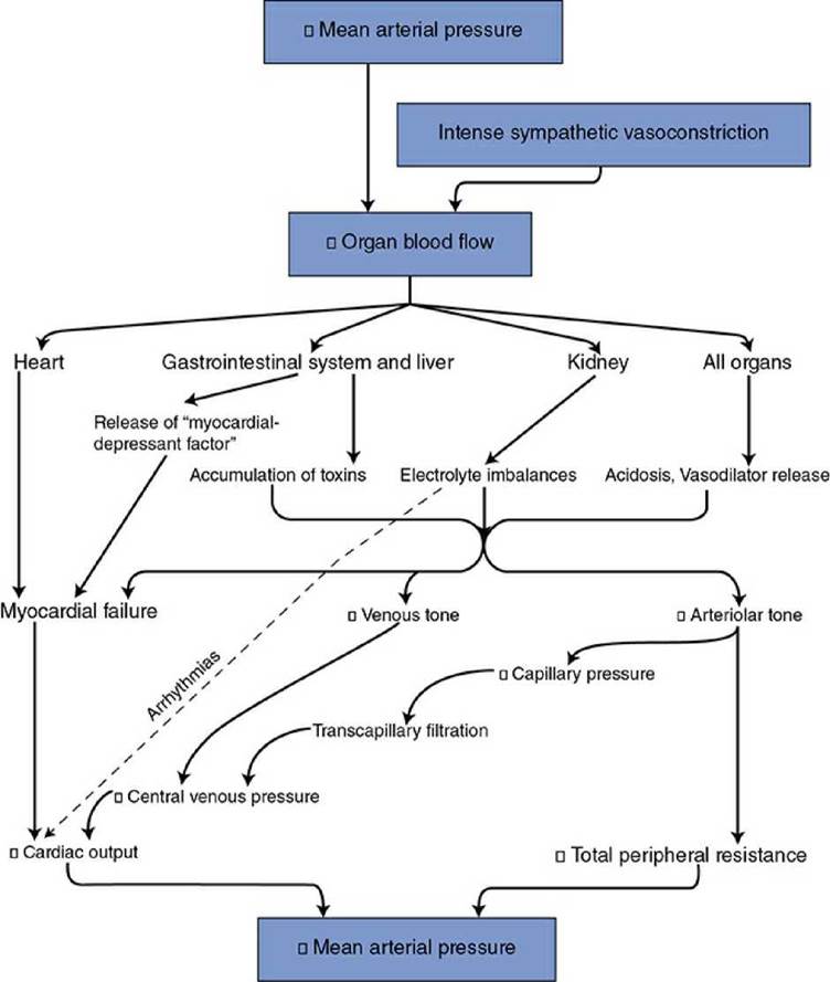
Figure 11-2. Decompensatory mechanisms in shock.
Cardiac disturbances
Coronary Artery Disease
DEFINITION AND PHYSIOLOGICAL CONSEQUENCES
Whenever coronary blood flow falls below that required to meet the metabolic needs of the heart, the myocardium is said to be ischemic and the pumping capability of the heart is impaired. The most common cause of myocardial ischemia is atherosclerotic disease of the large coronary arteries. In atherosclerotic disease, localized lipid deposits called plaques develop within the arterial walls. With severe disease, these plaques may become calcified and so large that they physically narrow the lumen of arteries (producing a chronic stenosis) and thus greatly and permanently increase the normally low vascular resistance of these large arteries. This extra resistance adds to the resistance of other coronary vascular segments and tends to reduce coronary flow. If the coronary artery stenosis is not too severe, local metabolic vasodilator mechanisms may reduce arteriolar resistance sufficiently to compensate for the abnormally large arterial resistance. Thus, an individual with coronary artery disease may have perfectly normal coronary blood flow when resting. A coronary artery stenosis of any significance will, however, limit the extent to which coronary flow can increase above its resting value by reducing maximum achievable coronary flow. This occurs because, even with very low arteriolar resistance, the overall vascular resistance of the coronary vascular bed is high if resistance in the large arteries is high.
In addition to atherosclerotic disease of large coronary arteries, there is another form of myocardial ischemia caused by abnormalities in the coronary microvasculature. Factors involved may include endothelial dysfunction and reduced flow (perhaps due to spasm) in the tiny “resistance” blood vessels of the heart. Because these changes are not characterized by major arterial blockages, they are harder to diagnose. Microvascular abnormalities were previously considered to be rather benign conditions, but studies now suggest that altered microvascular conditions are important pathophysiologic causes of ischemic heart disease especially in women.
Coronary artery disease can jeopardize cardiac function in several ways. Ischemic muscle cells are electrically irritable and unstable, and the danger of developing cardiac arrhythmias and fibrillation is enhanced. During a bout of ischemia, the normal cardiac electrical excitation pathways may be altered and often ectopic pacemaker foci develop. Electrocardiographic manifestations of myocardial ischemia may be observed in individuals with coronary artery disease during exercise stress tests. In addition, there is evidence that platelet aggregation and clotting function may be abnormal in atherosclerotic coronary arteries and the danger of thrombus or emboli formation is enhanced. It appears that certain platelet suppressants or anticoagulants such as aspirin may be beneficial in the treatment of this consequence of coronary artery disease. (The details of the blood clotting process are included in Appendix D.) Myocardial ischemia not only impairs the pumping ability of the heart but also may produce chest pain called angina pectoris. 3 Anginal pain is often absent in individuals with coronary artery disease when they are resting but is induced during physical exertion or emotional excitement. Both of these situations elicit an increase in sympathetic tone that increases myocardial oxygen consumption. Myocardial ischemia and chest pain will result if coronary blood flow cannot keep pace with the increase in myocardial metabolism.
Diagnosis
Coronary artery imaging techniques (described in Chapter 4) have proven very useful for determining the extent of coronary artery disease in the large arteries. For example, calcification of plaques (which is a significant indicator of advanced atherosclerosis) can be assessed noninvasively with specialized CT scans or magnetic resonance imaging. Specific information about the site(s) and degree of narrowing of the major coronary vessels can also be obtained invasively by angiography with injection of a radiopaque dye directly into the coronary arteries.
Treatments
Primary treatment of chronic coronary artery disease (and atherosclerosis, in general) should include attempts to lower blood lipids by dietary and pharmacological techniques that prevent (and possibly reverse) further development of the plaques. There are a number of recent strategies proven to be quite helpful. (1) Statins are well-accepted effective lipid- lowering drugs acting by blocking cholesterol production in the liver. (2) Newer drugs that prevent normal inactivation of certain lipid uptake receptors (LDL receptors in the liver) promote lipid removal from the circulation. (3) Because atherosclerotic heart disease is now recognized as a chronic inflammatory vascular disease, new anti-inflammatory drugs are being developed and show promising effects for treatment of plaque reduction and prevention of infarction. The interested student should consult medical biochemistry and pharmacology texts for a complete discussion of this very important topic.
Treatment of chest pain that is a result of coronary artery disease may involve several different pharmacological approaches. First, quick-acting vasodilator drugs such as nitroglycerin may be used to provide primary relief from an acute anginal attack. In addition to increasing myocardial oxygen delivery by dilating coronary vessels, nitrates reduce myocardial oxygen demand by dilating systemic veins, thereby reducing venous return, central venous filling, and cardiac preload, and by dilating systemic arterioles, which reduces arterial resistance, arterial pressure, and cardiac afterload. 4 Second, p-adrenergic blocking agents such as propranolol may be used to block the effects of cardiac sympathetic nerves on the heart rate and contractility. These agents limit myocardial oxygen consumption and prevent it from increasing above the level that the compromised coronary blood flow can sustain. Third, calcium channel blockers such as verapamil may be used to dilate coronary and systemic blood vessels. These drugs, which block entry of calcium into the vascular smooth muscle cell, interfere with normal excitation-contraction coupling. They have been found to be useful for treating the type of angina caused by vasoconstrictive spasms of large coronary arteries (Prinzmetal angina).
Invasive or surgical interventions are commonly used to eliminate a chronic coronary artery stenosis. X-ray techniques combined with radiopaque dye injections can be used to visualize a balloon-tipped catheter as it is threaded into the coronary artery to the occluded region. Rapid inflation of the balloon squeezes the plaque against the vessel wall and improves the patency of the vessel ( coronary angioplasty). A small tube-like expandible device called a stent is often implanted inside the vessel at the angioplasty site. This rigid implant promotes continued patency of the vessel over a longer period than angioplasty alone. Drugeluting stents are often used to slowly deliver drugs to the local area that limit the growth of neointimal scar formation which may lead to restenosis. If angioplasty and stent placement are inappropriate or unsuccessful, coronary bypass surgery may be performed. The stenotic coronary artery segments are bypassed by implanting parallel low- resistance pathways formed from either natural (e.g., saphenous vein or mammary artery) or artificial vessels.
Acute Coronary Occlusion—Myocardial Infarction
Myocardial infarction is a sudden ischemic crisis of cardiac tissue associated with acute interruption in coronary blood flow. This is most often a result of thrombotic occlusion of a coronary artery caused by clot formation at the site of a ruptured atherosclerotic plaque but may also result from the sudden arrival of an embolism released from a clot perhaps formed in a fibrillating atrium. Either of these events may abruptly occlude or significantly narrow a major coronary artery and its subsequent downstream flow. This is indeed a crisis situation and demands immediate attention. The physiological consequences of such an immediate occlusion are discussed in the preceding text under the topic of “Cardiogenic shock.” Treatments may include emergency coronary angioplasty (described earlier) with the placement of a stent, which may allow immediate restoration of flow to the ischemic area. Another method for treatment of acute myocardial infarction is the intravascular injection of thrombolytic substances (e.g., streptokinase or tissue plasminogen-activating factors) that dissolve blood clots. This approach is most successful when these “clot busters” are given within a few hours of the infarction.
To emphasize the 2 basic underlying physiological principles for treatment of an acute coronary event: (1) improve coronary blood flow to increase oxygen delivery and (2) minimize cardiac work to lower oxygen demand (by reducing preload, afterload, and contractility).
Cardiomyopathies
A cardiomyopathy is an abnormality of the cardiac muscle that occurs even though coronary perfusion is usually adequate. They are commonly further classified by specific structural and functional changes as noted below.
Dilated cardiomyopathy (DCM) is characterized by cardiac chamber enlargement with normal ventricular wall thickness. These patients may develop heart failure with a reduced ejection fraction. This type of cardiomyopathy is often a delayed consequence of infections (e.g., herpes, coxsackie, influenza, human immunodeficiency virus), cardiac toxins (e.g., alcohol, cocaine, amphetamine, cancer chemotherapeutics), and other chronic systemic challenges. Familial predisposition has also been noted and genetic coding for multiple protein abnormalities have been identified including those of the sarcomere, cytoskeleton, ion channels, and extracellular matrix proteins. Abnormalities in the giant protein, titin, that controls sarcomere stiffness, accounts for about 20% of familial cases of dilated cardiomyopathy.
Hypertrophic cardiomyopathy (HCM) is the most common genetic heart disease and characterized by a significant, often asymmetrical, left ventricular wall thickening often leading to ventricular outflow obstruction. This hypertrophy is associated with a disruption of normal muscle cell alignment and with various electrical malfunctions. It is often asymptomatic and is a leading cause of ventricular fibrillation and sudden cardiac death in young athletes. Because of its genetic predisposition, echocardiographic prescreening for susceptible populations may be lifesaving.
Takotsubo cardiomyopathy (aka stress cardiomyopathy or broken heart syndrome) is associated with a sudden, temporary, and often localized weakening of the ventricular musculature that is triggered by emotional stress or constant anxiety. Although it is classified as a non-ischemic form of cardiomyopathy in that no overt vascular occlusions are identifiable, it may be associated with transient (and reversible) vascular spasm or microcirculatory malfunction. Release of various stress hormones may also produce regional “stunning” of the cardiomyocytes so that pumping is impaired. It differs from an acute myocardial infarction in that it seems to be completely reversible with time and treatment. It is a now well- recognized cause of acute heart failure, lethal arrhythmias, and ventricular rupture.
Chronic Heart Failure
Heart (or cardiac or myocardial) failure is said to exist whenever ventricular function is depressed through myocardial damage, insufficient coronary flow, or any other condition that directly impairs the mechanical performance of the heart muscle. Cardiac failure can be categorized as being associated with systolic abnormalities (“Heart Failure with Reduced Ejection Fraction”—aka HFrEF) or with diastolic filling abnormalities (“Heart Failure with Preserved Ejection Fraction”—aka HFpEF).
Systolic heart failure (HFrEF)— By definition, systolic heart failure is associated with a left ventricular ejection fraction of less than 40%. This also implies that the heart is operating on a lower-than-normal cardiac function curve, that is, a reduced cardiac output at any given filling pressure. Acute heart failure has already been discussed in the context of sudden coronary artery occlusion, cardiogenic shock, and as part of the decompensatory mechanisms operating in progressive and irreversible shock. Often, however, sustained cardiac “challenges” may induce a chronic state of heart failure. Such challenges might include (1) progressive coronary artery disease, (2) sustained elevation in cardiac afterload as that which accompanies arterial hypertension or aortic valve stenosis, or (3) reduced functional muscle mass or contractility as a consequence of various cardiomyopathies or following myocardial infarction. Regardless of the precipitating cause, most forms of systolic failure are associated eventually with a reduced myocyte contractile function. Many specific structural, functional, and biochemical myocyte alterations accompany severe systolic heart failure. Some of the more well-documented abnormalities include (1) reduced calcium sequestration by the sarcoplasmic reticulum and upregulation of the sarcolemmal Na/Ca exchanger (leading to low intracellular calcium levels for excitationcontraction coupling), (2) low affinity of troponin for calcium (leading to reduced cross-bridge formation and contractile ability), (3) altered substrate metabolism from fatty acid to glucose oxidation, and (4) impaired respiratory chain activity (leading to impaired energy production).
The primary disturbance in systolic heart failure (acute or chronic) is depressed cardiac output and thus lowered arterial pressure. Consequently, all the compensatory responses important in shock ( Figure 11-1) are also important in heart failure. In chronic heart failure, however, the cardiovascular disturbances may not be sufficient to produce a state of shock. Moreover, long-term compensatory mechanisms are especially important in chronic heart failure.
The circumstances of chronic systolic heart failure are well illustrated by cardiac output and venous function curves such as those shown in Figure 11-3. The normal cardiac output and normal venous function curves intersect at point A in Figure 11-3. A cardiac output of 5 L/min at a central venous pressure of less than 2 mm Hg is indicated by the normal operating point (A). With heart failure, the heart operates on a much lower-than-normal cardiac output curve. Thus, acute heart failure alone (uncompensated) shifts the cardiovascular operation from the normal point (A) to a new position, as illustrated by point B in Figure 11-3—that is, cardiac output falls below normal while central venous pressure rises above normal. The decreased cardiac output leads to decreased arterial pressure and reflex activation of the cardiovascular sympathetic nerves. Increased sympathetic nerve activity tends to (1) increase heart rate and cardiac contractility to raise the cardiac function curve toward normal and (2) increase peripheral venous pressure through venous constriction, and thus raise the venous function curve above normal. Cardiovascular operation will shift from point B to point C in Figure 11-3. Thus, the depressed cardiac output is substantially improved by the immediate consequences of increased sympathetic nerve activity. Note, however, that the cardiac output at point C is still below normal. The arterial pressure associated with cardiovascular operation at point C is likely to be near normal, however, because higher-than-normal total peripheral resistance will accompany higher-than-normal sympathetic nerve activity.
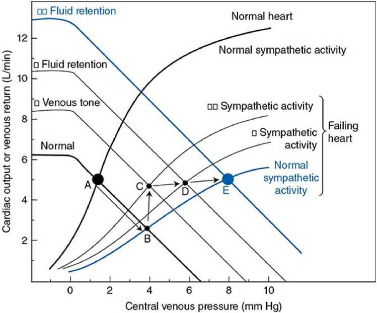
Figure 11-3. Cardiovascular alterations with compensated chronic systolic heart failure.
In the long term, cardiovascular operation cannot remain at point C in Figure 11-3. Operation at point C involves higher-than-normal sympathetic activity, and this will inevitably cause a gradual increase in blood volume by the mechanisms that are described in Chapter 9. Over several days, there is a progressive rise in the venous function curve as a result of increased blood volume and, consequently, increased mean circulatory filling pressure. Recall that this process involves a sympathetically induced release of renin from the kidney, which activates the renin-angiotensin-aldosterone system that promotes fluid retention.
This will progressively shift the cardiovascular operating point from C to D to E, as shown in Figure 11-3.
Note that increased fluid retention (C -+ D -+ E in Figure 11-3) causes a progressive increase in cardiac output toward normal and simultaneously allows a reduction in sympathetic nerve activity toward the normal value. Reduced sympathetic activity is beneficial for several reasons. First, decreased arteriolar constriction permits renal and splanchnic blood flow to return toward more normal values. Second, myocardial oxygen consumption may fall as sympathetic nerve activity falls, even though cardiac output tends to increase. Recall that the increased heart rate and increased cardiac contractility caused by sympathetic nerve activation greatly increase myocardial oxygen consumption. Reduced myocardial oxygen consumption is especially beneficial in situations where inadequate coronary blood flow is the cause of the heart failure. In any case, once enough fluid has been retained so that a normal cardiac output can be achieved with normal sympathetic nerve activity, the individual is said to be in a “compensated” state of chronic heart failure. 5 Note that the primary disturbance (i.e., the depressed cardiac function curve) is still present. Fluid retention did nothing to correct this. However, fluid retention does allow a “normal” cardiac output to now be achieved at the higher-than-normal central venous (i.e., cardiac filling) pressure.
Unfortunately, the consequences of fluid retention in chronic cardiac failure are not all beneficial. Note that in Figure 11-3 fluid retention (C -+ D -+ E) will cause both peripheral and central venous pressures to be much higher than their normal values. Chronically high central venous pressure causes chronically increased end-diastolic volume (cardiac dilation). Up to a point, cardiac performance is very much improved by increased cardiac filling volume. Excessive cardiac dilation, however, can impair cardiac function because increased total wall tension is required to generate pressure within an enlarged ventricular chamber ( T = P x r; Chapter 2). This increases the myocardial oxygen demand.
The high venous pressure associated with fluid retention also adversely affects organ function because high venous pressure produces transcapillary fluid filtration, edema formation, and congestion (hence the commonly used term congestive heart failure). Left-sided heart failure is accompanied by pulmonary edema with dyspnea (shortness of breath) and respiratory crisis. 6 Right-sided heart failure is associated with distended neck veins, ankle edema, and fluid accumulation in the abdomen (ascites) with liver congestion and dysfunction. 7
In the example shown in Figure 11-3, the depression in the cardiac output curve because of heart failure is only moderately severe. Thus, it is possible, through moderate fluid retention, to achieve a normal cardiac output with essentially normal sympathetic activity (point E). The situation at point E is relatively stable because the stimulus for further fluid retention (i.e., increased sympathetic drive) has been removed. If, however, the heart failure is more severe, the cardiac output curve may be so depressed that normal cardiac output cannot be achieved by any amount of fluid retention. In these cases fluid retention is extremely marked, as is the elevation in venous pressure, and the complications of congestion are very serious problems.
Another way of looking at the effects of left ventricular cardiac failure is given in Figure 11-4. The left ventricular pressure-volume loops describing the events of a cardiac cycle from a failing heart are displaced far to the right of those from normal hearts. The untreated patient described in this figure is in serious trouble with a reduced stroke volume and ejection fraction and high filling pressure. Furthermore, the slope of the line describing the end-systolic pressure-volume relationship is shifted downward and is less steep, indicating the reduced contractility of the cardiac muscle. However, because of this flatter relationship, small reductions in cardiac afterload (i.e., arterial blood pressure) will produce substantial increases in stroke volume, as indicated in Figure 11-4, and will significantly help this patient.
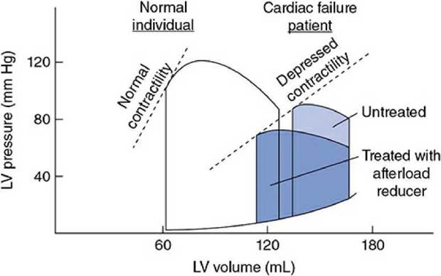
Figure 11-4. Left ventricular pressure-volume loops showing systolic heart failure (loops to the right) characterized by depressed contractility, increased end-diastolic volume, and reduced ejection fraction.
As might be expected from the previous discussion, the most common symptoms of patients with congestive heart failure are associated with the inability to increase cardiac output (low exercise tolerance and fatigue) and with the compensatory fluid accumulation (tissue congestion, shortness of breath, and peripheral swelling). In severe cases, the ability of the cardiac cells to respond to increases in sympathetic stimulation is diminished by a reduction in the effective number (downregulation) of the myocyte p 1- adrenergic receptors. This further reduces the ability of the myocytes to increase their contractility as well as the ability of the heart to increase its beating rate in response to sympathetic stimulation. Thus, low maximal heart rates contribute to the reduced exercise tolerance.
Treatment of the patient with congestive systolic heart failure is a difficult challenge. Treatment of the precipitating condition is of course the ideal approach, but often this cannot be done effectively. Cardiac glycosides (e.g., digitalis) 8 have been used to improve cardiac contractility (i.e., to increase the ejection fraction, shift the cardiac function curve upward, increasing contractile force of the myocyte at any given starting length). 9 These drugs are unfortunately quite toxic and often have undesirable side effects.
Treatment of the congestive symptoms involves balancing the need for enhanced cardiac filling with the problems of too much extracellular fluid. Drugs that promote renal fluid loss (diuretics such as furosemide or thiazides) are extremely helpful as are the angiotensin-converting enzyme (ACE) inhibitors and the angiotensin II receptor blockers (ARBs). 10 A potent diuretic can quickly save a patient from drowning in the pulmonary exudate and reduce diastolic volume of the dilated heart to acceptable levels, but it can also lower blood pressure to dangerous levels.
Chronic heart failure patients often have elevated sympathetic drive if they have not completely compensated for the depressed cardiac function by fluid retention and increased blood volume. Although high sympathetic drive is an important initial compensatory mechanism, the energy cost of a chronically elevated heart rate and contractility can put the heart with diminished coronary circulation at a disadvantage. Therefore, treatment with β-adrenergic receptor blockers can reduce metabolic demand to a level more easily met by a compromised vascular supply. Again, if elimination of the effect of elevated sympathetic drive on the heart is too aggressive, cardiac output may fall and worsen a failure state.
Diastolic heart failure (HFpEF)— Up to one-half of patients with heart failure have normal or near normal (i.e., “preserved”) ejection fraction (>50%). In these cases, the problem is not with the systolic pressure-developing processes but rather with the diastolic relaxation and passive distension processes. As shown in Figure 11-5, diastolic dysfunction implies a stiffened heart during diastole such that increases in cardiac filling pressure do not produce normal increases in end-diastolic volume.
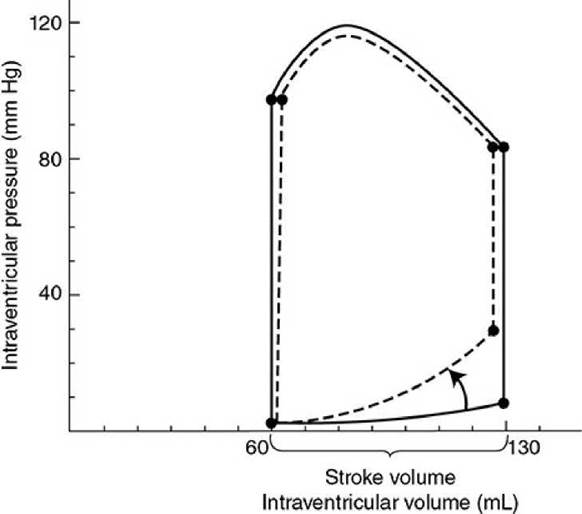
Figure 11-5. Left ventricular pressure-volume loops showing diastolic heart failure ( dashed lines) characterized by increased diastolic stiffness, increased end-diastolic pressure, and normal ejection fraction.
Some individuals (primarily elderly patients with hypertension and cardiac hypertrophy) who have some symptoms of cardiac failure (exertional dyspnea, fluid retention, pulmonary edema, and high end- diastolic pressures) seem to have normal systolic function (ejection fractions >50%), and normal or even reduced ventricular end-diastolic volumes despite increased cardiac filling pressure. Thus, the terms diastolic heart failure and heart failure with preserved systolic function have been used to describe this situation.
This condition is associated with many common risk factors (hypertension, atrial fibrillation, diabetes or metabolic syndrome, older age, female sex, inactivity, renal dysfunction, and/or obesity). Most of the time, angiographic evaluation of the coronary circulation does not show significant large vessel disease. The symptoms mimic those of systolic heart failure (i.e., with reduced ejection fraction) (e.g., exertional dyspnea, fatigue, congestion, chest pain). Some degree of diastolic dysfunction is also commonly present in patients with reduced ejection fraction and may precede systolic problems.
Potential causes of altered diastolic properties in heart failure include: (1) decreased cardiac tissue passive compliance due to extracellular remodeling, collagen cross-linking, and other extracellular matrix protein alterations often associated with left ventricular hypertrophy resulting from hypertension, (2) loss of myocardial elastic recoil that is partly responsible for early ventricular filling after systole, (3) increased myofibrillar passive stiffness due to alterations in the myofibrillar giant protein titin, (4) delayed myocyte relaxation early in diastole 238due to slow cytosolic calcium removal processes, (5) inadequate adenosine triphosphate levels required to disconnect the myofilament cross-bridges rapidly, and (6) residual, low-grade cross-bridge cycling during diastole due to calcium leaking from the sarcoplasmic reticulum. Metabolic comorbidities (diabetes, obesity) may trigger a systemic inflammatory state that leads to microvascular endothelial dysfunction allowing inflammatory cells to enter the myocardium. These trigger various paracrine signals that initiate various processes that lead to the passive compliance changes.
At this point, therapeutic strategies that directly influence diastolic properties are not well developed. Attempts to reduce interstitial fibrosis (with ACE inhibitors and/or angiotensin receptor antagonists) and to reduce diastolic calcium leak from the sarcoplasmic reticulum (with β- adrenergic blockers) have had limited success. Correcting underlying comorbidities and treatment with anti-inflammatory therapies are possibilities. Reduction of afterload seems to be most helpful, especially if it reduces left ventricular hypertrophy. In any case, “heart failure with preserved ejection fraction” is currently difficult to diagnose and to effectively treat.
Hypertension
Hypertension is defined as a chronic elevation in arterial blood pressure and can exist in either the pulmonary or the systemic vascular system.
Pulmonary hypertension is less common than systemic hypertension and less is known about its causes, progression, and treatments. Pulmonary hypertension is designated when mean pulmonary artery pressure is greater than 25 mm Hg. It can be caused by either structural alterations in the lung vasculature (associated with smoking, environmental toxins, left heart failure with pulmonary congestion) or by altered vascular smooth muscle reactivity. Right-sided heart failure resulting from chronic pulmonary hypertension is called cor pulmonale. Although there is a genetic component to its incidence, it seems to be highly correlated with conditions involving chronic hypoxia (e.g., chronic obstructive pulmonary disease, cystic fibrosis, and pneumoconiosis). 11 The increased prevalence of obesity with accompanying obstructive sleep apnea may account for a recent increase in reported incidence of this disease. It is relatively hard to diagnose until symptoms appear as the consequences of the elevated pulmonary arterial pressures. These symptoms may include systemic edema, pulmonary congestion, shortness of breath, chest pain, and fatigue.
There is no cure for pulmonary hypertension at present and strategies used for treatment of systemic hypertension have had little or no effect on the pulmonary bed. However, several pharmacological approaches are proving to be useful: (1) endothelin receptor antagonists block the biological activities of the vasoconstrictor, endothelin; (2) phosphodiesterase-5 inhibitors prevent breakdown of cyclic guanosine monophosphate, thus promoting the vasorelaxing effects of nitric oxide; and (3) prostacyclin derivatives provide an exogenous supply of the vasodilator, prostacyclin, or drugs that activates a prostacyclin receptor.
Systemic hypertension is defined as an elevation of mean systemic arterial pressure above 140/90 mm Hg. It is an extremely common cardiovascular problem, affecting more than 20% of the adult population of the western world. It has been established beyond doubt that hypertension increases the risk of coronary artery disease, myocardial infarction, heart failure, stroke, and many other serious cardiovascular problems. Moreover, it has been clearly demonstrated that the risk of serious cardiovascular incidents is reduced by proper treatment of hypertension. 12
In approximately 90% of cases, the primary abnormality that produces high blood pressure is unknown. This condition is sometimes referred to as primary or essential hypertension because the elevated level was thought to be “essential” to drive the blood through the systemic circulation. In the remaining 10% of hypertensive patients, the cause can be traced to a variety of sources, including epinephrine-producing tumors (pheochromocytomas), aldosterone-producing tumors (in primary hyperaldosteronism), certain forms of renal disease (e.g., renal artery stenosis, glomerular nephritis, and toxemia of pregnancy), certain neurological disorders (e.g., brain tumors that increase intracranial pressure), certain thyroid and parathyroid disorders, aortic coarctation, lead poisoning, drug side effects, abuse of certain drugs, obstructive sleep apnea, or even unusual dietary habits. The high blood pressure that accompanies such known causes is referred to as secondary hypertension. Most often, however, the true cause of the hypertension remains a mystery, and it is only the symptom of high blood pressure that is treated.
Facts about Systemic Hypertension
In the midst of an enormous amount of information about systemic hypertension, a few universally accepted facts stand out:
1. Genetic factors contribute importantly to the development of hypertension. Familial tendencies for high blood pressure are well documented. In addition, hypertension is generally more common in men than in women and racial differences in incidence have been identified.
2. Chronic conditions such as obesity, diabetes, kidney disease, and sleep disorders are strongly associated with systemic hypertension.
3. Environmental factors or behaviors can greatly influence the development of hypertension. Physical inactivity, use of tobacco, excess alcohol consumption, high-sodium or low-potassium diets, and/or certain forms of psychological stress may either aggravate or precipitate hypertension in genetically susceptible individuals.
4. Structural changes in the left side of the heart and arterial vessels occur in response to systemic hypertension. Early alterations include hypertrophy of muscle cells and thickening of the walls of the ventricle and systemic resistance vessels. Late changes associated with deterioration of function include increases in connective tissue and loss of elasticity.
5. The established phase of hypertension is associated with an increase in total peripheral resistance. Cardiac output and/or blood volume may be elevated during the early developmental phase, but these variables are usually normal after the hypertension is established.
6. The increased total peripheral resistance associated with established hypertension may be due to (a) rarefaction (decrease in density) of microvessels, (b) pronounced structural adaptations that occur in the peripheral vascular bed, (c) continuously increased activity of the vascular smooth muscle cells, 13 (d) increased sensitivity and reactivity of the vascular smooth muscle cells to external vasoconstrictor stimuli, an/or (e) diminished production and/or effect of endogenous vasodilator substances (e.g., nitric oxide).
7. The chronic elevation in blood pressure does not appear to be due to a sustained elevation in sympathetic vasoconstrictor neural discharge nor is it due to a sustained elevation of any blood-borne vasoconstrictive factor. (Both neural and hormonal influences, however, may help initiate primary hypertension.)
8. Blood pressure-regulating reflexes (both the short-term arterial and cardiopulmonary baroreceptor reflexes and the long-term, renal- dependent, pressure-regulating reflexes) become adapted or “reset” to regulate blood pressure at a higher-than-normal level.
9. Disturbances in renal function contribute importantly to the development and maintenance of primary hypertension. Recall that the urinary output rate is influenced by arterial pressure, and, in the long term, arterial pressure can stabilize only at the level that makes urinary output rate equal to fluid intake rate. As shown by point N in Figure 11-6, this pressure is approximately 100 mm Hg in a normal individual.
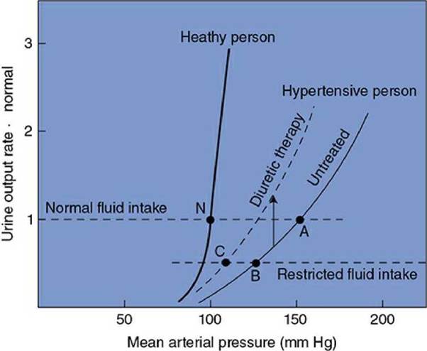
Figure 11-6. Renal function curves in healthy and hypertensive people.
All forms of hypertension involve an alteration somewhere in the chain of events by which changes in arterial pressure produce changes in urinary output rate (see Figure 9-6) such that the renal function curve is shifted rightward, as indicated in Figure 11-6. The important feature to note is that higher-than-normal arterial pressure is required to produce a normal urinary output rate in a hypertensive individual. Although this condition is always present with hypertension, it is not clear whether it could be the common cause of hypertension or simply another one of the many adaptations to it.
Consider that the untreated hypertensive individual in Figure 11-6 would have a very low urinary output rate at the normal mean arterial pressure of 100 mm Hg. Recall from Figure 9-5 that whenever the fluid intake rate exceeds the urinary output rate, fluid volume must rise and consequently so will cardiac output and mean arterial pressure. With a normal fluid intake rate, this untreated hypertensive patient will ultimately stabilize at point A (mean arterial pressure 150 mm Hg). Recall from Chapter 9 that the baroreceptors adapt within days so that they have a normal discharge rate at the prevailing average arterial pressure. Thus, once the hypertensive individual has been at point A for a week or more, even the baroreceptor mechanism will begin resisting acute changes from the 150-mm Hg pressure level.
A most important fact to realize is that, although either high cardiac output or high total peripheral resistance must always ultimately sustain high blood pressure, neither needs be the primary cause of the hypertension. A shift in the relationship between arterial pressure and urinary output rate, as illustrated in Figure 11-6, however, will always produce hypertension. The possibility that the kidneys actually “set” the blood pressure is supported by evidence accumulating from kidney transplant studies. In these studies, the blood pressure is shown to “follow” the kidney (i.e., putting a hypertensive kidney in a normotensive individual produces a hypertensive individual, whereas putting a normotensive kidney in a hypertensive individual produces a normotensive individual). Further support for an essential role of the kidney comes from recent studies showing that catheter-based, high-frequency radiowave ablation of renal sympathetic nerves effectively reduces hypertension in drug-resistant patients.
Therapeutic Strategies for Treatment of Systemic
Hypertension
In certain hypertensive individuals, restricting salt intake produces a substantial reduction in blood pressure because of the reduced requirement for water retention to osmotically balance the salt load. In the example in Figure 11-6, this effect is illustrated by a shift from point A to point B. The efficacy of lowering salt intake to lower arterial pressure depends heavily on the slope of the renal function curve in the hypertensive individual. The arterial pressure of a healthy individual, for example, is affected only slightly by changes in salt intake because the normal renal function curve is so steep.
A second common treatment of hypertension is diuretic therapy. Many diuretic drugs are available, but most have the effect of inhibiting renal tubular salt (and therefore fluid) reabsorption. The net effect of diuretic therapy, as shown in Figure 11-6, is that the urinary output rate for a given arterial pressure is increased; that is, diuretic therapy raises the renal function curve. The combined result of restricted fluid intake and diuretic therapy for the hypertensive individual in Figure 11-6 is illustrated by point C.
A third therapeutic intervention is treatment with β-adrenergic blockers that inhibit sympathetic influences on the heart and renal renin release. This approach is most successful in hypertensive patients who have high circulating renin levels.
A fourth antihypertensive strategy is to block the effects of the reninangiotensin system either with ACE inhibitors blocking the formation of the vasoconstrictor angiotensin II or with angiotensin II receptor blockers. Other pharmacological interventions may include use of a-adrenergic receptor blockers, which prevent the vasoconstrictive effects of catecholamines, and calcium channel blockers, which act directly to decrease vascular smooth muscle tone.
Alterations in lifestyle, including reduction of stress, decreases in caloric intake, limitation of the amount of saturated fats in the diet, and establishment of a regular exercise program, may help reduce blood pressure in certain individuals.
The possibility of catheter-based renal sympathetic denervation for treatment of refractory hypertension is quite intriguing and may become another approach to handling this condition in the future.
Perspectives
Medical knowledge is constantly expanding and it is difficult, if not impossible, to keep up. However, we hope that this chapter’s overview of some common cardiovascular diseases will reassure the student that memorizing everything is not necessary. Understanding the basic principles of normal cardiovascular operation should provide a firm foundation for identifying the underlying abnormalities, distinguishing the primary disturbances from the compensatory responses, understanding the mechanisms responsible for the symptoms, and appropriately treating the condition. What the future brings with new mechanistic insights, new diagnostic techniques, and new strategies for treatment (or better yet, for prevention) of these diseases will most assuredly be built on these basic underlying principles.
Key concepts
Circulatory shock is defined as a generalized, severe reduction in tissue blood flow so that metabolic needs are not met.
The primary disturbances that can lead to shock can be categorized as those that directly interfere with pump function, those that interfere with ventricular filling, or those that cause sustained vascular dilation.
Shock is usually accompanied by a compensatory increase in sympathetic activity aimed at maintaining arterial pressure via augmented cardiac output and vascular resistance.
Decompensatory processes precipitated by the shock state are generally caused by inadequate tissue blood flow, loss of local homeostasis, and tissue damage leading to a progressive and irreversible fall in arterial pressure.
Coronary artery disease, usually associated with development of atherosclerotic plaques, results in a progressive compromise in coronary blood flow that becomes inadequate to meet the tissue’s metabolic needs.
Acute heart failure due to an abrupt blockage of a major coronary artery is a form of cardiogenic shock.
Cardiomyopathies are primarily associated with deficits in myocyte function rather than inadequate coronary blood flow.
Systolic heart failure is defined as a reduction of cardiac muscle contractility and results in a depressed cardiac output at all preloads with reduced ejection fraction. Compensatory fluid retention mechanisms are evoked in heart failure to improve cardiac filling, but when fluid retention is excessive, congestive complications arise (e.g., pulmonary edema and abdominal ascites).
Diastolic dysfunction resulting from reduced cardiac compliance and impaired diastolic filling may precipitate heart failure even though ejection fraction is preserved.
Pulmonary hypertension that accompanies acute or chronic conditions of hypoxia is relatively uncommon but can lead to right-sided heart failure (cor pumonale).
Systemic hypertension is a common and serious condition influenced by multiple genetic and environmental factors and is usually associated with chronic elevation in total peripheral resistance.
Study questions
11-1. Clinical signs of hypovolemic shock often include pale and cold skin, dry mucous membranes, weak but rapid pulse, muscle weakness, and mental disorientation or unconsciousness. What are the physiological conditions that account for these signs?
11-2. Which of the following would be helpful to hemorrhagic shock victims?
a. Keep them on their feet.
b. Warm them up.
c. Give them fluids to drink.
d. Maintain their blood pressure with catecholamine-type drugs.
11-3. What happens to hematocrit?
a. during hypovolemic shock resulting from prolonged diarrhea
b. during acute cardiogenic shock
c. during septic shock
d. with chronic bleeding
11-4. Left ventricular chamber enlargement with congestive heart failure increases the wall tension required to generate a given systolic pressure. True or false?
11-5. Why are diuretic drugs often helpful in treating patients with congestive heart failure?
11-6. What is the potential danger of vigorous diuretic therapy for the patient with heart failure?
11-7. Why does renal artery stenosis produce hypertension?
11-8. Your 70-year-old, 70-kg patient has an ejection fraction of 70%. Left ventricular end-diastolic volume is 100 mL. Which of the following statements best fits these data?
a. Stroke volume is approximately 30 mL.
b. Left ventricular end-systolic volume is approximately 70 mL.
c. Your patient may be dehydrated.
d. Your patient may be suffering from chronic systolic heart failure.
e. These are normal values for someone this age.
11-9. All the following are compensatory processes that help maintain circulation during states of hypovolemic shock except
a. hepatic glycogenolysis to increase extracellular glucose concentration.
b. rapid respiratory effort to promote venous return of blood to the heart.
c. vasoconstrictive contributions from increases in circulating epinephrine.
d. autotransfusion of interstitial fluid into capillary beds.
e. increased blood flow to the kidney.
11-10. Predict the status of each of the following variables in most individuals with chronic systemic hypertension.
a. cardiac output
b. heart rate
c. arterial pulse pressure
d. total peripheral resistance
e. renal urinary output
11-11. Compression stockings are often used by people with congestive heart failure. Why?
11-12. Would compression stockings be helpful for someone who suffers from leg pain after walking a short distance?
11—13. Would compression stockings be helpful for someone with a large clot in a major vein (deep vein thrombosis).
1 In the case of cardiogenic shock, central venous pressure will increase; and in the case of neurogenic shock, central venous pressure cannot be predicted because both cardiac output and venous return are likely to be depressed. Thus, in these instances, it is not clear how the cardiopulmonary baroreceptors affect autonomic output.
2 Two primary exceptions to this statement include (1) neurogenic shock, where reflex responses may be absent or lead to further depression of blood pressure and (2) certain instances of cardiogenic shock associated with inferior or posterior wall myocardial infarctions, which elicit a reflex bradycardia and decrease sympathetic drive as well as apnea (the Bezold-Jarisch reflex).
3 Definition: “a strangling feeling in the chest.” Women are more likely to describe the ischemic episode as intense pressure, squeezing or chest discomfort, and men are more likely to describe it as intense, debilitating pain.
4 If blood pressure drops significantly, the beneficial effects of improved coronary circulation and reduced work load may be negated by a reflex increase in heart rate.
5 The extracellular fluid volume remains expanded after reaching the compensated state even though sympathetic activity may have returned to near-normal levels. Net fluid loss requires a period of less-than-normal sympathetic activity, which does not occur. For reasons not well understood, the cardiopulmonary baroreceptor reflexes apparently become less responsive to the increased central venous pressure and volume associated with heart failure.
6 Patients often complain of difficulty breathing especially during the night (paroxysmal nocturnal dyspnea). Being recumbent promotes a fluid shift from the extremities into the central venous pool and lungs, making the patient’s pulmonary problems worse. Such patients often sleep more comfortably when propped up.
7 Plasma volume expansion along with abnormal liver function reduces the concentration of plasma proteins by as much as 30%. This reduction in plasma oncotic pressure contributes to the development of interstitial edema that accompanies congestive heart failure.
8 A “tea” made from the leaves of the foxglove plant (Digitalis purpurea) was used for centuries as a common folk remedy for the treatment of “dropsy” (congestive heart failure with significant peripheral edema). With the formal recognition of its medicinal benefits in the late 18th century by the English physician Sir William Withering, digitalis became a valuable official pharmacological tool.
9 The mechanism of cardiac glycoside action is thought to involve the inhibition of the sodium/potassium adenosine triphosphatase (Na+/K+-ATPase) leading to increases in intracellular Na+, which is then exchanged for extracellular calcium via the Na+/Ca2+ exchanger. This results in “loading” of the sarcoplasmic reticulum during diastole and increased calcium release for subsequent excitation-contraction coupling.
10 ACE inhibitors can be helpful to the congestive heart failure patient for several reasons. By inhibiting the conversion of angiotensin I into its more active form, angiotensin II, peripheral vasoconstriction is reduced (which improves cardiac pumping by afterload reduction) and aldosterone levels are reduced (which promotes diuresis). In addition, ACE inhibitors as well as the ARBs seem to prevent some of the apparently inappropriate myocyte and collagen growth (i.e., remodeling) that occurs with cardiac overload and failure.
11 It is noteworthy that acute pulmonary hypertension and pulmonary edema are recognized risks of mountain climbing to extreme altitudes without the aid of supplemental oxygen.
12 Because an increased risk of cardiovascular complications with even mildly elevated blood pressure has been identified, a category designated prehypertension has been added to include blood pressures ranging from 120 to 139 mm Hg systolic and 80 to 89 mm Hg diastolic.
13 Continuous activation of vascular smooth muscle might be evoked by autoregulatory responses to increased blood pressure, as discussed in Chapter 6. A total body autoregulation could produce an increase in total peripheral resistance so that total systemic flow (i.e., cardiac output) would remain nearly normal in the presence of increased mean arterial pressure.