HEALTH HISTORY
Chief complaint
A patient with a cardiovascular problem typically cites specific complaints, such as:
|
· |
chest pain |
|
· |
irregular heartbeat or palpitations |
|
· |
shortness of breath on exertion, lying down, or at night |
|
· |
cough |
|
· |
cyanosis or pallor |
|
· |
weakness |
|
· |
fatigue |
|
· |
unexplained weight change |
|
· |
swelling of the extremities |
|
· |
dizziness |
|
· |
headache |
|
· |
|
· |
peripheral skin changes, such as decreased hair distribution, skin color changes, or a thin, shiny appearance to the skin |
|
· |
pain in the extremities, such as leg pain or cramps. |
Ask the patient how long he has had the problem, how it affects his daily routine, and when it began. Find out about any associated signs and symptoms. Ask about the location, radiation, intensity, and duration of any pain and any precipitating, exacerbating, or relieving factors. Ask him to rate the pain on a scale of 1 to 10, in which 1 means negligible and 10 means the worst pain imaginable.
Let the patient describe his problem in his own words. Avoid leading questions. Use expressions familiar to him rather than medical terms whenever possible. If the patient isn't in distress, ask questions that require more than a yes-or-no response. Try to obtain as accurate a description as possible of any chest pain.
 AGE AWARE
Even a child old enough to talk may have difficulty describing chest pain, so be alert for nonverbal clues, such as restlessness, facial grimaces, or holding of the painful area. Ask the child to point to the painful area and then to show you with his finger where the pain goes (to find out if it's radiating). Determine the pain's severity by asking the parents if the pain interferes with the child's normal activities and behavior. Because an elderly patient has a higher risk of developing life-threatening conditions, such as a myocardial infarction (MI), angina, and aortic dissection, carefully evaluate chest pain.
Current health history
In addition to checking for pain, ask the patient these questions:
|
· |
Are you ever short of breath? If so, what activities cause you to be short of breath? |
|
· |
Do you feel dizzy or fatigued? |
|
· |
Do your rings or shoes feel tight? |
|
· |
Do your ankles swell? |
|
· |
Have you noticed changes in the color or sensation in your legs? If so, what are those changes? |
|
· |
|
· |
Do you stand or sit in one place for long periods at work? |
|
· |
How many pillows do you sleep on at night? (See Key questions for assessing cardiac function, page 26.) |
Orthopnea or dyspnea that occurs when the patient is lying down and improves when he sits up suggests left ventricular heart failure or mitral stenosis. It can also accompany obstructive lung disease.
Pregnant women, especially those in the third trimester or those who stand for long periods of time, may report ankle edema. This is a common discomfort of pregnancy.
Past health history
Ask the patient about any history of cardiac-related disorders, such as hypertension, rheumatic fever, scarlet fever, diabetes mellitus, hyperlipidemia, congenital heart defects, and syncope. Other questions to ask include:
KEY QUESTIONS FOR ASSESSING CARDIAC FUNCTION
These questions and statements will help you to assess your patient's cardiac function more accurately:
|
· |
Are you still in pain? Where's it located? Point to where you feel it. |
|
· |
Describe what the pain feels like. (If the patient needs prompting, ask if he feels a burning, tightness, or squeezing sensation in his chest.) |
|
· |
Does the pain radiate to any other part of your body? Your arm? Neck? Back? Jaw? |
|
· |
When did the pain begin? What relieves it? What makes it feel worse? |
|
· |
Tell me about any other feelings you're experiencing. (If the patient needs prompting, suggest nausea, dizziness, or sweating.) |
|
· |
Tell me about any feelings of shortness of breath. Does a particular body position seem to bring this on? Which one? How long does the shortness of breath last? What relieves it? |
|
· |
Has sudden breathing trouble ever awakened you from sleep? Tell me more about this. |
|
· |
|
· |
Does your heart ever pound or skip a beat? If so, when does this happen? |
|
· |
Do you ever get dizzy or faint? What seems to bring this on? |
|
· |
Tell me about any swelling in your ankles or feet. At what time of day? Does anything relieve the swelling? |
|
· |
Do you urinate more frequently at night? |
|
· |
Tell me how you feel while you're doing your daily activities. Have you had to limit your activities or rest more often while doing them? |
|
· |
Have you ever had severe fatigue not caused by exertion? |
|
· |
Are you taking any prescription, over-the-counter, or illicit drugs? |
|
· |
Are you allergic to any drugs, foods, or other products? If yes, describe the reaction you experienced. |
In addition, ask a woman:
|
· |
Have you begun menopause? |
|
· |
Do you use hormonal contraceptives or estrogen? |
|
· |
Have you experienced any medical problems during pregnancy? Have you ever had gestational hypertension? |
Family history
Information about the patient's blood relatives may suggest a specific cardiac problem. Ask him if anyone in his family has ever had hypertension, MI, cardiomyopathy, diabetes mellitus, coronary artery disease (CAD), vascular disease, hyperlipidemia, or sudden death.
As you analyze a patient's problems, remember that age, gender, and race are essential considerations in identifying the risk for cardiovascular disorders. For example, CAD most commonly affects white men between ages 40 and 60. Hypertension occurs most commonly in blacks. Women are also vulnerable to heart disease, especially post-menopausal women and those with diabetes mellitus.
AGE AWARE
Many elderly people have increased systolic blood pressure because aging increases the rigidity of blood vessel walls. Overall, elderly people have a higher incidence of cardiovascular disease than younger people.
Psychosocial history
Obtain information about your patient's occupation, educational background, living arrangements, daily activities, and family relationships.
Also obtain information about:
|
· |
stress and how he deals with it |
|
· |
current health habits, such as smoking, alcohol intake, caffeine intake, exercise, and dietary intake of fat and sodium |
|
· |
environmental or occupational considerations |
|
· |
activities of daily living. |
During the history-taking session, note the appropriateness of the patient's responses, his speech clarity, and his mood to aid in better identifying changes later.
PHYSICAL ASSESSMENT
Cardiovascular disease affects people of all ages and can take many forms. Using a consistent, methodical approach to your assessment will help you identify abnormalities. The key to accurate assessment is regular practice, which will help improve technique and efficiency.
Before assessing the patient's cardiovascular system, assess the factors that reflect cardiovascular function. These include vital signs, general appearance, and related body structures.
Wash your hands and gather the necessary equipment. Choose a private room. Adjust the thermostat, if necessary; cool temperatures may alter the patient's skin temperature and color, heart rate, and blood pressure. Make sure the room is quiet. If possible, close the door and windows and turn off radios and noisy equipment.
Combine parts of the physical assessment, as needed, to conserve time and the patient's energy. If a female patient feels embarrassed about exposing her chest, explain each assessment step beforehand, use drapes appropriately, and expose only the area being assessed. If the patient experiences cardiovascular difficulties, alter the order of the assessment as needed.
 RED FLAG
If the patient develops chest pain and dyspnea, quickly check his vital signs and then auscultate the heart.
Vital sign assessment
Assessing vital signs includes measurement of temperature, blood pressure, pulse rate, and respiratory rate.
Temperature is measured and documented in degrees Fahrenheit (° F) or degrees Celsius (° C). Choose the method of obtaining the patient's temperature (oral, tympanic, rectal, or axillary) based on the patient's age and condition. Normal body temperature ranges from 96.8° F to 99.5° F (36° C to 37.5° C).
If the patient has a fever, anticipate these possibilities:
|
· |
cardiovascular inflammation or infection |
|
· |
heightened cardiac workload (Assess a febrile patient with heart disease for signs of increased cardiac workload such as tachycardia.) |
|
· |
MI or acute pericarditis (mild to moderate fever usually occurs 2 to 5 days after an MI when the healing infarct passes through the inflammatory stage) |
|
· |
infections, such as infective endocarditis, which causes fever spikes (high fever). |
In patients with lower than normal body temperatures, findings include poor perfusion and certain metabolic disorders.
BLOOD PRESSURE MEASUREMENT
First, palpate and then auscultate the blood pressure in an arm or a leg. Wait 5 minutes between measurements. Normally, blood pressure readings are less than 120/80 mm Hg in a resting adult and 78/46 to 114/78 mm Hg in a young child. (See Measuring blood pressure accurately.)
Emotional stress caused by physical examination may elevate blood pressure. If the patient's blood pressure is high, allow him to relax for several minutes and then measure again to rule out stress.
When assessing a patient's blood pressure for the first time, take measurements in both arms.
RED FLAG
A difference of 10 mm Hg or more between the patient's arms can indicate thoracic outlet syndrome or another form of arterial obstruction.
When taking the patient's blood pressure, begin by applying the cuff properly, as shown below. Then be alert for these common problems to avoid recording an inaccurate blood pressure measurement.
|
|
||
|
· |
Wrong-sized cuff. Select the appropriate-sized cuff for the patient. This ensures that adequate pressure is applied to compress the brachial artery during cuff inflation. If the cuff bladder is too narrow, a false-high reading will be obtained; too wide, a false-low reading. The cuff bladder width should be about 40% of the circumference of the midpoint of the limb; bladder length should be twice the width. If the arm circumference is less than 13″ (33 cm), select a regularsized cuff; if it's between 13″ and 16″ (33 to 40.5 cm), a large-sized cuff; if it's more than 16″, a thigh cuff. Pediatric cuffs are also available. |
|
|
· |
Slow cuff deflation, causing venous congestion. Don't deflate the cuff more slowly than 2 mm Hg/heartbeat; you'll get a false-high reading. |
|
· |
Cuff wrapped too loosely, reducing its effective width. Tighten the cuff to avoid a false-high reading. |
|
· |
Mercury column not read at eye level. Read the mercury column at eye level. If the column is below eye level, you may record a false-low reading; if it's above eye level, a false-high reading. |
|
· |
Poorly timed measurement |
|
· |
Incorrect position of the arm. Keep the patient's arm level with his heart to avoid a false-low reading. |
|
· |
Failure to notice an auscultatory gap (sound fades out for 10 to 15 mm Hg then returns). To avoid missing the top Korotkoff sounds, stimulate systolic pressure by palpation first. |
|
· |
Inaudibility of feeble sounds. Before reinflating the cuff, have the patient raise his arm to reduce venous pressure and amplify low-volume sounds. After inflating the cuff, lower the patient's arm; then deflate the cuff and listen. Or, with the patient's arm positioned at heart level, inflate the cuff and have the patient make a fist. Have him rapidly open and close his hand 10 times before you begin to deflate the cuff; then listen. Be sure to document that the blood pressure reading was augmented. |
If blood pressure is elevated in both arms, measure the pressure in the thigh. Wrap a large cuff around the patient's upper leg at least 1″ (2.5 cm) above the knee. Place the stethoscope over the popliteal artery, located on the posterior surface slightly above the knee joint. Listen for sounds when the bladder of the cuff is deflated.
High blood pressure in the patient's arms with normal or low pressure in the legs suggests aortic coarctation.
PULSE PRESSURE DETERMINATION
To calculate the patient's pulse pressure, subtract the diastolic pressure from the systolic pressure. This reflects arterial pressure during the resting phase of the cardiac cycle and normally ranges from 30 to 50 mm Hg.
Rising pulse pressure is seen with:
|
· |
increased stroke volume, which occurs with exercise, anxiety, and bradycardia |
|
· |
declined peripheral vascular resistance or aortic distention, which occurs with anemia, hyperthyroidism, fever, hypertension, aortic coarctation, and aging. |
Diminishing pulse pressure occurs with:
|
· |
mitral or aortic stenosis, which occurs with mechanical obstruction |
|
· |
constricted peripheral vessels, which occurs with shock |
|
· |
declined stroke volume, which occurs with heart failure, hypovolemia, cardiac tamponade, or tachycardia. |
RADIAL PULSE ASSESSMENT
If you suspect cardiac disease, palpate the radial pulse for 1 full minute to detect arrhythmias. Normally, an adult's pulse ranges from 60 to 100 beats/minute. Its rhythm should feel regular, except for a subtle slowing on expiration, caused by changes in intrathoracic pressure and vagal response. Note whether the pulse feels weak, normal, or bounding.
RESPIRATION EVALUATION
Observe for eupnea—a regular, unlabored, and bilaterally equal breathing pattern. In patients with irregular breathing, altered patterns may indicate:
|
· |
tachypnea with low cardiac output |
|
· |
dyspnea, a possible indicator of heart failure (not evident at rest; however, pausing occurs after only a few words to take breaths) |
|
· |
Cheyne-Stokes respirations, possibly accompanying severe heart failure (seen especially with coma) |
|
· |
shallow breathing, possibly seen with acute pericarditis (deep respirations occur in an attempt to reduce the pain associated with deep respirations). |
General appearance assessment
Begin by observing the patient's general appearance, particularly noting weight and muscle composition. Is he well developed, well nourished, alert, and energetic? Document any departures from normal. Does the patient appear older than his chronological age or seem unusually tired or slow-moving? Does the patient appear comfortable or does he seem anxious or in distress?
HEIGHT AND BODY WEIGHT MEASUREMENT
|
· |
determine risk factors |
|
· |
calculate hemodynamic indexes (such as cardiac index) |
|
· |
guide treatment plans |
|
· |
determine medication dosages |
|
· |
assist with nutritional counseling |
|
· |
Fluctuations in weight may prove significant, especially when extreme.
RED FLAG
Extreme weight fluctuation, for example, would occur if the patient with developing heart failure gains several pounds overnight.
Next, assess for cachexia—weakness and muscle wasting. Observe the amount of muscle bulk in the upper arms, thighs, and chest wall. For a more precise measurement, calculate the percentage of body fat. For men, this should be 12%; for women, it should be 18%. Loss of the body's energy stores slows healing and impairs immune function. A patient with chronic cardiac disease may develop cachexia. However, be aware that edema may mask these effects.
SKIN ASSESSMENT
Note the patient's skin color, temperature, turgor, and texture. Because normal skin color can vary widely among patients, ask him if his current skin tone is normal. Then inspect the skin color and note any cyanosis. Two types of cyanosis can occur in patients:
|
· |
central cyanosis, suggesting reduced oxygen intake or transport from the lungs to the bloodstream, which may occur with heart failure |
|
· |
peripheral cyanosis, suggesting constriction of peripheral arterioles, a natural response to cold or anxiety or a result of hypovolemia, cardiogenic shock, or a vasoconstrictive disease. |
Examine the underside of the tongue, buccal mucosa, and conjunctiva for signs of central cyanosis. Inspect the lips, tip of the nose, earlobes, and nail beds for signs of peripheral cyanosis. The color range for normal mucous membranes is narrower than that for the skin; therefore, it provides a more accurate assessment. In a darkskinned patient, inspect the oral mucous membranes, such as the lips and gingivae, which normally appear pink and moist but would appear ashen if cyanotic.
When evaluating the patient's skin color, also observe for flushing, pallor, and rubor. Flushing of a patient's skin can result from:
|
· |
medications |
|
· |
excess heat |
|
· |
anxiety |
|
· |
fear. |
Pallor can result from anemia or increased peripheral vascular resistance caused by atherosclerosis. Dependent rubor may be a sign of chronic arterial insufficiency.
Next, assess the patient's perfusion by evaluating the arterial flow adequacy. With the patient lying down:
|
· |
Elevate one of the patient's legs 12″ (30.5 cm) above heart level for 60 seconds. |
|
· |
Tell him to sit up and dangle both legs. |
|
· |
Compare the color of both legs. |
The leg that was elevated should show mild pallor compared with the other leg. Color should return to the pale leg in about 10 seconds, and the veins should refill in about 15 seconds. Suspect arterial insufficiency if the patient's foot shows marked pallor, delayed color return that ends with a mottled appearance, delayed venous filling, or marked redness.
Next, touch the patient's skin. It should feel warm and dry. If the patient's skin is cool and clammy, this is a sign of vasoconstriction, which occurs when cardiac output is low such as during shock. Warm, moist skin is a sign of vasodilation, which occurs when cardiac output is high such as during exercise.
Evaluate skin turgor by grasping and raising the skin between two fingers and then letting it go. Normally, the skin immediately returns to its original position. If the patient's skin is taut and shiny and can't be grasped, this may result from ascites or the marked edema that accompanies heart failure. Skin that doesn't immediately return to the original position exhibits tenting, a sign of decreased skin turgor, which may result from:
|
· |
dehydration, especially if the patient takes diuretics |
|
· |
age |
|
· |
malnutrition |
|
· |
adverse reaction to corticosteroid treatment. |
Observe the skin for signs of edema. Inspect the patient's arms and legs for symmetrical swelling. Because edema usually affects lower or dependent areas of the body first, be especially alert when assessing the arms, hands, legs, feet, and ankles of an ambulatory patient or the buttocks and sacrum of a bedridden patient. Determine the type of edema (pitting or nonpitting), its location, its extent, and its symmetry (unilateral or symmetrical). If the patient has pitting edema, assess the degree of pitting.
Edema can result from heart failure or venous insufficiency caused by varicosities or thrombophlebitis. Chronic right-sided heart failure may cause ascites, which leads to generalized edema and abdominal distention. Venous compression may result in localized edema along the path of the compressed vessel.
While inspecting the patient's skin, note the location, size, number, and appearance of any lesions. Dry, open lesions on the patient's lower extremities accompanied by pallor, cool skin, and lack of hair growth signify arterial insufficiency, possibly caused by arterial peripheral vascular disease. Wet, open lesions with red or purplish edges that appear on the patient's legs may result from the venous stasis associated with venous peripheral vascular disease.
EXTREMITY ASSESSMENT
Inspect the hair on the patient's arms and legs. Hair should be distributed symmetrically and should grow thicker on the anterior surface of the arms and legs. If the patient's hair isn't thicker on the anterior of the surface of the arms and legs, it may indicate diminished arterial blood flow to these extremities.
Note whether the length of the arms and legs is proportionate to the length of the trunk. A patient with long, thin arms and legs may have Marfan syndrome, a congenital disorder that causes cardiovascular problems, such as:
|
· |
aortic dissection |
|
· |
aortic valve incompetence |
|
· |
cardiomyopathy. |
FINGERNAIL ASSESSMENT
Fingernails normally appear pinkish with no markings. A bluish color in the nail beds indicates peripheral cyanosis.
Estimate the rate of peripheral blood flow; assess the capillary refill in the patient's fingernails (or toenails) by applying pressure to the nail for 5 seconds, then assessing the time it takes for color to return.
In a patient with a good arterial supply, color should return in less than 3 seconds.
To assess a patient for chronic tissue hypoxia, check his fingers for clubbing. Normally, the angle between the fingernail and the point where the nail enters the skin is about 160 degrees. Clubbing occurs when that angle increases to 180 degrees or more, as shown below.
|
|
Delayed capillary refill in the patient's fingernails suggests reduced circulation to that area, a sign of low cardiac output that may lead to arterial insufficiency.
Assess the angle between the nail and the cuticle. An angle of 180 degrees or greater indicates finger clubbing. Check for enlarged fingertips with spongy, slightly swollen nail bases. Normally, the nail bases feel firm; however, in early clubbing, they're spongy. Finger clubbing commonly indicates chronic tissue hypoxia. (See Checking for clubbed fingers.)
The shape of the patient's nails should be smooth and rounded. A concave depression in the middle of a thin nail indicates koilonychia (spoon nail), a sign of iron deficiency anemia or Raynaud's disease, whereas thick, ridged nails can result from arterial insufficiency.
Finally, check for splinter hemorrhages—small, thin, red or brown lines that run from the base to the tip of the nail. Splinter hemorrhages develop in patients with bacterial endocarditis.
EYE ASSESSMENT
Inspect the eyelids for xanthelasma—small, slightly raised, yellowish plaques that usually appear around the inner canthus. The plaques that occur in xanthelasma result from lipid deposits and may signal severe hyperlipidemia, a risk factor of cardiovascular disease.
Next, observe the color of the patient's sclerae. Yellowish sclerae may be the first sign of jaundice, which occasionally results from liver congestion caused by right-sided heart failure.
Next, check for arcus senilis—a thin grayish ring around the edge of the cornea. A normal occurrence in elderly patients, arcus senilis can indicate hyperlipidemia in patients younger than age 65.
Using an ophthalmoscope, examine the retinal structures, including the retinal vessels and background. The retina is normally light yellow to orange, and the background should be free from hemorrhages and exudates. Structural changes, such as narrowing or blocking of a vein where an arteriole crosses over, indicate hypertension. Soft exudates may suggest hypertension or subacute bacterial endocarditis.
HEAD MOVEMENT ASSESSMENT
Assess the patient's head at rest and be alert for abnormal positioning or movements. Also check range of motion and rotation of the neck. A slight, rhythmic bobbing of the patient's head in time with his heartbeat (Musset's sign) may accompany the high backpressure caused by aortic insufficiency or aneurysm.
Heart assessment
Ask the patient to remove all clothing except his underwear and to put on an examination gown. Have the patient lie on his back, with the head of the examination table at a 30- to 45-degree angle. Stand on the patient's right side if you're right-handed or his left side if you're left-handed so you can auscultate more easily.
When assessing the heart, as with assessing other body systems, use the following steps:
|
· |
inspect |
|
· |
palpate |
|
· |
percuss |
|
· |
auscultate. |
INSPECTION
First, inspect the patient's chest and thorax. Expose the anterior chest and observe its general appearance. Normally, the lateral diameter is twice the anteroposterior diameter. Note any deviations from typical chest shape. (See Identifying chest deformities, page 36.)
IDENTIFYING CHEST DEFORMITIES
When inspecting the patient's chest, note deviations in size and shape. These illustrations show a normal adult chest, along with four common chest deformities.
|
|
Note landmarks you can use to describe your findings as well as structures underlying the chest wall. (See Identifying cardiovascular landmarks.)
Look for pulsations, symmetry of movement, retractions, or heaves. A heave is a strong outward thrust of the chest wall and occurs during systole.
Position a light source, such as a flashlight or gooseneck lamp, so that it casts a shadow on the patient's chest. Note the location of the apical impulse. This is typically also the point of maximum impulse (PMI) and should be located in the fifth intercostal space medial to the left midclavicular line. The apical impulse gives an indication of how well the left ventricle is working because it corresponds to the apex of the heart. The impulse can be seen in about one-half of all adults.
IDENTIFYING CARDIOVASCULAR LANDMARKS
The anterior and lateral views of the thorax shown here identify where to locate critical landmarks while performing the cardiovascular assessment.
|
|
AGE AWARE
In children and patients with thin chest walls, the apical impulse is noted more easily. In these patients, you may see slight sternal movement and pulsations over the pulmonary arteries or the aorta as well as visible pulsations in the epigastric area. To find the apical impulse in a woman with large breasts, move the breasts during the examination.
On inspection, irregularities in the patient's heart may be noted. Some of these findings can impair cardiac output by preventing chest expansion and inhibiting heart muscle movement, whereas others can indicate cardiac disease:
|
· |
barrel chest, indicated by a rounded thoracic cage caused by chronic obstructive pulmonary disease |
|
· |
pectus excavatum, indicated by a depressed sternum |
|
· |
scoliosis, which is a lateral curvature of the spine |
|
· |
pectus carinatum, indicated by a protruding sternum |
|
· |
|
|
· |
retractions, indicated by visible indentations of the soft tissue covering the chest wall, or the use of accessory muscles to breathe, which typically results from a respiratory disorder, but may also indicate a congenital heart defect or heart failure |
|
· |
visible pulsation to the right of the sternum, a possible indication of aortic aneurysm |
|
· |
pulsation in the sternoclavicular or epigastric area, a possible indication of aortic aneurysm |
|
· |
sustained, forceful apical impulse, a possible indication of left ventricular hypertrophy, which increases blood pressure and may cause cardiomyopathy and mitral insufficiency |
|
· |
laterally displaced apical impulse, a possible sign of left ventricular hypertrophy. |
PALPATION
Maintain a gentle touch when palpating so you won't obscure pulsations or similar findings. Follow a systematic palpation sequence covering the sternoclavicular, aortic, pulmonary, right ventricular, left ventricular (apical), and epigastric areas. Use the pads of the fingers to effectively assess large pulse sites. Finger pads prove especially sensitive to vibrations.
Start at the sternoclavicular area and move methodically through the palpation sequence down to the epigastric area. At the sternoclavicular area, you may feel pulsation of the aortic arch, especially in a thin or average-build patient. In a thin patient, you may palpate a pulsation in the abdominal aorta over the epigastric area.
Starting with the ball of your hand then using your fingertips, palpate over the precordium to find the apical impulse. Note heaves or thrills, fine vibrations that feel like the purring of a cat. (See .)
Keep in mind that the apical impulse may be difficult to palpate in obese patients, pregnant women, and patients with thick chest walls.
PALPATING THE APICAL IMPULSE
To find the apical impulse, use the ball of your hand, then your fingertips, to palpate over the precordium. Note heaves or thrills, fine vibrations that feel like the purring of a cat.
|
|
If it's difficult to palpate with the patient lying on his back, have him lie on his left side or sit upright. It may also be helpful to have the patient exhale completely and hold his breath for a few seconds.
Palpation of the patient's heart may reveal:
|
· |
apical impulse that exerts unusual force and lasts longer than one-third of the cardiac cycle—a possible indication of increased cardiac output |
|
· |
displaced or diffuse impulse—a possible indication of left ventricular hypertrophy |
|
· |
pulsation in the aortic, pulmonary, or right ventricular area—a sign of chamber enlargement or valvular disease |
|
· |
pulsation in the sternoclavicular or epigastric area—a sign of aortic aneurysm |
|
· |
palpable thrill or fine vibration—an indication of blood flow turbulence, usually related to valvular dysfunction (Determine how far the thrill radiates and make a mental note to listen for a murmur at this site during auscultation.) |
|
· |
heave or a strong outward thrust during systole along the left sternal border—an indication of right ventricular hypertrophy |
|
· |
heave over the left ventricular area—a sign of a ventricular aneurysm (A thin patient may experience a heave with exercise, fever, or anxiety because of increased cardiac output and more forceful contraction.) |
|
· |
displaced PMI—a possible indication of left ventricular hypertrophy caused by volume overload from mitral or aortic stenosis, septal defect, acute MI, or other disorder. |
PERCUSSION
The sound changes from resonance to dullness over the left border of the heart, normally at the midclavicular line. If the cardiac border extends to the left of the midclavicular line, the patient's heart—and especially the left ventricle—may be enlarged.
The right border of the heart is usually aligned with the sternum and can't be percussed. In obese patients and women, percussion may be difficult because of the fat overlying the chest and because of breast tissue. In these cases, a chest X-ray can be used to provide information about the heart border.
AUSCULTATION
Auscultating for heart sounds provides a great deal of information about the heart. Cardiac auscultation requires a methodical approach and plenty of practice. Begin by warming the stethoscope in your hands, and then identify the sites where you'll auscultate: over the four cardiac valves and at Erb's point, the third intercostal space at the left sternal border. Use the bell to hear low-pitched sounds and the diaphragm to hear high-pitched sounds. (See Using auscultation sites.)
Auscultate for heart sounds with the patient in three positions:
|
· |
lying on his back with the head of the bed raised 30 to 45 degrees |
|
· |
|
· |
lying on his left side. |
Use the diaphragm of the stethoscope to listen as you go in one direction; use the bell as you come back in the other direction. Be sure to listen over the entire precordium, not just over the valves. Note the heart rate and rhythm.
Always identify the first heart sound (S1) and the second heart sound (S2), and then listen for adventitious sounds, such as third (S3) and fourth heart sounds (S4), murmurs, and rubs.
USING AUSCULTATION SITES
When auscultating for heart sounds, place the stethoscope over four different sites. Follow the same auscultation sequence during every cardiovascular assessment:
|
· |
Place the stethoscope in the second intercostal space along the right sternal border, as shown. In the aortic area, blood moves from the left ventricle during systole, crossing the aortic valve and flowing through the aortic arch. |
|
· |
Move to the pulmonic area, located in the second intercostal space at the left sternal border. In the pulmonic area, blood ejected from the right ventricle during systole crosses the pulmonic valve and flows through the main pulmonary artery. |
|
· |
In the third auscultation site, assess the tricuspid area, which lies in the fifth intercostal space along the left sternal border. In the tricuspid area, sounds reflect blood movement from the right atrium across the tricuspid valve, filling the right ventricle during diastole. |
|
· |
Finally, listen in the mitral area, located in the fifth intercostal space near the midclavicular line. (If the patient's heart is enlarged, the mitral area may be closer to the anterior axillary line.) In the mitral (apical) area, sounds represent blood flow across the mitral valve and left ventricular filling during diastole. |
|
|
Normal heart sounds
Start auscultating at the aortic area where S2 is loudest. S2 is best heard at the base of the heart at the end of ventricular systole. This sound corresponds to closure of the pulmonic and aortic valves and is generally described as sounding like “dub.” It's a shorter, higherpitched, louder sound than S1. When the pulmonic valve closes later than the aortic valve during inspiration, you'll hear a split S2.
From the base of the heart, move to the pulmonic area and down to the tricuspid area. Then move to the mitral area, where S1 is the loudest. S1 is best heard at the apex of the heart. This sound corresponds to closure of the mitral and tricuspid valves and is generally described as sounding like “lub.” It's low-pitched and dull. S1 occurs at the beginning of ventricular systole. It may be split if the mitral valve closes just before the tricuspid.
QUICK GUIDE TO EXTRA HEART SOUNDS
This chart lists some common extra heart sounds along with their characteristics.
|
SOUND |
LOCATION |
PITCH |
TIMING |
PATIENT POSITION |
CAUSE |
|
S3 |
Apex |
Low |
Early to middiastole |
Supine or left lateral |
Noncompliant ventricle |
|
S4 |
Mitral or tricuspid area (with bell of stethoscope) |
Low |
Late diastole |
Supine |
Atrial contraction into a noncompliant ventricle |
|
Summation gallop |
Apex |
Low |
Middiastole |
Left lateral |
S3 and S4 |
|
Quadruple rhythm |
Apex |
Low |
Throughout cardiac cycle |
Left lateral |
Hearing S1, S2, S3, and S |
|
Click |
Apex or lower left sternal border |
High |
Mid to late systole |
Sitting or standing |
Tensing of the chordae tendineae and mitral valve cusps |
|
Snap |
Apex along lower left sternal border |
High |
Mid to late diastole |
Left lateral |
Stenotic valve attempting to open |
|
Rub |
Third left intercostal space at the lower left sternal border |
High |
Throughout systole, diastole, or both |
Leaning forward |
Auscultation may detect S1 and S2 that are accentuated, diminished, or inaudible. These abnormalities may result from:
|
· |
pressure changes |
|
· |
valvular dysfunctions |
|
· |
conduction defects. |
A prolonged, persistent, or reversed split sound may result from a mechanical or electrical problem.
Abnormal heart sounds
Auscultation may reveal an S3, an S4, or both. Other abnormal sounds include a summation gallop, click, opening snap, rubs, and murmur. (See Quick guide to extra heart sounds.)
S3
Also known as a ventricular gallop, S3 is a low-pitched noise that's best heard by placing the bell of the stethoscope at the apex of the heart. Its rhythm resembles a horse galloping, and its tempo resembles the word “Ken-tuc-ky” (lub-dub-by). Listen for S with the patient in a supine or left-lateral decubitus position.
S3 usually occurs during early diastole to middiastole, at the end of the passive-filling phase of either ventricle. Listen for this sound immediately after S2. It may signify that the ventricle isn't compliant enough to accept the filling volume without additional force. If the right ventricle is noncompliant, the sound will occur in the tricuspid area; if the left ventricle is noncompliant, in the mitral area. A heave may be palpable when the sound occurs.
AGE AWARE
In a child or young adult, S3 may occur normally. It may also occur during the last trimester of pregnancy. In a patient over age 30, it usually indicates a disorder, such as right-sided heart failure, left-sided heart failure, pulmonary congestion, intracardiac shunting of blood, MI, anemia, or thyrotoxicosis.
S4
4 is an abnormal heart sound that occurs late in diastole, just before the pulse upstroke. It immediately precedes the S1 of the next cycle and is associated with acceleration and deceleration of blood entering a chamber that resists additional filling. Known as the atrial or presystolic gallop, it occurs during atrial contraction.
S44 may occur in the tricuspid or mitral area, depending on which ventricle is dysfunctional.
AGE AWARE
In elderly patients, S4 commonly appears with age-related systolic hypertension and aortic stenosis.
Although rare, S4 may occur normally in a young patient with a thin chest wall. More commonly, it indicates cardiovascular disease, such as:
|
· |
acute MI |
|
· |
hypertension |
|
· |
CAD |
|
· |
cardiomyopathy |
|
· |
angina |
|
· |
anemia |
|
· |
elevated left ventricular pressure |
|
· |
aortic stenosis. |
Summation gallop
Occasionally, a patient may have both S3 and S4. Auscultation may reveal two separate abnormal heart sounds and two normal sounds. Usually, the patient has tachycardia and diastole is shortened. S3 and S4 occur so close together that they appear to be one sound—a summation gallop.
Clicks
Clicks are high-pitched abnormal heart sounds that result from tensing of the chordae tendineae structures and mitral valve cusps. Initially, the mitral valve closes securely, but a large cusp prolapses into the To detect the high-pitched click of mitral valve prolapse in the patient, place the stethoscope diaphragm at the apex and listen during midsystole to late systole. To enhance the sound, change the patient's position to sitting or standing, and listen along the lower left sternal border.
Snaps
Upon placing the stethoscope diaphragm at the apex along the lower left sternal border, you may detect an opening snap immediately after S2. The snap resembles the normal S1 and S in quality; its high pitch helps differentiate it from an S3. Because the opening snap may accompany mitral or tricuspid stenosis, it usually precedes a middiastolic to late diastolic murmur—a classic sign of stenosis. It results from the stenotic valve attempting to open.
Rubs
To detect a pericardial friction rub, use the diaphragm of the stethoscope to auscultate in the third left intercostal space along the lower left sternal border. Listen for a harsh, scratchy, scraping, or squeaking sound that occurs throughout systole, diastole, or both. To enhance the sound, have the patient sit upright and lean forward or exhale. A rub usually indicates pericarditis.
Murmurs
Longer than a heart sound, a murmur occurs as a vibrating, blowing, or rumbling noise. Just as turbulent water in a stream babbles as it passes through a narrow point, turbulent blood flow produces a murmur.
If you detect a murmur, identify where it's loudest, pinpoint the time it occurs during the cardiac cycle, and describe its pitch, pattern, quality, intensity, and implications.
Location and timing
Murmurs may occur in any cardiac auscultatory site and may radiate from one site to another. To identify the radiation area, auscultate from the site where the murmur seems loudest to the farthest site it's still heard. Note the anatomic landmark of this farthest site.
Determine if the murmur occurs during systole (between S1 and S2) or diastole (between S2 and the next S1). Pinpoint when in the cardiac cycle the murmur occurs—for example, during middiastole or late systole. A murmur that's heard throughout systole is called holosystolic or pansystolic, whereas a murmur heard throughout diastole is called a pandiastolic murmur. Occasionally murmurs occur during both portions of the cycle, known as continuous murmur.
IDENTIFYING MURMUR PATTERNS
To help classify a murmur, begin by identifying its configuration (pattern). Shown here are four basic patterns of murmurs.
|
|
CRESCENDO/DECRESCENDO (diamond-shaped)
|
||||
|
|
DECRESCENDO
|
||||
|
|
PANSYSTOLIC (holosystolic or plateau-shaped)
|
||||
|
|
CRESCENDO
|
Pitch
Depending on rate and pressure of blood flow, pitch may be high, medium, or low. You can best hear a low-pitched murmur with the bell of the stethoscope, a high-pitched murmur with the diaphragm, and a medium-pitched murmur with both.
Pattern
describes a murmur with increasing loudness followed by increasing softness. (See Identifying murmur patterns.)
GRADING MURMURS
Use the system outlined here to grade the intensity of a murmur. When recording your findings, use Roman numerals as part of a fraction, always with VI as the denominator. For example, a grade III murmur would be recorded as “grade III/VI.”
|
· |
Grade I is barely audible. |
|
· |
Grade II is audible but quiet and soft. |
|
· |
Grade III is moderately loud, without a thrust or thrill. |
|
· |
Grade IV is loud, with a thrill. |
|
· |
Grade V is very loud, with a thrust or a thrill. |
|
· |
Grade VI is loud enough to be heard before the stethoscope comes into contact with the patient's chest. |
Quality
The volume of blood flow, the force of the contraction, and the degree of valve damage all contribute to murmur quality. Terms used to describe quality include:
|
· |
musical |
|
· |
blowing |
|
· |
harsh |
|
· |
rasping |
|
· |
rumbling |
|
· |
machinelike. |
Intensity
Use a standard, six-level grading scale to describe the intensity of the murmur. (See Grading murmurs.)
Implications
An innocent or functional murmur may appear in a patient without heart disease. Best heard in the pulmonic area, it occurs early in systole and seldom exceeds grade II in intensity. When the patient changes from a supine to a sitting position, the murmur may disappear. If fever, exercise, anemia, anxiety, pregnancy, or other factors increase cardiac output, the murmur may increase in intensity.
AGE AWARE
Innocent murmurs affect up to one-fourth of children but usually disappear by adolescence. Similarly, elderly patients who experience changes in the aortic valve structures and the aorta also experience a nonpathologic murmur. This murmur occurs as a short systolic murmur, best heard at the left sternal border.
POSITIONING THE PATIENT FOR AUSCULTATION
Forward-leaning position
The forward-leaning position is best suited for hearing high-pitched sounds related to semilunar valve problems, such as aortic and pulmonic valve murmurs. To auscultate for these sounds, place the diaphragm of the stethoscope over the aortic and pulmonic areas in the right and left second intercostal spaces, as shown.
|
|
Left lateral recumbent position
The left lateral recumbent position is best suited for hearing low-pitched sounds, such as mitral valve murmurs and extra heart sounds. To hear these sounds, place the bell of the stethoscope over the apical area, as shown.
|
|
Pathologic murmurs in a patient may occur during systole or diastole and may affect any heart valve. These murmurs may result from:
|
· |
valvular stenosis (inability of the heart valves to open properly) |
|
· |
valvular insufficiency (inability of the heart valves to close properly, allowing regurgitation of blood |
|
· |
a septal defect (a defect in the septal wall separating two heart chambers). |
The best way to hear murmurs is with the patient sitting up and leaning forward. You can also have him lie on his left side. (See Positioning the patient for auscultation, andDifferentiating murmurs
DIFFERENTIATIN GMURMURS
|
WHAT YOU'LL HEAR |
WHERE YOU'LL HEAR IT |
WHAT CAUSES IT |
||||||||||||
|
|
Aortic stenosis |
||||||||||||
|
|
Aortic insufficiency |
||||||||||||
|
|
Mitral insufficiency |
||||||||||||
|
|
Tricuspid insufficiency |
||||||||||||
|
Key
|
||||||||||||||
|
|
Mitral stenosis |
||||||||||||
|
Key
|
Vascular assessment
Assessment of the vascular system is an important part of a full cardiovascular assessment. Examination of the patient's arms and legs can reveal arterial or venous disorders. Examine the patient's arms when you take his vital signs. Check the legs later during the physical examination, when the patient is lying on his back. Remember to evaluate leg veins when the patient is standing.
INSPECTION
Start the vascular assessment in the same way as starting the cardiac assessment—by making general observations. Are the patient's arms equal in size? Are the legs symmetrical?
Inspect the patient's skin color. Note how body hair is distributed. Note lesions, scars, clubbing, and edema of the extremities. If the patient is bedridden, check the sacrum for swelling. Examine the fingernails and toenails for abnormalities.
|
· |
cyanosis, pallor, or cool or cold skin, indicating poor cardiac output and tissue perfusion |
|
· |
warm skin caused by fever or increased cardiac output |
|
· |
absence of body hair on the patient's arms or legs, indicating diminished arterial blood flow to those areas (see Differentiating arterial and chronic venous insufficiency) |
|
· |
swelling or edema, indicating heart failure or venous insufficiency, or varicosities or thrombophlebitis |
|
· |
ascites and generalized edema suggesting chronic right-sided heart failure |
|
· |
localized swelling due to compressed veins |
|
· |
lower leg swelling indicating right-sided heart failure. |
DIFFERENTIATING ARTERIAL AND CHRONIC VENOUS INSUFFICIENCY
Assessment findings differ in patients with arterial insufficiency and those with chronic venous insufficiency. These illustrations show those differences.
Arterial insufficiency
In a patient with arterial insufficiency, pulses may be decreased or absent. His skin will be cool, pale, and shiny, and he may have pain in his legs and feet. Ulcerations typically occur in the area around the toes, and the foot usually turns deep red when dependent. Nails may be thick and ridged.
In a patient with chronic venous insufficiency, check for ulcerations around the ankle. Pulses are present but may be difficult to find because of edema. The foot may become cyanotic when dependent.
|
|
Observe the vessels in the patient's neck. The carotid artery should appear as a brisk, localized pulsation. The internal jugular vein has a softer, undulating pulsation. The carotid pulsation doesn't decrease when the patient is upright, when he inhales, or when you palpate the carotid artery. The internal jugular pulsation, on the other hand, changes in response to position, breathing, and palpation.
EVALUATING JUGULAR VEIN DISTENTION
With the patient in a supine position, position him so that you can see his jugular vein with pulsations reflected from the right atrium.
|
· |
|
|
· |
Locate the angle of Louis (sternal notch)-the reference point for measuring venous pressure. To do so, palpate the clavicles where they join the sternum (the suprasternal notch). Place your first two fingers on the suprasternal notch. Then, without lifting them from the skin, slide them down the sternum until you feel a bony protuberance-this is the angle of Louis. |
|
· |
Find the internal jugular vein, which indicates venous pressure more reliably than the external jugular vein. |
|
· |
Shine a penlight across the patient's neck to create shadows that highlight his venous pulse. |
|
· |
Distinguish jugular vein pulsations from carotid artery pulsations. One way to do this is to palpate the vessel: Arterial pulsations continue, but venous pulsations disappear with light finger pressure. Also, venous pulsations increase or decrease with changes in body position; arterial pulsations remain constant. |
|
· |
Locate the highest point along the vein where you can see pulsations. |
|
· |
Using a centimeter ruler, measure the distance between the highest point and the sternal notch. Record this finding as well as the angle at which the patient was lying. A finding greater than 1¼″ to 1½″ (3 to 4 cm) above the sternal notch, with the head of the bed at a 45-degree angle, indicates jugular vein distention. |
|
|
|
Check carotid artery pulsations. Are they weak or bounding? Inspect the jugular veins. Inspection of these vessels can provide information about blood volume and pressure in the right side of the heart.
To check jugular pulsation, have the patient lie on his back. Elevate the head of the bed 30 to 45 degrees, and turn the patient's head slightly away from you. Normally, the highest pulsation occurs no more than 1½″ (3.8 cm) above the sternal notch.
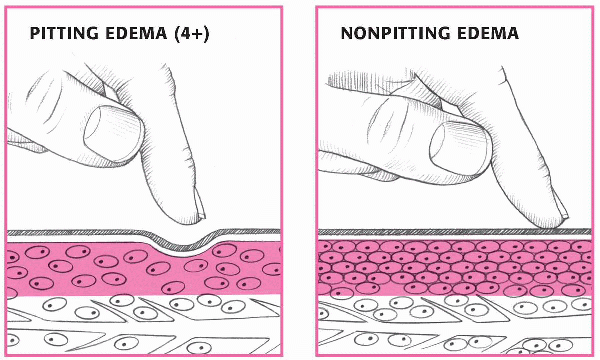
EDEMA: PITTING OR NONPITTING?
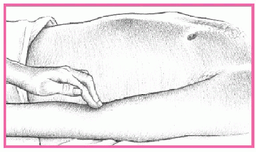
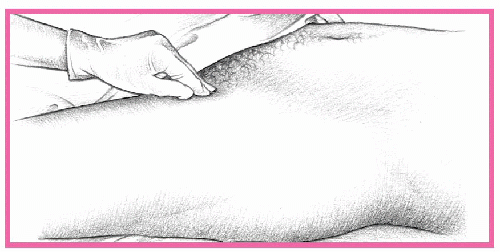
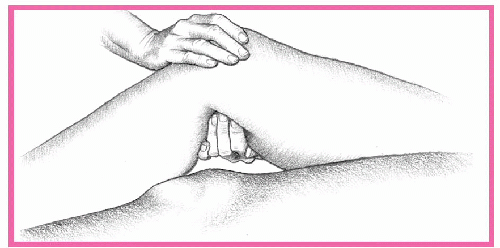
To differentiate pitting from nonpitting edema, press your finger against a swollen area for 5 seconds, then quickly remove it.
With pitting edema, pressure forces fluid into the underlying tissues, causing an indentation that slowly fills. To determine the severity of pitting edema, estimate the indentation's depth in centimeters: 1+ (1 cm), 2+ (2 cm), 3+ (3 cm), or 4+ (4 cm).
With nonpitting edema, pressure leaves no indentation because fluid has coagulated in the tissues. Typically, the skin feels unusually tight and firm.
|
|
If the patient's pulsations appear higher, this indicates elevation in central venous pressure and jugular vein distention. Characterize this distention as mild, moderate, or severe. Determine the level of distention in fingerbreadths above the clavicle or in relation to the jaw or clavicle. Also note the amount of distention in relation to head elevation. (See Evaluating jugular vein distention.)
PALPATION
The first step in palpating the vascular system is to assess skin temperature, texture, and turgor. Then check capillary refill time by assessing the nail beds on the fingers and toes. Refill time should be no more than 3 seconds, or long enough to say “capillary refill.”
Palpate the patient's arms and legs for temperature and edema. Edema is graded on a four-point scale. (See Edema: Pitting or nonpitting?)
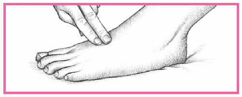
Palpate for arterial pulses by gently pressing with the pads of your index and middle fingers. Start at the top of the patient's body at the temporal artery, and work your way down. Check the carotid, brachial, radial, femoral, popliteal, posterior tibial, and dorsalis pedis pulses. Palpate for the pulse on each side, comparing pulse volume and symmetry.
RED FLAG
Don't palpate both carotid arteries at the same time or press too firmly. If you do, the patient may faint or become bradycardic.
Put on gloves for the examination when you palpate the femoral arteries.
All pulses should be regular in rhythm and equal in strength. Pulses are graded on the following scale: 4+ is bounding, 3+ is increased, 2+ is normal, 1+ is weak, and 0 is absent. (See Assessing arterial pulses.)
Strong or bounding pulsations usually occur in a patient with a condition that causes increased cardiac output, such as hypertension, hypoxia, anemia, exercise, or anxiety. (SeeIdentifying pulse waveforms, pages 56 and 57.)
AUSCULTATION
After you palpate, use the bell of the stethoscope to begin auscultating the vascular system; then follow the palpation sequence and listen over each artery. You shouldn't hear sounds over the carotid arteries. A hum, or bruit, sounds like buzzing or blowing and could indicate arteriosclerotic plaque formation.
Assess the upper abdomen for abnormal pulsations, which could indicate the presence of an abdominal aortic aneurysm. Finally, auscultate the femoral and popliteal pulses, checking for bruits or other abnormal sounds. (See Locating abdominal auscultation points, page 56
If you hear a bruit during arterial auscultation, the patient may have occlusive arterial disease or an arteriovenous fistula. Various high cardiac output conditions, such as anemia, hyperthyroidism, and pheochromocytoma, may also cause bruits.
ASSESSING ARTERIAL PULSES
Carotid pulse
Lightly place your fingers just medial to the trachea and below the jaw angle. Never palpate both carotid arteries at the same time.
|
|
Brachial pulse
Position your fingers medial to the biceps tendon.
|
|
Radial pulse
Apply gentle pressure to the medial and ventral side of the wrist, just below the base of the thumb.
|
|
Femoral pulse
Press relatively hard at a point inferior to the inguinal ligament. For an obese patient, palpate in the crease of the groin, halfway between the pubic bone and the hip bone.
|
|
Popliteal pulse
Press firmly in the popliteal fossa at the back of the knee.
|
|
Posterior tibial pulse
Apply pressure behind and slightly below the malleolus of the ankle.
|
|
Dorsalis pedis pulse
Place your fingers on the medial dorsum of the foot while the patient points his toes down. (Note: The pulse is difficult to detect here and may be nonpalpable in healthy patients.)
|
|
IDENTIFYING PULSE WAVEFORMS
To identify abnormal arterial pulses, check the waveforms below and see which one matches the patient's peripheral pulse.
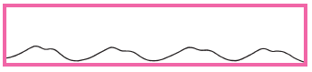
Weak pulse
A weak pulse has decreased amplitude with a slower upstroke and downstroke. Possible causes of a weak pulse include increased peripheral vascular resistance (from cold weather or severe heart failure) and decreased stroke volume (from hypovolemia or aortic stenosis).
|
|
Bounding pulse
A bounding pulse has a sharp upstroke and downstroke with a pointed peak. The amplitude is elevated. Possible causes of a bounding pulse include increased stroke volume, as with aortic insufficiency; or stiffness of arterial walls, as with aging.
|
|
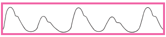
Pulsus alternans
Pulsus alternans has a regular, alternating pattern of a weak and a strong pulse. This pulse is associated with left-sided heart failure.
|
|
Pulsus bigeminus
Pulsus bigeminus is similar to alternating pulse but occurs at irregular intervals. This pulse is caused by premature atrial or ventricular beats.
|
|
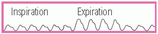
Pulsus paradoxus
Pulsus paradoxus has increases and decreases in amplitude associated with the respiratory cycle. Marked decreases occur when the patient inhales. Pulsus paradoxus is associated with pericardial tamponade, advanced heart failure, and constrictive pericarditis.
|
|
Pulsus biferiens
Pulsus biferiens shows an initial upstroke, a subsequent downstroke, and then another upstroke during systole. Pulsus biferiens is caused by aortic stenosis and aortic insufficiency.
|
|
LOCATING ABDOMINAL AUSCULTATION POINTS
|
|
ABNORMAL FINDINGS
A patient's chief complaint may be due to any of the signs and symptoms related to the cardiovascular system. Common findings include:
|
· |
decreased or increased blood pressure |
|
· |
bruits |
|
· |
increased capillary refill time |
|
· |
chest pain |
|
· |
fatigue |
|
· |
atrial or ventricular gallop |
|
· |
intermittent claudication |
|
· |
jugular vein distention |
|
· |
palpitations |
|
· |
absent or weak pulse |
|
· |
peripheral edema. |
The following history, physical assessment, and analysis summaries will help you interpret each finding quickly and accurately. (See Interpreting your findings, pages 58 to 66.)
INTERPRETING YOUR FINDINGS
After you assess the patient, a group of findings may lead you to a particular disorder of the cardiovascular system. This chart shows some common groups of findings for major signs and symptoms related to the cardiovascular assessment, along with their probable causes.
|
SIGN OR SYMPTOM AND FINDINGS |
PROBABLE CAUSE |
||||||||||||||||||||||
|
BLOOD PRESSURE, DECREASED |
|||||||||||||||||||||||
|
Adrenal insufficiency, acute |
||||||||||||||||||||||
|
Cardiogenic shock |
||||||||||||||||||||||
|
Hypovolemic shock |
||||||||||||||||||||||
|
Septic shock |
||||||||||||||||||||||
|
BLOOD PRESSURE, ELEVATED |
|||||||||||||||||||||||
|
Aldosteronism, primary |
||||||||||||||||||||||
|
Cushing's syndrome |
||||||||||||||||||||||
|
Hypertension |
||||||||||||||||||||||
|
BRUITS |
|||||||||||||||||||||||
|
Aortic aneurysm, abdominal |
||||||||||||||||||||||
|
Carotid artery stenosis |
||||||||||||||||||||||
|
Peripheral vascular disease |
||||||||||||||||||||||
|
CAPILLARY REFILL TIME, INCREASED |
|||||||||||||||||||||||
|
Arterial occlusion, acute |
||||||||||||||||||||||
|
Hypothermia |
||||||||||||||||||||||
|
Raynaud's disease |
||||||||||||||||||||||
|
CHEST PAIN |
|||||||||||||||||||||||
|
Angina |
||||||||||||||||||||||
|
|||||||||||||||||||||||
|
Myocardial infarction |
||||||||||||||||||||||
|
Rib fracture |
||||||||||||||||||||||
|
FATIGUE |
|||||||||||||||||||||||
|
Anemia |
||||||||||||||||||||||
|
Depression |
||||||||||||||||||||||
|
Valvular heart disease |
||||||||||||||||||||||
|
Angina |
||||||||||||||||||||||
|
Aortic insufficiency |
||||||||||||||||||||||
|
Hypertension |
||||||||||||||||||||||
|
GALLOP, VENTRICULAR |
|||||||||||||||||||||||
|
Aortic insufficiency |
||||||||||||||||||||||
|
Cardiomyopathy |
||||||||||||||||||||||
|
Mitral insufficiency |
||||||||||||||||||||||
|
INTERMITTENT CLAUDICATION |
|||||||||||||||||||||||
|
Arteriosclerosis obliterans |
||||||||||||||||||||||
|
Buerger's disease |
||||||||||||||||||||||
|
JUGULAR VEIN DISTENTION |
|||||||||||||||||||||||
|
Cardiac tamponade |
||||||||||||||||||||||
|
|||||||||||||||||||||||
|
Pericarditis, chronic constrictive |
||||||||||||||||||||||
|
PALPITATIONS |
|||||||||||||||||||||||
|
Acute anxiety attack |
||||||||||||||||||||||
|
Arrhythmias |
||||||||||||||||||||||
|
Hypoglycemia |
||||||||||||||||||||||
|
PERIPHERAL EDEMA |
|||||||||||||||||||||||
|
Heart failure |
||||||||||||||||||||||
|
Superior vena cava syndrome |
||||||||||||||||||||||
|
Venous insufficiency |
||||||||||||||||||||||
|
PULSE, ABSENT OR WEAK |
|||||||||||||||||||||||
|
|||||||||||||||||||||||
|
Arterial occlusion |
||||||||||||||||||||||
|
Peripheral vascular disease |
||||||||||||||||||||||
Blood pressure, decreased
Normal blood pressure varies considerably; what qualifies as low blood pressure for one person may be perfectly normal for another. Consequently, every blood pressure reading must be compared against the patient's baseline. Typically, a reading below 90/60 mm Hg, or a drop of 30 mm Hg from the baseline, is considered low blood pressure, which could result in an inadequate intravascular pressure to maintain the oxygen requirements of the body's tissues.
HISTORY
If the patient is conscious, ask him about associated symptoms. For example, find out if he feels unusually weak or fatigued; if he has had nausea, vomiting, or dark or bloody stools; if his vision is blurred; or if his gait is unsteady. Ask him if he has palpitations or chest or abdominal pain or difficulty breathing. Then ask if he has had episodes of dizziness or fainting. Find out if these episodes occur when he stands up suddenly. If so, take the patient's blood pressure while he's lying down, sitting, and then standing; compare readings. A drop in systolic or diastolic pressure of 10 to 20 mm Hg or more and an increase in heart rate of more than 15 beats/minute between position changes suggest orthostatic hypotension.
PHYSICAL ASSESSMENT
Next, continue with a physical examination. Inspect the skin for pallor, sweating, and clamminess. Palpate peripheral pulses. Note paradoxical pulse, an accentuated fall in systolic pressure during inspiration, which suggests pericardial tamponade. Then auscultate for abnormal heart sounds, such as gallops and murmurs; heart rate, signaling bradycardia or tachycardia; and heart rhythm. Auscultate the lungs for abnormal breath sounds, such as diminished sounds, crackles, wheezing; breath rate, signaling bradypnea or tachypnea; and breath rhythm, such as agonal or Cheyne-Stokes respirations. Look for signs of hemorrhage, including:
|
· |
visible bleeding |
|
· |
palpable masses |
|
· |
bruising |
|
· |
tenderness. |
Assess the patient for abdominal rigidity and rebound tenderness; auscultate for abnormal bowel sounds. Carefully assess the patient for possible sources of infection such as open wounds.
ANALYSIS
Although commonly linked to shock, decreased blood pressure may also result from a cardiovascular, respiratory, neurologic, or metabolic disorder. Hypoperfusion states especially affect the kidneys, brain, and heart, and may lead to renal failure, change in level of consciousness (LOC), or myocardial ischemia. Low blood pressure may be drug-induced or may accompany diagnostic tests—typically those using contrast media. It may stem from stress or change of position—specifically, rising abruptly from a supine or sitting position to a standing position (orthostatic hypotension). Low blood pressure can reflect an expanded intravascular space, as in severe infections, allergic reactions, or adrenal insufficiency; reduced intravascular volume, as in dehydration and hemorrhage; or decreased cardiac output, as in impaired cardiac muscle contractility. Because the body's pressureregulating mechanisms are complex and interrelated, a combination of these factors usually contributes to low blood pressure.
AGE AWARE
In children, normal blood pressure is lower than blood pressure in adults. Because accidents occur frequently in children, suspect trauma or shock first as a possible cause of low blood pressure. Remember that low blood pressure typically doesn't accompany head injury in adults because intracranial hemorrhage is insufficient to cause hypovolemia. However, it does accompany head injury in infants and young children because their expandable cranial vaults allow significant blood loss into the cranial space, resulting in hypovolemia. Another common cause of low blood pressure in children is dehydration, which results from failure to thrive or from persistent diarrhea or vomiting occurring for as few as 24 hours.
In elderly patients, low blood pressure commonly results from the use of multiple drugs with hypotension as a potential adverse effect. Orthostatic hypotension due to autonomic dysfunction is another common cause.
Blood pressure, elevated
Elevated blood pressure—an intermittent or sustained increase in blood pressure exceeding 140/90 mm Hg—strikes more men than women and twice as many Blacks as Whites. By itself, this common sign is easily ignored by the patient because he can't see or feel it. However, its causes can be life-threatening.
Hypertension has been reported to be two to three times more common in women taking hormonal contraceptives than those not taking them. Women age 35 and older who smoke cigarettes should be strongly encouraged to stop. If they continue to smoke, they should be discouraged from using hormonal contraceptives.
HISTORY
If you detect sharply elevated blood pressure, act quickly to rule out possible life-threatening causes. After ruling out life-threatening causes, complete a more leisurely history and physical examination. Determine if the patient has a history of diabetes or cardiovascular, cerebrovascular, or renal disease. Ask about a family history of high blood pressure—a likely finding with essential hypertension, pheochromocytoma, and polycystic kidney disease. Then ask about its onset and if the high blood pressure appeared abruptly. Ask the patient's age. Sudden onset of high blood pressure in middle-aged or elderly patients suggests renovascular stenosis. Although essential hypertension may begin in childhood, it typically isn't diagnosed until near age 35.
Note headache, palpitations, blurred vision, and sweating. Ask about wine-colored urine and decreased urine output; these signs suggest glomerulonephritis, which can cause elevated blood pressure.
Obtain a drug history, including past and present prescriptions, herbal preparations, and over-the-counter drugs especially decongestants.
RED FLAG
Ephedra (ma huang), ginseng, and licorice may cause high blood pressure or irregular heartbeat. St. John's wort can also raise blood pressure, especially when taken with substances that antagonize hypericin, such as amphetamines, cold and hay fever medications, nasal decongestants, pickled foods, beer, coffee, wine, and chocolate.
If the patient is already taking an antihypertensive, determine how well he complies with the regimen. Ask about his perception of the elevated blood pressure. Find out how serious he believes it is and if he expects drug therapy will help. Explore psychosocial or environmental factors that may impact blood pressure control.
PHYSICAL ASSESSMENT
Obtain vital signs and check for orthostatic hypotension. Take the patient's blood pressure with him laying down, sitting, and then standing. Normally, systolic pressure falls and diastolic pressure rises on standing. With orthostatic hypotension, both pressures fall.
Using a funduscope, check for intraocular hemorrhage, exudate, and papilledema, which characterize severe hypertension. Perform a thorough cardiovascular assessment. Check for carotid bruits and jugular vein distention. Assess skin color, temperature, and turgor. Palpate peripheral pulses. Auscultate for abnormal heart sounds, including gallops, louder S2, and murmurs; heart rate, including bradycardia and tachycardia; and heart rhythm. Then auscultate for abnormal breath sounds, such as crackles and wheezing; abnormal respiratory rate, such as bradypnea and tachypnea; and breath rhythm.
Palpate the abdomen for tenderness, masses, and liver enlargement. Auscultate for abdominal bruits. Renal artery stenosis produces bruits over the upper abdomen or in the costovertebral angles. Easily palpable, enlarged kidneys and a large, tender liver suggest polycystic kidney disease. Obtain a urine sample to check for microscopic hematuria.
ANALYSIS
Elevated blood pressure may develop suddenly or gradually. A sudden, severe rise in pressure (exceeding 180/110 mm Hg) may indicate life-threatening hypertensive crisis. However, even a less dramatic rise may be equally significant if it heralds a dissecting aortic aneurysm, increased intracranial pressure, MI, eclampsia, or thyrotoxicosis.
Usually associated with essential hypertension, elevated blood pressure may also result from a renal or endocrine disorder, a drug's adverse effect, or a treatment that affects fluid status, such as dialysis. Ingestion of large amounts of certain foods, such as black licorice and cheddar cheese, may temporarily elevate blood pressure.
Sometimes elevated blood pressure may simply reflect inaccurate blood pressure measurement. However, careful measurement alone doesn't ensure a useful reading. To be useful, each blood pressure reading must be compared with the patient's baseline. Also, serial readings may be needed to establish elevated blood pressure.
The patient may experience elevated blood pressure in a health care provider's office (known as “white-coat hypertension”). In such cases, 24-hour blood pressure monitoring is indicated to confirm elevated readings in other settings. In addition, other risk factors for CAD, such as smoking and elevated cholesterol levels, need to be addressed.
AGE AWARE
Normally, blood pressure in children is lower than it is in adults. In children, elevated blood pressure may result from lead or mercury poisoning, essential hypertension, renovascular stenosis, chronic pyelonephritis, coarctation of the aorta, patent ductus arteriosus, glomerulonephritis, adrenogenital syndrome, or neuroblastoma. In elderly patients, atherosclerosis commonly produces isolated systolic hypertension.
Bruits
Typically an indicator of life- or limb-threatening vascular disease, bruits are swishing sounds caused by turbulent blood flow. They're characterized by location, duration, intensity, pitch, and time of onset in the cardiac cycle. Loud bruits produce intense vibration and a palpable thrill. A thrill, however, doesn't provide any further clue to the causative disorder or to its severity.
HISTORY
If you detect a bruit, be sure to check for further vascular damage and perform a thorough cardiac assessment.
PHYSICAL ASSESSMENT
If you detect bruits over the abdominal aorta, check for a pulsating mass or a bluish discoloration around the umbilicus (Cullen's sign). Either of these signs—or severe, tearing pain in the abdomen, flank, or lower back—may signal life-threatening dissection of an aortic aneurysm. Also check peripheral pulses, comparing intensity in the arms versus the legs.
If you detect bruits over the thyroid gland, ask the patient if he has a history of hyperthyroidism or signs and symptoms, such as nervousness, tremors, weight loss, palpitations, heat intolerance, and (in women) amenorrhea.
RED FLAG
Watch for signs and symptoms of life-threatening thyroid storm, such as tremors, restlessness, diarrhea, abdominal pain, and hepatomegaly.
If you detect carotid artery bruits, be alert for signs and symptoms of a transient ischemic attack, including dizziness, diplopia, slurred speech, flashing lights, and syncope. These findings may indicate impending stroke. Evaluate the patient frequently for changes in LOC and muscle function.
If you detect bruits over the femoral, popliteal, or subclavian artery, watch for signs and symptoms of decreased or absent peripheral circulation—edema, weakness, and paresthesia. Ask the patient if he has a history of intermittent claudication. Frequently check distal pulses and skin color and temperature. Also watch for the sudden absence of pulse, pallor, or coolness, which may indicate a threat to the affected limb.
ANALYSIS
Bruits are most significant when heard over the abdominal aorta; the renal, carotid, femoral, popliteal, or subclavian artery; or the thyroid gland. They're also significant when heard consistently despite changes in patient position and when heard during diastole.
AGE AWARE
In young children, bruits are common but are usually of little significance-for example, cranial bruits are normal until age 4. However, certain bruits may be significant. Because birthmarks commonly accompany congenital arteriovenous fistulas, carefully auscultate for bruits in a child with port-wine spots or cavernous or diffuse hemangioma.
Capillary refill time, increased
Capillary refill time is the duration required for color to return to the nail bed of a finger or toe after application of slight pressure, which causes blanching. This duration reflects the quality of peripheral vasomotor function. Normal capillary refill time is less than 3 seconds.
Capillary refill time is typically tested during a routine cardiovascular assessment. It isn't tested with suspected life-threatening disorders because other, more characteristic signs and symptoms appear earlier.
HISTORY
Take a brief medical history, especially noting previous peripheral vascular disease. Find out which drugs the patient is taking.
PHYSICAL EXAMINATION
If you detect increased capillary refill time, take the patient's vital signs and check pulses in the affected limb. Does the limb feel cold or look cyanotic? Ask the patient about pain or any unusual sensations in his fingers or toes, especially after exposure to cold.
ANALYSIS
Increased refill time isn't diagnostic of any disorder but must be evaluated along with other signs and symptoms. However, this sign usually signals obstructive peripheral arterial disease or decreased cardiac output.
Chest pain
Chest pain can arise suddenly or gradually, and its cause may be difficult to ascertain initially. The pain can radiate to the arms, neck, jaw, or back. It can be steady or intermittent and mild or acute. It can range from a sharp shooting sensation to a feeling of heaviness, fullness, or even indigestion. It can be provoked or aggravated by stress, anxiety, exertion, deep breathing, or eating certain foods.
HISTORY
If the patient's chest pain isn't severe, proceed with the health history. Ask if the patient feels diffuse pain or can point to the painful area. Sometimes a patient won't perceive the sensation he's feeling as pain, so ask whether he has any discomfort radiating to the neck, jaw, arms, or back. If he does, ask him to describe it, such as a dull, aching, pres-sure-like sensation or a sharp, stabbing, knifelike pain. Find out if he feels it on the surface or deep inside.
Next, find out whether the pain is constant or intermittent. If it's intermittent, ask him how long it lasts. Ask if movement, exertion, breathing, position changes, or the eating of certain foods worsens or helps relieve the pain. Find out what in particular seems to bring on the pain.
Review the patient's history for cardiac or pulmonary disease, chest trauma, intestinal disease, or sickle cell anemia. Find out what medication he's taking, if any, and ask about recent dosage or schedule changes.
PHYSICAL ASSESSMENT
When taking the patient's vital signs, note the presence of tachycardia, paradoxical pulse, and hypertension or hypotension. Also look for jugular vein distention and peripheral edema. Observe the patient's breathing pattern, and inspect his chest for asymmetrical expansion. Auscultate his lungs for pleural friction rub, crackles, rhonchi, wheezing, and diminished or absent breath sounds. Next, auscultate for murmurs, clicks, gallops, and pericardial friction rubs. Palpate for lifts, heaves, thrills, gallops, tactile fremitus, and abdominal mass or tenderness.
ANALYSIS
Chest pain usually results from disorders that affect thoracic or abdominal organs, such as the heart, pleurae, lungs, esophagus, rib cage, gallbladder, pancreas, or stomach. An important indicator of several acute and life-threatening cardiopulmonary and GI disorders, chest pain can also result from musculoskeletal and hematologic disorders, anxiety, and drug therapy.
Keep in mind that cardiac-related pain may not always occur in the chest. Pain originating in the heart is transmitted through the thoracic region via the upper five thoracic spinal cord segments. Thus, it may be referred to areas served by the cervical or lower thoracic segments, such as the neck and arms. (See Understanding chest pain, pages 74and 75.)
Fatigue
Fatigue is a feeling of excessive tiredness, lack of energy, or exhaustion, accompanied by a strong desire to rest or sleep. This common symptom is distinct from weakness, which involves the muscles, but may occur with it.
UNDERSTANDING CHEST PAIN
This table outlines the different types of chest pain including location, exacerbating factors, causes, and alleviating measures. Use it to accurately assess your patients with chest pain.
|
DESCRIPTION |
LOCATION |
EXACERBATING FACTORS |
CAUSES |
ALLEVIATING MEASURES |
|
Aching, squeezing, pressure, heaviness, burning pain, tightness; usually subsides within 10 minutes |
Substernal; may radiate to jaw, neck, arms, and back |
Eating, physical effort, smoking, cold weather, stress, anger, hunger |
Angina pectoris |
Rest, nitroglycerin, oxygen (Note:Unstable angina appears even at rest.) |
|
Tightness or pressure; burning, aching pain; possible dyspnea, diaphoresis, weakness, anxiety, or nausea; sudden onset; lasts ½ to 2 hours |
Substernal; may radiate to jaw, neck, arms, or back |
Exertion, anxiety |
Acute myocardial infarction (MI) |
|
|
Sharp and continuous; may be accompanied by friction rub; sudden onset; increased with inspiration |
Substernal; may radiate to neck, arm, or back |
Deep breathing; lying in a supine position |
Pericarditis |
Sitting up, leaning forward, anti-inflammatory drugs |
|
Excruciating, tearing pain; may be accompanied by blood pressure difference between right and left arms; sudden onset |
Retrosternal, upper abdominal, or epigastric; may radiate to back, neck, or shoulders |
Hypertension |
Dissecting aortic aneurysm |
Analgesics, surgery |
|
Sudden, stabbing pain, pressure, deep ache; may be accompanied by cyanosis, dyspnea, or cough with hemoptysis |
Substernal; lateral chest; may radiate to neck or shoulders |
Inspiration; venous status |
Pulmonary embolus |
Analgesics, high Fowler's position |
|
Sudden and severe pain; possible dyspnea, increased pulse rate, decreased breath sounds, or deviated trachea |
Lateral thorax; may radiate to back, shoulders, or arms |
Coughing |
Pneumothorax |
Analgesics, chest tube insertion |
|
Dull, pressurelike, squeezing pain |
Substernal, epigastric areas |
Food, cold liquids, exercise |
Esophageal spasm |
Nitroglycerin, calcium channel blockers |
|
Sharp, severe pain; usually occurs shortly after eating |
Lower chest or upper abdomen |
Eating a heavy meal, bending, lying down |
Hiatal hernia |
Antacids, walking, semi-Fowler's position |
|
Burning feeling after eating; possible hematemesis or tarry stools; sudden onset that generally subsides within 15 to 20 minutes |
Epigastric |
Lack of food or highly acidic foods |
Peptic ulcer |
Food, antacids |
|
Gripping, sharp pain; possible nausea and vomiting |
Right epigastric or abdominal areas; possible radiation to shoulders |
Eating fatty foods, lying down |
Cholecystitis |
Rest and analgesics, surgery |
|
Continuous or intermittent sharp pain; possible tenderness to touch; gradual or sudden onset |
Anywhere in chest |
Movement, palpation |
Time, analgesics, heat applications |
|
|
Dull or stabbing pain usually with hyperventilation or breathlessness; sudden onset; lasting less than 1 minute or as long as several days |
Anywhere in chest |
Increased respiratory rate, stress or anxiety |
Acute anxiety |
Slowing of respiratory rate, stress relief |
Obtain a history to identify the patient's fatigue pattern. Ask about related symptoms and any recent viral illness or stressful changes in lifestyle.
Explore nutritional habits and appetite or weight changes. Carefully review the patient's medical and psychiatric history for chronic disorders that commonly produce fatigue. Ask about a family history of such disorders.
PHYSICAL ASSESSMENT
Observe the patient's general appearance for overt signs of depression or organic illness. Is he unkempt or expressionless? Does he appear tired or sickly, or have a slumped posture? If warranted, evaluate his mental status, noting especially mental clouding, attention deficits, agitation, or psychomotor retardation.
ANALYSIS
Fatigue that worsens with activity and improves with rest generally indicates a physical disorder; the opposite pattern, a psychological disorder. Fatigue lasting longer than 4 months, constant fatigue that's unrelieved by rest, and transient exhaustion that quickly gives way to bursts of energy are other findings associated with psychological disorders.
Fatigue reflects hypermetabolic and hypometabolic states in which nutrients needed for cellular energy and growth are lacking because of overly rapid depletion, impaired replacement mechanisms, insufficient hormone production, or inadequate nutrient intake or metabolism. Cardiac causes include heart failure, MI, and valvular heart disease.
AGE AWARE
When evaluating a child for fatigue, ask his parents if they've noticed any change in his activity level. Fatigue without an organic cause occurs normally during accelerated growth phases in preschool age and prepubescent children. However, psychological causes of fatigue must be considered—for example, a depressed child may try to escape problems at home or school by taking refuge in sleep. In the pubescent child, consider the possibility of drug abuse, particularly of hypnotics and tranquilizers. Always ask elderly patients about fatigue because this symptom may be insidious and mask more serious underlying conditions in this age-group.
Gallop, atrial
An atrial or presystolic gallop is an extra heart sound (known as S4) that's heard or often palpated immediately before S1, late in diastole, as described in “Abnormal heart sounds,” page 42.
HISTORY
When the patient's condition permits, ask about a history of hypertension, angina, valvular stenosis, or cardiomyopathy. If appropriate, have him describe the frequency and severity of anginal attacks.
PHYSICAL ASSESSMENT
Carefully auscultate the chest for S4. Use the bell of the stethoscope. Note any murmurs or abnormalities in S1 and S2
ANALYSIS
An atrial S4 gallop typically results from hypertension, conduction defects, valvular disorders, or other problems such as ischemia. Occasionally, it helps differentiate angina from other causes of chest pain. It results from abnormal forceful atrial contraction caused by augmented ventricular filling or by decreased left ventricular compliance. An atrial gallop usually originates from left atrial contraction, is heard at the apex, and doesn't vary with inspiration. A left-sided S4 can occur in hypertensive heart disease, coronary artery disease, aortic stenosis, and cardiomyopathy. It may also originate from right atrial contraction. A right-sided S4 is indicative of pulmonary hypertension and pulmonary stenosis. If so, it's heard best at the lower left sternal border and intensifies with inspiration.
An atrial gallop seldom occurs in normal hearts; however, it may occur in athletes with physiologic hypertrophy of the left ventricle.
In children, an atrial gallop may occur normally, especially after exercise. However, it may also result from congenital heart diseases, such as atrial septal defect, ventricular septal defect, patent ductus arteriosus, and severe pulmonary valvular stenosis.
Because the absolute intensity of an atrial gallop doesn't decrease with age, as it does with an S1, the relative intensity of S4 increases compared with S1. This explains the increased frequency of an audible S4
Gallop, ventricular
A ventricular gallop is a heart sound known as S3, associated with rapid ventricular filling in early diastole. Usually palpable, this lowfrequency sound occurs about 0.15 second after S2. It may originate in either the left or right ventricle. A right-sided gallop usually sounds louder on inspiration and is heard best along the lower left sternal border or over the xiphoid region. A left-sided gallop usually sounds louder on expiration and is heard best at the apex.
HISTORY
Focus the history on the cardiovascular system. Begin the history by asking the patient if he has had any chest pain. If so, have him describe its character, location, frequency, duration, and any alleviating or aggravating factors. Also ask about palpitations, dizziness, or syncope. Find out if the patient has difficulty breathing after exertion, while lying down, or at rest. Ask the patient if he has a cough. Also ask about a history of cardiac disorders. Find out if the patient is currently receiving any treatment for heart failure. If so, find out which medications he's taking.
PHYSICAL ASSESSMENT
During the physical examination, carefully auscultate for murmurs or abnormalities in S1 and S2. Then listen for pulmonary crackles. Next, assess peripheral pulses, noting an alternating strong and weak pulse. Finally, palpate the liver to detect enlargement or tenderness, and assess for jugular vein distention and peripheral edema.
ANALYSIS
Ventricular gallops are easily overlooked because they're usually faint. Fortunately, certain techniques make their detection more likely. These include auscultating in a quiet environment; examining the patient in the supine, left lateral, and semi-Fowler's positions; and having the patient cough or raise his legs to augment the sound.
A physiologic ventricular gallop normally occurs in children and adults younger than age 40; however, most people lose this S3 by age 40. Ventricular gallop may also occur during the third trimester of pregnancy. Abnormal S3 (in adults older than age 40) can be a sign of decreased myocardial contractility, myocardial failure, and volume overload of the ventricle, as in mitral and tricuspid valve regurgitation.
Although the physiologic S3 has the same timing as the pathologic S, its intensity waxes and wanes with respiration. It's also heard more faintly if the patient is sitting or standing.
A pathologic ventricular gallop may be one of the earliest signs of ventricular failure. It may result from one of two mechanisms: rapid deceleration of blood entering a stiff, noncompliant ventricle, or rapid acceleration of blood associated with increased flow into the ventricle. A gallop that persists despite therapy indicates a poor prognosis.
Patients with cardiomyopathy or heart failure may develop both a ventricular gallop and an atrial gallop—a condition known as a summation gallop.
Intermittent claudication
Typically occurring in the legs, intermittent claudication is cramping limb pain brought on by exercise and relieved by 1 to 2 minutes of rest. This pain may be acute or chronic. Without treatment, it may progress to pain at rest.
HISTORY
If the patient has chronic intermittent claudication, gather history data first. Ask how far he can walk before pain occurs and how long he must rest before it subsides. Find out if he can walk less further now than before, or if he needs to rest longer. Next, ask if the pain-rest pattern varies and if this symptom has affected his lifestyle.
RED FLAG
Obtain a history of risk factors for atherosclerosis, such as smoking, diabetes, hypertension, and hyperlipidemia. Next, ask about associated signs and symptoms, such as paresthesia in the affected limb and visible changes in the color of the fingers (white to blue to pink) when he's smoking, exposed to cold, or under stress. If the patient is male, ask if he experiences impotence.
PHYSICAL ASSESSMENT
Focus the physical examination on the cardiovascular system. Palpate for femoral, popliteal, dorsalis pedis, and posterior tibial pulses. Note character, amplitude, and bilateral equality. Note diminished or absent popliteal and pedal pulses with the femoral pulse present.
Examine his feet, toes, and fingers for ulceration, and inspect his hands and lower legs for small, tender nodules and erythema along blood vessels. Note the quality of his nails and the amount of hair on his fingers and toes. Physical findings include pallor on elevation, rubor on dependency (especially the toes and soles), loss of hair on the toes, and diminished arterial pulses.
If the patient has arm pain, inspect his arms for a change in color (to white) on elevation. Next, palpate for changes in temperature, muscle wasting, and a pulsating mass in the subclavian area. Palpate and compare the radial, ulnar, brachial, axillary, and subclavian pulses to identify obstructed areas.
ANALYSIS
When acute, this pain may signal acute arterial occlusion. Intermittent claudication is most common in men ages 50 to 60 with a history of diabetes mellitus, hyperlipidemia, hypertension, or tobacco use. With chronic arterial occlusion, limb loss is uncommon because collateral circulation usually develops.
With occlusive artery disease, intermittent claudication results from an inadequate blood supply. Pain in the calf (the most common area) or foot indicates disease of the femoral or popliteal arteries; pain in the buttocks and upper thigh, disease of the aortoiliac arteries. During exercise, the pain typically results from the release of lactic acid due to anaerobic metabolism in the ischemic segment, secondary to obstruction. When exercise stops, the lactic acid clears and the pain subsides. Diminished femoral and distal pulses may indicate disease of the terminal aorta or iliac branches. Absent pedal and popliteal pulses with normal femoral pulses may suggest atherosclerotic disease of the femoral artery. Absent pedal pulses with normal femoral and popliteal pulses may indicate Buerger's disease.
Intermittent claudication may also have a neurologic cause: narrowing of the vertebral column at the level of the cauda equina. This condition creates pressure on the nerve roots to the lower extremities.
Walking stimulates circulation to the cauda equina, causing increased pressure on those nerves and resultant pain.
AGE AWARE
In children, intermittent claudication rarely occurs. Although it sometimes develops in patients with coarctation of aorta, extensive compensatory collateral circulation typically prevents manifestation of this sign. Muscle cramps from exercise and growing pains may be mistaken for intermittent claudication in children.
Jugular vein distention
Jugular vein distention is the abnormal fullness and height of the pulse waves in the internal or external jugular veins. For a patient in a supine position with his head elevated 45 degrees, a pulse wave height greater than 1¼″ to 1½″ (3 to 4 cm) above the angle of Louis indicates distention.
HISTORY
If the patient isn't in severe distress, obtain a personal history. Find out if he has recently gained weight, has difficulty putting on his shoes, and if his ankles are swollen. Ask about chest pain, shortness of breath, paroxysmal nocturnal dyspnea, anorexia, nausea or vomiting, and a history of cancer or cardiac, pulmonary, hepatic, or renal disease. Obtain a drug history noting diuretic use and dosage. Find out if the patient is taking drugs as prescribed. Ask the patient about his regular diet patterns, noting a high sodium intake.
PHYSICAL ASSESSMENT
Next, perform a physical examination, beginning with vital signs. Tachycardia, tachypnea, and increased blood pressure indicate fluid overload that's stressing the heart. Inspect and palpate the patient's extremities and face for edema. Then weigh the patient and compare that weight to his baseline.
Auscultate his lungs for crackles and his heart for gallops, a pericardial friction rub, and muffled heart sounds. Inspect his abdomen for distention, and palpate and percuss for an enlarged liver. Finally, monitor urine output and note any decrease.
ANALYSIS
Engorged, distended veins reflect increased venous pressure in the right side of the heart, which in turn, indicates an increased central venous pressure. This common sign characteristically occurs in heart failure and other cardiovascular disorders, such as constrictive pericarditis, tricuspid stenosis, and obstruction of the superior vena cava.
AGE AWARE
In most infants and toddlers, jugular vein distention is difficult (sometimes impossible) to evaluate because of their short, thick necks. Even in school-age children, measurement of jugular vein distention can be unreliable because the sternal angle may not be the same distance (2″ to 2¾″ [5 to 7 cm]) above the right atrium as it is in adults.
Palpitations
Defined as a conscious awareness of one's heartbeat, palpitations are usually felt over the precordium or in the throat or neck. The patient may describe them as pounding, jumping, turning, fluttering, or flopping, or as missing or skipping beats. Palpitations may be regular or irregular, fast or slow, paroxysmal or sustained.
HISTORY
If the patient isn't in distress, obtain a complete cardiac history. Ask about cardiovascular or pulmonary disorders, which may produce arrhythmias. Find out if the patient has a history of hypertension or hypoglycemia.
Obtain a drug history. Ask the patient if he has recently started digoxin (Lanoxin) therapy. In addition, ask about caffeine, tobacco, and alcohol consumption. Ask about associated symptoms, such as weakness, fatigue, and angina.
To help characterize the palpitations, ask the patient to simulate their rhythm by tapping his finger on a hard surface. An irregular “skipped beat” rhythm points to premature ventricular contractions, whereas an episodic racing rhythm that ends abruptly suggests paroxysmal atrial tachycardia.
PHYSICAL ASSESSMENT
If the patient isn't in distress, perform a complete physical assessment of the cardiovascular system. Auscultate for gallops, murmurs, and abnormal breath sounds.
ANALYSIS
Although frequently insignificant, palpitations are a common chief complaint that may result from a cardiac or metabolic disorder or from the adverse effects of certain drugs. Nonpathologic palpitations may occur with a newly implanted prosthetic valve because the valve's clicking sound heightens the patient's awareness of his heartbeat. Transient palpitations may accompany emotional stress, such as fright, anger, and anxiety, or physical stress, such as exercise and fever. They can also accompany the use of stimulants, such as tobacco and caffeine.
AGE AWARE
In children, palpitations commonly result from fever and congenital heart defects, such as patent ductus arteriosus and septal defects. Because many children have difficulty describing this symptom, focus your attention on objective measurements, such as cardiac monitoring, physical examination, and laboratory test results.
Peripheral edema
The result of excess interstitial fluid in the arms or legs, peripheral edema may be unilateral or bilateral, slight or dramatic, or pitting or nonpitting.
HISTORY
Begin by asking how long the patient has had the edema and if it developed suddenly or gradually. Find out if the edema decreases if the patient elevates his arms or legs, if it's worse in the mornings, and if it gets progressively worse during the day.
Find out if the patient recently injured the affected extremities or had surgery or an illness that may have immobilized him. Ask about a history of cardiovascular disease. Find out what medication he's taking and which drugs he has taken in the past.
PHYSICAL ASSESSMENT
Begin the assessment by examining each extremity for pitting edema. In pitting edema, pressure forces fluid into the underlying tissues, causing an indentation that slowly fills. In nonpitting edema, pressure leaves no indentation in the skin, but the skin may feel unusually firm. Because edema may compromise arterial blood flow, palpate peripheral pulses to detect insufficiency. Observe the color of the extremity and look for unusual vein patterns. Then palpate for warmth, tenderness, and cords and gently squeeze the muscle against the bone to check for deep pain. Finally, note any skin thickening or ulceration in the edematous areas.
ANALYSIS
Peripheral edema signals a localized fluid imbalance between the vascular and interstitial spaces. It may result from trauma, a venous disorder, or a bone or cardiac disorder.
Cardiovascular causes of arm edema are superior vena cava syndrome, which leads to slowly progressing arm edema accompanied by facial and neck edema with dilated veins marking these edematous areas, and thrombophlebitis, which may cause arm edema, pain, and warmth.
P.84
Leg edema is an early sign of right-sided heart failure. It can also signal thrombophlebitis and chronic venous insufficiency.
AGE AWARE
Uncommon in children, arm edema can result from trauma. Leg edema, also uncommon, can result from osteomyelitis, leg trauma or, rarely, heart failure.
Pulse, absent or weak
An absent or weak pulse may be generalized or affect only one extremity.
HISTORY
RED FLAG
If you detect an absent or weak pulse, quickly palpate the remaining arterial pulses to distinguish between localized or generalized loss or weakness. Then quickly check other vital signs and evaluate cardiopulmonary status, obtain a brief history, and intervene accordingly.
PHYSICAL ASSESSMENT
Carefully check the rate, amplitude, and symmetry of all pulses. Note any confusion or restlessness, hypotension, and cool, pale clammy skin.
ANALYSIS
When generalized, absent or weak pulse is an important indicator of such life-threatening conditions as shock and arrhythmia. Localized loss or weakness of a pulse that's normally present and strong may indicate acute arterial occlusion, which could require emergency surgery. However, the pressure of palpation may temporarily diminish or obliterate superficial pulses, such as the posterior tibial or the dorsalis pedis. Thus, bilateral weakness or absence of these pulses doesn't necessarily indicate an underlying disorder.
AGE AWARE
In infants and small children, radial, dorsalis pedis, and posterior tibial pulses aren't easily palpable, so be careful not to mistake these normally hard-to-find pulses for weak or absent pulses. Instead, palpate the brachial, popliteal, or femoral pulses to evaluate arterial circulation to the extremities. In children and young adults, weak or absent femoral and more distal pulses may indicate coarctation of the aorta.
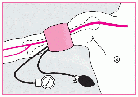

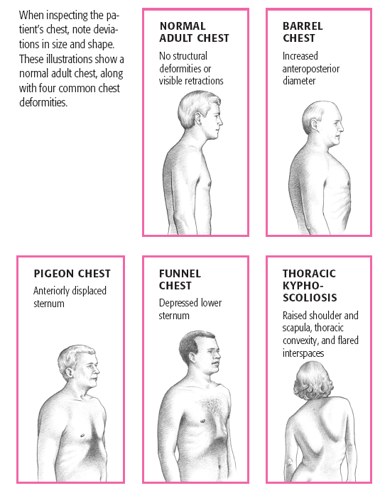
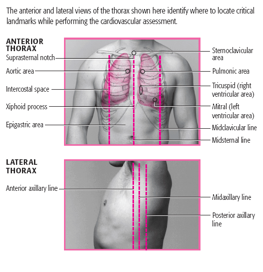

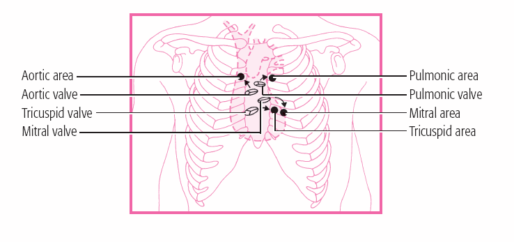
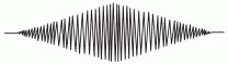
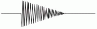
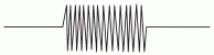


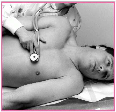
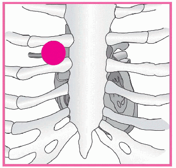
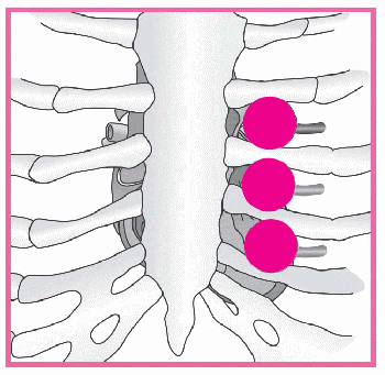
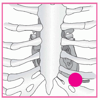
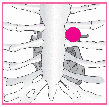
 Diaphragm
 Bell
Bell