
TERMINOLOGY
The ventriculoarterial connection is considered “double-outlet” when more than 50% of each great artery arises from one ventricle. The origin of both great arteries from the right ventricle (RV) is termed double-outlet right ventricle(DORV). Conversely, the origin of both great arteries from the left ventricle (LV) is termed double-outlet left ventricle (DOLV).
DOUBLE-OUTLET RIGHT VENTRICLE
Basically recognized by the origin of both great arteries from the morphologic RV, DORV encompasses hemodynamic features of a variety of entities, ranging from simple ventricular septal defect (VSD) with excessive pulmonary blood flow, to tetralogy of Fallot (TOF) with reduced pulmonary artery flow secondary to pulmonary stenosis, to transposition of the great arteries (TGA) with systemic desaturation and pulmonary overcirculation. This congenital malformation is a rare anomaly. Its frequency has been reported as approximately 0.09 per 1000 births and represents 1% to 1.5% of patients with congenital heart disease. No racial/ethnic or sexual predilection is evident.
There are 16 possible variations of DORV based on the great artery relationships and the location of the VSD. Figure 17.1 illustrates these variations but shows that only nine types were observed clinically. However, this series includes only cases with situs solitus of the atria and viscera, atrioventricular (AV) concordance, and two well-developed ventricles and AV valves. The location of the VSD is described as subaortic, subpulmonary, doubly-committed (below both great arteries), noncommitted, or remote (Fig. 17.2). In addition, an intact ventricular septum (very rare) allows four other possible types of DORV, depending on the great artery relationships. Multiple other variations and combinations are possible if one also includes situs inversus and situs ambiguous, AV discordance, and AV valve atresia.

Figure 17.1. Relationships of the great arteries and locations of the ventricular septal defect (VSD) in 70 patients with double-outlet right ventricle (DORV). Ao, aorta; d-MGA, dextro-malposed great arteries; l-MGA, levo-malposed great arteries; P, pulmonary artery. (Reprinted with permission from Hagler DJ, Tajik AJ, Seward JB, et al. Double-outlet right ventricle: wide-angle two-dimensional echocardiographic observations. Circulation. 1981;63:419–428.)

Figure 17.2. Schematic illustration of three ventricular septal defect (VSD) locations in double-outlet right ventricle. Ao, aorta; APM, apical papillary muscle; IL, inferior limb; IS, conus septum; MB, moderator band; PB, parietal band; PT, pulmonary artery; SB, septal band; SL, superior limb. (Courtesy of Dr. William Edwards, Mayo Clinic.)
Double-Outlet Right Ventricle with Side-by-Side Great Arteries and Subaortic Ventricular Septal Defect
This represents the most common and typical form of DORV. The presence of bilateral conus separates both semilunar valves from both AV valves. The VSD is the only outlet from the LV, and the aortic conus is a muscular structure between the aortic valve and the anterior leaflet of the mitral valve. Figure 17.3 illustrates the pathologic findings of this type of DORV. The RV has been opened illustrating both great arteries originating from the RV. Figure 17.4illustrates the angiographic findings in a patient with DORV, subaortic VSD, and side-by-side great arteries.
Two-dimensional echocardiographic findings for the diagnosis of DORV have been reported. Three observations were noted for the diagnosis: (a) origin of both great arteries from the anterior RV, (b) mitral–semilunar valve discontinuity, and (c) absence of left ventricular outflow other than the VSD. In a series of 36 patients studied by two-dimensional echocardiography, both great arteries were observed to originate predominantly from the anterior RV. If one great artery overrode the VSD, nearly exclusive or predominant commitment to the RV was required for diagnosis. Figures 17.5, 17.6, and 17.7 illustrate the most common forms of DORV with side-by-side or normally related, right anterior aorta, and left anterior aorta, respectively. In addition, Figure 17.7 illustrates the associated finding of left-juxtaposed atrial appendages. Figure 17.8 illustrates the pathologic features of a patient with subpulmonary VSD with right anterior aorta. Most patients with DORV will have situs solitus of the atria and viscera, but situs ambiguous with bilateral right- or left-sidedness and situs inversus may be present. Most patients with DORV will have AV concordance, but less commonly AV discordance (ventricular inversion) may be present.
IMAGING PEARLS
Position of the Great Arteries
In two-thirds of the cases, both great arteries can be observed simultaneously originating from the anterior RV. In the subcostal sagittal plane, both semilunar valves may not be visualized simultaneously. However, each semilunar valve may be demonstrated with slight right or left transducer angulation. The semilunar valves are positioned in a more anterior and superior location relative to the rest of the ventricle because of conus muscle beneath the valves. It is important to sweep in the short-axis scans from apex to base to demonstrate the commitment of each great artery to the right ventricular cavity (Fig. 17.9). With the use of a short-axis scan at the cardiac base, a double-circle appearance of the great arteries is consistent with a parallel orientation of the great arteries. A superior short-axis scan demonstrates the pulmonary artery bifurcation.

Figure 17.3. Pathologic specimen of double-outlet right ventricle. The right ventricle (RV) has been opened, demonstrating the origin of both great arteries from the RV. AC, aortic conus; CS, conus septum; D, defect (ventricular septal defect [VSD]); PA, pulmonary artery.

Figure 17.4. Right ventricular (RV) angiogram of double-outlet right ventricle. The frontal and lateral views illustrate features of DORV with subaortic VSD and side-by-side great arteries. Both semilunar valves are at the same horizontal plane and are side by side. In the lateral view, the VSD (D) is below the conus and allows some filling of the left ventricle (LV). Ao, aorta; PA, pulmonary artery. (With permission of the Mayo Foundation.)

Figure 17.5. Parasternal long-axis scan in a neonate with double-outlet right ventricle, with side-by-side great arteries and subaortic ventricular septal defect (VSD). With the aorta (Ao) anterior and rightward, it is the only great artery observed in this scan. Arrow,subaortic conus separating the aortic valve from the mitral valve. LA, left atrium; LV, left ventricle; RV, right ventricle.

Figure 17.6. Parasternal long-axis scan in a patient with double-outlet right ventricle, with the aorta anterior and right of the pulmonary artery with a subpulmonary ventricular septal defect (VSD). There is a relatively small amount of conus tissue (arrow) separating the pulmonary valve (PV) from the mitral valve (MV). Both great arteries are entirely committed to the right ventricle (RV). LA, left atrium; LV, left ventricle.

Figure 17.7. Parasternal long-axis scan in a patient with double-outlet right ventricle, with the aorta (Ao) anterior and to the left of the pulmonary artery (PA). Both great arteries are entirely committed to the right ventricular cavity and are observed in parallel orientation originating from the right ventricle (RV). In this standard long-axis scan, all four cardiac chambers and both great arteries are observed simultaneously. In this example, findings of left-juxtaposed atrial appendages are also evident. The right atrial (RA) appendage courses posterior to the great arteries to lie next to the left atrial appendage. The pulmonary valve appears slightly thickened and, in real time, the valve was dome shaped during systole, consistent with pulmonary stenosis. The mitral valve (MV) is markedly separated from the semilunar valves. AS, atrial septum (black arrow head); LA, left atrium; LV, left ventricle; TV, tricuspid valve; VS, ventricular septum. (Reprinted with permission from Hagler DJ, Tajik AJ, Seward JB, et al. Double-outlet right ventricle: wide-angle two-dimensional echocardiographic observations. Circulation. 1981;63:419–428.)
Parasternal long-axis scans are often obtained from a slightly more superior position at the left sternal edge. This view demonstrates the initial parallel course of the great arteries (Figs. 17.6, 17.7, and 17.10; Videos 17.1 to 17.6). The pulmonary artery may be recognized by its posterior course to the lungs and by its bifurcation into the right and left pulmonary arteries, as noted with short-axis (see Fig. 17.10) or subcostal scans. The more anterior and superior great artery is the aorta. Parasternal long-axis scans demonstrate mitral–semilunar valve discontinuity with the presence of muscular conus separation (see Figs. 17.5, 17.6, and 17.10). Mitral–semilunar valve discontinuity is demonstrated by two-dimensional echocardiography as a dense echo (fibromuscular) or muscular conus separating the two valves. As observed in Figure 17.4, the degree of separation is variable, but with high-resolution imaging (7- and 10-MHz transducers), it can be demonstrated even when 2 to 3 mm in size.
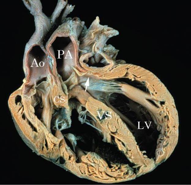
Figure 17.8. Pathologic specimen illustrating DORV with subpulmonary VSD. A sagittal section of DORV with subpulmonary VSD. The conus septum (CS) is malaligned with the lower ventricular septum (VS). The outlet from the left ventricle (LV) is directed into the pulmonary artery (PA). The aorta (Ao) is anterior and to the right.
Position of the Ventricular Septal Defect
Two-dimensional echocardiography accurately predicts the position of the VSD in reference to the great arteries. In most patients, typical subaortic or subpulmonary defects can be demonstrated by parasternal and subcostal scans (see Figs. 17.5, 17.6, 17.7, and 17.10). Doubly-committed defects appear nearly equally committed to both great arteries. Remote or noncommitted defects are isolated or multiple muscular VSDs or complete AV septal defects. Figure 17.11 illustrates a remote posterior muscular VSD in DORV. Neither great artery is committed to the VSD. Similarly, remote complete AV septal defects are best recognized on apical four-chamber or subcostal views (Fig. 17.12).
Doppler echocardiography and color flow Doppler may be helpful adjuncts for demonstrating these abnormalities as well as associated muscular VSDs. Continuous-wave Doppler interrogation of the VSD may demonstrate a high-velocity jet consistent with an LV-to-RV pressure gradient from a restrictive VSD. Although some associated muscular VSDs may be appreciated with color flow imaging, many centers recommend complete angiographic assessment if multiple muscular (“Swiss cheese septum”) VSDs are suspected.
DORV may be associated with a number of AV valve anomalies, including complete AV septal defect, isolated cleft of the anterior mitral leaflet, and overriding (atrial and ventricular septal malalignment) or straddling left or right AV valves. Apical and subcostal four-chamber views and short-axis scans easily demonstrate these AV valve abnormalities at the crux of the heart. It is particularly important to accurately delineate the location and points of insertion of the AV valve chordal apparatus. Abnormal chordal insertions of the tricuspid valve into the conus septum may prohibit surgical efforts to direct the left ventricular outflow anteriorly toward the aorta. Isolated clefts of the anterior mitral leaflet often have chordal attachments to the ventricular septum or attachments that straddle the ventricular septum into the RV. Figure 17.13 illustrates a pathologic specimen of DORV with subpulmonary VSD and straddling mitral valve. Associated anomalies with DORV include left-juxtaposed atrial appendage (see Fig. 17.7), ASD, anomalous systemic (left SVC to coronary sinus) and pulmonary venous connections, and coarctation of the aorta. Multiple imaging planes must be used to exclude these associated anomalies.
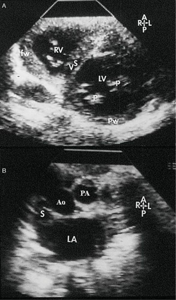
Figure 17.9. Short-axis scans of the heart from apex (top) to base (bottom) in a patient with double-outlet right ventricle. A: Short-axis scan at the mid-ventricular level. This image illustrates the plane of the ventricular septum (VS) below the level of the ventricular septal defect (VSD). B: Short-axis scan at the level of the great arteries. The great arteries are related normally. The superior vena cava (S) is to the right of the aorta (Ao). The pulmonary artery (PA) is to the left and anterior. Thus, the VSD is noted to be subaortic at the cardiac base. (Reprinted with permission from Hagler DJ, Tajik AJ, Seward JB, et al. Double-outlet right ventricle: wide-angle two-dimensional echocardiographic observations. Circulation. 1981;63:419–428.)
CORONARY ANOMALIES
Coronary artery patterns in DORV predominately are of three types: normal; abnormal, as in tetralogy of Fallot; and abnormal, as in transposition of the great arteries. The tetralogy of Fallot-like anomalies may have anomalous origin of the left anterior descending coronary artery from the right coronary artery. The pattern observed in transposition of the great arteries would have origin of the right coronary artery from the right posterior aortic cusp and origin of the left coronary artery from the left posterior cusp. They should be carefully assessed as described previously in these entities.
PHYSIOLOGY
The physiology observed in patients with DORV depends on the four positions of the VSD and the great artery relationships. It is also affected by other associated conditions, most notably PS, the presence of pulmonary vascular obstructive disease, ASD, and the size of the VSD.
Figure 17.14 presents data from 62 cases of DORV reported by Sridaromont et al. There were three categories of patients: those with pulmonary arterial saturation greater than systemic arterial saturation; those with systemic arterial saturation greater than pulmonary arterial saturation; and those with equal systemic and pulmonary arterial saturations. From these data, one can conclude that patients who have systemic arterial saturation higher than pulmonary arterial saturation do not have subpulmonary VSDs, regardless of the presence or absence of PS or pulmonary vascular obstructive disease.
SURGICAL TREATMENT
Because of the complexity of intracardiac repair of these anomalies, it may be necessary to palliate some infants and small children who become symptomatic in the first year of life. Simple forms of DORV with subaortic VSD have been successfully repaired in infancy. For patients with DORV and subpulmonary VSD, the arterial switch operation appears to be the procedure of choice and can be performed in the neonatal period. Pulmonary arterial banding in DORV without PS will reduce pulmonary flow and protect the pulmonary arterioles from obstructive arteriopathy but may induce or aggravate subaortic stenosis due to hypertrophy of the subaortic conus. Pulmonary artery banding may be appropriate in the setting of DORV with multiple muscular VSDs or a remote VSD. Conversely, in patients with DORV and pulmonary stenosis (PS), pulmonary blood flow may need to be augmented. Systemic–to–pulmonary artery shunts will increase pulmonary flow and reduce cyanosis in patients with PS and complex associated anomalies. Forms of DORV that have large VSD physiology or tetralogy of Fallot physiology can be corrected in infancy.

Figure 17.10. Neonate with double-outlet right ventricle (DORV) and right anterior aorta. A: Parasternal long-axis image in a neonate. The scan demonstrates commitment of the aorta (Ao) to the right ventricle (RV) and mitral-aortic discontinuity. The ventricular septal defect (VSD) is subaortic in location. The aorta courses directly superior. B: Parasternal long-axis image in the same patient showing the origin of the pulmonary artery from the right ventricle but with no relationship to the VSD or left ventricle. C: Subcostal scan in the same neonate illustrating the anterior location of the aorta and clear origin of both great arteries from the right ventricle (RV). Prominent conus septum (CS) separates the great arteries and is mal-aligned with the rest of the ventricular septum (VS). There is subpulmonary stenosis. The ventricular septal defect is subaortic in location. AV, aortic valve; LV, left ventricle; PA, pulmonary artery. D: Parasternal short-axis image of same neonate showing the ventricular septal defect (arrow) that is subaortic in location. E: High parasternal short-axis image of both great arteries with the aorta (Ao) anterior and to the right of the pulmonary artery identified by its bifurcation into right (RPA) and left (LPA) pulmonary artery branches. (See Videos 17.1 to 17.6.)

Figure 17.11. Double-outlet right ventricle (DORV). Parasternal short-axis image in a patient with DORV with a remote or noncommitted VSD that is posterior in location (arrows). Neither great artery could be connected through the VSD to the left ventricle (LV). A, anterior; L, left; RV, right ventricle; VS, ventricular septum.
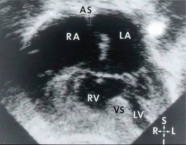
Figure 17.12. Apical four-chamber view of a complete atrioventricular (AV) septal defect in a patient with double-outlet right ventricle (DORV). In some patients, the common AV valve may be unbalanced, favoring one or the other ventricular chamber. In conotruncal defects, the common AV valve is usually undivided and free floating as described with a Type C complete AV septal defect. The more posterior defect in complete AV septal defects may be too remote from the great arteries to allow patch direction of blood from the left ventricle (LV) to one of the great arteries. AS, atrial septum (black arrow); I, inferior; L, left; LA, left atrium; R, right; RA, right atrium; RV, right ventricle; S, superior.

Figure 17.13. Double-outlet right ventricle with subpulmonary VSD and straddling mitral valve. The aorta (Ao) is anterior and right of the pulmonary artery (PA). A cord (arrow) from the mitral valve (MV) crosses over the VSD and inserts along the lower ventricular septum in the right ventricle (RV). CS, conus septum; LV, left ventricle.
Complete correction of DORV depends on the complexity of the intracardiac anatomy. In the surgical correction of DORV, the position of the VSD and its relationship to the great vessels are of paramount importance. The classic form of repair is possible only in those patients with a subaortic VSD. Ideally, in complex anatomy this may be attempted when the patient is about two years old or if an extracardiac conduit is anticipated.
The objectives of the operation are as follows.
1. Establishment of LV-to-aorta continuity. In general, this is accomplished by creating a tunnel between the VSD and the subaortic outflow tract by means of a patch. Care must be exercised to prevent obstruction of this connection. Some VSDs may need to be enlarged to avoid LVOT obstruction and care is needed to avoid injury to the conduction tissue.
2. Establishment of RV–to–pulmonary artery continuity. In the simple forms of the anomaly—with situs solitus and AV concordance in which PS is absent—this requires care in preventing the subaortic tunnel from encroaching on the subpulmonary outflow tract. But, in patients with PS, this may require pulmonary valvotomy, infundibular resection, patch enlargement of the RV outflow tract, or insertion of an extracardiac valved conduit to bring the RV into communication with the pulmonary artery.
3. Repair of complex DORV.
3a:Subpulmonary VSD: When the defect is positioned at a distance from the aortic outflow tract, in the muscular septum posteriorly or below the pulmonary outflow tract, then tunneling from the VSD to the aortic outflow tract is not possible. Under these circumstances, other surgical solutions are required. In subpulmonary VSD, closure of the defect in such a manner as to divert LV blood to the pulmonary artery creates TGA, which is then corrected by an inflow procedure (Mustard or Senning) or by an outflow procedure (Jatene, Kaye-Damus-Stansel, or Aubert). In DORV with left anterior aorta, LV blood should be diverted to the subaortic outflow tract, establishing RV–to–pulmonary artery continuity.

Figure 17.14. Hemodynamic Data. Schematic illustration of hemodynamic data recorded in 62 cases of DORV reported by Sridaromont et al. There are three categories of patients: those with pulmonary arterial saturation greater than systemic arterial saturation, those with systemic arterial saturation greater than pulmonary arterial saturation, and those with pulmonary arterial saturation equal to systemic arterial saturation.
3b:Straddling AV valves: This complicates the anatomy of the VSD and septation may not be possible without replacement of the straddling valve. Alternatively, if the pulmonary pressure and resistance are low, definitive palliation (as opposed to correction) can be achieved by obliteration of the right-sided AV valve and closure of any ASD, interruption of ventriculopulmonary arterial continuity, and establishment of an cavopulmonary connection (bidirectional Glenn and modified Fontan procedures).
3c:DORV with AV discordance can be corrected by closure of the VSD, transection of the pulmonary artery, and establishment of morphologic LV–to–pulmonary artery continuity with an extracardiac conduit. In these patients, the RV remains as the systemic ventricle. Kiser et al. reported repair of dextrocardia, AV discordance, VSD, and DORV. More recently, efforts have been directed to establish continuity between the morphologic LV and the ascending aorta through the VSD to allow physiologic function of the LV as the systemic ventricle. However, this also requires a concomitant atrial switch procedure to direct the pulmonary venous blood to the morphologic LV. Gomes et al. reported the results of complete repair of DORV without PS in 18 patients. The overall operative mortality rate was 22%; higher mortality was encountered in patients with elevated pulmonary arterial resistance and in those with associated lesions, especially AV septal defects. These researchers concluded that patients with DORV and no PS should have early operation before the onset of severe obstructive pulmonary arteriopathy. The same group reported on 22 patients with DORV and PS who underwent complete repair. The overall mortality rate was 32%, but in patients who had surgery after 1960, the mortality rate decreased to 16%. More recent reports of repair in the neonatal period have demonstrated surgical mortality rates of 4% to 8%. Patients with anomalies of coronary distribution, multiple VSDs, or residual PS are at higher risk. Extracardiac conduits may be necessary in cases with complex anatomy.
DOUBLE-OUTLET LEFT VENTRICLE
DOLV is a very rare anomaly most accurately defined as a malformation in which the aorta and the main pulmonary artery both arise predominantly from the morphologic LV. As a conotruncal anomaly, it has features that are very similar to those observed in DORV. As in DORV, the clinical and pathologic features described with DOLV also encompass features of a variety of entities: large VSD, TOF, or complete TGA. DOLV occurs far less frequently than DORV.
Van Praagh et al. provided a complete review of 109 cases of DOLV based on autopsy material, personal communications, and a literature review. This review pointed out the various anatomic conditions associated with DOLV and attempted a categorization of anatomic types of DOLV. It included associated cardiovascular and noncardiovascular anomalies. Similar to the classification scheme for DORV, in DOLV the relationship of the VSD (if present) to the great arteries was of primary importance. Associated anomalies included pulmonary stenosis, subaortic stenosis, and AV valve abnormalities.
DOLV with subaortic VSD was the most common and was observed in 52 (48%) cases, and in 73% of the cases with situs solitus and AV concordance. The patients with subaortic VSD were further categorized based on their great artery relationships as having a right anterior or a left anterior aorta. Figure 17.15 illustrates the most common form of DOLV with a subaortic VSD and right/anterior aorta. The second most frequent form of DOLV was characterized by a subaortic VSD, left/anterior aorta, and PS. The third most common form of DOLV in the Van Praagh et al. series occurred in 11 patients having a subpulmonary VSD (Fig. 17.16). The VSD in this subgroup of patients is described as a high and anterior type of VSD involving the outlet and, specifically, the conus septum. This VSD is in a typical supracristal location, allowing the pulmonary artery to override the ventricular septum and to have a varying degree of commitment to both RV and LV cavities. Because the VSD involves the conus septum, the subpulmonary conus is relatively deficient. In patients with a subpulmonary VSD, the great artery relationships were normal or had a right/anterior aorta (Fig. 17.17).

Figure 17.15. Double-outlet left ventricle (DOLV) with subaortic ventricular septal defect (VSD) and right anterior aorta. Schematic illustration of the VSD and great artery relationships. Note that the VSD involves the membranous and outlet septum. There is fibrous continuity between both semilunar valves and the mitral valve. In DOLV, patients with subaortic VSD and right anterior aorta, 83% had associated pulmonary stenosis. LV, left ventricle; MV, mitral valve; RV, right ventricle; TV, tricuspid valve.
Malalignment of the aortic and pulmonary conus septum relative to the ventricular septum often results in narrowing of the outflow tract with associated aortic valve hypoplasia and stenosis (Fig. 17.18). Arch hypoplasia and coarctation of the aorta may also occur in these patients. Patients with a deficient subpulmonary conus associated with a supracristal type of VSD lack PS and present with typical clinical or hemodynamic findings of a large VSD. Rarely, the VSD in DOLV is doubly committed. As opposed to the rather striking conal development observed in patients with DORV and doubly-committed VSD, patients described with a DOLV and doubly-committed VSD have markedly underdeveloped subarterial conus, which allows aortic–mitral and pulmonary–mitral fibrous continuity. Absence of subarterial conal septum would be consistent with the embryologic explanation proposed by Van Praagh et al. in the morphologic development of DOLV. With a virtual absence of conus septum, none of these patients had PS. Unlike DORV, a remote or noncommitted VSD has been an unusual observation in patients with DOLV. In the Van Praagh et al. series, there were no patients with classic findings of complete AV septal defects and DOLV, nor were any described with muscular VSDs and DOLV.
As noted in the Van Praagh et al. series of DOLV, the vast majority of patients have situs solitus of the atria and viscera and with AV concordance. But, similar to DORV, DOLV has been observed with situs inversus and AV discordance. More commonly, tricuspid atresia or stenosis in association with hypoplastic RV was described in 20 cases of DOLV. These cases were associated with a subaortic VSD. Three patients with DOLV and VSD also had Ebstein anomaly of the tricuspid valve. Rarely, DOLV has been associated with the following: mitral atresia, double-inlet LV, situs ambiguous, or crisscross AV relationships in association with large doubly-committed inlet-to-outlet VSDs.
Surgical Correction of Double-Outlet Left Ventricle
Early surgical reports by Sakakibara et al. and Pacifico et al. emphasized surgical correction of DOLV with two developed ventricles by VSD closure and placement of an RV–to–pulmonary artery conduit. Most patients with DOLV with two ventricles in association with situs solitus should be able to undergo a “two-ventricle” repair. Unlike DORV, the VSD may be simply closed regardless of its relationship to the great arteries. The major exception to this is in those patients with an anterior subpulmonary VSD involving the conal septum. In this example, all the patients who have been reported had a right lateral or right anterior aorta, and therefore patch closure of the VSD excluding the pulmonary artery from the LV, would represent those who received the easiest and most direct type of repair. However, this also may be altered by the presence of subaortic or aortic valvar stenosis. If severe subaortic stenosis cannot be relieved by resection, VSD closure leaving the pulmonary artery to the LV with creation of an aortopulmonary window and placement of an RV–to–pulmonary artery conduit may be an option for repair. The reports by Pacifico et al. and others suggested that other forms could be repaired by VSD closure but that most also required closure of the native LV–to–pulmonary outflow and placement of an RV–to–pulmonary artery conduit. DeLeon et al. described pulmonary root translocation as an alternative for biventricular repair of DOLV. More complex forms of DOLV with functional single ventricle or AV valve atresia will require a Fontan type of correction.

Figure 17.16. Transesophageal echocardiography images of double-outlet left ventricle (DOLV) with subaortic ventricular septal defect (VSD) and normally related great arteries. A: Scan in the left ventricular long axis showing aortic–mitral continuity and patch closure (arrow) of a large VSD. Ao, aorta; LA, left atrium; LV, left ventricle; RV, right ventricle; VS, ventricular septum. B: A slightly rotated leftward scan illustrating the anterior pulmonary outflow (arrow) also committed to the left ventricle (LV). AV, aortic valve; MV, mitral valve. C: Short-axis scans demonstrate the great artery relationship with the aorta posterior and to the right. The aortic valve (AV) and pulmonary valve (PV) are on the same level. D: Slightly lower short-axis scan also demonstrating the VSD patch position (arrow) in the membranous-to-outlet portion of the septum medial to the aortic valve (AV).
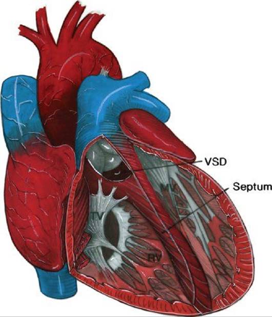
Figure 17.17. Schematic illustration demonstrating double-outlet left ventricle (DOLV) with subpulmonary ventricular septal defect (VSD) and right posterior aorta (normally related great arteries). The VSD is anterior or supracristal and involves the outlet conus septum. The pulmonary artery overrides the VSD but is predominantly committed to the left ventricle. Without pulmonary stenosis, it is evident that this defect would be hemodynamically similar to a large VSD with pulmonary hyperperfusion. MV, mitral valve; RV, right ventricle; TV, tricuspid valve.
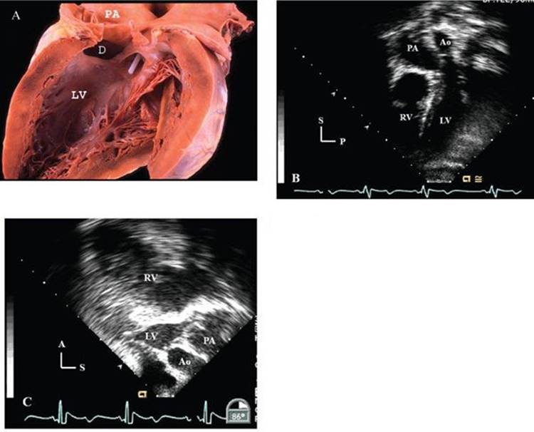
Figure 17.18. Double-outlet LV (DOLV) with aortic stenosis. A: Pathologic example of DOLV with subpulmonary VSD with aortic stenosis and aortic annular hypoplasia. The white probe is exiting the aortic outflow tract and both outflow tracts are predominantly committed to the left ventricle. B: TTE image of a 12-year-old patient with features suggestive of DOLV and aortic stenosis. Although the pulmonary outflow overrides the ventricular septum in this subcostal image, the aortic outflow clearly exits the LV and is moderately hypoplastic. C:TEE image of the same patient at the time of RV conduit replacement shows a typical long-axis image. The pulmonary outflow again is noted to override the ventricular septum; however, the aortic outflow is committed to the LV and there is mitral–aortic continuity. There is severe aortic hypoplasia and subaortic stenosis. These images could also be classified as normally related great arteries with AS. A, anterior; Ao, aorta; D, defect; LV, left ventricle; P, posterior; PA, pulmonary artery; RV, right ventricle; S, superior.
SUGGESTED READING
Ciaravella JM Jr, McGoon DC, Hagler DJ, et al. Caplike double-horned double-outlet right ventricle: report of two cases. J Thorac Cardiovasc Surg. 1979;77:536–542.
Davachi F, Möller JH, Edwards JF. Origin of both great vessels from right ventricle with intact ventricular septum. Am Heart J. 1968;75:790–794.
DeLeon SY, Ow EP, Chiemmongkoltip P, et al. Alternatives in biventricular repair of double-outlet left ventricle. Ann Thorac Surg. 1995;60:213–216.
Gomes MMR, Weidman WH, McGoon DC, Danielson GK. Double-outlet right ventricle with pulmonic stenosis: surgical considerations and results of operation. Circulation. 1971;43: 131–136.
Gomes MMR, Weidman WH, McGoon DC, et al. Double-outlet right ventricle with pulmonic stenosis: surgical considerations and results of operation. Circulation. 1971;43:889–894.
Goor DA, Edwards JE. The spectrum of transposition of the great arteries: with specific reference to developmental anatomy of the conus. Circulation. 1973;48:406–415.
Grant RP. The morphogenesis of transposition of the great vessels. Circulation. 1962;26:819–840.
Hagler DJ, Tajik AJ, Seward JB, et al. Double-outlet right ventricle: wide-angle two-dimensional echocardiographic observations. Circulation. 1981;63:419–428.
Hagler DJ, Tajik AJ, Seward JB, et al. Wide-angle two-dimensional echocardiographic profiles of conotruncal abnormalities. Mayo Clin Proc. 1980;55:73–82.
Judson JP, Danielson GK, Ritter DG, et al. Successful repair of co-existing double-outlet right ventricle and two-chamber right ventricle. J Thorac Cardiovasc Surg. 1982;84:113–121.
Kiser JC, Ongley PA, Kirklin JW, et al. Surgical treatment of dextrocardia with inversion of ventricles and double-outlet right ventricle. J Thorac Cardiovasc Surg. 1968;55:6–15.
Kleinert S, Sano T, Weintraub RB, et al. Anatomic features and surgical strategies in double-outlet right ventricle. Circulation. 1997;96:1233–1239.
Lev M, Bharati S, Meng L, et al. A concept of double-outlet right ventricle. J Thorac Cardiovasc Surg. 1972;64:271–281.
Manner J, Seidl W, Steding G. Embryological observations on the morphogenesis of double-outlet right ventricle with subaortic ventricular septal defect and normal arrangement of the great arteries. Thorac Cardiovasc Surg. 1995;43:307–312.
Mitchell SC, Korones SB, Berendes HW. Congenital heart disease in 56,109 births: incidence and natural history. Circulation. 1971;43:323–332.
Neufeld HN, DuShane JW, Edwards JE. Origin of both great vessels from the right ventricle. II: With pulmonary stenosis. Circulation. 1961;23:603–612.
Pacifico AD, Kirklin JW, Bargeron LM Jr. Complex congenital malformations: surgical treatment of double-outlet right ventricle and double-outlet left ventricle. In: Kirklin JW, ed. Advanced Cardiovascular Surgery. New York: Grune & Stratton, 1973:57.
Pacifico AD, Kirklin JW, Bargeron LM, et al. Surgical treatment of double-outlet LV. Report of four cases. Circulation. 1973;48(suppl III):III19–III23.
Paul MH, Sinha SN, Muster AJ, et al. Double-outlet left ventricle with an intact ventricular septum: Clinical and autopsy diagnosis and developmental implications. Circulation. 1970;41:129–139.
Ruttenberg HD, Anderson RC, Elliott LP, et al. Origin of both great vessels from the arterial ventricle: a complex with ventricular inversion. Br Heart J. 1964;26:631–641.
Sridaromont S, Feldt RH, Ritter DG, et al. Double-outlet right ventricle: hemodynamic and anatomic correlations. Am J Cardiol. 1976;38:85–94.
Sridaromont S, Ritter DG, Feldt RH, et al. Double-outlet right ventricle: anatomic and angiocardiographic correlations. Mayo Clin Proc. 1978;53:555–577.
Stellin G, Ho SY, Anderson RH, et al. The surgical anatomy of double-outlet right ventricle with concordant atrioventricular connection and noncommitted ventricular septal defect. J Thorac Cardiovasc Surg. 1991;102:849–855.
Taussig HB, Bing RJ. Complete transposition of the aorta and a levoposition of the pulmonary artery: clinical, physiological and pathological findings. Am Heart J. 1949;37;551–559.
Uemura H, Yagihara T, Kawashima Y, et al. Coronary arterial anatomy in double-outlet right ventricle with subpulmonary VSD. Ann Thorac Surg. 1995;59:591–597.
Van Mierop LHS, Wiglesworth FW. Pathogenesis of transposition complexes. II: Anomalies due to faulty transfer of the posterior great artery. Am J Cardiol. 1963;12:226–232.
Van Praagh R, Weinberg PM, Srebro JP. Double-outlet left ventricle. In: Adams FH, Emmanouilides GC, Riemenschneider JA, eds. Moss’ Heart Disease in Infants, Children, and Adolescents, 4th ed. Baltimore: Williams & Wilkins, 1989:461–485.
Van Praagh S, Davidoff A, Chin A, et al. Double-outlet right ventricle: anatomic types and developmental implications based on a study of 101 cases. Coeur (Paris). 1982;12:389–439.
Vierordt H. Die angeborenen herzkrankheiten. In: Nothnagel’s Spez., ed. Path Therapie. 1898;15:244.
Wilcox BR, Ho SY, Macartney FJ, et al. Surgical anatomy of double-outlet right ventricle with situs solitus and atrioventricular concordance. J Thorac Cardiovasc Surg. 1981;82:405–417.
Questions
1.What would the definition of Double Outlet Right Ventricle (DORV)best include?
A.Transposed origin of both great arteries
B.Origin of both great arteries from the morphologic right ventricle
C.All patients with subaortic ventricular septal defect
D.Patients who lack subaortic conus
2.What would the definition of Double Outlet Left Ventricle (DOLV) best include?
A.All patients with hypoplastic left-heart syndrome
B.All patients who lack subaortic conus
C.Origin of both great arteries from the morphologic left ventricle
D.Transposed origin of both great arteries
3.What typical hemodynamic features are found in patients with Double Outlet Right Ventricle?
A.Variable (excessive or reduced) pulmonary blood flow.
B.Severe left ventricular outflow tract obstruction
C.Severe right ventricular outflow tract obstruction
D.Multiple sites of left ventricular outflow obstruction
4.Classification of the types of Double Outlet Right Ventricle is based on:
A.echocardiographic features of left ventricular outflow tract obstruction.
B.presence or absence of aortic valve stenosis.
C.great artery locations and the location of the ventricular septal defect.
D.types of orientation of the great artery conus muscle.
5.What type of ventricular septal defect is most common in patients with atrioventricular septal defects?
A.Remote or noncommited VSD
B.Subaortic VSD
C.Subpulmonary VSD
D.Doubly committed VSD
6.What is the most typical and common type of Double Outlet Right Ventricle?
A.Transposition of the great arteries
B.Left anterior aorta
C.Complete atrioventricular septal defect
D.Side-by-side great arteries and subaortic VSD
7.How would a muscular ventricular septal defect in Double Outlet Right Ventricle best be classified?
A.Subaortic VSD
B.Doubly committed VSD
C.Remote VSD
D.Subpulmonary VSD
8.What would Double Outlet Right Ventricle patients with systemic saturations higher than the pulmonary artery saturation typically not have?
A.Subaortic VSD
B.Subpulmonary VSD
C.Remote VSD
D.Double committed VSD
9.What would the most common form of Double Outlet Left Ventricle have?
A.Remote VSD
B.Subpulmonary VSD
C.Subaortic VSD
D.Doubly committed VSD
10.With what is Double Outlet Left Ventricle most commonly associated?
A.Criss-cross atrioventricular relationships
B.Situs solitus
C.Situs inversus
D.Situs ambiguous
Answers
1.Answer: B. While the other answers may occasionally be correct, the classic definition of DORV is that both great arteries nearly entirely originate from the right ventricle.
2.Answer: C. The correct definition of DOLV would be the origin of both great arteries from the morphologic left ventricle.
3.Answer: A. Outflow tract obstruction can occur with DORV, but not in all cases. Therefore, the correct answer primarily varies according to the volume of pulmonary flow.
4.Answer: C. Since surgical correction eventually is the planned management of DORV patients, their classification usually depicts those features that are important for the surgical correction. Therefore, this classification is based on the great artery location and the VSD location relative to the great arteries—features that will determine the surgical correction.
5.Answer: A. AV septal defects are posterior in location, therefore they are usually remote from the great arteries.
6.Answer: D. The most common type of DORV occurs with side-by-side great arteries with a subaortic VSD. The next most common occurs with transposition, but the others are less common.
7.Answer: C. Since muscular VSD would be separated from the great arteries, by definition they would be remote from them.
8.Answer: B. This hemodynamic observation is noted in Fig. 17.14, showing that no patient with subpulmonary VSD had a systemic saturation higher than the pulmonary artery saturation. They behave like patients with transposition of the great arteries.
9.Answer: C. The data shows that the most common form has a subaortic VSD.
10.Answer: B. While DOLV can occur with situs abnormalities, most have situs solitus.