
David D. Spragg  Gordon F. Tomaselli
HISTORY AND INTRODUCTION
The field of cardiac electrophysiology was ushered in with the development of the electrocardiogram (ECG) by Einthoven at the turn of the twentieth century. The recording of cellular membrane currents revealed that the body surface ECG is the timed sum of the cellular action potentials in the atria and ventricles. In the late 1960s, the development of intracavitary recording, in particular the ability to record His bundle electrograms with programmed stimulation of the heart, marked the beginning of contemporary clinical electrophysiology. Adoption of radio frequency technology to ablate cardiac tissue in the early 1990s heralded the birth of interventional cardiac electrophysiology.
The clinical problem of sudden death caused by ventricular arrhythmias, most commonly in the setting of coronary obstruction, was recognized as early as the late nineteenth century. The problem was vexing and led to the development of pharmacologic and nonpharmacologic therapies, including transthoracic defibrillators, cardiac massage, and, most recently, implantable defibrillators. Over time the limitations of antiarrhythmic drug therapy have been highlighted repeatedly in clinical trials, and now ablation and devices are first-line therapy for a number of cardiac arrhythmias.
In the last two decades, the genetic basis of a number of heritable arrhythmias has been elucidated, revealing important insights into the mechanisms not only of these rare arrhythmias but also of similar rhythm disturbances observed in more common forms of heart disease.
DESCRIPTIVE PHYSIOLOGY
The normal cardiac impulse is generated by pacemaker cells in the sinoatrial node situated at the junction of the right atrium and the superior vena cava (see Fig. 11-1). This impulse is transmitted slowly through nodal tissue to the anatomically complex atria, where it is conducted more rapidly to the atrioventricular node (AVN), inscribing the P wave of the ECG (see Fig. 11-2). There is a perceptible delay in conduction through the anatomically and functionally heterogeneous AVN. The time needed for activation of the atria and the AVN delay is represented as the PR interval of the ECG. The AVN is the only electrical connection between the atria and the ventricles in the normal heart. The electrical impulse emerges from the AVN and is transmitted to the His-Purkinje system, specifically the common bundle of His, then the left and right bundle branches, and then to the Purkinje network, facilitating activation of ventricular muscle. In normal circumstances, the ventricles are activated rapidly in a well-defined fashion that is determined by the course of the Purkinje network, and this inscribes the QRS complex (see Fig. 11-2). Recovery of electrical excitability occurs more slowly and is governed by the time of activation and duration of regional action potentials. The relative brevity of epicardial action potentials in the ventricle results in repolarization that occurs first on the epicardial surface and then proceeds to the endocardium, which inscribes a T wave normally of the same polarity as the QRS complex. The duration of activation and recovery is determined by the action potential duration represented on the body surface ECG by the QT interval (see Fig. 11-2).
Cardiac myocytes exhibit a characteristically long action potential (200–400 ms) compared with neurons and skeletal muscle cells (1–5 ms). The action potential profile is sculpted by the orchestrated activity of multiple distinctive time- and voltage-dependent ionic currents (Fig. 14-1A). The currents in turn are carried by transmembrane proteins that passively conduct ions down their electrochemical gradients through selective pores (ion channels), actively transport ions against their electrochemical gradient (pumps, transporters), or electrogenically exchange ionic species (exchangers).
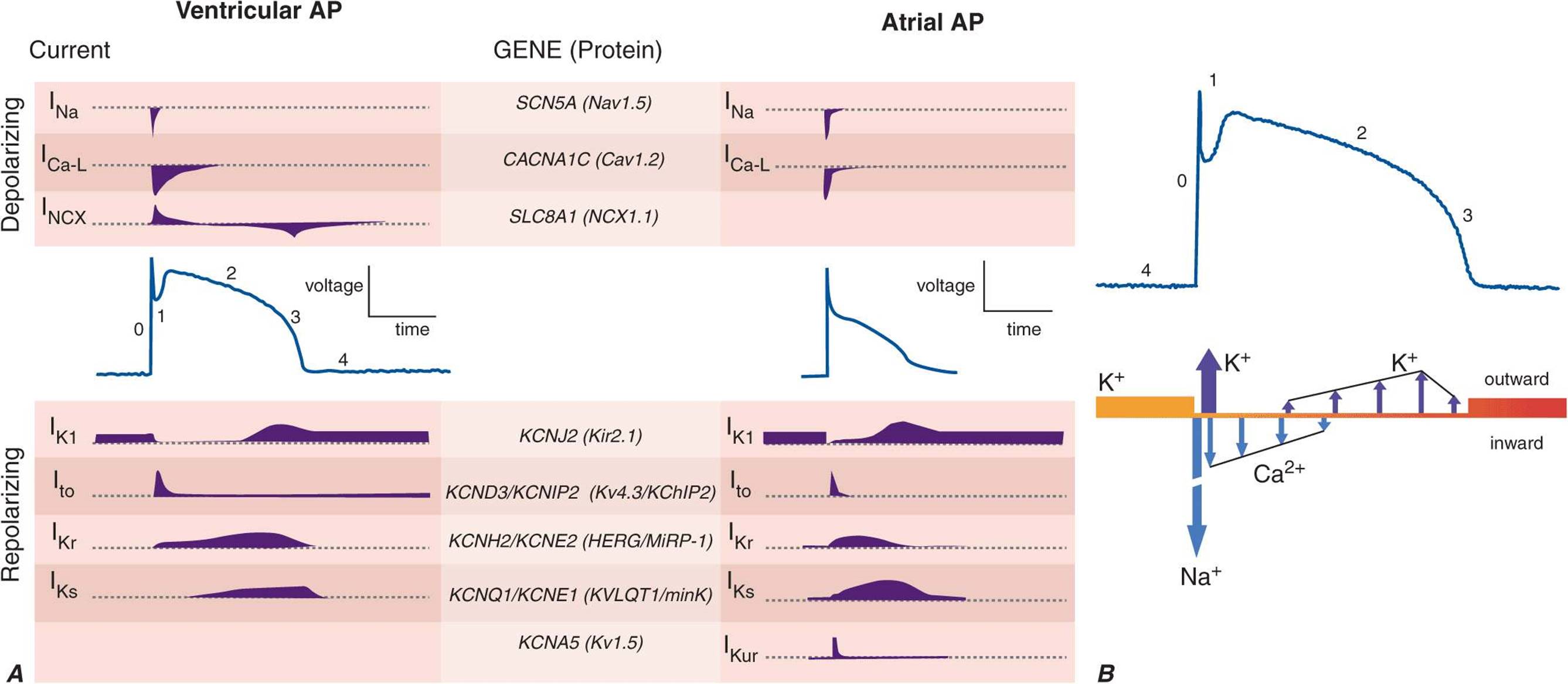
FIGURE 14-1
A. Cellular atrial and ventricular action potentials. Phases 0–4 are the rapid upstroke, early repolarization, plateau, late repolarization, and diastole, respectively. The ionic currents and their respective genes are shown above and below the action potentials. The currents that underlie the action potentials vary in atrial and ventricular myocytes. B. A ventricular action potential with a schematic of the ionic currents flowing during the phases of the action potential. Potassium current (IK1) is the principal current during phase 4 and determines the resting membrane potential of the myocyte. Sodium current generates the upstroke of the action potential (phase 0); activation of Ito with inactivation of the Na current inscribes early repolarization (phase 1). The plateau (phase 2) is generated by a balance of repolarizing potassium currents and depolarizing calcium current. Inactivation of the calcium current with persistent activation of potassium currents (predominantly IKr and IKs) causes phase 3 repolarization.
Action potentials in the heart are regionally distinct. The regional variability in cardiac action potentials is a result of differences in the number and types of ion channel proteins expressed by different cell types in the heart. Further, unique sets of ionic currents are active in pacemaking and muscle cells, and the relative contributions of these currents may vary in the same cell type in different regions of the heart (Fig. 14-1A).
Ion channels are complex, multisubunit transmembrane glycoproteins that open and close in response to a number of biologic stimuli, including a change in membrane voltage, ligand binding (directly to the channel or to a G protein–coupled receptor), and mechanical deformation (Fig. 14-2). Other ion motive exchangers and transporters contribute importantly to cellular excitability in the heart. Ion pumps establish and maintain the ionic gradients across the cell membrane that serve as the driving force for current flow through ion channels. Transporters or exchangers that do not move ions in an electrically neutral manner (e.g., the sodium-calcium exchanger transports three Na+ for one Ca2+) are termed electrogenic and contribute directly to the action potential profile.

FIGURE 14-2
Topology and subunit composition of the voltage-dependent ion channels. Potassium channels are formed by the tetramerization of α or pore-forming subunits and one or more β subunits; only single β subunits are shown for clarity. Sodium and calcium channels are composed of α subunits with four homologous domains and one or more ancillary subunits. In all channel types the loop of protein between the fifth and sixth membrane-spanning repeat in each subunit or domain forms the ion-selective pore. In the case of the sodium channel, the channel is a target for phosphorylation, the linker between the third and fourth homologous domain is critical to inactivation, and the sixth membrane-spanning repeat in the fourth domain is important in local anesthetic antiarrhythmic drug binding.
The most abundant superfamily of ion channels expressed in the heart is voltage gated. Several structural themes are common to all voltage-dependent ion channels. First, the architecture is modular, consisting either of four homologous subunits (e.g., K channels) or of four internally homologous domains (e.g., Na and Ca channels). Second, the proteins fold around a central pore lined by amino acids that exhibit exquisite conservation within a given channel family of like selectivity (e.g., jellyfish, eel, fruit fly, and human Na channels have very similar P segments). Third, the general strategy for activation gating (opening and closing in response to changes in membrane voltage) is highly conserved: the fourth trans-membrane segment (S4), studded with positively charged residues, lies within the membrane field and moves in response to depolarization, opening the channel. Fourth, most ion channel complexes include not only the pore-forming proteins (α subunits) but also auxiliary subunits (e.g., β subunits) that modify channel function (Fig. 14-2).
Na and Ca channels are the primary carriers of depolarizing current in both the atria and the ventricles; inactivation of these currents and activation of repolarizing K currents hyperpolarize the heart cells, reestablishing the negative resting membrane potential (Fig. 14-1B). The plateau phase is a time when little current is flowing, and relatively minor changes in depolarizing or repolarizing currents can have profound effects on the shape and duration of the action profile. Mutations in subunits of these channel proteins produce arrhythmogenic alterations in the action potentials that cause long and short QT syndrome, idiopathic ventricular fibrillation, familial atrial fibrillation, and some forms of conduction system disease.
MECHANISMS OF CARDIAC ARRHYTHMIAS
Cardiac arrhythmias result from abnormalities of electrical impulse generation, conduction, or both. Bradyarrhythmias typically arise from disturbances in impulse formation at the level of the sinoatrial node or from disturbances in impulse propagation at any level, including exit block from the sinus node, conduction block in the AV node, and impaired conduction in the His-Purkinje system. Tachyarrhythmias can be classified according to mechanism, including enhanced automaticity (spontaneous depolarization of atrial, junctional, or ventricular pacemakers), reentry (circus propagation of a depolarizing wavefront), or triggered arrhythmias (initiated by afterdepolarizations) occurring during or immediately after cardiac repolarization, during phase 3 or 4 of the action potential. A variety of mapping and pacing maneuvers typically performed during invasive electrophysiologic testing can often determine the underlying mechanism of a tachyarrhythmia (Table 14-1).
TABLE 14-1
ARRHYTHMIA MECHANISMS

Alterations in impulse initiation: Automaticity
Spontaneous (phase 4) diastolic depolarization underlies the property of automaticity (pacemaking) characteristic of cells in the sinoatrial (SA) and atrioventricular (AV) nodes, His-Purkinje system, coronary sinus, and pulmonary veins. Phase 4 depolarization results from the concerted action of a number of ionic currents, including K+ currents, Ca2+ currents, electrogenic Na, K-ATPase, the Na-Ca exchanger, and the so-called funny, or pacemaker, current (If); however, the relative importance of these currents remains controversial.
The rate of phase 4 depolarization and, therefore, the firing rates of pacemaker cells are dynamically regulated. Prominent among the factors that modulate phase 4 is autonomic nervous system tone. The negative chronotropic effect of activation of the parasympathetic nervous system is a result of the release of acetylcholine that binds to muscarinic receptors, releasing G protein βγ subunits that activate a potassium current (IKACh) in nodal and atrial cells. The resulting increase in K+ conductance opposes membrane depolarization, slowing the rate of rise of phase 4 of the action potential. Conversely, augmentation of sympathetic nervous system tone increases myocardial catecholamine concentrations, which activate both α– and β–adrenergic receptors. The effect of β1-adrenergic stimulation predominates in pacemaking cells, augmenting both L-type Ca current (ICa-L) and If, thus increasing the slope of phase 4. Enhanced sympathetic nervous system activity can dramatically increase the rate of firing of SA nodal cells, producing sinus tachycardia with rates >200 beats/min. By contrast, the increased rate of firing of Purkinje cells is more limited, rarely producing ventricular tachyarrhythmias >120 beats/min.
Normal automaticity may be affected by a number other factors associated with heart disease. Hypokalemia and ischemia may reduce the activity of Na, K-ATPase, thereby reducing the background repolarizing current and enhancing phase 4 diastolic depolarization. The end result would be an increase in the spontaneous firing rate of pacemaking cells. Modest increases in extracellular potassium may render the maximum diastolic potential more positive, thereby also increasing the firing rate of pacemaking cells. A more significant increase in [K+]o, however, renders the heart inexcitable by depolarizing the membrane potential.
Normal or enhanced automaticity of subsidiary latent pacemakers produces escape rhythms in the setting of failure of more dominant pacemakers. Suppression of a pacemaker cell by a faster rhythm leads to an increased intracellular Na+ load ([Na+]i), and extrusion of Na+ from the cell by Na, K-ATPase produces an increased background repolarizing current that slows phase 4 diastolic depolarization. At slower rates, [Na+]i is decreased, as is the activity of the Na, K-ATPase, resulting in progressively more rapid diastolic depolarization and warm-up of the tachycardia rate. Overdrive suppression and warm-up are characteristic of, but may not be observed in, all automatic tachycardias. Abnormal conduction into tissue with enhanced automaticity (entrance block) may blunt or eliminate the phenomena of overdrive suppression and warm-up of automatic tissue.
Abnormal automaticity may underlie atrial tachycardia, accelerated idioventricular rhythms, and ventricular tachycardia, particularly that associated with ischemia and reperfusion. It has also been suggested that injury currents at the borders of ischemic myocardium may depolarize adjacent nonischemic tissue, predisposing to automatic ventricular tachycardia.
Afterdepolarizations and triggered automaticity
Triggered automaticity or activity refers to impulse initiation that is dependent on afterdepolarizations (Fig. 14-3). Afterdepolarizations are membrane voltage oscillations that occur during (early afterdepolarizations, EADs) or after (delayed afterdepolarizations, DADs) an action potential.
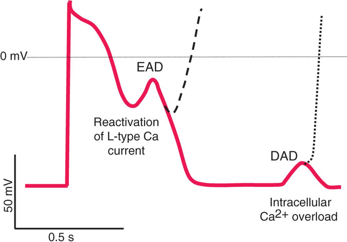
FIGURE 14-3
Schematic action potentials with early afterdepolarizations (EADs) and delayed afterdepolarizations (DADs). Afterdepolarizations are spontaneous depolarizations in cardiac myocytes. EADs occur before the end of the action potential (phases 2 and 3), interrupting repolarization. DADs occur during phase 4 of the action potential after completion of repolarization. The cellular mechanisms of EADs and DADs differ (see text).
The cellular feature common to the induction of DADs is the presence of an increased Ca2+ load in the cytosol and sarcoplasmic reticulum. Digitalis glycoside toxicity, catecholamines, and ischemia all can enhance Ca2+ loading sufficiently to produce DADs. Accumulation of lysophospholipids in ischemic myocardium with consequent Na+ and Ca2+ overload has been suggested as a mechanism for DADs and triggered automaticity. Cells from damaged areas or cells that survive a myocardial infarction may display spontaneous release of calcium from the sarcoplasmic reticulum, and this may generate “waves” of intracellular calcium elevation and arrhythmias.
EADs occur during the action potential and interrupt the orderly repolarization of the myocyte. Traditionally, EADs have been thought to arise from action potential prolongation and reactivation of depolarizing currents, but more recent experimental evidence suggests a previously unappreciated interrelationship between intracellular calcium loading and EADs. Cytosolic calcium may increase when action potentials are prolonged. This, in turn, appears to enhance L-type Ca current, further prolonging action potential duration as well as providing the inward current driving EADs. Intracellular calcium loading by action potential prolongation may also enhance the likelihood of DADs. The interrelationship among intracellular [Ca2+], EADs, and DADs may be one explanation for the susceptibility of hearts that are calcium loaded (e.g., in ischemia or congestive heart failure) to develop arrhythmias, particularly on exposure to action potential–prolonging drugs.
EAD-triggered arrhythmias exhibit rate dependence. In general, the amplitude of an EAD is augmented at slow rates when action potentials are longer. Indeed, a fundamental condition that underlies the development of EADs is action potential and QT prolongation. Hypokalemia, hypomagnesemia, bradycardia, and, most commonly, drugs can predispose to the generation of EADs, invariably in the context of prolonging the action potential. Antiarrhythmics with class IA and III action (see later) produce action potential and QT prolongation intended to be therapeutic but frequently causing arrhythmias. Noncardiac drugs such as phenothiazines, nonsedating antihistamines, and some antibiotics can also prolong the action potential duration and predispose to EAD-mediated triggered arrhythmias. Decreased [K+]o paradoxically may decrease membrane potassium currents (particularly the delayed rectifier current, IKr) in the ventricular myocyte, explaining why hypokalemia causes action potential prolongation and EADs. In fact, potassium infusions in patients with the congenital long QT syndrome (LQTS) and in those with drug-induced acquired QT prolongation shorten the QT interval.
EAD-mediated triggered activity probably underlies initiation of the characteristic polymorphic ventricular tachycardia, torsades des pointes, seen in patients with congenital and acquired forms of LQTS. Structural heart disease such as cardiac hypertrophy and failure may also delay ventricular repolarization (so-called electrical remodeling) and predispose to arrhythmias related to abnormalities of repolarization. The abnormalities of repolarization in hypertrophy and heart failure are often magnified by concomitant drug therapy or electrolyte disturbances.
Abnormal impulse conduction: Reentry
The most common arrhythmia mechanism is reentry. Fundamentally, reentry is defined as circulation of an activation wave around an inexcitable obstacle. Thus, the requirements for reentry are two electrophysiologically dissimilar pathways for impulse propagation around an inexcitable region such that unidirectional block occurs in one of the pathways and a region of excitable tissue exists at the head of the propagating wavefront (Fig. 14-4). Structural and electrophysiologic properties of the heart may contribute to the development of the inexcitable obstacle and that of unidirectional block. The complex geometry of muscle bundles in the heart and spatial heterogeneity of cellular coupling or other active membrane properties (i.e., ionic currents) appear to be critical.

FIGURE 14-4
Schematic diagram of reentry. A. The circuit contains two limbs, one with slow conduction. B. A premature impulse blocks in the fast pathway and conducts over the slow pathway, allowing the fast pathway to recover so that the activation wave can reenter the fast pathway from the retrograde direction. C. During sustained reentry utilizing such a circuit, a gap (excitable gap) exists between the activating head of the wave and the recovering tail. D. One mechanism of termination of reentry occurs when the conduction and recovery characteristics of the circuit change and the activating head of the wave collides with the tail, extinguishing the tachycardia.
A key feature in classifying reentrant arrhythmias, particularly for therapy, is the presence and size of an excitable gap. An excitable gap exists when the tachycardia circuit is longer than the tachycardia wavelength (λ = conduction velocity × refractory period, representing the size of the circuit that can sustain reentry), allowing appropriately timed stimuli to reset propagation in the circuit. Reentrant arrhythmias may exist in the heart in the absence of an excitable gap and with a tachycardia wavelength nearly the same size as the path length. In this case, the wavefront propagates through partially refractory tissue with no anatomic obstacle and no fully excitable gap; this is referred to as leading circle reentry, a form of functional reentry (reentry that depends on functional properties of the tissue). Unlike excitable gap reentry, there is no fixed anatomic circuit in leading circle reentry, and it may therefore not be possible to disrupt the tachycardia with pacing or destruction of a part of the circuit. Furthermore, the circuit in leading circle reentry tends to be less stable than that in excitable gap reentrant arrhythmias, with large variations in cycle length and a predilection to termination.
Anatomically determined, excitable gap reentry can explain several clinically important tachycardias, such as AV reentry, atrial flutter, bundle branch reentry ventricular tachycardia, and ventricular tachycardia in scarred myocardium. There is strong evidence to suggest that other, less organized arrhythmias, such as atrial and ventricular fibrillation, are associated with more complex activation of the heart and are due to functional reentry.
Structural heart disease is associated with changes in conduction and refractoriness that increase the risk of reentrant arrhythmias. Chronically ischemic myocardium exhibits a downregulation of the gap junction channel protein (connexin 43) that carries intercellular ionic current. The border zones of infarcted and failing ventricular myocardium exhibit not only functional alterations of ionic currents but also remodeling of tissue and altered distribution of gap junctions. The changes in gap junction channel expression and distribution, in combination with macroscopic tissue alterations, support a role for slowed conduction in reentrant arrhythmias that complicate chronic coronary artery disease (CAD). Aged human atrial myocardium exhibits altered conduction, manifest as highly fractionated atrial electrograms, producing an ideal substrate for the reentry that may underlie the very common development of atrial fibrillation in the elderly.
APPROACH TO THE PATIENT Cardiac Arrhythmias
The evaluation of patients with suspected cardiac arrhythmias is highly individualized; however, two key features—the history and ECG—are pivotal in directing the diagnostic workup and therapy. Patients with cardiac arrhythmias exhibit a wide spectrum of clinical presentations that range from asymptomatic ECG abnormalities to survival from cardiac arrest. In general, the more severe the presenting symptoms are, the more aggressive the evaluation and treatment are. Loss of consciousness that is believed to be of cardiac origin typically mandates an exhaustive search for the etiology and often requires invasive, device-based therapy. The presence of structural heart disease and prior myocardial infarction dictates a change in the approach to the management of syncope or ventricular arrhythmias. The presence of a family history of serious ventricular arrhythmias or premature sudden death will influence the evaluation of presumed heritable arrhythmias.
The physical examination is focused on determining whether there is cardiopulmonary disease that is associated with specific cardiac arrhythmias. The absence of significant cardiopulmonary disease often, but not always, suggests benignity of the rhythm disturbance. In contrast, palpitations, syncope, or near syncope in the setting of significant heart or lung disease have more ominous implications. In addition, the physical examination may reveal the presence of a persistent arrhythmia such as atrial fibrillation.
The judicious use of noninvasive diagnostic tests is an important element in the evaluation of patients with arrhythmias, and there is no test more important than the ECG, particularly if recorded at the time of symptoms. Uncommon but diagnostically important signatures of electrophysiologic disturbances may be unearthed on the resting ECG, such as delta waves in Wolff-Parkinson-White (WPW) syndrome, prolongation or shortening of the QT interval, right precordial ST-segment abnormalities in Brugada syndrome, and epsilon waves in arrhythmogenic right ventricular dysplasia. Variants of body surface ECG recording can provide important information about arrhythmia substrates and triggers. Holter monitoring and event recording, either continuous or intermittent, record the body surface ECG over longer periods, enhancing the possibility of observing the cardiac rhythm during symptoms. Implantable long-term monitors and commercial ambulatory ECG monitoring services permit prolonged telemetric monitoring both for diagnosis and to assess the efficacy of therapy.
Long-term recordings permit the assessment of the time-varying behavior of the heart rhythm. Heart rate variability (HRV) and QT interval variability (QTV) provide noninvasive methods to assess autonomic nervous system influence on the heart. A decrease in HRV has been associated with increased sympathetic nervous system tone and increased mortality rates in patients after myocardial infarction. Signal-averaged electrocardiography (SAECG) uses signal-averaging techniques to amplify small potentials in the body surface ECG that are associated with slow conduction in the myocardium. The presence of these small potentials, referred to as late potentials because of their timing with respect to the QRS complex, and prolongation of the filtered (or averaged) QRS duration are indicative of slowed conduction in the ventricle and have been associated with an increased risk of ventricular arrhythmias after myocardial infarction. Exercise electrocardiography is important in determining the presence of myocardial demand ischemia; more recently, analysis of the morphology of the QT interval with exercise has been used to assess the risk of serious ventricular arrhythmias. Microscopic alterations in the T wave (T wave alternans, TWA) at low heart rates may identify patients at risk for ventricular arrhythmias. Cardiac imaging plays an important role in the detection and characterization of myocardial structural abnormalities that may render the heart more susceptible to arrhythmia. Ventricular tachyarrhythmias, for instance, occur more frequently in patients with ventricular systolic dysfunction and chamber dilation, in hypertrophic cardiomyopathy, and in the setting of infiltrative diseases such as sarcoidosis. Supraventricular arrhythmias may be associated with particular congenital conditions, including AV reentry in the setting of Ebstein’s anomaly. Echocardiography is a frequently employed imaging technique to screen for disorders of cardiac structure and function. Increasingly, magnetic resonance (MR) imaging of the myocardium is being used to screen for scar burden, fibrofatty infiltration of the myocardium as seen in arrhythmogenic right ventricular cardiomyopathy, and other structural changes that affect arrhythmia susceptibility.
Head-up tilt (HUT) testing is useful in the evaluation of some patients with syncope. The physiologic response to HUT is incompletely understood; however, redistribution of blood volume and increased ventricular contractility occur consistently. Exaggerated activation of a central reflex in response to HUT produces a stereotypic response of an initial increase in heart rate, then a drop in blood pressure followed by a reduction in heart rate characteristic of neurally mediated hypotension. Other responses to HUT may be observed in patients with orthostatic hypotension and autonomic insufficiency. HUT is used most often in patients with recurrent syncope, although it may be useful in patients with single syncopal episodes with associated injury, particularly in the absence of structural heart disease. In patients with structural heart disease, HUT may be indicated in those with syncope, in whom other causes (e.g., asystole, ventricular tachyarrhythmias) have been excluded. HUT has been suggested as a useful tool in the diagnosis of and therapy for recurrent idiopathic vertigo, chronic fatigue syndrome, recurrent transient ischemic attacks, and repeated falls of unknown etiology in the elderly. Importantly, HUT is relatively contraindicated in the presence of severe CAD with proximal coronary stenoses, known severe cerebrovascular disease, severe mitral stenosis, and obstruction to left ventricular outflow (e.g., aortic stenosis). The method of HUT is variable, but the angle of tilt and the duration of upright posture are central to the diagnostic utility of the test. Pharmacologic provocation of orthostatic stress with isoproterenol, nitrates, adenosine, and edrophonium has been used to shorten the test and enhance specificity.
Electrophysiologic testing is central to the understanding and treatment of many cardiac arrhythmias. Indeed, most frequently, electrophysiologic testing is interventional, providing both diagnosis and therapy. The components of the electrophysiologic test are baseline measurements of conduction under resting and stressed (rate or pharmacologic) conditions and maneuvers, both pacing and pharmacologic, to induce arrhythmias. A number of sophisticated electrical mapping and catheter-guidance techniques have been developed to facilitate catheter-based therapeutics in the electrophysiology laboratory.
TREATMENT Cardiac Arrhythmias
Antiarrhythmic Drug Therapy The interaction of antiarrhythmic drugs with cardiac tissues and the resulting electrophysiologic changes are complex. An incomplete understanding of the effects of these drugs has produced serious missteps that have had adverse effects on patient outcomes and the development of newer pharmacologic agents. Currently, antiarrhythmic drugs have been relegated to an ancillary role in the treatment of most cardiac arrhythmias.
There are several explanations for the complexity of antiarrhythmic drug action: the structural similarity of target ion channels; regional differences in the levels of expression of channels and transporters, which change with disease; time and voltage dependence of drug action; and the effect of these drugs on targets other than ion channels. Because of the limitations of any scheme to classify antiarrhythmic agents, a shorthand that is useful in describing the major mechanisms of action is of some utility. Such a classification scheme was proposed in 1970 by Vaughan-Williams and later modified by Singh and Harrison. The classes of antiarrhythmic action are class I, local anesthetic effect due to blockade of Na+ current; class II, interference with the action of catecholamines at the β-adrenergic receptor; class III, delay of repolarization due to inhibition of K+ current or activation of depolarizing current; class IV, interference with calcium conductance (Table 14-2). The limitations of the Vaughan-Williams classification scheme include multiple actions of most drugs, overwhelming consideration of antagonism as a mechanism of action, and the fact that several agents have none of the four classes of action in the scheme.
TABLE 14-2
ANTIARRHYTHMIC DRUG ACTIONS

Catheter Ablation The use of catheter ablation is based on the principle that there is a critical anatomic region of impulse generation or propagation that is required for the initiation and maintenance of cardiac arrhythmias. Destruction of such a critical region results in the elimination of the arrhythmia. The use of radio frequency (RF) energy in clinical medicine is nearly a century old. The first catheter ablation using a DC energy source was performed in the early 1980s by Scheinman and colleagues. By the early 1990s, RF had been adapted for use in catheter-based ablation in the heart (Fig. 14-5).

FIGURE 14-5
Catheter ablation of cardiac arrhythmias. A. A schematic of the catheter system and generator in a patient undergoing radio frequency catheter ablation (RFCA); the circuit involves the catheter in the heart and a dispersive patch placed on the body surface (usually the back). The inset shows a diagram of the heart with a catheter located at the AV valve ring for ablation of an accessory pathway. B. A right anterior oblique fluoroscopic image of the catheter position for ablation of a left-sided accessory pathway. A catheter is placed in the atrial side of the mitral valve ring (abl) via a transseptal puncture. Other catheters are placed in the coronary sinus, in the right atrium (RA), and in the right ventricular (RV) apex to record local electrical activation. C. Body surface ECG recordings (I, II, V1) and endocardial electrograms (HRA, high right atrium; HISp, proximal His bundle electrogram; CS 7,8, recordings from poles 7 and 8 of a decapolar catheter placed in the coronary sinus) during RFCA of a left-sided accessory pathway in a patient with Wolff-Parkinson-White syndrome. The QRS narrows at the fourth complex; the arrow shows the His bundle electrogram, which becomes apparent with elimination of ventricular preexcitation over the accessory pathway.
The RF frequency band (300–30,000 kHz) is used to generate energy for several biomedical applications, including coagulation and cauterization of tissues. Energy of this frequency will not stimulate skeletal muscle or the heart and heats tissue by a resistive mechanism, with the intensity of heating and tissue destruction being proportional to the delivered power. Alternative, less frequently used energy sources for catheter ablation of cardiac arrhythmias include microwaves (915 MHz or 2450 MHz), lasers, ultrasound, and freezing (cryoablation). Of these alternative ablation techniques, cryoablation is being used clinically with the most frequency, especially ablation in the region of the AV node. At temperatures just below 32°C, membrane ion transport is disrupted, producing depolarization of cells, decreased action potential amplitude and duration, and slowed conduction velocity (resulting in local conduction block)—all of which are reversible if the tissue is rewarmed in a timely fashion. Tissue cooling can be used for mapping and ablation. Cryomapping can be used to confirm the location of a desired ablation target, such as an accessory pathway in WPW syndrome, or can be used to determine the safety of ablation around the AV node by monitoring AV conduction during cooling. Another advantage of cryoablation is that once the catheter tip cools below freezing, it adheres to the tissue, increasing catheter stability independent of the rhythm or pacing.
Device Therapy Bradyarrhythmias due either to primary sinus node dysfunction or to atrioventricular conduction defects are readily treated through implantation of a permanent pacemaker. Clinical indications for pacemaker implantation often depend on the presence either of symptomatic bradycardia or of an unreliable endogenous escape rhythm and are more fully reviewed in Chap. 15.
Ventricular tachyarrhythmias, particularly those occurring in the context of progressive structural heart diseases such as ischemic cardiomyopathy or arrhythmogenic right ventricular cardiomyopathy, may recur despite therapy with antiarrhythmic drugs or catheter ablation. In appropriate candidates, implantation of an internal cardioverter-defibrillator (ICD) may reduce mortality rates from sudden cardiac death. In a subset of patients with congestive heart failure (CHF) and ventricular mechanical dyssynchrony, ICD or pacemaker platforms can be used to provide cardiac resynchronization therapy, typically through implantation of a left ventricular pacing lead. In patients with dyssynchronous CHF, such therapy has been shown to improve both morbidity and mortality rates.