
Stuart Rich
Pulmonary hypertension, an abnormal elevation in pulmonary artery pressure, may be the result of left heart failure, pulmonary parenchymal or vascular disease, thromboembolism, or a combination of these factors. Whether the pulmonary hypertension arises from cardiac, pulmonary, or intrinsic vascular disease, it generally is a feature of advanced disease. Because the causes of pulmonary hypertension are so diverse, it is essential that the etiology underlying the pulmonary hypertension be clearly determined before beginning treatment.
PATHOPHYSIOLOGY
The right ventricle responds to an increase in pulmonary vascular resistance by increasing right ventricular (RV) systolic pressure to preserve cardiac output. In some patients, chronic changes occur in the pulmonary circulation, resulting in progressive remodeling of the vasculature, which can sustain or promote pulmonary hypertension even if the initiating factor is removed.
The ability of the RV to adapt to increased vascular resistance is influenced by several factors, including age and the rapidity of the development of pulmonary hypertension. For example, a large acute pulmonary thromboembolism can result in RV failure and shock, whereas chronic thromboembolic disease of equal severity may result in only mild exercise intolerance. Coexisting hypoxemia can impair the ability of the ventricle to compensate. Studies support the concept that RV failure occurs in pulmonary hypertension when the RV myocardium becomes ischemic as a result of excessive demands and inadequate RV coronary blood flow. The onset of RV failure, often manifest by peripheral edema, is associated with a poor outcome.
DIAGNOSIS
The most common symptom attributable to pulmonary hypertension is exertional dyspnea. Other common symptoms are fatigue, angina pectoris, syncope, near syncope, and peripheral edema.
The physical examination typically reveals increased jugular venous pressure, a reduced carotid pulse, and a palpable RV impulse. Most patients have an increased pulmonic component of the second heart sound, a right-sided fourth heart sound, and tricuspid regurgitation (Chap. 9). Peripheral cyanosis and/or edema tend to occur in later stages of the disease.
Laboratory findings
(Fig. 40-1) The chest x-ray generally shows enlarged central pulmonary arteries. The lung fields may reveal other pathology. The electrocardiogram usually shows right axis deviation and RV hypertrophy. The echocardiogram commonly demonstrates RV and right atrial enlargement, a reduction in left ventricular (LV) cavity size, and a tricuspid regurgitant jet that can be used to estimate RV systolic pressure by Doppler. Pulmonary function tests are helpful in documenting underlying obstructive airways disease, whereas high-resolution chest computed tomography (CT) is preferred to diagnose restrictive lung disease. Hypoxemia and an abnormal diffusing capacity for carbon monoxide occur with pulmonary hypertension of many causes. A perfusion lung scan is almost always abnormal in patients with thromboembolic pulmonary hypertension. However, diffuse defects of a nonsegmental nature often can be seen in long-standing pulmonary hypertension in the absence of thromboemboli. Laboratory tests should include antinuclear antibody and HIV testing. Because of the high frequency of thyroid abnormalities in patients with idiopathic pulmonary hypertension, it is recommended that the thyroid-stimulating hormone level be determined periodically.

FIGURE 40-1
An algorithm for the workup of a patient with unexplained pulmonary hypertension. All potential etiologies and associated conditions must be investigated in a patient with clinical findings consistent with pulmonary hypertension. COLD, chronic obstructive lung disease; CBC, complete blood count; ANA, antinuclear antibodies; HIV, human immunodeficiency virus; TSH, thyroid-stimulating hormone; LFTs, liver function tests.
Cardiac catheterization
Cardiac catheterization is mandatory for accurate measurement of pulmonary artery pressure, cardiac output, and LV filling pressure as well as documentation of an underlying cardiac shunt. Care should be taken to record pressures only at end expiration. It is recommended that patients with pulmonary arterial hypertension undergo drug testing with a short-acting pulmonary vasodilator to determine the extent of pulmonary vasodilator reactivity. Inhaled nitric oxide, intravenous adenosine, and intravenous epoprostenol have comparable effects in reducing pulmonary artery pressure acutely. Nitric oxide is administered via inhalation in 10–20 parts per million. Adenosine is given in doses of 50 μg/kg per min and increased every 2 min until side effects develop. Epoprostenol is given in doses of 2 ng/kg per min and increased every 30 min until side effects develop. Patients who respond usually can be treated with calcium channel blockers and have a more favorable prognosis.
PULMONARY ARTERIAL HYPERTENSION
Pulmonary arterial hypertension (PAH) refers to a variety of diseases that include idiopathic PAH, as noted in Table 40-1. Patients with PAH have a common histopathology characterized by medial hypertrophy, eccentric and concentric intimal fibrosis, recanalized thrombi appearing as fibrous webs, and plexiform lesions.
TABLE 40-1
A CLINICAL CLASSIFICATION OF PULMONARY HYPERTENSION


PATHOBIOLOGY
Vasoconstriction, vascular proliferation, thrombosis, and inflammation appear to underlie the development of PAH (Fig. 40-2). Abnormalities in multiple molecular pathways and genes that regulate the pulmonary vascular endothelial and smooth-muscle cells have been identified. These abnormalities include decreased expression of the voltage-regulated potassium channel, mutations in the bone morphogenetic protein-2 receptor, increased tissue factor expression, overactivation of the serotonin transporter, transcription factor activation of hypoxia-inducible factor-1 alpha, and activation of nuclear factor of activated T cells. As a result, there appears to be loss of apoptosis of the smooth-muscle cells that allows their proliferation and the emergence of apoptosis-resistant endothelial cells that can obliterate the vascular lumen. In addition, thrombin deposition in the pulmonary vasculature from a procoagulant state that develops as an independent abnormality or as a result of endothelial dysfunction may amplify the vascular proliferation.
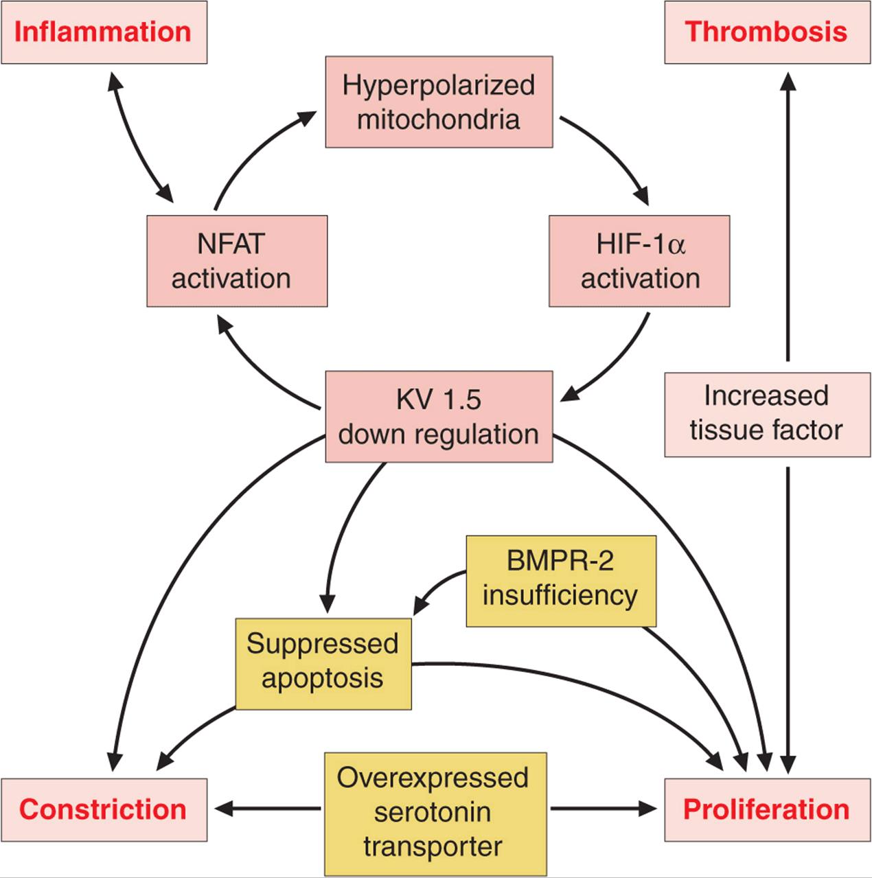
FIGURE 40-2
Multiple biologic pathways that can lead to pulmonary arterial hypertension. Some of the better-characterized ones are illustrated. Because of the redundancy in these pathways and the spectrum of abnormalities that may coexist, it is unlikely that a single agent will produce disease reversal. BMPR-2, bone morphogenetic protein receptor-2; HIF, hypoxia inducible factor; KV 1.5, voltage-regulated potassium channel 1.5; NFAT, nuclear factor of activated T cells.
IDIOPATHIC PULMONARY ARTERIAL HYPERTENSION
Idiopathic pulmonary arterial hypertension (IPAH), formerly referred to as primary pulmonary hypertension, is uncommon, with an estimated incidence of two cases per million. There is a female predominance, with most patients presenting in the fourth and fifth decades, although the age range is from infancy to >60 years.
Familial IPAH accounts for up to 20% of cases of IPAH and is characterized by autosomal dominant inheritance and incomplete penetrance. The clinical and pathologic features of familial and sporadic IPAH are identical. Heterozygous germ-line mutations involving the gene that code the type II bone morphogenetic protein receptor (BMPR II), a member of the transforming growth factor (TGF) β superfamily, appear to account for most cases of familial IPAH. The TGF-β superfami-lies include multifunctional proteins that initiate diverse cellular responses by binding to and activating serine/threonine kinase receptors. The low gene penetrance indicates that other risk factors or abnormalities are necessary to manifest clinical disease. Germ-line mutations in the activin-like kinase gene and endoglin gene, which have been linked to hereditary hemorrhagic telangiectasia, coexist in some patients with familial IPAH.
NATURAL HISTORY
The natural history of IPAH is uncertain, but the disease typically is diagnosed late in its course. Before current therapies, a mean survival of 2–3 years from the time of diagnosis was reported. Functional class remains a strong predictor of survival, with patients who are in New York Heart Association (NYHA) functional class IV having a mean survival of <6 months. The cause of death is usually RV failure, which is manifest by progressive hypoxemia, tachycardia, hypotension, and edema.
TREATMENT Pulmonary Arterial Hypertension
Because the pulmonary artery pressure in PAH increases with exercise, patients should be cautioned against participating in activities that impose physical stress. Diuretic therapy relieves peripheral edema and may be useful in reducing RV volume overload. Pulse oximetry should be monitored, as O2 supplementation helps alleviate dyspnea and RV ischemia in patients whose arterial O2 saturation is reduced. Anticoagulant therapy is advocated for all patients with PAH based on studies demonstrating that warfarin increases survival of patients with PAH. The dose of warfarin generally is titrated to achieve an international normalized ratio (INR) of 2–3 times control.
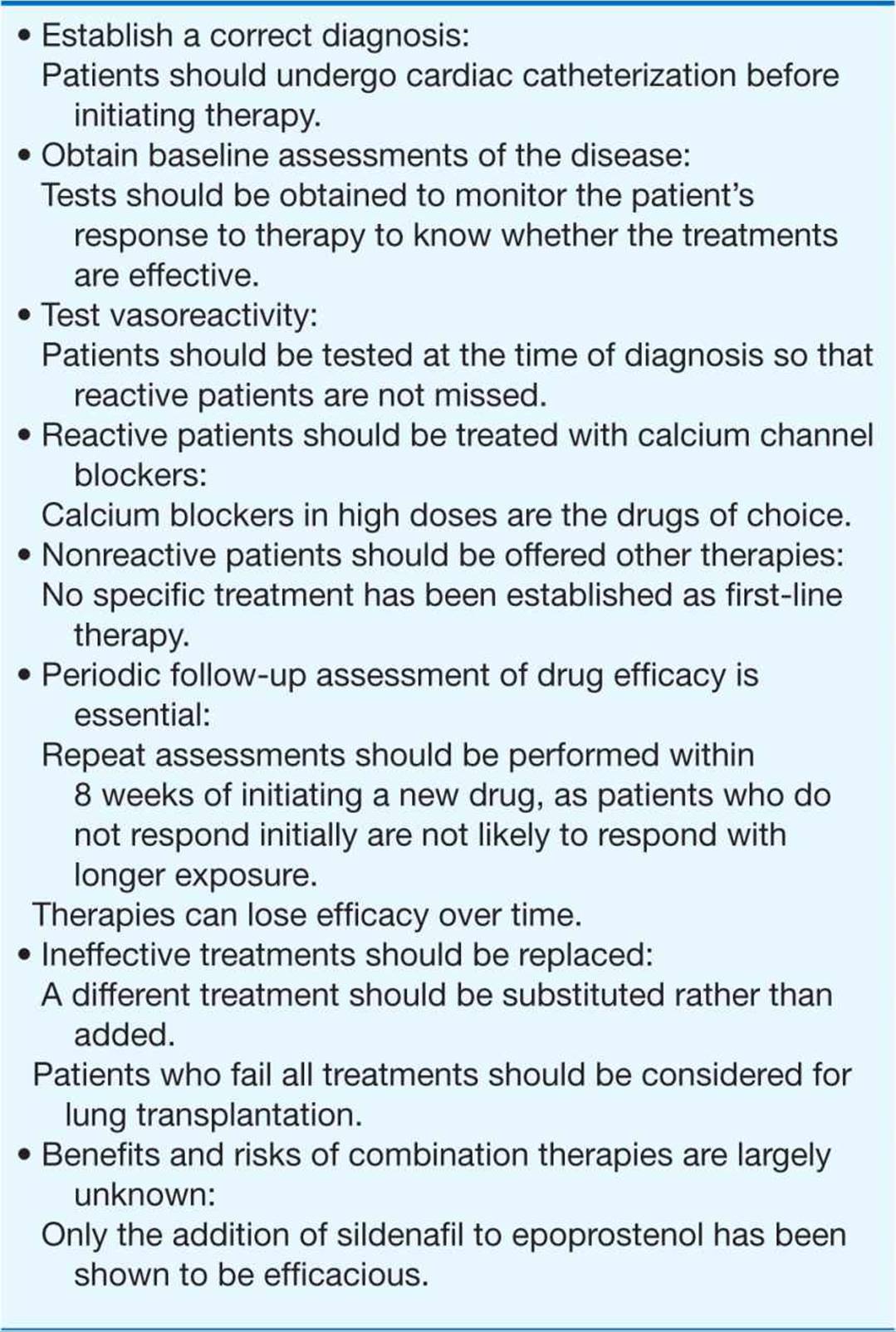
Several treatments are approved for PAH; they are reviewed below without making a distinction among the different types. However, the efficacy and side effects of these drugs may not be the same in all types of PAH. Other than calcium channel blockers, none of the drugs produce a significant lowering of the pulmonary arterial pressure, and their long-term effects on survival are undefined. The principles for the selection and use of the approved drug treatments are reviewed in Table 40-2.
TABLE 40-2
PRINCIPLES OF DRUG TREATMENT OF PULMONARY ARTERIAL HYPERTENSION
CALCIUM CHANNEL BLOCKERS Patients who respond to short-acting vasodilators at the time of cardiac catheterization (a fall in mean pulmonary arterial pressure ≥10 mmHg and a final mean pressure <40 mmHg) should be treated with calcium channel blockers. Typically, these patients require high doses (e.g., nifedipine, 240 mg/d, or amlodipine, 20 mg/d). Patients may have dramatic reductions in pulmonary artery pressure and pulmonary vascular resistance associated with improved symptoms, regression of RV hypertrophy, and improved survival now documented to exceed 20 years. However, <20% of patients respond to calcium channel blockers in the long term. These drugs are not effective in patients who are not vasoreactive. They also have not been approved for the treatment of PAH by the U.S. Food and Drug Administration.
ENDOTHELIN RECEPTOR ANTAGONISTS The endothelin receptor antagonists bosentan and ambrisentan are approved treatments of PAH. In randomized clinical trials, both improved exercise tolerance as measured by an increase in 6-min walking distance. Therapy with bosentan is initiated at 62.5 mg bid for the first month and increased to 125 mg bid thereafter. Ambrisentan is initiated as 5 mg once daily and can be increased to 10 mg daily. Because of the high frequency of abnormal hepatic function tests associated with these drugs, primarily an increase in transaminases, it is recommended that liver function be monitored monthly throughout the duration of use. Bosentan is contraindicated in patients who are on cyclosporine or glyburide concurrently.
PHOSPHODIESTERASE-5 INHIBITORS Sildenafil and tadalafil, phosphodiesterase-5 inhibitors, are approved for the treatment of PAH. Phosphodiesterase-5 is responsible for the hydrolysis of cyclic GMP in pulmonary vascular smooth muscle, the mediator through which nitric oxide lowers pulmonary artery pressure and inhibits pulmonary vascular growth. Clinical trials have shown that both drugs improve exercise tolerance in patients with PAH. The effective dose for sildenafil is 20–80 mg tid. The effective dose for tadalafil is 40 mg once daily. The most common side effect is headache. Neither drug should be given to patients who are taking nitrovasodilators.
PROSTACYCLINS Iloprost, a prostacyclin analogue, is approved via inhalation for PAH. It has been shown to improve a composite measure of symptoms and exercise tolerance by 10%. Therapy can be given at either 2.5 or 5 μg per inhalation treatment via a dedicated nebulizer. The most common side effects are flushing and cough. Because of the very short half-life (<30 min) it is recommended that treatments be administered as often as every 2 h.
Epoprostenol is approved as a chronic IV treatment of PAH. Clinical trials have demonstrated an improvement in symptoms, exercise tolerance, and survival even if no acute hemodynamic response to drug challenge occurs. Drug administration requires placement of a permanent central venous catheter and infusion through an ambulatory infusion pump system. Side effects include flushing, jaw pain, and diarrhea, which are tolerated by most patients.
Treprostinil, an analogue of epoprostenol, is approved for PAH and may be given intravenously, subcutaneously, or via inhalation. Clinical trials have demonstrated an improvement in symptoms with exercise. Local pain at the infusion site with subcutaneous administration has caused most patients to switch to another therapy. Side effects are similar to those seen with epoprostenol.
The intravenous prostacyclins have the greatest efficacy as treatments for PAH and are often effective in patients who have failed all other treatments. Favorable properties include vasodilation, platelet inhibition, inhibition of vascular smooth muscle growth, and inotropic effects. It generally takes several months to titrate the dose of epoprostenol or treprostinil upward to achieve optimal clinical efficacy, which can be determined by symptoms, exercise testing, and catheterization. The optimal doses of these drugs have not been determined, but the typical doses of epoprostenol range from 25 to 40 ng/kg per min, and from 75 to 150 ng/kg per min for treprostinil. The major problem with intravenous therapy is infection related to the indwelling venous catheter, which requires close monitoring and diligence on the part of the patient. In addition, abrupt discontinuation of intravenous prostacyclins can lead to a rebound increase in pulmonary pressure.
It is recommended that every patient diagnosed with PAH be treated. Although no drug has been demonstrated to be superior as first-line therapy, many prefer to initiate treatment with an oral or inhaled form of therapy. Patients who fail to improve adequately within the first 2 months should be switched to a different therapy, as there is concern that delaying a more effective treatment may allow the disease to progress and become less responsive. The use of these drugs in combination has become popular, but the only randomized clinical trial demonstrating beneficial effects added sildenafil to patients treated with epoprostenol.
LUNG TRANSPLANTATION Lung transplantation is considered for patients who, while on an intravenous prostacyclin, continue to manifest right heart failure. Acceptable results have been achieved with heart-lung, bilateral lung, and single-lung transplantation. The availability of donor organs often influences the choice of procedure.
CONDITIONS ASSOCIATED WITH PULMONARY HYPERTENSION
COLLAGEN VASCULAR DISEASE
All the collagen vascular diseases may be associated with PAH. This complication occurs commonly with the CREST syndrome (calcinosis, Raynaud’s phenomenon, esophageal involvement, sclerodactyly, and telangiectasia) and in scleroderma and less frequently in systemic lupus erythematosus, Sjögren’s syndrome, dermatomyositis, polymyositis, and rheumatoid arthritis. Often these patients have coexistent interstitial pulmonary fibrosis even though it may not be apparent on chest x-ray, CT, or pulmonary function tests. Consequently, they tend to have hypoxemia as an important clinical feature, along with the other classic findings of pulmonary hypertension. A fall in diffusing capacity may precede the development of pulmonary hypertension. Treatment of these patients is identical to that of patients with IPAH (see earlier) but is less effective. The treatment of the pulmonary hypertension, however, does not affect the natural history of the underlying collagen vascular disease.
CONGENITAL SYSTEMIC TO PULMONARY SHUNTS
It is common for large post-tricuspid cardiac shunts (e.g., ventricular septal defect, patent ductus arteriosus) to produce severe PAH (Chap. 19). Although less common, this also may occur in pretricuspid shunts (e.g., atrial septal defect, anomalous pulmonary venous drainage). In patients with uncorrected shunts, the clinical features include those associated with right-to-left shunting, such as hypoxemia and peripheral cyanosis, which worsen dramatically with exertion (Chap. 6). PAH may also occur years or even decades after surgical correction in the absence of right-to-left shunting. These patients present similarly to patients with IPAH but tend to have better long-term survival. The treatments are similar to those for IPAH.
PORTAL HYPERTENSION
Portal hypertension is associated with PAH, but the mechanism is unknown. Patients with advanced cirrhosis can have the combined features of a high-output cardiac state in association with the features of pulmonary hypertension and RV failure. Thus, a normal cardiac output may actually reflect a marked impairment of RV function. The etiology of ascites and edema can be confusing in these patients because this condition can have both cardiac and hepatic causes. Overall, these patients have a worse prognosis than do patients with IPAH. Patients with mild pulmonary hypertension who have a favorable response to epoprostenol have undergone successful liver transplantation with improvement of the pulmonary vascular disease.
ANOREXIGENS
A causal relationship has been established between exposure to several anorexigens, including aminorex and the fenfluramines, and the development of PAH. Often the pulmonary hypertension will develop years after the last exposure. Although the clinical features are identical to those of IPAH, the patients appear to be less responsive to medical treatments.
PULMONARY CAPILLARY HEMANGIOMATOSIS
Pulmonary capillary hemangiomatosis is a very rare form of pulmonary hypertension. Histologically it is characterized by the presence of infiltrating thin-walled blood vessels throughout the pulmonary interstitium and walls of the pulmonary arteries and veins. The presenting symptoms are those of IPAH but often with hypoxemia or hemoptysis as a clinical feature. The diagnosis may be suggested by findings on chest CT. The clinical course is usually one of progressive deterioration leading to death. There is no established therapy.
PULMONARY VENOUS HYPERTENSION
Pulmonary hypertension occurs as a result of increased resistance to pulmonary venous drainage. It is associated with diastolic dysfunction of the left ventricle, diseases affecting the pericardium or mitral or aortic valves, and rare entities such as cor triatriatum, left atrial myxoma, extrinsic compression of the central pulmonary veins from fibrosing mediastinitis, and pulmonary venoocclusive disease. Pulmonary venous hypertension affects the pulmonary veins and venules, producing arterialization of the external elastic lamina, medial hypertrophy, and focal eccentric intimal fibrosis. Microcirculatory lesions include capillary congestion, focal alveolar edema, and dilation of the interstitial lymphatics. Although these lesions are potentially reversible, regression may take years after the underlying cause is removed. Pulmonary venous hypertension often triggers reactive vasoconstriction in the pulmonary arterial bed and results in proliferative changes of the intima and media that can produce severe elevations in pulmonary artery pressure. Clinically it may be confusing and appear as if two separate disease processes are occurring simultaneously. The distinction is important, however, as treatments that are effective in PAH may make patients with pulmonary venous hypertension worse.
LEFT VENTRICULAR DIASTOLIC DYSFUNCTION
Pulmonary hypertension as a result of LV diastolic failure is common but often unrecognized (Chap. 17). It can occur with or without LV systolic failure. The most common risk factors are hypertensive heart disease; coronary artery disease; and impaired LV compliance related to age, diabetes, obesity, and hypoxemia. Symptoms of orthopnea and paroxysmal nocturnal dyspnea are prominent. Many patients improve considerably if LV end-diastolic pressure is lowered, but current treatments are unsatisfactory.
MITRAL VALVE DISEASE
Mitral stenosis and mitral regurgitation represent important causes of pulmonary hypertension (Chap. 20) from reactive pulmonary vasoconstriction resulting in marked elevations in pulmonary artery pressures. An echocardiogram usually shows abnormalities such as thickened mitral valve leaflets with reduced mobility or severe mitral regurgitation documented by Doppler echocardiography (Chap. 12). At cardiac catheterization, a pressure gradient between the pulmonary capillary wedge pressure and LV end-diastolic pressure is diagnostic of mitral stenosis.
In patients with mitral stenosis, corrective surgery of the mitral valve or mitral balloon valvuloplasty predictably results in a reduction in pulmonary artery pressure and pulmonary vascular resistance. Patients with mitral regurgitation, however, may not have as dramatic a response to surgery because of persistent elevations in LV end-diastolic pressure.
PULMONARY VENOOCCLUSIVE DISEASE
Pulmonary venoocclusive disease is a rare and distinct pathologic entity found in <10% of patients who present with unexplained pulmonary hypertension. Histologically it is manifest by intimal proliferation and fibrosis of the intrapulmonary veins and venules, occasionally extending to the arteriolar bed. A CT scan may reveal septal thickening, diffuse or mosaic ground-glass opacities, multiple small nodules, or areas of alveolar consolidation. Advanced pulmonary venous obstruction explains the orthopnea that can mimic LV failure, pulmonary edema noted on chest x-ray, and the increase in pulmonary capillary wedge pressure at catheterization. Effective therapy for this condition has not been established.
PULMONARY HYPERTENSION ASSOCIATED WITH LUNG DISEASE AND HYPOXEMIA
The acute hypoxic response of the pulmonary arterial smooth-muscle cells involves inhibition of the potassium current, membrane depolarization, and calcium entry through L-type calcium channels. Hypoxia, acting through the small G protein RhoA, stimulates Rho kinase, which inhibits myosin vs. heavy chain in light chain phosphatase, thereby increasing phosphorylation of the light chain and augmenting contraction. Chronic hypoxia results in muscularization of the arterioles with minimal effects on the intima. When it occurs as an isolated entity, the changes produced are potentially reversible.
Although chronic hypoxia is an established cause of pulmonary hypertension, it rarely leads to an increase in the systolic pulmonary artery pressure >50 mmHg. Polycythemia in response to the hypoxemia is a characteristic finding. Hypoxia also may occur in conjunction with other causes of pulmonary hypertension associated with more extensive vascular changes. Clinically, the hypoxia has an added adverse effect. Patients with chronic hypoxia who have a marked elevation in pulmonary pressure should be evaluated for the other causes of the pulmonary hypertension.
CHRONIC OBSTRUCTIVE LUNG DISEASE
Chronic obstructive lung disease (COLD) is associated with mild pulmonary hypertension in the advanced stages. The factors leading to an increase in pulmonary vascular resistance are numerous, but alveolar hypoxia is considered the predominant one. The presence of pulmonary hypertension in patients with COLD confers a worse outcome. The only effective therapy is supplemental oxygen. Clinical trials have documented that continuous oxygen therapy relieves the pulmonary vasoconstriction, reverses chronic ischemia throughout the systemic and pulmonary vascular beds, and improves survival. Long-term oxygen therapy is indicated if the resting arterial Po2 remains <55 mmHg. Pulmonary vasodilators can worsen gas exchange and should not be used.
INTERSTITIAL LUNG DISEASE
Pulmonary hypertension is common in interstitial lung disease that results from parenchymal and vascular remodeling. Coexisting hypoxemia occurs frequently and contributes to morbidity. Interstitial lung disease often is associated with the collagen vascular diseases. Many patients have pulmonary fibrosis of unknown etiology. Patients are commonly older than 50 years and report an insidious onset of progressive dyspnea and cough for months to years. It is uncommon for the mean pulmonary artery pressure to exceed 40 mmHg. The pulmonary vasodilators approved for PAH have not been shown to be helpful.
SLEEP-DISORDERED BREATHING
The incidence of pulmonary hypertension in the setting of obstructive sleep apnea, a common condition, is <20% and is generally mild. Some patients have severe pulmonary hypertension in conjunction with sleep apnea, which may be unrelated. It is recommended that the sleep apnea and the PAH be treated as coexisting problems.
ALVEOLAR HYPOVENTILATION
Pulmonary hypertension can occur in patients with chronic hypoventilation and hypoxia secondary to thoracovertebral deformities. Symptoms are slowly progressive and are related to hypoxemia. In patients with advanced disease, intermittent positive-pressure breathing and supplemental oxygen have been used successfully.
Pulmonary hypertension secondary to hypoxemia has been reported in patients with neuromuscular disease as a result of generalized weakness of the respiratory muscles and in patients with diaphragmatic paralysis, generally from trauma to the phrenic nerve. Patients with nontraumatic bilateral diaphragmatic paralysis may go unrecognized until they present with either respiratory failure or pulmonary hypertension.
PULMONARY HYPERTENSION DUE TO THROMBOEMBOLIC DISEASE
CHRONIC THROMBOEMBOLIC PULMONARY HYPERTENSION
Most patients treated for acute pulmonary thromboembolism with intravenous heparin and chronic oral warfarin do not develop chronic pulmonary hypertension. However, some patients have impaired fibrinolytic resolution of the thromboembolism, which leads to organization and incomplete recanalization and chronic obstruction of the pulmonary vascular bed. Because the initial pulmonary thromboembolism goes undetected or untreated, many patients are misdiagnosed as having IPAH. These patients may have underlying thrombophilic disorders, such as the lupus anticoagulant/anticardiolipin antibody syndrome, prothrombin gene mutation, or factor V Leiden.
Diagnosis
The physical examination is characteristic of pulmonary hypertension but may include bruits heard over areas of the lung, representing blood flow through vessels with partial occlusion. A perfusion lung scan or contrast-enhanced spiral CT scan should reveal multiple thromboemboli. High-resolution CT scanning is necessary to document the location and proximal extent of the thromboemboli and hence the potential for operability.
TREATMENT Chronic Thromboembolic Pulmonary Hypertension
Pulmonary thromboendarterectomy is an established surgical treatment in patients whose thrombi are accessible to surgical removal. The operative mortality is <10% in experienced centers. Postoperative survivors can expect an improvement in functional class and exercise tolerance. Lifelong anticoagulation using warfarin is mandatory. Thrombolytic therapy is rarely helpful in patients with chronic thromboembolic pulmonary hypertension and may expose them to the increased risk of bleeding without potential benefit.
OTHER DISORDERS AFFECTING THE PULMONARY VASCULATURE
SARCOIDOSIS
Sarcoidosis can produce pulmonary hypertension as a result of fibrocystic lung involvement or direct cardiovascular involvement. Consequently, patients with sarcoidosis who present with progressive dyspnea and pulmonary hypertension require a thorough evaluation. There is a subset of patients with sarcoidosis and severe pulmonary hypertension who exhibit a favorable response to epoprostenol therapy.
SICKLE CELL DISEASE
Cardiovascular system abnormalities are prominent in the clinical spectrum of sickle cell disease, including pulmonary hypertension. The etiology is multifactorial, including hemolysis, hypoxemia, thromboembolism, chronic high cardiac output, and chronic liver disease. The presence of pulmonary hypertension in patients with sickle cell disease is associated with higher mortality. Intensification of sickle cell disease–specific therapy appears to reduce the morbidity. Clinical trials assessing drugs to treat pulmonary hypertension are ongoing, but the efficacy of those drugs is unknown.
SCHISTOSOMIASIS
 Although extremely rare in North America, schistosomiasis is one of the most common causes of pulmonary hypertension worldwide. The development of pulmonary hypertension occurs in the setting of hepatosplenic disease and portal hypertension. Studies suggest that inflammation from the infection triggers the pulmonary vascular changes that occur. The diagnosis is confirmed by finding the parasite ova in the urine or stools of patients with symptoms, which can be difficult. The efficacy of therapies directed toward pulmonary hypertension in these patients is unknown.
HIV INFECTION
The mechanism by which HIV infection produces pulmonary hypertension is unknown. Although the incidence is estimated at 1 per 200 cases, the marked rise in the prevalence of HIV infection worldwide could have a significant impact on the frequency with which these entities are seen in combination. The evaluation and treatments are identical to those for IPAH. Treatment of the HIV infection does not appear to affect the severity or natural history of the underlying pulmonary hypertension.