
■ INTRODUCTION
■ OVERVIEW OF CARDIAC SURGICAL PROCEDURES
■ TIMING OF SURGERY
■ POSTOPERATIVE COMMUNICATION
■ MONITORING
■ GENERAL PRINCIPLES OF POSTOPERATIVE CARE BY SYSTEM
Cardiovascular
Pulmonary Arterial Hypertension
Respiratory
Renal Function and Fluids and Electrolytes
Nutrition
Analgesia and Sedation Hematologic
■ SELECTED COMPLICATIONS
Excessive Bleeding Open Sternum
Cardiac Tamponade
Infection
Postpericardiotomy Syndrome Diaphragmatic Paralysis Chylothorax
■ SUGGESTED READINGS
■ INTRODUCTION
To avoid the cumulative morbidity and mortality associated with initial palliative procedures followed by later repair, primary corrective surgery has become increasingly common for patients with congenital heart disease. One result of this trend is that an increasing number of cardiac surgical procedures are performed on neonates and even on premature infants. Optimal care of these infants requires specialized knowledge of the unique structural and functional characteristics of neonatal organ systems and is best accomplished by a multidisciplinary specialty team including cardiology, cardiac surgery, nursing, neonatology, anesthesia, and critical care. The purpose of this chapter is to review general principles of care for these infants. The physiology and surgical procedures pertinent to specific defects are discussed in Chapters 6 through 8. Medications commonly used in the postoperative period are discussed in Chapter 12.
■ OVERVIEW OF CARDIAC SURGICAL PROCEDURES
Cardiac surgical procedures are classified as to whether they are open or closed and whether they are corrective or palliative (Table 13-1). “Open” refers to a procedure in which cardiopulmonary bypass is used; bypass is not used in “closed” procedures. Palliative procedures are performed in patients in whom complete correction of the cardiac defects is not possible or not feasible because of comorbidities. Palliated patients have residual intracardiac shunting or other hemodynamic abnormalities. In most institutions, palliative procedures are performed only on infants with a functional single ventricle (eg, hypoplastic left heart syndrome, tricuspid atresia) or on those with poorly developed pulmonary arteries.
TABLE 13-1. Classification of Cardiac Surgery Procedures
Closed: Cardiopulmonary bypass is not used.
Palliative—Pulmonary artery band, systemic- to-pulmonary artery shunt
Corrective—Repair of aortic coarctation, ligation of patent ductus arteriosus
Open: Cardiopulmonary bypass is used.
Palliative—Stage I Norwood procedure, Damus- Kaye-Stansel procedure
Corrective—Repair of transposition of the great arteries, tetralogy of Fallot, total anomalous pulmonary venous connection, truncus arteriosus, ventricular septal defect
Examples of palliative and corrective procedures are provided, but these lists are not comprehensive.
Most corrective procedures, such as for truncus arteriosus or transposition of the great arteries, are performed by use of cardiopulmonary bypass. Bypass diverts blood from the operative field while maintaining circulation, adds oxygen and removes carbon dioxide from the blood, and facilitates cooling and subsequent warming. Venous blood from both superior and inferior vena cavae is siphoned to a reservoir of the heart-lung bypass machine, which also collects blood drained from the operative field by suction catheters (Figure 13-1). Blood is pumped through an oxygenator, a heat exchanger (for cooling or warming), and a filter and then is returned to the patient’s ascending aorta through an aortic cannula. The patient is always fully anticoagulated with heparin while on bypass.
Hypothermia extends the safe duration of cardiopulmonary bypass in neonates and infants. Metabolic activity and thus oxygen consumption decrease at cooler temperatures. “Deep” hypothermia involves cooling from 17°C to 22°C. For many years, surgeons combined deep hypothermia with either low-flow bypass (25% to 50% of normal flow) or, more commonly, with no bypass flow, arresting the heart (deep hypothermic circulatory arrest). Deep hypothermic circulatory arrest provided the surgeon with a bloodless and relaxed heart free of multiple cannulas that may distort the surgical field. This technique allowed intricate surgical procedures to be performed and markedly improved survival of infants with complex congenital defects. Unfortunately, neurologic and developmental morbidities are increasingly being recognized in survivors of deep hypothermic circulatory arrest (Chapter 14), and longer duration of circulatory arrest is associated with a greater incidence of adverse neurologic outcomes. Alternate perfusion strategies include moderate hypothermia (22°C to 30°C) with normal or increased pump flow or deep hypothermia with intermittent perfusion. Another approach used during aortic arch reconstruction is antegrade cerebral perfusion, which involves directing blood flow specifically to the cerebral circulation via a cannula in the innominate artery. Further clinical studies and continued refinements in technique are necessary to optimize neurologic outcomes.
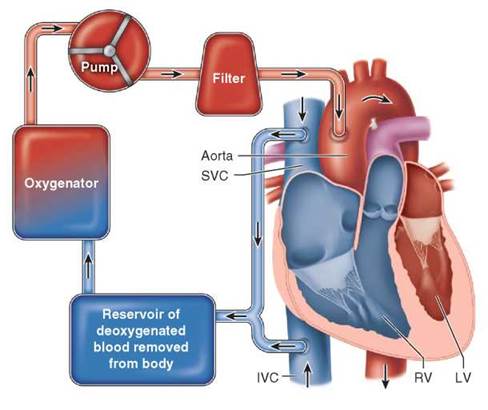
FIGURE 13-1. Diagram of basic cardiopulmonary bypass system.
Although cardiopulmonary bypass allows for correction of complex cardiac defects, morbidity associated with its use impacts care of the postoperative patient, particularly neonates. Blood cells are damaged by exposure to excessive shear forces and to artificial surfaces. Despite filtration, microembolization of gas bubbles and platelet clumps occurs. Nonpulsatile perfusion and hypothermia per se may also incite injury. Tissue ischemia is present to some extent, and subsequent reperfusion injury occurs. These and other factors combine to stimulate a systemic inflammatory response, consisting of activation of the complement system, leukocytes and the endothelium. Together with induction of cytokines, chemokines, and endotoxin, this results in increased capillary permeability, tissue edema, and multisystem dysfunction. Total body water increases, transient myocardial dysfunction occurs, pulmonary vascular resistance may increase, gas exchange is impaired, and stress and hormonal responses often cause fluid and electrolyte abnormalities. This response is exaggerated in newborn infants compared to older children and results in a decrease in cardiac output that often peaks 6 to 12 hours after bypass. To limit this systemic inflammatory response, steroids are usually administered in the operating room, and various ultrafiltration techniques are used during rewarming or after cardiopulmonary bypass to remove excess water and small-molecular- weight inflammatory mediators. Hemodilution may occur in part because of the priming volume required in the bypass circuit. This decreases oxygen delivery, decreases oncotic pressure resulting in further fluid extravasation, and dilutes clotting factors, contributing to postoperative coagulopathy. Maintenance of a hematocrit of about 25% to 30% during bypass seems to be optimal for hypothermia-induced alterations in blood viscosity and also for maintaining oncotic pressure and oxygen-carrying capacity.
■ TIMING OF SURGERY
Many infants diagnosed with congenital heart disease are managed as outpatients before undergoing surgery, but those with ductal-dependent circulations need supportive critical care before proceeding with surgical intervention. In general, cyanotic infants can be stabilized by administration of PGE1 and other supportive measures before intervention. Surgery or catheter-based intervention can be scheduled on a semielective basis after careful evaluation is complete. Two exceptions are noteworthy: (1) emergency surgery is necessary for neonates with obstructed total anomalous pulmonary venous return, and (2) neonates with d-transposition of the great arteries and a restrictive patent foramen ovale often need emergency balloon atrial septostomy because of profound hypoxemia. This procedure can be performed at the bedside with echocardiographic guidance. After successful septostomy, these infants are usually stable, and corrective surgery can be performed soon thereafter if end-organ damage has not occurred.
Infants who present with shock because of left heart obstructive lesions (eg, coarctation of the aorta, hypoplastic left heart syndrome) can usually be stabilized by administration of PGE1 and other supportive care. These infants may have sustained end-organ damage because of decreased perfusion, most commonly to the kidneys or liver. Deferring surgery while organ function recovers allows for complete evaluation (including neurologic) of the infant and decreases surgical morbidity and risk. However, surgical intervention should be undertaken if there is evidence of ongoing organ injury despite maximal medical therapy.
■ POSTOPERATIVE COMMUNICATION
All personnel responsible for care of the infant after surgery must understand the anatomic defects and be familiar with the preoperative evaluation and course. Upon return from the operating room, information must be provided regarding the following:
• Intraoperative findings, especially if the anatomy is different than what was on the preoperative diagnosis list
• The exact procedure(s) performed
• Length of time on cardiopulmonary bypass, including aortic cross-clamp time, level of hypothermia, and circulatory arrest times
• Available postoperative hemodynamic and echocardiographic data, especially regarding residual lesions
• Complications, including arrhythmias and bleeding (including blood product administration)
• Location of catheters, tubes, and temporary pacing wires
• Vasoactive infusions and other medications
• Airway and ventilatory status (anesthetic agents and status of neuromuscular blockade are also summarized, as is the timing of the last doses of analgesic and sedative agents)
The personnel transporting the infant must work harmoniously with staff in the cardiac intensive care unit (ICU) to ensure a careful and efficient admission of the patient to the ICU. A standard template for handoff of care from the operating room to ICU is helpful. Maintenance of adequate oxygenation and ventilation and uninterrupted delivery of vasoactive drugs are critically important. About 15 minutes after the patient is placed on the ventilator (or earlier if clinically indicated), initial laboratory studies should be obtained. This often includes arterial blood gas, complete blood count, serum electrolytes and glucose, ionized calcium, and coagulation profile. A chest radiograph and often an electrocardiogram are obtained as soon as possible.
A comprehensive assessment should be performed as soon as possible, including a physical examination focusing on cardiovascular and respiratory function, as should a review of data from bedside monitors, postoperative orders, and laboratory data as available. The family should be updated and allowed to visit as soon as is feasible.
■ MONITORING
Although variation exists among institutions, most infants have continuous monitoring of their heart rate, rhythm, blood pressure, oxygen saturation, and respiratory rate after cardiac surgery. Most infants have an arterial line. End-tidal CO2 monitoring is valuable in patients who are mechanically ventilated. Near-infrared spectroscopy (NIRS) monitoring estimates the oxygen saturation in tissues (regional oxygen saturation [rSO2]) by comparing the tissue’s absorption of two wavelengths of light corresponding to hemoglobin-carrying oxygen and hemoglobin without oxygen. NIRS leads placed on the forehead and in the paravertebral region over a kidney allow assessment of oxygen delivery to the brain and kidneys, respectively. Changes in head rSO2 correlate closely with changes in mixed venous saturation.
Infants who have been on cardiopulmonary bypass usually have one or more intracardiac catheters for monitoring and infusions. Temporary pacing wires are often placed on the epicardial surface of the atrium and/or ventricle. The data obtained allow more precise measurement of hemodynamic variables, thereby providing a rational basis for therapy. Nevertheless, the information obtained should be viewed as an adjunct to rather than a substitute for careful serial physical examinations to assess cardiac output.
A right atrial (RA) catheter may be advanced via a central vein or placed through the RA appendage. RA catheters are used to measure central venous pressure, which may reflect intravascular volume status. In addition to volume overload, other causes of increased RA pressure include decreased right (or single) ventricular compliance, tricuspid valve regurgitation or a residual left ventricular to RA shunt, and cardiac tamponade. RA saturation can also be measured but may not represent a true mixed venous sample because of streaming of venous inflow within the atrium and because it is mixed with left atrial blood in the single-ventricle patient. When blood can be obtained from a site that is a reasonable estimate of mixed venous saturation (the superior vena cava is usually the best source), an arteriovenous oxygen saturation difference of less than 30% indicates adequate cardiac output.
A left atrial (LA) catheter may be placed via the LA appendage or through the right superior pulmonary vein. The LA pressure provides indirect data regarding functioning of the systemic ventricle as long as the systemic atrioventricular valve is neither regurgitant nor stenotic. LA pressure also rises and falls with intravascular volume status. The oxygen saturation in the LA should be about 100%. Decreased LA saturation can be caused by right-to- left atrial shunting or by pulmonary venous desaturation secondary to abnormal gas exchange (eg, atelectasis).
Echocardiography is used frequently in the ICU to assess postoperative status. Intraoperative transesophageal examination evaluates residual lesions as well as ventricular function, outflow tract obstruction, valve regurgitation, and possibly pulmonary arterial pressure. At times of clinical deterioration in the ICU, emergent echocardiography is useful to assess all of the above as well as shunt function and the presence of pericardial effusion.
■ GENERAL PRINCIPLES OF POSTOPERATIVE CARE BY SYSTEM
Cardiovascular
Decreased Cardiac Output
Myocardial dysfunction is common after surgery in young infants, especially neonates. One or more of the following factors may be involved:
• Preoperative myocardial dysfunction—This is most common in patients with left heart obstructive lesions, such as aortic coarctation.
• Effects of cardiopulmonary bypass—As discussed in the preceding text, a prominent systemic inflammatory response frequently occurs and may adversely affect myocardial function. Inadequate myocardial perfusion can occur during surgery. Finally, a ventriculotomy, if performed, may lead to regional myocardial dysfunction. If these intraoperative events lead to a low-output state, their effects usually peak 6 to 12 hours after surgery. Physical examination will disclose tachycardia, hypotension, and cool extremities with decreased perfusion. Decreased urine output, lactic acidosis, low head rSO2, edema, and pleural effusions may be present. It is critically important to anticipate this reproducible and potentially lethal phenomenon and to intervene immediately. Infusion of volume and administration of inotropic and afterload reducing agents are often necessary. Serial physical examinations and frequent evaluation of hemodynamic and metabolic data are necessary to assess the response to therapeutic interventions.
• Residual anatomic lesions—The operative assessment of cardiac anatomy and function is done by direct inspection, blood gas and pressure measurements, and imaging. The use of transesophageal echocardiography in the operating room after surgery has reduced the incidence of unsuspected residual anatomic lesions. Nevertheless, if the postoperative course does not conform to that expected, investigation should be undertaken to assess for residual or previously undiagnosed anatomic defects. This certainly includes echocardiography but may also involve magnetic resonance imaging and/or cardiac catheterization.
• Arrhythmias—Abnormal heart rhythms, such as atrioventricular block and tachycardias, are relatively common in the postoperative period and may contribute to inadequate cardiac output. Diagnosis and treatment of rhythm disorders is discussed in Chapter 10. The neonate is relatively dependent on a physiologic heart rate and an atrial contribution to ventricular filling (“atrial kick”) to maintain cardiac output. Therefore, bradycardia associated with atrioventricular block will markedly decrease cardiac output, and pacing should be instituted promptly via the transthoracic pacing wires. Junctional ectopic tachycardia is the most common tachyarrhythmia and results in loss of atrioventricular synchrony. Infants who are symptomatic from rapid heart rates should be treated immediately.
• Electrolyte and hormonal disturbances—Hypocalce- mia and hypomagnesemia depress myocardial contractility and should be treated appropriately. It has been postulated that administration of glucocorticosteroids improves refractory hemodynamic instability in stressed neonates who may have an inappropriate or abnormal adrenal response to stress after cardiac surgery. Similarly, decreased concentrations of triiodothyronine, the biologically active hormone in cardiac myocytes, may contribute to low cardiac output syndrome after bypass; some have advocated administration of triiodothyronine. Neither of these therapies has been shown to be efficacious in a clinical trial, but both are used empirically at times.
• Anemia—Increasing the hemoglobin concentration improves the oxygen-carrying capacity of blood and may benefit patients with low levels of cardiac output. Transfusing cyanotic patients up to a hemoglobin concentration of 14 to15 g/dL may be helpful.
Support of the Circulation
The primary goals in the initial postoperative period are to optimize cardiac output and blood flow distribution by maintaining optimal heart rate, preload, afterload, and contractility. As discussed earlier, bradycardia adversely affects cardiac output in neonates and should be treated, as should tachyarrhythmias. Preload must be optimized, based on assessment of intravascular volume. Vasodilator agents can be used to decrease afterload as indicated. Contractility is increased by administration of inotropic agents as needed, but care must be taken to avoid their adverse effects. Increased inotropy is associated with increased myocardial energy requirements. Tachycardia increases myocardial energy demand and also reduces diastolic filling time, which can decrease myocardial blood flow. Administration of sympathomimetic amines may predispose to junctional ectopic tachycardia. Despite these concerns, inotropic agents are quite useful for treating myocardial dysfunction and are discussed in detail in Chapter 12. Treatment should be titrated to maintain adequate end-organ perfusion and not to just a certain level of blood pressure. The physical examination, urine output, plasma acid load, and systemic arteriovenous difference should be monitored to evaluate the adequacy of the cardiac output. The choice of inotropic agent is often empiric. The combination of milrinone and dopamine is frequently used to increase cardiac output, maintain appropriate perfusion pressure without an excessive increase in afterload, and improve myocardial relaxation. If increased doses of these agents fail to adequately improve cardiac output, epinephrine in low doses (0.01 to 0.05 pg/kg/min) that do not activate a-receptors is often effective, especially for infants with relative bradycardia or who are chronically stressed. The dose is increased as indicated, but the need for doses higher than 0.1 to 0.2 pg/kg/min should raise consideration of mechanical circulatory support.
Mechanical support of the circulation Venoarterial extracorporeal membrane oxygenation (ECMO) is the most commonly used mode of mechanical circulatory support for neonates and infants with heart disease both before and after surgery. The venous cannula drains blood from the inferior vena cava and right atrium. The blood is pumped through an oxygenator and heat exchanger and then returned to the ascending aorta through the arterial cannula. ECMO should be considered for patients thought to have reversible causes of low cardiac output who are not responding to maximal medical therapy. In addition to maintaining adequate systemic blood flow, ECMO facilitates myocardial recovery, as ventricular wall tension is decreased and coronary perfusion is improved. This modality can also serve as a bridge to cardiac transplantation. The reported survival rates to hospital discharge or transfer after ECMO cannulation as reported by the Extracorporeal Life Support Organization is 41% for neonates who are placed on ECMO after cardiac surgery or as a bridge to transplantation. The relatively low survival rate reflects the facts that neonates needing ECMO for cardiac reasons most often have complex congenital heart defects and that there is limited availability of donor hearts for transplantation. Patients who fail to wean from bypass are at increased risk of life-threatening bleeding when placed on ECMO. Typical indications for ECMO are shown in Table 13-2. Contraindications include significant neurologic deficit, end-stage or irreversible systemic disease, major bleeding, and inaccessible vessels for cannulation. Some consider significant renal or hepatic dysfunction to be a relative contraindication. Aggressive evaluation for residual anatomic lesions, including possible cardiac catheterization, is critical for patients who are placed on ECMO because of failure to wean from bypass.
Implantation of durable ventricular assist devices (VADs) is increasingly available in some centers for those patients who fail to wean from ECMO and who are awaiting cardiac transplantation. The size of pumps and cannulas needed for neonates and infants is a critical issue. The Berlin heart is the most common device used in infants. This is a pneumatically driven pulsatile device that can provide univentricular (right or left) or biventricular support (Figure 13-2). Survival with this device in older children is far superior to that of historical control patients supported on ECMO, though the frequency of complications remains relatively high. Other devices that rely on continuous rather than pulsatile flow are being investigated in animal models and clinical trials are planned. The 5- and 10-year survival rates after transplantation are higher for children who have received a VAD as compared to those who were on ECMO at the time of transplantation; similar data are not available for neonates and infants. Advantages of a VAD compared to ECMO include decreased trauma to blood cells (which decreases the need for anticoagulation), decreased risk of stroke and infection, and greater patient mobility. The major disadvantage is that these devices do not provide respiratory support.
TABLE 13-2. Indications for ECMO
Severe ventricular dysfunction (pre- or postoperative)
Failure to wean from cardiopulmonary bypass
Pulmonary arterial hypertension refractory to medical therapy
Acute systemic-pulmonary shunt malfunction
Arrhythmias refractory to medical management and associated with hemodynamic compromise
Cardiac arrest
Abbreviation: ECMO, extracorporeal membrane oxygenation.
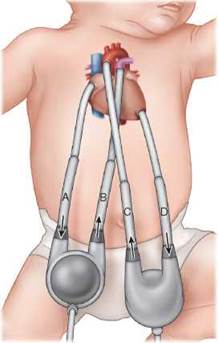
FIGURE 13-2. Berlin Heart EXCO (Berlin Heart Inc., Berlin, Germany). Air is pumped into and out of the air chamber in the pump. This creates positive or negative pressure that results in blood flowing through the pump. A. Right atrial cannula takes system venous blood from patient to pump. B. Pulmonary artery cannula delivers systemic venous blood to pulmonary artery. Oxygenation occurs as blood passes through the pulmonary vasculature and returns to the left atrium and then to the left ventricle. C. Cannula in the apex of the left ventricle takes pulmonary venous blood from patient to pump. D. Aortic cannula delivers pulmonary venous blood to patient.
Pulmonary Arterial Hypertension
Pulmonary arterial (PA) hypertension is an important cause of morbidity and mortality in infants after cardiac surgery. The pulmonary vascular bed is very reactive in young infants, especially neonates during the first few days after cardiac surgery. Acute increases in PA pressure (pulmonary hypertensive crisis) result in decreased pulmonary venous return and left ventricular filling and may precipitate life-threatening hypotension, poor perfusion, and metabolic acidosis. Distended pulmonary arterioles compromise the lumen of small airways and cause poor ventilation. Acidosis and hypoxemia can trigger increased pulmonary arterial pressures with right ventricular failure and cardiac arrest. Patients at highest risk include those with documented increased pulmonary vascular resistance, systemic ventricular dysfunction associated with increased ventricular end-diastolic pressure, and pulmonary venous hypertension from other causes. Conditions known to increase PA pressure, such as acidosis, hypoxia, hypercar- bia, cold stress, and agitation from pain, should be prevented if possible and be treated aggressively if they occur. Concomitant pulmonary disease, including infection, atelectasis, and reactive airways disease, further predisposes the infant to acute increases in PA pressure and also prolongs the period of time that the infant is vulnerable. Sedation, mild hyperventilation, ventilation to maintain normal functional residual capacity, inspired oxygen as needed to avoid alveolar hypoxia, adequate hematocrit, and infusion of agents such as milrinone are often efficacious.
Infants who fail to respond to these treatments may benefit from inhaled nitric oxide (NO). When administered via inhalation, NO acts as a selective pulmonary vasodilator. It does not cause systemic hypotension that often results from intravenously administered vasodilators. High-risk infants may benefit from starting NO prophylactically in the operating room. Patients with a history of pulmonary venous hypertension (eg, obstructive total anomalous pulmonary venous connection, congenital mitral stenosis) are particularly responsive to NO, but this agent is also beneficial in patients after repair of defects associated with PA hypertension (eg, truncus arteriosus, atrioventricular septal defect). Weaning of NO can be facilitated by treatment with sildenafil, which can be continued for weeks to months as indicated clinically.
Less severe PA hypertension may increase intracardiac right-to-left shunting, causing hypoxemia and thereby limiting the ability to wean a patient off the ventilator or off of supplemental oxygen. Echocardiography is valuable for assessing right ventricular function and PA pressure.
Respiratory
Ventilation and respiratory mechanics have important effects on the hemodynamic status of neonates, especially after cardiac surgery. Infants with preexisting myocardial dysfunction as a result of lesions such as severe aortic coarctation and those who have undergone open-heart procedures benefit from a period of maximal ventilatory support while myocardial function recovers. Sedation and, occasionally, neuromuscular blockade are continued for a period of time after arrival in the ICU.
Differentiating between corrective or palliative surgical intervention is essential to setting goals for systemic oxygenation. After corrective surgery, systemic oxygen saturation should be normal (>95%). The oxygen saturation should be about 75% to 85% in patients after a palliative procedure such as a systemic-to-pulmonary artery shunt. Infants with a single ventricle (such as hypoplastic left heart syndrome) and intracardiac mixing in whom the systemic blood flow is entirely dependent on a surgically constructed shunt following a stage I Norwood procedure are exquisitely sensitive to the ratio of pulmonary to systemic blood flow. Both hyperventilation and hyperoxygenation cause pulmonary vasodilation and decreased pulmonary vascular resistance. This can result in an adverse redistribution of blood flow with pulmonary overcirculation and reduced systemic flow, leading to systemic hypotension, poor systemic perfusion, and metabolic acidosis.
For infants at risk for PA hypertension, ventilation is often adjusted to maintain a mild respiratory alkalosis, and the amount of inspired oxygen remains relatively high for a significant amount of time. Caution must be used when manually ventilating or suctioning these infants, as these maneuvers may precipitate an acute increase in PA pressures and lead to hemodynamic instability. Some advocate for the administration of additional sedation before suctioning and for increasing the level of inspired oxygen during suctioning.
Relatively high levels of positive end-expiratory pressure and large tidal volumes are sometimes necessary to recruit end-expiratory lung volumes after bypass in neonates or in those with heart failure. Overinflation should be avoided because this compresses the pulmonary microcirculation and increases pulmonary vascular resistance, and it is best to decrease tidal volume and peak pressures as soon as possible to avoid excessive trauma to the airways and lungs.
Mechanical ventilation is continued until postoperative bleeding has ceased, the sternum is closed, the hemodynamics are stable, postoperative diuresis has occurred (if the patient became fluid overloaded), atelectasis and secretions are minimal, and the patient is capable of protecting the airway. Failure to wean from the ventilator can result from several different problems. Residual hemodynamic abnormalities, such as shunts or valve regurgitation, may play a role and should be evaluated by echocardiography and possibly by magnetic resonance imaging or cardiac catheterization. Severe fluid overload decreases chest wall compliance, compromising ventilation. Airway abnormalities (eg, bronchomalacia), pleural effusions, and alveolar disease are relatively common. Occasionally, paralysis or paresis of a hemidiaphragm may be present, particularly after a reoperation (see following text). Finally, prolonged mechanical ventilation and poor nutrition can weaken respiratory muscles.
Renal Function and Fluids and Electrolytes
Postoperative renal dysfunction related to low cardiac output and ischemia (and the inflammatory response to bypass) causes many infants to develop excess total body water during and immediately after surgery. Total fluid intake should be restricted often to 50% to 60% of maintenance requirements. Urine output is usually at least 1 mL/kg/h immediately after surgery but often decreases markedly 6 to 12 hours later. Total fluid intake and output must be monitored carefully to maintain an adequate intravascular volume. Losses that occur as a result of capillary leak and bleeding may be replaced using infusions of isotonic crystalloids, 5% albumin, or blood products.
Fluids containing 10% dextrose are generally administered, and serum glucose should be measured frequently. Sodium chloride needs are minimal (1 to 2 mEq/kg/d). Hyponatremia usually reflects excess total body water rather than a sodium deficit. Potassium chloride should not be administered until urine output is adequate. Calcium is an important determinant of myocardial contractility, and hypocalcemia impairs myocardial function. Ionized calcium should be monitored frequently. Transfusion of citrate-anticoagulated blood and parathyroid deficiency (secondary to DiGeorge syndrome) are other important causes of hypocalcemia. Calcium is administrated routinely by constant infusion after surgery in many centers.
Diuretics such as furosemide are usually begun 12 to 24 hours after surgery. Maintenance of an adequate cardiac output by judicious volume replacement and administration of inotropic agents and vasodilators is important to achieve a good response to diuretic agents. At times, a continuous infusion of furosemide (0.1 to 0.4 mg/kg/h) promotes a slow but steady diuresis that is often better tolerated than the sudden large diuresis that may result from a bolus dose of diuretic. Diuretic-induced electrolyte losses always occur, and the resulting electrolyte abnormalities, such as hypokalemia, hypocalcemia, and metabolic alkalosis, must be treated aggressively. Rarely, fulminant renal failure related to persistent low output occurs. Peritoneal dialysis, hemofiltration, or hemodialysis may be necessary and will assist in removing excess body water.
Nutrition
Maintaining adequate nutrition is notoriously difficult in many infants with heart disease. The incidence of gastroesophageal reflux is high. Prolonged endotracheal intubation can lead to feeding aversion. Fragile infants with single-ventricle physiology and other serious conditions may not have been fed orally before surgery. Those with DiGeorge syndrome and trisomy 21 often have poor suck and swallow coordination. All of these situations can lead to feeding aversion, poor nippling, emesis, and/or aspiration. Many patients will need some form of tube feeding, at least transiently, to achieve adequate caloric intake.
Fat reserves are frequently limited and energy demands increased after cardiac surgery. Parenteral nutrition may be necessary to provide adequate nutrition for patients too unstable to tolerate enteral feeding within 2 to 3 days after surgery. Continuous nasogastric feeding is often begun at very low rates of 1 to 2 mL/h (“trophic feeds”) to prevent involution of intestinal villi and then advanced as tolerated. Feedings should be advanced to 110 to 130 kcal/kg/d as tolerated. Caloric supplementation up to 30 kcal/oz of breast milk or formula may be necessary to achieve this level of caloric intake in patients who are sensitive to fluid administration. Care must be taken in infants who are at risk for bowel ischemia because necrotizing enterocolitis can occur, especially in neonates who have (1) left-sided obstructive lesions, (2) systemic- to-pulmonary artery shunts with wide pulse pressures associated with retrograde flow in the mesenteric arteries during diastole, or (3) a history of hypotension or severe hypoxemia.
Infants who have undergone major aortic arch reconstruction, including the Norwood Stage 1 palliation procedure, are at risk for recurrent laryngeal nerve injury (15% to 25%) that results in vocal cord dysfunction. Many centers routinely evaluate vocal cord function and do a feeding readiness evaluation to assess oral-motor coordination and risk of aspiration before beginning oral feeding in high-risk infants. Infants at risk for aspiration may be fed with thickened feedings or with a continuous infusion of expressed breast milk or formula via a trans- pyloric tube.
Malrotation occurs frequently in patients with hetero- taxy syndrome; feeding intolerance should be investigated early in these patients. Those diagnosed with malrotation may eventually need a Ladd procedure, but life-threatening volvulus is rare in the first year of life and is not a major cause for emesis during this time.
Analgesia and Sedation
Stress responses to pain and noxious stimuli adversely affect hemodynamics and may precipitate an acute increase in PA pressure. The choice of agent is empiric, but relatively large doses of narcotic and sedative drugs, often fentanyl and midazolam, are usually administered and then weaned when the patient is hemodynamically stable to allow effective spontaneous ventilation. Precise control of pCO2 and pH and, therefore, pulmonary vascular resistance is much easier in a well-sedated patient. All infants who are receiving neuromuscular blocking agents must also be given narcotic and sedative medications. Unexplained tachycardia is often a sign of inadequate sedation or analgesia in these patients. Infants who have a long course in the ICU will need increased doses of medications as they become tolerant. Administration of longer-acting drugs (methadone, valium, or lorazepam) and/or use of agents with opioid-sparing effects, such as clonidine or dexmedetomidine, is often beneficial.
Hematologic
Blood should be administered to neonates who are cyanotic after palliative procedures to keep the hematocrit greater than 40% to 45%. An adequate hematocrit should also be maintained for all critically ill infants to maximize systemic oxygen delivery. Irradiated blood products should be used for infants with known or suspected DiGeorge syndrome because of the risk of graft-versus- host disease in this population.
Coagulopathy is often present in neonates after cardiopulmonary bypass and can be caused by incomplete reversal of anticoagulation, dilution of clotting factors, increased consumption of clotting factors, or loss of clotting factors in the presence of significant pleural drainage. This should be monitored closely in the immediate postoperative period by following clotting studies and platelet counts and by assessing bleeding. Fresh frozen plasma and platelets should be transfused as indicated.
■ SELECTED COMPLICATIONS
Excessive Bleeding
Bleeding, especially from the chest tubes, should be monitored closely. A volume of more than 10 mL/kg/h is cause for concern and may reflect bleeding from a discrete site but more often is caused by diffuse oozing as a result of coagulopathy. Frequent stripping of chest tubes is essential in an actively bleeding patient to prevent life-threatening complications, such as tamponade. Coagulopathy is treated with transfusion of fresh frozen plasma and platelets and administration of cryoprecipitate and/or antifibrinolytic agents. Reoperation is sometimes necessary if bleeding does not resolve after coagulation studies have normalized. Chest tube output should become serous within 8 to 12 hours after surgery.
Open Sternum
At times, closing the sternum causes marked increases in diastolic pressure because of edema of the heart and lungs after complex open procedures in young infants. In these cases, the sternum is left open, and the overlying skin is closed or a barrier dressing is placed. This also allows rapid access to the mediastinum if bleeding and tamponade occur or if cannulation for ECMO is necessary. Neuromuscular blockade and mechanical ventilation are continued until after the sternum is closed. The sternum is usually closed 2 to 4 days after surgery when the edema resolves and the infant is hemodynamically stable.
Cardiac Tamponade
Fluid accumulation within the pericardial space eventually causes the pericardial pressure to exceed atrial and ventricular diastolic pressures. This impairs cardiac filling and causes decreased stroke volume. Blood pressure and perfusion decrease, changes in the oximetry or arterial blood pressure tracing during the inspiratory and expiratory phases of respiration are noted, and atrial filling pressure equalizes. A chest radiograph may show a widened mediastinum. Echocardiography shows fluid around the heart and may show collapse of the right ventricle during diastole.
In the immediate postoperative period, pericardial compliance is low, and myocardial edema further limits the potential space. Cardiac tamponade because of undrained intrapericardial bleeding may occur precipitously. Often, an abrupt cessation in mediastinal chest tube output signals obstruction by a clot. Emergency surgical intervention to open the sternum and drain the pericardial collection of blood is required.
Later in the postoperative period, fluid accumulation is often serous and may be related to postpericardiotomy syndrome (see following text). Slow fluid accumulation gradually distends the pericardial sac and allows the pericardial space to accommodate a great deal of fluid. Cardiomegaly may be seen on the chest radiograph. Sometimes, effusions are seen on a routine echocardiographic examination in asymptomatic patients. Other patients develop signs and symptoms of tamponade, and pericardiocentesis is necessary.
Infection
Infection, especially nosocomial infection, is unfortunately common in neonates and infants after surgery. The most serious infections include septicemia, mediastinitis, and endocarditis, but any infection can prolong hospital stay and increase morbidity. Neonates, especially those with compromised immune systems or genetic syndromes, are especially susceptible to these infections. When confronted with a febrile postoperative neonate or infant, the following should be considered:
• Intravascular catheter/endocarditis—Bacteremia and sepsis from intravascular catheters occur in about 5% of infants under 1 year of age after cardiac surgery. Careful adherence to procedures for sterile placement and maintenance of central catheters has decreased infection rates. The need for each indwelling catheter should be assessed daily, and these should be removed as soon as possible. The catheter usually should be removed in patients with infection, but sometimes intravenous antibiotics alone are effective in patients with limited vascular access. Any patient with positive blood cultures should be assessed according to the institutional central line associated blood stream infection protocol as well as careful evaluation for endocarditis.
• Surgical site infection—Reoperation increases the risk of postoperative wound infections. Staphylococcus aureus is the most common causative organism. Erythema and seropurulent drainage is suggestive of a superficial wound infection. These can usually be treated with antibiotics alone. Mediastinitis is a much more serious infection and is characterized by persistent fever, purulent drainage from the sternotomy incision, leukocytosis, and possibly instability of the sternum. In unclear cases, imaging the mediastinum with computed tomography or ultrasound may demonstrate an abscess or accumulation of fluid. Surgical debridement and irrigation are required in conjunction with antibiotic therapy directed at the specific organism. If sternal osteomyelitis is present, antibiotics must be administered for 4 to 6 weeks.
• Respiratory—Infection may be caused by nosocomial organisms, aspiration, or preexisting subclinical infection.
• Urinary tract infection—Urinary catheters are routinely placed to facilitate monitoring of urine output after surgery. These catheters should be removed as soon as possible. Urinalysis and urine culture should be obtained in postoperative patients with fever or other signs of infection.
Postpericardiotomy Syndrome
Postpericardiotomy syndrome is an inflammatory process possibly mediated by an autoimmune response to cardiac antigens exposed during surgery. Fever, anorexia, and malaise occur usually 5 to 10 days after surgery. The erythrocyte sedimentation rate and C-reactive protein are increased. A pericardial effusion is often present. If the effusion is large, pericardial tamponade with associated respiratory distress, tachycardia, narrow pulse pressure, jugular venous distention, and hepatomegaly may occur. Mildly affected patients are placed on nonsteroidal anti-inflammatory agents. Patients with symptomatic effusions require pericardiocentesis. All patients should have an echocardiogram as part of the discharge process to screen for the presence of a pericardial effusion and to obtain a baseline postoperative assessment. The infant’s caregivers should be advised to contact their cardiologist for fever or any flulike symptoms up to 4 to 6 weeks postoperatively.
Diaphragmatic Paralysis
Hemidiaphragmatic paresis or paralysis caused by phrenic nerve injury during cardiac surgery may not be obvious while the patient is receiving positive pressure ventilation. This problem is most common after aortic arch reconstruction, systemic-to-pulmonary artery shunts, and procedures requiring hilar dissection. This condition should be considered in any patient who fails extubation and does not have other problems. Ultrasonography of diaphragmatic motion will establish the diagnosis. Treatment is conservative initially, as many patients will gradually recover enough function to tolerate extubation. The patient who requires prolonged ventilator support may need plication of the diaphragm.
Chylothorax
Disruption of the thoracic duct and intrathoracic lymphatic channels at the time of surgery as well as increased superior vena caval pressures can result in a chylothorax. A chylous effusion will not be apparent until the patient begins taking a reasonable amount of fat enterally. Chyle often appears milky and usually contains 2000 to 200 000 lymphocytes/pL, >3 g/dL protein, and >110 mg/ dL triglyceride.
Therapy involves evacuation of the pleural space using chest tubes and elimination of long-chain fatty acids in the diet. Patients are usually placed on a low-fat diet. Several formulas contain primarily medium-chain triglycerides and can be useful. The low-fat diet is generally continued for at least 6 weeks. If large volumes of effusion are present, parenteral nutrition or other interventions may be implemented until the volume decreases. Thoracic duct ligation is sometimes necessary.
SUGGESTED READINGS
General Postoperative Care
Hoffman TM, Wernovsky G, Atz AM, et al. Efficacy and safety of milrinone in preventing low cardiac output syndrome in infants and children after corrective surgery for congenital heart disease. Circulation. 2003;107(7):996-1002. Hovels-Gurich HH, Vazquez-Jimenez JF, Silvestri A, et al. Production of proinflammatory cytokines and myocardial dysfunction after arterial switch operation in neonates with transposition of the great arteries. J Thorac Cardiovasc Surg. 2002;124(4):811-820.
Lee JE, Hillier SC, Knoderer CA. Use of sildenafil to facilitate weaning from inhaled nitric oxide in children with pulmonary hypertension following surgery for congenital heart disease. J Intensive Care Med. 2008;23(5):329-334.
Levy JH, Tanaka KA. Inflammatory response to cardiopulmonary bypass. Ann Thorac Surg. 2003;75(2):S715-S720.
Mascio CE, Myers JA, Edmonds HL, Austin EH III. Nearinfrared spectroscopy as a guide for an intermittent cerebral perfusion strategy during neonatal circulatory arrest. ASAIO J. 2009;55(3):287-290.
Murray MT, Krishnamurthy G, Corda R, et al. Surgical site infections and bloodstream infections in infants after cardiac surgery. J Thorac Cardiovasc Surg. 2014;148(1):259-265.
Ricci MF, Alton GY, Ross DB, et al. Gastrostomy tube feeding after neonatal complex cardiac surgery identifies the need for early developmental intervention. J Pediatr. 2016;169:160-165.
Schwalbe-Terilli CR, Hartman DH, Nagle ML, et al. Enteral feeding and caloric intake in neonates after cardiac surgery. Am J Crit Care. 2009;18(1):52-57.
Slicker J, Hehir DA, Horsley M, et al. Nutrition algorithms for infants with hypoplastic left heart syndrome; birth through the first interstage period. Congenit Heart Dis. 2013;8(2):89-102.
Smith BM, Ezeokoli NJ, Kipps AK, Azakie A, Meadows JJ. Course, predictors of diaphragm recovery after phrenic nerve injury during pediatric cardiac surgery. Ann Thorac Surg. 2013;96(3):938-994.
Suominen PK, Dickerson HA, Moffett BS, et al. Hemodynamic effects of rescue protocol hydrocortisone in neonates with low cardiac output syndrome after cardiac surgery. Pediatr Crit Care Med. 2005;6(6):655-659.
Wessel DL, Berger F, Li JS, et al. Clopidogrel in infants with systemic-to-pulmonary-artery shunts. N Engl J Med. 2013;368(25):2377-2384.
Mechanical Support of the Circulation
During Surgery
Algra SO, Jansen NJG, van der Tweel I, et al. Neurological injury after neonatal cardiac surgery: a randomized trial of 2 perfusion techniques. Circulation. 2014;129:224-233.
Andropoulos DB, Easley RB, Brady K, et al. Neurodevel- opmental outcomes after regional cerebral perfusion with neuromonitoring for neonatal aortic arch reconstruction. Ann Thorac Surg. 2013;95(2):648-654.
Dominguez TE, Wernovsky G, Gaynor JW. Cause and prevention of central nervous system injury in neonates undergoing cardiac surgery. Semin Thorac Cardiovasc Surg. 2007;19(3):269-277.
Gaynor JW, Stopp C, Wypij D, et al. Neurodevelopmen- tal outcomes after cardiac surgery in infancy. Pediatrics. 2015;135(5):816-825.
Goldberg CS, Bove EL, Devaney EJ, et al. A randomized clinical trial of regional cerebral perfusion versus deep hypothermic circulatory arrest: outcomes for infants
with functional single ventricle. J Thorac Cardiovasc Surg. 2007;133(4):880-887.
Hirsch JC, Jacobs ML, Andropoulos D, et al. Protecting the infant brain during cardiac surgery: a systematic review. Ann Thorac Surg. 2012;94(4):1365-1373.
Malhotra SP, Hanley FL. Routine continuous perfusion for aortic arch reconstruction in the neonate. Semin Thorac Cardiovasc Surg Pediatr Card Surg Annu. 2008: 57-60.
Newburger JW, Jonas RA, Soul J, et al. Randomized trial of hematocrit 25% versus 35% during hypothermic cardiopulmonary bypass in infant heart surgery. J Thorac Cardiovasc Surg. 2008;135(2):347-354.
Ohye RG, Goldberg CS, Donohue J, et al. The quest to optimize neurodevelopmental outcomes in neonatal arch reconstruction: the perfusion techniques we use and why we believe in them. J Thorac Cardiovasc Surg. 2009;137(4):803-806.
Use of Ventricular Assist Devices in Pediatrics
Allan CK, Thiagarajan RR, del Nido PJ, et al. Indication for initiation of mechanical circulatory support impacts survival of infants with shunted single-ventricle circulation supported with extracorporeal membrane oxygenation. J Thorac Cardiovasc Surg. 2007;133(3):660-667.
Blume ED, Thiagarajan RR, Laussen PC. Cardiac mechanical support therapies. In: Allen HD, Driscoll DJ, Shaddy RE, Feltes TF, eds. Moss and Adams’ Heart Disease in Infants, Children and Adolescents. 8th ed. Philadelphia, PA: Lippincott Williams & Wilkins; 2013:530-545.
Conway J, St Louis J, Morales DL, et al. Delineating survival outcomes in children <10 kg bridged to transplant or recovery with the Berlin Heart EXCOR Ventricular Assist Device. JACC Heart Fail. 2015;3(1):70-77.
Mansfield RT, Lin KY, Zaoutis T. The use of pediatric ventricular assist devices in children’s hospitals from 2000 to 2010: morbidity, mortality, and hospital charges. Pediatr Crit Care Med. 2015;16(6):522-528.
Morales DL, Almond CS, Jaquiss RD, et al. Bridging children of all sizes to cardiac transplantation: the initial multicenter North American experience with the Berlin Heart EXCOR ventricular assist device. J Heart Lung Transplant. 2011;30:1-8.