
■ INTRODUCTION
■ EPIDEMIOLOGY
■ INCIDENCE OF CONGENITAL CARDIOVASCULAR DISEASE
■ RISK FACTORS FOR CONGENITAL CARDIOVASCULAR DISEASE Genetic Factors
Parental Characteristics
Attributes of the Infants
■ GENETIC COUNSELING
Indications for Genetics/Dysmorphology Evaluation
Recurrence Risks
■ SUGGESTED READINGS
■ INTRODUCTION
Knowledge of the occurrence and etiology of congenital defects is essential to improvements in diagnosis, management, and genetic counseling. Congenital cardiovascular disease refers to structural abnormalities of the heart or intrathoracic great vessels that impact the function of the cardiovascular system. This chapter summarizes current knowledge regarding epidemiology and etiology of congenital cardiovascular disease. Information regarding the etiology of inherited cardiomyopathies and arrhythmias is presented in Chapters 9 and 10, respectively.
■ EPIDEMIOLOGY
Epidemiologic studies seek to measure disease frequency and to establish associations between disease states and a multitude of other variables, such as cardiovascular defects and maternal diabetes. These observational studies establish statistical associations—but not causality—that are useful for (1) developing diagnostic screening studies, (2) defining heritability and recurrence risk, (3) evaluating the contribution of candidate genes identified in high-risk families or experimental models to disease in the general population, (4) characterizing environmental risk factors, (5) developing testable hypotheses regarding etiology and pathogenesis, and (6) planning for effective delivery of health care services.
All epidemiologic studies begin with measures of disease frequency (ie, how often a disease is found). The two most common measures are prevalence and incidence:
• Prevalence is the proportion of the population at risk affected by disease at a given point in time. Prevalence excludes those who have already died from the disease, those in whom the disease has been cured or has spontaneously resolved, and those with undetected disease. Prevalence answers the question, “How many people have this disease in this place, at this time?”
• Incidence is expressed as a rate and is defined as the number of new cases among those at risk within a population over a certain period of time. Incidence answers the question, “How often does this disease occur?” For congenital cardiovascular disease, the total population at risk includes all embryos. However, even with advances in fetal echocardiography, the true incidence of congenital cardiovascular disease is difficult to measure. Cardiovascular defects are associated with spontaneous abortion, elective pregnancy termination, and stillbirth so that many of those embryos are never known to have cardiovascular disease. Congenital cardiovascular disease is estimated to occur in about 15% of fetuses that have been spontaneously aborted and in about 8% of stillborn infants. Based on these estimates, it is likely that the true incidence of congenital cardiovascular disease is much greater than that reported in studies of the incidence of congenital cardiovascular disease at birth. Although incidence at birth is most frequently reported and is probably the most useful concept for the clinician, this figure must be interpreted with caution because the entire population with congenital cardiovascular disease is not considered.
• Counting congenital cardiovascular disease cases after birth depends on the accurate detection of persons with various cardiac defects, and the accuracy of detection depends on the method used. No one method is completely accurate. Some studies have relied on data from medical records and birth/death certificates, which are known to be inaccurate. Others have relied on physical examination alone, in which case the training and skill of the examiner will certainly affect the results. More recently, some investigators have included results of echocardiograms, cardiac catheterizations, cardiac surgeries, and autopsies to increase diagnostic accuracy.
Once a case of congenital cardiovascular disease has been identified, the method of naming and classifying the defects will affect the results of epidemiologic studies. Classifying defects based on developmental mechanisms may reveal important pathophysiologic relationships among heterogeneous lesions. Unfortunately, no universally agreed-on nomenclature and classification scheme exists. It is difficult, if not impossible, to compare results from studies that have used different systems. For example, the same infant with pulmonary stenosis, a ventricular septal defect, and a malaligned ventricular septum may be classified as having tetralogy of Fallot or as having double-outlet right ventricle with pulmonary stenosis. This will affect the relative proportion of patients having each of these defects. Currently, international collaborations of clinicians are working to develop consensusbased nomenclature and classification systems and to map defects across different existing systems.
Difficulties in calculating prevalence also result from determining the denominator, or reference, population. Characterizing an entire population at risk for developing congenital cardiovascular disease is quite difficult, so a representative sample or subgroup is often selected. The process of selection must ensure that bias is not introduced. For example, studying only patients seen at a tertiary care center will be biased toward patients with more serious conditions.
■ INCIDENCE OF CONGENITAL CARDIOVASCULAR DISEASE
Multiple studies have been published reporting the birth incidence of congenital cardiovascular disease. The overall incidence ranges from about 2 to 20 cases per 1000 live births and depends largely on the number of trivial lesions included. In general, the incidence is 3 per 1000 for clinically severe conditions that require surgical or catheter intervention, excluding atrial septal defects and noncriti- cal coarctation. The incidence of less severe defects (atrial septal defect, mild to moderate aortic stenosis/insuf- ficiency, moderate pulmonic stenosis or insufficiency, complicated but not large ventricular septal defects) is another 3 per 1000. The reported incidence increases to a total of 9 to 20 per 1000 when minor conditions, such as small septal defects and mild pulmonic stenosis, are included. Of note, this does not include isolated bicuspid aortic valve, which has an estimated incidence of 9 to 14 per 1000.
Attempts have also been made to define the birth incidence of individual cardiovascular defects. As noted above, these efforts have been hampered by the lack of a uniform naming and classification scheme. The results also are affected by whether patients with chromosomal abnormalities such as trisomy 21 and other important noncardiac defects are included. Multiple studies have shown that the prevalence of ventricular septal defect has increased as a result of detection of tiny defects by color Doppler imaging. Indeed, differences in the relative frequency and prevalence of various defects in more recent studies are driven primarily by inclusion of minor conditions, such as small septal defects and mild valve stenosis, that are detected by echocardiographic examination. A decline noted in the incidence of hypoplastic left heart syndrome may be the result of an increase in therapeutic pregnancy terminations or direct referral of prenatally detected cases to a surgical center outside of the study area. Prevalence data for specific defects from a large study that used the Society of Thoracic Surgeons nomenclature and classification system are shown in Table 15-1.
■ RISK FACTORS FOR CONGENITAL CARDIOVASCULAR DISEASE
Genetic Factors
Epidemiologic data indicate that genetic factors play an important role in the pathogenesis of congenital cardiovascular defects. First, several studies have shown that the relative risk for any defect in first-degree relatives is >3. Second, parental consanguinity significantly increases the risk of congenital cardiovascular defects. Third, rare families have been reported that show similar defects in multiple members across two or more generations. Finally, specific congenital cardiovascular defects are associated with specific chromosomal syndromes, such as atrioventricular septal defect in patients with trisomy 21.
The search for genetic alterations contributing to congenital cardiovascular disease either as part of a syndrome or in isolation has progressed rapidly over the past decade. These efforts have been aided by advances in molecular genetic techniques (Table 15-2). Beginning in the 1980s, classical linkage analyses identified congenital cardiovascular defect loci in rare families with multiple affected members; sequence analyses of candidate genes or those identified by positional cloning identified disease-related mutations. Rapidly improving sequencing techniques that allow detection of smaller chromosomal deletions and duplications as well as automated sequencing and mutation detection have accelerated these efforts. Other techniques, such as genomewide association studies and whole-genome and exome sequencing, are also contributing to our knowledge of disease-associated novel genetic abnormalities.
Many of the disease-causing mutations are thought to directly or indirectly impact cardiac developmental genes or related signaling molecules. The list of genetic abnormalities contributing to congenital cardiovascular disease continues to grow quickly. Clinicians are advised to consult the gene tests website (http://www.genetests.org) for updates on currently available testing. Specific chromosomal abnormalities (Table 15-3) and single-gene disorders (Table 15-4) associated with syndromic congenital cardiovascular disease have been described. The specific genetic abnormality has not been identified for other examples of syndromic congenital cardiovascular disease (Table 15-5). Additionally, the genetic etiology of some nonsyndromic defects has also been described (Chapter 1), but this accounts for a small minority of cases.
TABLE 15-1. Live Birth Incidence (per 10 000) of Selected Defects: Atlanta, 1998-2005a
|
Prevalence |
|
|
Left-to-right shunts |
|
|
Ventricular septal defect (all) |
41.8 |
|
Membranous |
10.6 |
|
Muscular |
27.5 |
|
Atrial septal defect |
13.1 |
|
Secundum atrial septal defect |
10.3 |
|
Sinus venosus atrial septal defect |
0.4 |
|
Atrioventricular septal defects |
4.1 |
|
Complete atrioventricular septal defectb |
2.2 |
|
Patent ductus arteriosusc |
2.9 |
|
Cyanotic congenital heart defects |
|
|
Tetralogy of Fallot |
4.7 |
|
Transposition of the great arteries |
2.3 |
|
Total anomalous pulmonary venous connection |
0.8 |
|
Truncus arteriosus |
0.6 |
|
Tricuspid atresia |
0.5 |
|
Ebstein anomaly |
0.6 |
|
Functional single ventricle |
1.0 |
|
Heterotaxy syndrome |
1.7 |
|
Discordant atrioventricular connections |
0.3 |
|
Left heart obstructive defects |
|
|
Coarctation of the aorta |
4.4 |
|
Valvar aortic stenosis |
1.1 |
|
Interrupted aortic arch type B |
0.4 |
|
Hypoplastic left heart syndrome |
2.3 |
|
Right heart obstructive defects |
|
|
Valvar pulmonary stenosis |
5.5 |
|
Pulmonary atresia |
0.4 |
|
All congenital heart defects |
81.4 |
|
aAdapted from Reller MD,et al. J Pediatr 2008;153:807-813. b80% with trisomy 21 after patients with heterotaxy syndrome excluded. cPatent ductus arteriosus excluded in premature infants, in newborn infants <6 weeks of age, and when part of a complex congenital heart defect with obligate ductal shunting. |
|
TABLE 15-2. Molecular Genetic Approaches Available to Assist in Identification of Congenital Heart Disease Genes
|
Technique |
Comments |
|
Standard metaphase karyotype analysis |
Widely available. Useful for evaluation of chromosome number, eg, trisomy or monosomy (Turner syndrome, 45,X). Small chromosomal abnormalities may be missed. |
|
High-resolution banding (Giemsa staining) |
Detects relatively large duplications, translocations between chromosomes, and interstitial or terminal deletions. |
|
Fluorescence in situ hybridization (FISH) |
Biotinylated test and control DNA probes are hybridized with metaphase chromosomes. Useful for smaller structural abnormalities such as microdeletions. Newly developed fluorescent DNA probes for interstitial chromosomal regions allow detection of abnormalities in the subtele- more-telomere regions. Not practical for application on a genomewide level. |
|
Multiplex ligation-dependent probe amplification Linkage analysis |
Multiplex PCR method capable of detecting abnormal copy numbers of up to 50 different loci simultaneously. Linkage analysis previously used to identify chromosomal location of a disease gene by mapping of a gene by analysis of its proximity to another locus on the same chromosome. A large family pedigree (usually at least 10 affected family members) was required. This technique has largely been superseded by direct next-generation sequencing. |
|
Genomewide association studies |
Compares the frequency of a specific allele (single-nucleotide polymorphisms or haplotype) in affected individuals and in unaffected controls. Large cohorts (>1000 cases) are needed. |
|
Chromosomal microarray analysis |
Includes array comparative genomic hybridization and single nucleotide polymorphism (SNP) arrays. Used to identify submicroscopic chromosome copy number variations and SNPs across the entire genome. |
|
Next-generation sequencing |
Allows assessment of either the exome (“whole-exome sequencing”) or entire genome (“whole-genome sequencing”) for disease-causing variants without a priori knowledge of sequence information. |
In general, chromosomal abnormalities and single gene defects likely account for less than 20% of congenital cardiovascular disease. However, even for these so-called sporadic defects, epidemiologic studies show an increased precurrence (number of affected relatives at the time of birth) and a recurrence risk for congenital cardiovascular disease within families (see following text), both of which support the concept of genetic predisposition. Nevertheless, only 2% to 4% of patients with isolated defects have a family history of congenital cardiovascular disease. Malformations in the vast majority of these patients are therefore likely multifactorial in origin and result from the interaction of complex environmental and genetic factors (Figure 15-1). These complex conditions are likely caused by one or more “susceptibility genes,” which may be influenced by various “modifier genes,” epigenetic factors, hemodynamic phenomena, and environmental factors. The presence of different polymorphisms of these genes may directly affect normal development or may induce biological alterations that predispose to the adverse effects of other factors.
TABLE 15-3. Selected Chromosomal Abnormalities Causing Genetic Syndromes Associated with Congenital Heart Disease3
|
Syndrome |
Incidence |
Prevalence of CHD (%) |
Types of CHD |
Noncardiac features |
Comments |
|
Aneuploidy syndromes |
|||||
|
Trisomy 21 (Down syndrome) |
1/800 |
40-50 |
AVSD (60%), VSD, ASD, TOF, PDA |
Hypotonia, developmental delay, flat facial profile, slanted palpebral fissures, dysplasia of midphalanx of fifth finger, GI malformations, endocrine abnormalities |
|
|
Trisomy 18 |
1/6000 |
>95 |
VSD, polyvalve disease, DORV, TOF |
Intrauterine growth retardation, polyhydramnios, hypertonicity, severe mental retardation, prominent occiput, short sternum, rockerbottom feet |
Both proximal and distal regions of 18Q are required for full expression of the phenotype. |
|
Trisomy 13 |
1/10 000-1/15 000 |
80-100 |
DORV, TOF, ASD, VSD, PDA, polyvalve disease |
Holoprosencephaly, apnea, microcephaly with sloping forehead, microphthalmia, colobomata of iris, cleft lip and palate, polydactyly |
|
|
Turner (monosomy X) |
1/2500-1/5000 |
25-50 |
BAV (30%), coarctation (10% to 20%), mitral valve anomaly, HLHS |
Short stature, short and webbed neck, broad chest, lymphedema, cubitus valgus, horseshoe kidney |
CHD critical region not defined. |
|
Deletion syndromes Deletion 22q11.2b |
1/6000 |
80-100 |
TOF, IAA-B, trun- cus arteriosus, VSD, aortic arch abnormalities |
Cleft palate, velopharyngeal incompetence, long and slender limbs, hypocalcemia, T-cell dysfunction |
Microdeletion in TBX1 (90%) of whom 6% to 28% inherit the deletion. Marked variability in expression within families. |
|
Deletion 1p36 |
1/5000-1/10 000 |
40-70 |
Noncompaction CM, dilated CM, PDA, TOF |
Severe intellectual disability, microcephaly with deep set eyes and mid-face hypoplasia, seizures, hypotonia, hearing loss |
|
(Continued)
TABLE 15-3. Selected Chromosomal Abnormalities Causing Genetic Syndromes Associated with Congenital Heart Disease3 (Continued)
|
Syndrome |
Incidence |
Prevalence of CHD (%) |
Types of CHD |
Noncardiac features |
Comments |
|
Deletion 7q11.23 (Williams-Beuren syndrome) |
1/10 000 |
80-100 |
Supravalvar AS and PS, coronary artery stenosis, multiple arterial stenoses |
Depressed nasal bridge, epicanthal folds, long philtrum, large mouth, stellate pattern in the iris, hypercalcemia, loquacious personality, renal disorders |
90% have de novo submicroscopic deletion that encompasses elastin and >25 other genes. Abnormalities in elastin account for vascular manifestations. |
|
Deletion 4p16.3 (Wolf-Hirschhorn syndrome) |
1/40 000 |
50 |
ASD, VSD, |
Intrauterine growth retardation, microcephaly, hypertelorism, hypotonia, hypospadias |
|
|
Deletion 11q23 (Jacobsen syndrome) |
1/100 000 |
55 |
Left ventricular outflow tract obstruction including hypoplastic left heart syndrome |
Intrauterine growth retardation, hypotonia, trigonocephaly, epicanthal folds, hypertelorism, camp- todactyly, urologic abnormalities, isoimmune thrombocytopenia |
|
|
Deletion 5p15 (Cri-du-chat) |
<1/200 000 |
20-60 |
VSD, ASD, PDA, TOF |
Low birth weight, microcephaly, catlike cry, hypotonia, hypertelorism, downward slanting palpebral fissures |
aSelected examples of more common conditions are shown in the table. The reader is urged to consult online resources, such as Online Mendelian Inheritance in Man (http://www.ncbi. nlm.nih.gov/omim), for new data.
bIncludes DiGeorge, velocardiofacial, and CATCH-22 syndromes.
Abbreviations: AS, aortic stenosis; ASD, atrial septal defect; AVSD, atrioventricular septal defect; BAV, bicuspid aortic valve; CHD, congenital heart disease; CM, cardiomyopathy; DORV, double-outlet right ventricle; GI, gastrointestinal; HLHS, hypoplastic left heart syndrome; IAA-B, type B interrupted aortic arch; PDA, patent ductus arteriosus; PS, pulmonary stenosis; TOF, tetralogy of Fallot; TBX, T-box transcription factor; VSD, ventricular septal defect.
TABLE 15-4. Selected Single Gene Disorders Associated with Syndromal Congenital Heart Disease3
|
Disorder |
Incidence |
Inheritance |
Prevalence of CHD (%) |
Types of CHD |
Noncardiac features |
Comments |
|
Alagille |
1/70,000 |
AD |
>90 |
Peripheral and valvar PS, TOF |
Decreased intrahepatic interlobular bile ducts, chronic cholestasis, typical facial features, vertebral arch defects |
90% have mutations in JAG-1; mutations rarely seen in NOTCH2. Both are involved in the Notch signaling pathway. |
|
Cardio-facio- cutaneous |
AD |
75 |
Valvar PS, hypertrophic cardiomyopathy |
Macrocephaly with prominent forehead, bitemporal narrowing, shallow orbits, sparse, curly hair, skin abnormalities |
Mutations in BRAF (75% to 80%), MAP2K or MAP2K2 (15%), and KRAS (5%). All involved in RAS- MAPK pathway. |
|
|
CHAR |
AD |
100 |
PDA |
Supernumerary nipple, fifth-finger anomalies |
Mutations in TFAP2B gene which encodes a transcription factor expressed in neural crest cells. |
|
|
CHARGE association |
1/12 000 |
AD |
75-90 |
TOF, DORV, AVSD, aortic arch abnormalities |
Colobomata of iris, choanal atresia, retardation of mental and somatic development, genital and ear anomalies |
Mutations in CHD7 (70%, regulates neurocrest gene expression) or SEMA3E. |
|
Costello |
AD |
60-75 |
Valvar PS, hypertrophic cardiomyopathy, atrial tachycardia |
Curly hair, coarse facies, hyperextensibility |
Gain of function mutations in HRAS gene involved in the RAS/ MAPK pathway. |
|
|
Ellis-van Creveld |
AR |
60 |
ASD, AVSD, common atrium |
Short stature, hypoplastic nails, dental anomalies, polydactyly |
Mutations in EVC and EV2genes (66%); may regulate the Sonic Hedgehog signaling pathway. |
|
|
Holt-Oram |
1/100 000 |
AD |
85 |
ASD, VSD, atrioventricular conduction delay |
Upper limb defects |
Mutations in TBX5 gene (75%), which encodes a transcription factor. |
|
Kabuki |
De novo, AR |
30-50 |
Septal defects, TOV, single ventricle, coarctation, PDA |
Postnatal dwarfism, facial features, spinal deformities, cleft palate |
Mutations in MLL2 (KMT2D) (75%) and KDM6A (6%) genes. Both disrupt normal histone methylation. |
(Continued)
TABLE 15-4. Selected Single Gene Disorders Associated with Syndromal Congenital Heart Disease3 (Continued)
|
Disorder |
Incidence |
Inheritance |
Prevalence of CHD (%) |
Types of CHD |
Noncardiac features |
Comments |
|
LEOPARD |
AD |
85 |
Hypertrophic cardiomyopathy, PS, arrhythmias |
Cafe au lait macules, multiple lentigines |
Mutations in PTPN11 (90%), RAF (<5%), and BRAF (<5%) all of which are involved in RAS/MAPK pathway. |
|
|
Loeys Dietz |
AD |
>95 |
Arterial tortuosity and aneurysm, PDA, mitral valve prolapse |
Craniosynostosis, hypertelorism, bifid uvula, micrognathia |
Mutations in genes encoding type I and II receptors for transforming growth factor beta and SMAD3. |
|
|
Marfan |
1/5000 |
AD |
80 |
Aortic dilation and dissection, mitral valve prolapse |
Arachnodactyly, pectus deformities, lens dislocation often not present in infancy |
Mutations in fibrillin-1 (FBN1) gene. |
|
Noonan |
1/1000 1/2500 |
AD, AR |
80-90 |
Valvar PS, hypertrophic cardiomyopathy |
Short stature, downslanting palpebral fissures, short and webbed neck, pectus deformities, bleeding diathesis |
Mutations in 11 genes encoding proteins in the RAS-MAPK pathway. |
|
Smith Lemli Opitz |
1/30 000 |
AR |
45 |
ASD, VSD, AVSD, TAPVC |
Intrauterine growth retardation, microcephaly, ptosis of eyelids, genital abnormalities, syndactyly of second and third toes |
Mutations in DHCR7, which encodes a protein involved in cholesterol synthesis. Role in cardiogenesis unknown. |
|
aSelected examples of more common conditions are shown in the table. Newer data are available through resources such as Online Mendelian Inheritance in Man (http://www.ncbi.nlm. nih.gov/omim). Abbreviations: AD, autosomal dominant; AR, autosomal recessive; AS, aortic stenosis; ASD, atrial septal defect; AVSD, atrioventricular septal defect; BAV, bicuspid aortic valve; CHD, congenital heart disease; DORV, double-outlet right ventricle; HLHS, hypoplastic left heart syndrome; IAA-B, type B interrupted aortic arch; PDA, patent ductus arteriosus; PS, pulmonary stenosis; RAS/MAPK, rat sarcoma/mitogen-activated protein kinase (regulates cell differentiation and proliferation); TAPVC, total anomalous pulmonary venous connection; TBX, T-box transcription factor; TOF, tetralogy of Fallot; VSD, ventricular septal defect. |
||||||
TABLE 15-5. Disorders Associated with Syndromal Congenital Heart Disease3
|
Disorder |
Prevalence of CHD (%) |
Types of CHD |
Noncardiac features |
Comments |
|
PHACES association |
90 |
Coarctation, aortic arch anomalies including interruption |
Hemangiomas, posterior fossa malformations |
|
|
Goldenhar syndrome (hemifacial microsomia) |
30 |
TOF, VSD |
Asymmetric facies, malformed ears including microtia, vertebral and renal anomalies |
Part of a spectrum of craniofacial anomalies. |
|
Heterotaxy |
95 |
Ambiguous situs, transposition, AVSD, pulmonary and systemic venous anomalies |
Malrotation, spleen abnormalities |
Abnormalities in right-left axis. Various chromosome abnormalities observed in isolated cases. |
|
VATER association |
50 |
Multiple |
Vertebral defects, tracheoesophageal fistula with esophageal atresia, radial dysplasia, renal anomalies |
Likely several pathogenetic mechanisms. |
aSelected examples of more common conditions are shown in the table. The reader is urged to consult online resources, such as Online Mendelian Inheritance in Man (http://www.ncbi.nlm.nih.gov/omim), for new data.
Abbreviations as in Table 15-4.
Parental Characteristics
Both advanced and young maternal and paternal ages are associated with an increased risk of offspring with congenital cardiovascular disease. The specific defects associated with advanced and younger maternal age vary among studies.
Maternal pregestational insulin-dependent diabetes increases the risk of fetal malformation. Multiple studies show that the overall risk of cardiovascular defects is two to four times higher if maternal pregestational diabetes is present. The most common defects are double-outlet right ventricle, truncus arteriosus, and tetralogy of Fallot. In addition, offspring of mothers with overt diabetes are 18 times more likely to have cardiomyopathy (Chapter 9) than offspring of mothers without diabetes. Lack of adequate glycemic control is correlated with the risk of congenital cardiovascular disease. Offspring of diabetic women who practice good glycemic control do not have an increased risk of cardiovascular defects. No definite increased risk of congenital structural cardiovascular disease is associated with gestational diabetes.
A variety of other factors have been variably associated with congenital cardiovascular disease. Caution is necessary when interpreting these studies because associations identified from observational studies may be spurious based on chance, bias, or confounding factors. Weak associations are often noted in small studies. Recall bias is a problem because exposure to most factors is assessed after the birth of the child. Mothers of affected infants often describe a much more detailed list of exposures than mothers of normal infants. Confounding occurs when an association between an exposure and outcome is distorted by the presence of another exposure. For example, an apparent association between a maternal condition and congenital cardiovascular disease in her infant may actually be the result of the medication taken by the mother for that condition. Exposure conditions may not be rigorously defined in some studies. Further investigation is necessary to more precisely define the parental factors that contribute to risk for congenital cardiovascular disease in offspring.
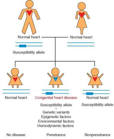
FIGURE 15-1. Model of multifactorial origin of congenital heart disease. A parent may harbor a genetic predisposition to disease (susceptibility allele) and transmit this genetic risk to offspring. However, this would result in heart defects only in conjunction with variants in other genetic loci or with epigenetic factors, resulting in disease penetrance. The susceptibility allele alone may not be sufficient to cause disease in offspring (nonpenetrance), but the individual would still transmit increased risk to offspring. Used with permission from Shieh J, Srivastava D. Circulation. 2009;120(4):269-271. © 2009 Wolters Kluwer Health, Inc.
Attributes of the Infants
Infants with complex cardiovascular defects often have lower birth weights than those in the normal population, but this observation is confounded by the observation that infants with these defects are often born prematurely. Additionally, decreased fetal growth is commonly found in infants with any cardiac defect who also have noncardiac malformations or chromosomal abnormalities.
Gender differences in the prevalence of specific defects have been noted consistently. Transposition of the great arteries, tetralogy of Fallot, and total anomalous pulmonary venous return occur more frequently in boys. Patent ductus arteriosus, atrial septal defect, muscular ventricular septal defect, atrioventricular septal defect, heterotaxy syndrome, and Ebstein anomaly of the tricuspid valve occur more often in girls. The reasons for these gender differences are not known.
Racial and ethnic group differences in the prevalence of congenital cardiovascular disease have been reported in some studies. Most of these are likely the result of incomplete case ascertainment and differences in the distribution of risk factors in the populations; true racial or ethnic differences have not been identified.
■ GENETIC COUNSELING
Indications for Genetics/Dysmorphology Evaluation
Clinicians caring for patients with congenital cardiovascular disease should be aware of the need to assess a newly diagnosed patient for relevant associated anomalies. Identification of a recognizable syndrome will assist in determining the need for genetic testing and in evaluating the patient for other anomalies. Additionally, identification of a syndrome may indicate the need for genetic testing of other family members and will allow more accurate risk assessment of recurrence of congenital cardiovascular disease in future children.
A complete physical examination should include an assessment for dysmorphic features. Genetics evaluation including examination by a geneticist and laboratory testing is often indicated (Table 15-6). A complete family history should be taken and should include pregnancy loss, ethnic origin, and consanguinity. Chest and abdominal radiographs may identify abnormalities in situs. Upper gastrointestinal series, abdominal ultrasound, liver-spleen scan, and head imaging studies are obtained as indicated. Recent studies have shown that first-degree family members may have subclinical cardiovascular defects. For example, the parents of a male child with hypoplastic left heart syndrome have a greater than 25% likelihood of having a bicuspid aortic valve. Thus, echocardiographic study of first-degree relatives may be indicated.
TABLE 15-6. Indications for Genetic Evaluation
Patient
Multiple congenital anomalies
Dysmorphic facial features or skeletal abnormalities
Abnormal prenatal diagnostic test
Abnormal newborn screening test
Perinatal death
Family history
Congenital heart disease
Genetic disorder
Congenital anomalies
Miscarriages, stillbirths, neonatal deaths
Certain congenital cardiovascular defects are frequently associated with genetic abnormalities. For example, 60% of infants with atrioventricular septal defects have trisomy 21. Tetralogy of Fallot is associated with more than 50 syndromes, the most common being deletion 22q11 syndrome. This syndrome is present even more frequently in patients with defects such as interrupted aortic arch type B (Table 15-7). Patients with cardiovascular defects highly associated with syndromes may benefit from evaluation for the presence of a genetic abnormality even in the absence of dysmorphic features.
Recurrence Risks
Questions regarding causality are directly relevant to the risk of recurrence, and the parents of any child with congenital cardiovascular disease will have questions about the risk of recurrence in subsequent pregnancies or in their grandchildren. Additionally, as more patients with cardiovascular defects survive to reproductive age, questions about recurrence come directly from patients. Unfortunately, accurate genetic counseling requires knowledge regarding causality, and the direct cause of most congenital cardiovascular disease is not known. The availability of prenatal diagnosis, including amniocentesis and chorionic villus sampling for chromosomal diagnosis and fetal echocardiography (beginning at about 16 weeks’ gestation) for evaluation of cardiac structure, should be communicated. The long-term goal of genetic counseling is educational; information regarding risk and prenatal diagnosis must be presented in a balanced manner, and informed decisions regarding reproduction should be left to the patient and family.
Many studies have attempted to assess the recurrence risk to siblings and offspring of patients with congenital cardiovascular disease. Study results often conflict, and interpretation is confounded by methodologic differences in patient ascertainment, diagnostic techniques, morphologic classification of defects, and identification of chromosomal abnormalities and syndromic features. Clearly, the recurrence risk will vary among specific types of defects and within different kindred. Risk will be determined in part by the specific genetic and environmental contributions to each defect, such as the presence of additional affected family members and the presence of known genetic or syndromic risk factors.
The recurrence risk for a sibling is at least 2% to 3% and increases substantially if more than one sibling is affected. The recurrence risk to offspring of parents with congenital cardiovascular disease averages 2% to 4%. However, multiple studies have shown that recurrence risks vary depending on the exact defect. 0yen et al. reported recurrence risk ratios from 0 among 228 patients with anomalous pulmonary veins to 79 in 359 patients with heterotaxy (Table 15-8). For persons with isolated cardiovascular defects, the overall relative risk of recurrence for the same defect was 8.15. Interestingly, same-sex twins (some of which are monozygotic) showed about a threefold higher relative risk of recurrence than unlike-sex twins. These data strongly suggest a genetic component even to sporadic congenital cardiovascular disease. Future research that characterizes genes and gene defects in family pedigrees will provide more definitive information.
TABLE 15-7. Estimated Frequency of 22q11 Deletions in Congenital Heart Disease
|
Defect |
Estimated deletion frequency (%) |
|
Interrupted aortic arch type B |
50-89 |
|
Ventricular septal defect |
10 |
|
With normal aortic arch |
3 |
|
With aortic arch anomalya |
45 |
|
Truncus arteriosus |
34-41 |
|
Tetralogy of Fallot |
8-35 |
|
Isolated aortic arch anomalies |
24 |
|
Double-outlet right ventricle |
<5 |
|
Transposition of the great arteries |
<1 |
|
aIncludes right aortic arch, abnormal branching pattern, cervical location, and/or discontinuous pulmonary arteries. Reprinted with permission: Circulation. 2007;115:3015-3038. ©2007 American Heart Association, Inc. |
|
TABLE 15-8. Relative Risk of Recurrence by Congenital Heart Defect (CHD) by Family History of CHDa
|
Heart defect |
Relative riskb |
|
Heterotaxy |
79.1 |
|
Conotruncal defect |
11.7 |
|
Atrioventricular septal (canal) defect |
24.3 |
|
Anomalous pulmonary venous return |
0 |
|
Left ventricular outflow tract obstruction |
12.9 |
|
Right ventricular outflow tract obstruction |
48.6 |
|
Atrial septal defect |
7.07 |
|
Ventricular septal defect |
3.41 |
|
Patent ductus arteriosus (term) |
4.80 |
|
Overall same heart defect |
8.15 |
|
aAdapted from 0yen N., et al. Circulation. 2009;120:295-301. bReference was index persons with a heart defect who had a first- degree relative without a heart defect. |
|
SUGGESTED READINGS
Biesecker LG, Green RC. Diagnostic clinical genome and exome sequencing. N Engl J Med. 2014;370(25):2418-2425.
Botto LD, Correa A, Erickson JD. Racial and temporal variations in the prevalence of heart defects. Pediatrics. 2001;107(3):E32.
Carey AS, Liang L, Edwards J, et al. Effect of copy number variants on outcomes for infants with single ventricle heart defects. Circ Cardiovasc Genet. 2013;6(5)444-451.
Cordell HJ, Clayton DG. Genetic association studies. Lancet. 2005;366(9491):1121-1131.
Fahed AC, Gelb BD, Seidman JG, Seidman CE. Genetics of congenital heart disease: the glass half empty. Circ Res. 2013;112(4):707-720.
Ferencz C, Loffredo CA, Correa-Villasenor A, Wilson PD. Perspectives in Pediatric Cardiology. Vol. 5. Genetic and Environmental Risk factors of Major Cardiovascular Malformations: The Baltimore-Washington Infant Study 1981-1989. Armonk, NY: Futura; 1997.
Ferencz C, Loffredo CA, Rubin JD, Magee CA. Perspectives in Pediatric Cardiology. Vol. 4. Epidemiology of Congenital Heart Disease: The Baltimore-Washington Infant Study 1981-1989. Mount Kisco, NY: Futura; 1993.
Gelb, BD. Genetics of congenital heart disease. Curr Opin Cardiol. 2016;31(3):235-241.
George AL. Use of contemporary genetics in cardiovascular diagnosis. Circulation. 2014;130(22):1971-1980.
Goldmuntz E, Dirscoll DA, Emanuel B, et al. Evaluation of potential modifiers of the cardiac phenotype in the 22q11.2 deletion syndrome. Birth Defects Res A Clin Mol Teratol. 2009;85(2):125-129.
Hoffman JI, Kaplan S. The incidence of congenital heart disease. J Am Coll Cardiol. 2002;39(12):1890-1900.
Homsy J, Zaidi S, Shen Y, et al. De novo mutations in congenital heart disease with neurodevelopmental and other congenital anomalies. Science. 2015;350(6265):1262-1266.
Huybrechts KF, Palmsten K, Avorn J, et al. Antidepressant use in pregnancy and risk of cardiac defects. N Engl J Med. 2014;370(25):2397-2407.
Jenkins KJ, Correa A, Feinstein JA, et al. Noninherited risk factors and congenital cardiovascular defects: current knowledge: a scientific statement from the American Heart Association Council on Cardiovascular Disease in the Young: endorsed by the American Academy of Pediatrics. Circulation. 2007;115(23):2995-3014.
Jones KL, Jones MC, del Campo M. Smith’s Recognizable Patterns of Human Malformation. 7th ed. Philadelphia, PA: Elsevier Saunders; 2013.
Liu S, Joseph KS, Linokova S, et al. Association between maternal chronic conditions and congenital heart defects. Circulation. 2013;128(6):583-589.
0yen N, Poulsen G, Boyd HA, et al. Recurrence of congenital heart defects in families. Circulation. 2009;120(4):295-301.
Patel SS, Burns TL. Nongenetic risk factors and congenital heart defects. Pediatr Cardiol. 2013;34(7):1535-1555.
Peyvandi S, Lupo PJ, Garbarini J, et al. 22q11.2 deletions in patients with conotruncal defects: data from 1,610 consecutive cases. Pediatr Cardiol. 2012;34(7):1687-1694.
Reller MD, Strickland MJ, Riehle-Colarusso T, Mahle WT, Correa A. Prevalence of congenital heart defects in metropolitan Atlanta, 1998-2005. J Pediatr. 2008;153(6):807-813.
Tegnander E, Williams W, Johansen OJ, Blaas HG, Eik-Nes SH. Prenatal detection of heart defects in a non-selected population of 30,149 fetuses—detection rates and outcome. Ultrasound Obstet Gynecol. 2006;27(3):252-265.
Vinet E, Pineau CA, Scott, S, et al. Increased congenital heart defects in children born to women with systemic lupus erythematosus: results from the offspring of systemic lupus erythematosus mothers registry study. Circulation. 2015;131(2):149-156.
Reference Websites
Clinical Teratology Web. http://depts.washington.edu/ter- isweb. University of Washington, Seattle; 2014.
Gene Reviews. http://www.ncbi.nlm.nih.gov/sites/Gene- Tests/review?db=genetests. University of Washington, Seattle; 2014.
GeneTests. Medical Genetics Information Resource. http:// www.genetest.org. University of Washington, Seattle; 2014.
National Newborn Screening and Genetics Resource Center. http://genes-r-us.uthscsa.edu. University of Texas San Antonio, San Antonio; 2014.
Online Mendelian Inheritance in Man (OMIM) http:// www.ncbi.nlm.nih.gov/omim. Johns Hopkins University, Baltimore; 2014.