
■ INTRODUCTION
Critical Congenital Cardiovascular Disease: Extent of the Problem
Presentation of Congenital Cardiovascular Disease: An Overview
■ HISTORY
■ FAMILY HISTORY
■ PHYSICAL EXAMINATION
Pulse Oximetry as a Screening Test
The Cardiovascular Examination
■ ANCILLARY TESTS
Arterial Blood Gases
Electrocardiogram
Chest Radiograph
Echocardiography
Genetics Evaluation
Evaluation of Other Organ Systems
■ INITIAL TREATMENT
General Principles
■ SUGGESTED READINGS
■ INTRODUCTION
Critical Congenital Cardiovascular Disease: Extent of the Problem
Congenital cardiovascular disease occurs in about 1% of live births (excluding bicuspid aortic valve and hemo- dynamically insignificant lesions, such as very small secundum atrial and muscular ventricular septal defects). Congenital cardiovascular disease is considered to be critical if it will predictably result in death or severe morbidity if unrecognized and not treated in early infancy. These conditions often depend on patency of the ductus arteriosus to maintain either pulmonary or systemic blood flow. Signs of severe cyanosis or progressive heart failure leading to cardiovascular collapse develop as the ductus arteriosus constricts during the first few days or weeks after birth. It is important to recognize that a newborn with critical congenital cardiovascular disease may show little evidence of cardiovascular compromise on physical examination in the first 24 to 48 hours of life. Cardiovascular instability may not occur until after the infant is discharged from the hospital. The recent addition of routine pulse oximetry screening in many hospitals should greatly enhance our ability to diagnose such infants before discharge and thus decrease the number who present critically ill in the days and weeks after discharge.
The National Vital Statistic Report reported 3,932,181 registered births in the United States in 2013, which means that about 40,000 infants were born with heart defects. Of the 25% to 30% of infants with heart defects, at least 10,000 are predicted to have critical disease. Diagnosis before the onset of cardiovascular decompensation is essential for optimal outcome. In the absence of detection during fetal life by ultrasound examination or pulse oximetry screening after birth, about 70% of infants with critical congenital cardiovascular disease are not diagnosed before 2 days of age. This leads to severe morbidity and mortality in many hundreds of newborns each year in the United States alone, at a large social and economic cost. Thus, it is incumbent on all physicians and other health care professionals who care for newborns to rigorously evaluate every newborn for the possibility of critical cardiovascular disease. Furthermore, if there is any indication that such disease might exist, the infant must be referred for further evaluation without delay.
Presentation of Congenital Cardiovascular Disease: An Overview
The evaluation of the infant for critical cardiovascular disease should focus on the three cardinal signs of neonatal cardiovascular distress: cyanosis, decreased systemic perfusion, and tachypnea. Cyanosis may be appreciated by careful visual inspection and pulse oximetry, decreased systemic perfusion is identified by examination of the extremities, and tachypnea is noted by observing the respiratory rate and pattern. The presence of a congenital cardiovascular defect (or, less commonly, a cardiomyopathy or arrhythmia) must be considered in the differential diagnosis of any infant with one or more of these findings. A cyanotic infant most likely has underlying cardiovascular disease and almost certainly does in the absence of significant respiratory distress. An infant with decreased systemic perfusion may be septic or have a primary metabolic abnormality, but about one-half of such infants have symptomatic cardiovascular disease. An infant who has tachypnea without either cyanosis or decreased perfusion most often has primary parenchymal or extraparen- chymal lung disease but could also have cardiovascular disease associated with excessive pulmonary blood flow. The respiratory symptoms are often subtle and develop slowly, and there may be only mild distress so that the most notable manifestation may not be the respiratory distress but, rather, poor weight gain, particularly after a few weeks of life. A thoughtful and rational approach to the differential diagnosis of all three of these signs is important for prompt recognition and appropriate management. Moreover, the clinician must be cognizant of the fact that the transition from a fetal to a mature postnatal circulation occurs not immediately but rather over the first several days or weeks of life so that serial evaluations are necessary, each as rigorous as the first.
Each cardinal sign of neonatal cardiovascular disease can be attributed to one of at least two pathophysiologic causes:
Cyanosis
1. Decreased pulmonary blood flow
2. Normal to increased pulmonary blood flow but with a transposed aorta; for example, d-transposition of the great arteries
Decreased systemic perfusion
1. Obstruction of the left heart (inflow or outflow)
2. Cardiac dysfunction without obstruction (primary cardiomyopathy or secondary dysfunction)
Tachypnea/poor weight gain (due to excessive pulmonary blood flow)
1. Exclusive left-to-right shunt
2. Dominant left-to-right shunt with a lesser right-to-left shunt
The cardiac defects in each hemodynamic category are discussed in Chapters 6 (cyanosis), 7 (excessive pulmonary blood flow), and 8 (decreased systemic perfusion). This chapter will describe the general approach to the cardiovascular evaluation of the newborn, including history, physical examination, and ancillary tests. The information obtained from each step of the evaluation should follow a logical progression, from recognizing the primary sign to defining the underlying pathophysiologic process and eventually to determining the specific diagnosis. The latter is rarely important at the initial evaluation. Understanding the pathophysiologic process allows effective initiation of therapy and stabilization of the patient. The specific diagnosis is usually important only to implement the definitive therapeutic plan.
From these considerations, a focused and rational approach to the newborn with symptomatic congenital cardiovascular disease can be developed. Admittedly, this approach is imperfect; some lesions are complex with overlapping manifestations (eg, an infant with hypoplastic left heart syndrome who has decreased systemic perfusion may also be tachypneic and mildly cyanotic). However, even in the most complex cases, one of the major signs usually predominates and provides a clue to the most likely category of disease. The concepts described in this and other chapters emphasize the importance of a simple, clear, logical, and stepwise approach to the evaluation and treatment of each infant with cardiovascular disease.
■ HISTORY
The prenatal evaluation of the newborn for congenital cardiovascular disease is reviewed in Chapter 4, including the evaluation of the fetus with a family history of congenital cardiovascular disease or a genetic syndrome that is associated with cardiovascular disease. If the perinatal history is completely benign (eg, no history of perinatal asphyxia), it is more likely that a problem causing symptoms in a neonate is cardiac in origin.
Cyanosis is caused by either congenital cardiovascular disease, pulmonary hypertension of the newborn, or intra- or extraparenchymal lung disease. The primary differential diagnosis of congenital cardiovascular disease is persistent pulmonary hypertension of the newborn. Persistent pulmonary hypertension must be excluded when a diagnosis of congenital heart disease is being considered. Infants with either of these conditions often have mild or moderate respiratory distress. In contrast, cyanotic infants with primary parenchymal lung disease usually have severe respiratory distress requiring mechanical ventilation and have a chest radiograph showing abnormal lung parenchyma. The infant with persistent pulmonary hypertension, however, often has a perinatal history of birth asphyxia, with or without meconium aspiration. Additionally, the infant may be small for gestational age, or the mother may have taken nonsteroidal antiinflammatory medications over the weeks before birth, which can cause intrauterine constriction of the ductus arteriosus and subsequent pulmonary hypertension.
The infant with cyanotic cardiovascular disease typically has a benign birth history with a normal or nearly normal Apgar score. The ductus arteriosus usually maintains adequate blood flow and mixing immediately after birth, and cyanosis is not readily apparent. It may not be until hours or days after birth that the infant becomes cyanotic, frequently during feeding or crying. The increased physical effort associated with feeding or crying increases oxygen consumption and decreases pulmonary blood flow, accentuating the cyanosis. Despite the presence of cyanosis, respiratory distress is typically not part of the history. The chemoreceptor response to hypoxemia is intact so that mild tachypnea often occurs but respiratory distress (eg, retractions, nasal flaring, grunting) is usually absent because ventilation is normal. Use of pulse oximetry screening should allow recognition of nearly all infants with cyanotic cardiovascular disease who do not show visible cyanosis.
At times when a newborn infant cries, a transient increase in right atrial pressure occurs and results in a small right-to-left shunt through the foramen ovale, causing the infant to appear dusky. This transient desaturation must be distinguished from infants with cyanotic cardiovascular disease who may also appear dusky initially only with crying or feeding. Although not apparently cyanotic at rest, infants with cyanotic congenital cardiovascular disease show some degree of oxygen desaturation by pulse oximetry at all times, whereas the normal newborn infant has normal oxygen saturation at rest. This is important to appreciate during pulse oximetry screening, which should be repeated when an infant is quiet if an abnormal value is obtained when the infant is crying.
The differential diagnosis of decreased systemic perfusion includes obstructive cardiovascular lesions and myocardial dysfunction from sepsis, hematological abnormalities (anemia and polycythemia), or endocrine/metabolic disorders, such as hypocalcemia, hypoglycemia, and metabolic acidosis. Neonatal sepsis is common, especially in the setting of prolonged rupture of the membranes. Hematological abnormalities are associated with placenta abruptio, twin-to-twin transfusion, placental insufficiency, postterm delivery, or small-for-gestational age infants. A positive family history is often present in newborns with endocrine/metabolic diseases. Newborns with obstructive cardiovascular disease rarely have a positive perinatal history. These infants typically are stable during the first hours of life, but eventually develop poor feeding, pallor, diaphoresis, and tachypnea with respiratory distress. This may occur as late as 3 to 4 weeks after birth, so it is extremely important for every infant to be carefully assessed at the time of discharge and at subsequent visits during the first month of life. Subtle findings of irritability, pallor, poor feeding, or diaphoresis may reflect inadequate systemic perfusion. Particularly concerning is that many of these infants may “pass” the pulse oximetry screen, unless right-to-left shunting of desaturated blood to the lower body via the ductus arteriosus is present and lower body pulse oximetry is included a part of the screening. Tachypnea is often a subtle finding that develops over days or weeks, as pulmonary vascular resistance and hemoglobin concentration decline during the first 6 weeks of life. Although some patients show dramatic shunting and respiratory symptoms in the first few days of life (particularly those with trisomy 21), tachypnea during this time period, in the absence of signs of cyanosis or decreased systemic perfusion, often points to pulmonary disease rather than cardiovascular disease. Parents rarely appreciate that an infant is breathing more rapidly than normal. Poor feeding with associated failure to thrive and diaphoresis is common; murmurs may be absent. Thus, an infant with unexplained failure to thrive, particularly in association with tachypnea and diaphoresis, should be evaluated for possible congenital cardiovascular disease. Many of these infants “pass” pulse oximetry screening, supporting the approach that the clinician must evaluate every infant over the first weeks of life for congenital cardiovascular disease at each encounter and not rely on the newborn evaluation only.
■ FAMILY HISTORY
A family history of congenital cardiovascular defects or cardiomyopathy is relevant to the outcome of the fetus, as a positive family history increases the risk for subsequent children. Genetic abnormalities are increasingly recognized to contribute to congenital cardiovascular disease and are discussed in Chapter 15.
■ PHYSICAL EXAMINATION
Pulse Oximetry as a Screening Test
Pulse oximetry screening for all newborn infants is accepted and has been implemented widely. In 2010, the US Health and Human Services Secretary’s Advisory Committee on Heritable Disorders in Newborns and Children recommended universal screening, and a working group selected from that committee as well as the American Academy of Pediatrics, the American College of Cardiology, and the American Heart Association published a joint document outlining how pulse oximetry screening should be performed. As of May 2016, only five states have neither active nor pending legislation or regulations mandating universal screening (Figure 5-1). Unfortunately, screening is not yet mandated in Canada, and only four European countries (Switzerland, Ireland, Poland, and Norway) have national recommendations to screen. Despite this, many hospitals in Canada and European countries have instituted screening, and a recent study has shown it to be nearly universal throughout the Nordic countries.
The Cardiovascular Examination
The physical examination should be performed systematically (Ch5An1). Each step determines if the infant falls into a specific mode of presentation (cyanosis, decreased systemic perfusion, or excessive pulmonary blood flow) and, once defined, into a specific hemodynamic category. Ancillary tests assist in establishing specific diagnoses and defining the most appropriate therapy for each infant.
The general examination includes vital signs and observation of the unclothed and warm infant. The vital signs of temperature, heart rate, respiratory rate, and blood pressure are measured in conjunction with respiratory status, perfusion, and color. We consider that pulse oximetry is also a vital sign in the infant, not just to be performed as part of the initial screen but also to be included in any evaluation during the transitional circulatory period. Weight, length, and head circumference are measured and plotted on growth charts to aid in identifying growth impairment. Any postnatal decrease in weight percentiles compared to length and head circumference should raise the possibility of cardiovascular disease. This is of particular importance in lesions of excessive pulmonary blood flow—the other signs of cyanosis and hypoperfusion are not present, and there may be no murmurs or discernible respiratory distress.
The first sign to assess on general observation is cyanosis. Peripheral cyanosis (acrocyanosis) is common in newborn infants and reflects their normally unstable peripheral vasomotor tone. Central cyanosis, which is indicative of arterial oxygen desaturation, is the important sign to recognize. Thus, vascular beds with little vasoconstrictor tone, such as the tongue, gums, and the buccal mucosa, should be evaluated (not the hands, feet, or perioral region). It is also important to evaluate the patient during conditions such as feeding or crying, which are most likely to produce central cyanosis. Cyanosis is difficult to perceive until arterial oxygen saturation is less than about 85%, and the physiologic decrease in hemoglobin concentration in infants 4 to 12 weeks of age makes detecting cyanosis more difficult. Thus, oxygen saturation should be measured if there is any question of cyanosis. Measuring oxygen saturation simultaneously in the right hand and a lower extremity by use of two pulse oximeters is necessary to evaluate whether the upper and lower bodies are perfused, at least partially, by different ventricles. A difference in oxygen saturation of only 3% to 5% may be significant, but most oximeters are accurate only to within ±2% to 3%. For this reason, it may be helpful to reverse the probes to ensure that any difference (or absence of a difference) is real and not just related to inherent variations in the probes or oximeters. Less commonly, it may be necessary to measure the oxygen saturation in an earlobe if aortic arch and arch vessel anomalies are suspected (Chapter 6).

FIGURE 5-1. Status of pulse oximetry screening for critical congenital heart disease in the United States. As of May 2, 2016 legislation has been fully enacted in 40 states and the District of Columbia, and in only three states has no action been taken. Image extracted from https://www.aap.org/en-us/ advocacy-and-policy/state-advocacy/Documents/2016%20CCHD%20Newborn%20Screening%20 Bills,%20Regulations,%20and%20Executive%20Orders%20-%20AAP%20Division%20of%20State%20 Govt%20Affairs.pdf
Next, the respiratory status should be carefully evaluated. Infants who have isolated cyanosis are usually tachypneic but do not otherwise exhibit increased work of breathing. In contrast, the increased pulmonary venous pressures and pulmonary edema seen in patients with hypoperfusion cause respiratory distress in addition to tachypnea. In that case, variable intercostal and/or subcostal retractions, nasal flaring, and grunting may be observed.
Signs of decreased systemic perfusion, including the temperature and color of the skin, blood pressure, peripheral pulses, and capillary refill in each extremity, should be assessed next. Lower extremity pulses are more easily palpated in the feet than in the inguinal area. If the infant has a normal dorsalis pedis or posterior tibial pulse, then pulsatile blood flow to the lower extremity is not impaired. Blood pressure should be measured in the upper and lower extremities; normally, the lower extremity blood pressure is slightly greater than that in the upper extremity. The left subclavian artery arises from the aortic isthmus and may be involved in a coarctation. Thus, the systolic pressures in the right arm and either leg should be measured simultaneously. If the pulses are decreased and no blood pressure differential is detected, the carotid arteries should be palpated. If they are increased, the infant may have a coarctation or interruption of the aorta in the presence of a right subclavian artery arising anomalously from the descending aorta.
The periphery, head, and neck should be examined for dysmorphic features of syndromes associated with cardiovascular disease, such as Down syndrome, 22q11 deletion (DiGeorge) syndrome, Turner syndrome, Noonan syndrome, Williams syndrome, and trisomy 21 (Chapter 15).
At this point in the examination, it has been determined whether the infant has cyanosis, decreased systemic perfusion, or tachypnea. Examination of the abdomen, lungs, and heart then assists in defining the hemodynamic category. Heart murmurs are common in many normal infants and are absent in about 50% of infants with symptomatic cardiovascular disease. Thus, the mere presence of a murmur is of little value to the examination, as is its absence. However, specific murmurs are much more likely to be appreciated if the clinician has a differential diagnosis in mind at the time when auscultation is performed. Moreover, the presence of a nonspecific murmur is of much less concern in an infant who has an otherwise normal examination.
The abdomen should be percussed and palpated. Palpation for liver size is important, as the liver is often a sign of right atrial hypertension or increased circulating volume from excessive pulmonary blood flow. The location of the liver and stomach is reversed in situs inversus and is anomalously positioned in patients with heterotaxy syndromes (see Chapter 6).
Examination of the lungs includes inspection of the pattern and work of breathing, the symmetry of chest movement, and auscultation. Because of the small size of the patient, normal breath sounds in one lung field may reflect ventilation of the other lung.
The cardiac examination begins with palpation of the precordium to assess right ventricular pressure and volume load. Unlike the older patient, the normal newborn infant has a parasternal and subxiphoid impulse because the sternum is thin and the right ventricle is thick walled. The parasternal and subxiphoid impulses are increased in most infants with cyanotic cardiovascular disease because the right ventricle is ejecting at or above systemic pressure against right ventricular outflow obstruction or into a transposed aorta. A decreased right ventricular impulse in a cyanotic patient is suggestive of inflow obstruction to the right ventricle, either tricuspid atresia or hypoplastic right heart syndrome. A parasternal thrill suggests the presence of a ventricular septal defect, but only a small minority of infants who have ventricular septal defects have thrills at birth. Therefore, the absence of a thrill is not useful in excluding the presence of a ventricular septal defect. In contrast, the presence of a thrill is very helpful in a cyanotic newborn, as this is diagnostic of tricuspid atresia with ventricular septal defect because this is the only form of cyanotic cardiovascular disease in which the ventricular shunt is directed anteriorly toward the sternum, from the left ventricle to the right ventricle. In cyanotic cardiovascular disease with outflow obstruction, such as tetralogy of Fallot, the ventricular shunt is directed posteriorly, from the right ventricle through the left ventricle to the aorta, so that a thrill is not present. The left ventricular apical impulse is not usually palpable in a normal newborn infant because the dominant right ventricle displaces the left ventricle posteriorly. A palpable left ventricular impulse usually indicates increased left ventricular volume load as the ventricular cavity dilates and extends anteriorly and laterally. In contrast, increased left ventricular pressure load often does not cause a palpable impulse. A suprasternal notch thrill is suggestive of turbulence from valvar or supravalvar aortic stenosis.
Auscultation should be performed in a systematic manner. The first heart sound is rarely helpful but may be louder than normal in the infant with a complete atrioventricular septal defect. The quality of the second heart sound provides important information. Although it is often difficult to appreciate splitting of the second heart sound in the normal newborn because of the rapid heart rate, the presence of a clearly split second heart sound is suggestive of markedly increased pulmonary blood flow. This may lead the clinician toward the diagnosis of total anomalous pulmonary venous connection or a large arteriovenous malformation. Most cyanotic infants have a single heart sound because the pulmonary valve is either diminutive or atretic or because it is malposed, posterior to the aorta. Thus, the presence of a split second heart sound in a cyanotic infant strongly suggests total anomalous pulmonary venous connection.
After listening to the normal heart sounds, the presence of clicks and gallops should be evaluated. Clicks may be difficult to hear, but when present usually indicate a bicuspid aortic valve or persistent truncus arteriosus. A click is not present in patients with severe aortic or pulmonic stenosis because valve mobility is greatly decreased. In contrast, truncus arteriosus can frequently be diagnosed in the patient with tachypnea and modest desaturation based on the presence of an ejection click. Such clicks are commonly heard because the truncal valve is almost always dysplastic. The click is often heard close to the apex rather than in the region of the semilunar valve. Mid-systolic clicks are rarely heard but may be present in Ebstein anomaly or in newborns with severe mitral valve prolapse, such as occurs in neonatal patients with Marfan syndrome. Gallop rhythms may be present in newborn infants with severe left ventricular dysfunction.
Auscultation of heart murmurs may define specific diagnoses based on unique features of the murmurs. A murmur is best localized by determining the location of its highest-frequency components because high-frequency sounds radiate much shorter distances than do lower- frequency sounds. Loudness may be a poor indicator of the site of origin because most clinicians hear low-frequency components as being louder than high-frequency components. Thus, when the highest-frequency components of a murmur are heard in the left axilla, the source is likely extracardiac in origin, and the diagnosis of peripheral pulmonary artery stenosis in a normal newborn infant can be made. In addition to localization of the source of a murmur, focusing on its frequency allows the clinician to determine whether more than one murmur is present. While following the radiation of a murmur, if there is an increase in the frequency after a decrease, it is likely that a second systolic murmur is present.
The frequency of a murmur reflects the degree of turbulence, which correlates directly with the pressure gradient. A high-frequency murmur indicates a high- pressure gradient, and a low-frequency murmur indicates a low-pressure gradient. Mid-diastolic murmurs are difficult to appreciate because they are of very low frequency and are often noticed as the absence of silence in diastole. Conversely, early diastolic murmurs caused by semilunar valve insufficiency are usually easy to hear because the pressure gradient is greater and the murmur is thus in an easily audible frequency range. If a murmur of semilunar valve insufficiency is heard in a newborn without significant respiratory distress, the diagnosis is usually truncus arteriosus. Stenotic aortic and pulmonary valves are rarely insufficient at birth. If the newborn has severe respiratory distress in addition to the diastolic murmur of semilunar valve insufficiency, the diagnosis is usually absent pulmonary valve syndrome. In this lesion, the patient has very large pulmonary arteries that compress the bronchi, leading to severe respiratory distress. The cardiac disease is usually a mild form of tetralogy of Fallot, and the absence of pulmonary valve tissue leads to a characteristic to-and-fro murmur. Finally, it is important to recognize that newborns with large ventricular septal defects or atrioventricular septal defects often do not present with murmurs in the first few days of life. Pulmonary vascular resistance is relatively high so that flow across the defect is fairly low.
■ ANCILLARY TESTS
Arterial Blood Gases
As mentioned earlier, measurement of oxygen saturation by pulse oximetry is now performed in almost all newborns at some time in the first 1 to 2 days of life. It should be done at least in one foot, and many centers will also measure it in the right hand. In a newborn with any findings suggestive of cardiovascular disease, oxygen saturation should definitely be measured in the right hand and one foot, and any abnormality should lead to further investigation, including the measurement of arterial blood gases. It is also important to measure arterial blood gases in any infant who has respiratory distress, even with normal pulse oximetry. Blood gases determine not only the oxygen content of arterial blood but also the extent of the oxygen debt and an infant’s ability to compensate for a metabolic acidosis by decreasing CO2, which is particularly important in patients who might have decreased systemic perfusion.
Electrocardiogram
Unless an arrhythmia is present (Chapter 10), an electrocardiogram is of limited value in making a specific diagnosis at birth for many defects. The right ventricle is dominant in utero, and thus a pattern of right ventricular hypertrophy is present in the normal newborn: right axis deviation, prominent R waves and upright T waves in the right precordium, and septal Q waves positioned very laterally, between V5 and V7 (Figure 5-2A). The electrocardiogram recorded at birth is normal in patients with common symptomatic cardiovascular disease, such as d-transposition of the great arteries, coarctation of the aorta, and tetralogy of Fallot. It is not until 5 to 10 days after birth, when the pattern of right ventricular hypertrophy regresses in the normal infant (the T waves become inverted in the right precordium, Figure 5-2B), that the electrocardiogram in such patients is abnormal. However, as discussed in subsequent chapters, some defects are associated with abnormal and, in some cases, pathognomonic electrocardiograms so that electrocardiography can be of value in assisting in the diagnosis of certain defects. It is important to emphasize that a normal electrocardiogram does not exclude cardiovascular disease in the newborn.
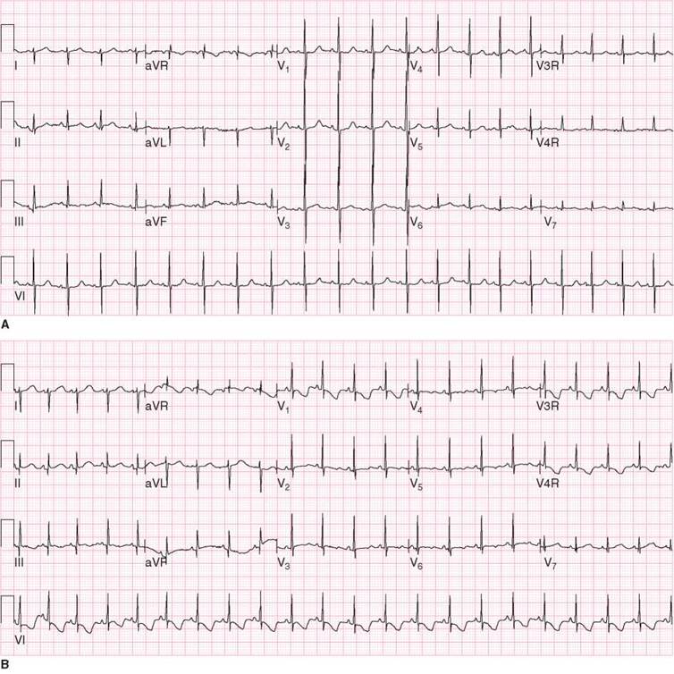
FIGURE 5-2. The normal electrocardiograms over the first week of life. A. On the first day of life there is right axis deviation, the right precordial forces are prominent and the right precordial T waves are upright. B. By 5-10 days of age the right precordial forces remain prominent but the T-waves have become inverted.
Chest Radiograph
The chest radiograph provides information about the heart size and contour, pulmonary blood flow, and the lung parenchyma. Absence of a thymic shadow may contribute to the diagnosis of 22q11 deletion (DiGeorge) syndrome. The absence of a rightward deviation of the trachea and a contour on the right side of the spine are suggestive of a right aortic arch, which is highly associated with this syndrome. The main pulmonary artery is often malposed (as in d-transposition of the great arteries) or diminutive (as in tetralogy of Fallot), and thus the normal convexity in the left upper mediastinum is absent. Other defects associated with abnormalities of the contour or size of the heart include l-transposition of the great arteries, truncus arteriosus, and Ebstein anomaly, among others.
Increased pulmonary blood flow is often difficult to assess by conventional chest radiography at birth because, even when pulmonary blood flow is three to four times systemic flow, the pulmonary vessels may not appear large. Small proximal vessels can conduct large quantities of blood as long as the distal vasculature is well developed. It is not until many days or weeks later that the arteries enlarge in response to increased flow. Conversely, pulmonary venous congestion can be appreciated very soon after birth, as pulmonary blood flow increases dramatically. That increased flow rapidly increases pulmonary venous pressures when the left side of the heart is critically obstructed. Lesions such as total anomalous pulmonary venous connection with obstruction show increased venous markings and pulmonary edema within hours of birth.
Finally, the upper abdomen is usually seen on chest radiography in the newborn. Thus, liver size and situs can be evaluated. As noted above, this is of value when hetero- taxy syndrome is being considered.
Echocardiography
Echocardiography is the mainstay of the diagnosis of the newborn with symptomatic cardiovascular disease and has largely replaced cardiac catheterization. However, to understand the pathophysiology and to offer the best care, it is important to take a systematic approach at each point of the evaluation and to use each piece of information to build on that understanding rather than focusing solely on the echocardiogram. Two-dimensional echocardiography clearly visualizes the anatomy of the heart and the central great vessels, even in the smallest premature infants. Demonstrating blood flow by use of color Doppler studies is essential for evaluating the atrial septum, the aortopulmonary septum, and the pulmonary veins because imaging alone may fail to identify these very thin structures.
Pulsed and continuous wave Doppler studies add information about the physiology of the heart and great vessels beyond two-dimensional imaging. For example, peak instantaneous pressure differences can be estimated from the Bernoulli equation:

where P is pressure difference in mm Hg and v is velocity measured by Doppler study in meters per second. This equation is based on nonpulsatile flow through a discrete narrowing in a rigid tube. Thus, Doppler estimates of pressure differences are approximations only. If the obstruction is relatively long or the geometry of the structures is not linear, the gradient is often overestimated by the Doppler equation. Acquiring the velocity at angles greater than about 30 degrees from parallel tends to underestimate velocity and thus underestimate the pressure difference. Within these limitations, however, Doppler studies are invaluable for estimating the severity of defects and pressures in chambers and arteries not accessible by other noninvasive methods. Right ventricular systolic pressure can be estimated by the peak instantaneous Doppler velocity of the tricuspid insufficiency jet, which correlates with the peak systolic pressure difference between the right ventricle and right atrium. In addition, the velocity of the flow across a defect in the ventricular septum provides an estimate of the peak systolic pressure difference between the left and right ventricles. Pulmonary arterial mean pressure can be estimated by the velocity of a pulmonary insufficiency jet. Doppler studies are also helpful in evaluating patients with noncardiac disease. For example, right ventricular pressure and pulmonary arterial mean pressure can be estimated in patients with pulmonary hypertension of the newborn.
In some cases, magnetic resonance imaging and magnetic resonance angiography can be very helpful in defining the anatomy and physiology. These imaging techniques are especially useful in defining the extracardiac and great vessel anatomy. Magnetic resonance imaging may be an important component of the evaluation of infants with coarctation of the aorta, interrupted aortic arch, and anomalous pulmonary venous connections. The application of these techniques is expanding as the technology improves to provide accurate information regarding blood flow, ventricular volume and function, and pressure gradients in the small infant.
Genetics Evaluation
Every infant with congenital cardiovascular disease should be carefully evaluated for evidence of dysmorphic features. As more extensively described in Chapter 15, a number of relatively common syndromes, such as DiGeorge syndrome, trisomy 21, trisomy 18, Williams syndrome, Turner syndrome, and Alagille syndrome, have characteristic features that may be evident on physical examination. These conditions have a high incidence of significant congenital cardiovascular defects. It is often helpful to involve specialists in clinical genetics to assist in the diagnostic evaluation of the infant and the family. Even a relatively low index of suspicion should prompt a genetics evaluation. Chromosome analysis or other specialized genetic testing may be indicated and should be performed in a rational and targeted manner. This will become increasingly important in the near future as more information becomes available regarding the molecular genetics of congenital cardiovascular disease.
Evaluation of Other Organ Systems
Abnormalities of other organ systems are present in about 25% of newborns with congenital cardiovascular disease, and the frequency is higher in newborns with critical congenital cardiovascular disease. In addition, many such infants undergo cardiac surgery in the first few days of life, often with low-flow bypass or circulatory arrest. Thus, it is important to evaluate major organ function in all infants who either have lesions associated with other organ involvement (such as known genetic syndromes or associations such as CHARGE) or are undergoing interventional catheterization or surgery. Noncardiac features of specific genetic syndromes and associations are discussed in Chapter 15. All newborns with critical congenital disease should undergo head and abdominal ultrasounds and laboratory evaluations of hematologic, renal, and liver function prior to intervention.
■ INITIAL TREATMENT
Timely initiation of medical therapy in patients with congenital cardiovascular defects is necessary to prevent and/ or reverse clinical deterioration. The general approach to a term or preterm infant with cardiovascular disease should follow the usual guidelines for management of a critically ill (or potentially critically ill) infant. It is beyond the scope of this text to review specific details of ventilator use, fluid and electrolyte management, monitoring, maintenance of a neutral thermal environment, and general supportive care of newborns. However, it is useful to review a few commonly applied guidelines that are particularly relevant to infants with cardiovascular disease. More detailed information regarding specific medical and surgical care is provided in the chapters that describe individual defects and postoperative care.
General Principles
Oxygen
Supplemental oxygen is often administered to infants with known or suspected cardiovascular disease without full consideration of the goals of therapy and of the possible adverse effects. As with any therapy, rational use of oxygen must be founded on sound principles of pathophysiology and include setting end points of efficacy and toxicity. The detrimental effects of supplemental oxygen are particularly notable in infants with critical congenital cardiovascular disease.
Depending on the specific pathophysiology, newborns with cardiovascular disease may be prone to overcirculation of the pulmonary vascular bed, which can be exacerbated by oxygen administration. As noted below, many newborns with critical congenital cardiovascular disease receive prostaglandin E1 (PGE1) to maintain ductal patency. Administration of PGE1 to infants who have single-ventricle physiology causes very high pulmonary blood flow either through a large patent ductus arteriosus, as in the case of pulmonary atresia with intact ventricular septum, or antegrade from the heart, as in hypoplastic left heart syndrome. In this setting, the balance of pulmonary to systemic blood flow is determined by the relative resistances of the pulmonary and systemic vascular beds. The normal decrease in pulmonary vascular resistance that occurs after birth is augmented by the vasodilator effects of PGE1. Pulmonary overcirculation occurs, and this can result in inadequate systemic perfusion. This leads to deterioration in pulmonary mechanics, a fall in urine output because of poor renal perfusion, and evidence of decreased systemic perfusion (metabolic acidosis or diminished cutaneous perfusion). A common response in this setting is to increase the fraction of inspired oxygen in an attempt to “normalize” oxygen saturation or arterial pO2. However, this response will almost certainly result in further deterioration because it will cause further pulmonary vasodilation and systemic vasoconstriction; thus, oxygen administration may cause a further in increase in pulmonary blood flow at the expense of systemic perfusion.
A vicious cycle is perpetuated unless proper steps are taken to reduce pulmonary and increase systemic blood flow. Simply reducing the fraction of inspired oxygen to 0.21 (room air) is the first step in reversing this pathophysiologic spiral. It is often better to accept lower oxygen saturations, in the 70s to 80s, to maintain effective systemic circulation and limit pulmonary blood flow. It must be remembered at all times that systemic blood flow and hemoglobin concentration are as important as arterial oxygen saturation in determining systemic oxygen delivery. Transfusion of packed red blood cells will increase systemic oxygen delivery without the deleterious effects of pulmonary vasodilation caused by oxygen.
In the patient with suspected cardiovascular disease, an oxygen challenge test is sometimes performed. This is only rarely of value in assisting in the diagnosis and may promote constriction of the ductus arteriosus, which can be deleterious to the newborn with an undiagnosed ductal-dependent cardiac defect. If performed, it should be done over a few minutes, oxygen saturation should be measured by pulse oximetry in both the upper and the lower bodies, and if oximetry shows a saturation of 97% or higher, arterial blood from the upper body should be drawn for measurement of arterial oxygen tension.
Mechanical Ventilation
Mechanical ventilation of the infant whose primary symptom is cyanosis is often not necessary. If arterial blood gases show adequate spontaneous ventilation, intubation and mechanical ventilation usually will not improve oxygenation significantly. Pulmonary venous blood is already fully saturated—the patient is cyanotic not because of poor pulmonary mechanics but because of the abnormal blood flow patterns caused by the heart disease (see Chapter 6). In contrast, mechanical ventilation and sedation are often very beneficial to the infant with decreased systemic perfusion and inadequate systemic oxygen delivery because decreasing the work of breathing decreases oxygen consumption. Also, decreased systemic perfusion is associated with increased pulmonary venous pressures and alveolar edema, which can be reversed with positive pressure ventilation.
Although it is generally desirable to maintain normal acid-base and electrolyte status to optimize cardiac function, in some settings it may be appropriate to allow pCO2 to rise (permissive hypercapnia). This strategy may be particularly useful in manipulating the ratio of pulmonary to systemic blood flow by promoting pulmonary vasoconstriction.
Infants often require intermittent sedation to achieve appropriate control of ventilation, and some even require muscle relaxants. Monitoring ventilatory status, blood gases, urine output, and the clinical examination are all important in caring for neonates with cardiovascular disease who are being mechanically ventilated.
Fluids
Careful attention to fluid status and urine output is essential when managing newborns with congenital cardiovascular disease. In general, on the first day or two of life, newborn infants with congenital cardiovascular disease manifest the same fluid, glucose, and electrolyte requirements as infants without congenital cardiovascular disease. Depending on the particular defect, however, fluid and electrolyte management may change dramatically. For example, an infant with a single-ventricle or large unrestricted ventricular septal defect and no pulmonary stenosis will exhibit progressively greater left-to-right shunting as the pulmonary vascular resistance falls postnatally. Consequently, signs and symptoms of heart failure may develop because of increased pulmonary blood flow, reduced systemic output, and compensatory sodium and water retention. Free water restriction and diuretic therapy are indicated to reduce total body sodium and water. It is not unusual for mild hyponatremia to occur, which should not prompt administration of additional sodium. Instead, this most likely represents dilution due to water retention and should be treated with further reduction of free water intake. In contrast to hyponatremia, hypokalemia and hypocalcemia should be treated with supplemental administration of potassium and calcium. Hypochloremia may impair diuretic function and should be replaced but not with sodium. If there is an associated significant metabolic alkalosis, a carbonic anhydrase inhibitor can be given judiciously. Urine output, serum electrolytes, and body weight must be monitored and used to guide water and electrolyte administration. Requirements may change rapidly during the first few days and weeks after birth since circulatory patterns and hemodynamics may change considerably during this time period. Thus, it is important to constantly reassess and be willing to modify therapy as conditions change.
Just as with oxygen therapy, fluid resuscitation must be considered carefully from a physiologic perspective in the treatment of the newborn with critical cardiovascular disease. Reflex response of administering large volumes of fluid to the hypotensive infant may be deleterious. Unlike hypovolemic or septic shock, venous pressures in the infant with decreased systemic perfusion are usually increased, not decreased. As is discussed in Chapter 8, decreased systemic perfusion is caused by either impaired myocardial function or obstruction to the left heart. In both instances, pulmonary venous pressures increase. In the newborn, this leads to a marked increase in pulmonary arterial pressures because the arteries are still highly muscularized. The subsequent elevation in right ventricular pressures, in concert with decreased oxygen delivery to the heart, results in secondary right ventricular failure and elevation of right atrial pressures. In the presence of elevated right and left atrial pressures, fluid resuscitation could be extremely harmful, increasing pulmonary interstitial fluid and further dilating an already dilated right ventricle, leading to further ventricular failure. In the newborn with signs of cardiovascular collapse that might be caused by critical congenital cardiovascular disease, the clinician must evaluate filling pressures by palpating the liver and looking for evidence of pulmonary edema. If venous filling pressures are found to be increased, excess fluid administration is contraindicated; only the other measures to improve oxygen delivery or decrease oxygen demand (mechanical ventilation, inotropic support, vasomodulating agents, PGE1, bicarbonate, calcium, etc.) should be considered.
PGE1
More than any other therapy, PGE1 has been lifesaving for newborns with critical congenital cardiovascular disease. The ductus arteriosus normally begins to close soon after birth, but many critical cardiovascular defects require patency of the ductus arteriosus to maintain either pulmonary or systemic blood flow. Although oxygen is the primary effector of ductal closure, the ductus arteriosus still constricts within the first day of life in most patients with systemic arterial desaturation. Thus, effective therapy to maintain ductal patency is necessary for the healthy survival of infants with ductal-dependent cardiovascular defects. PGE1 is the initial therapy in most instances. Its appropriate use not only is lifesaving but also allows time for careful diagnosis, evaluation, and formulation of a rational treatment plan. The clinical pharmacology of PGE1 is presented in Chapter 12.
The decision to initiate therapy with PGE1 is usually not very difficult. The two general indications are either inadequate pulmonary blood flow due to pulmonary outflow obstruction (eg, critical pulmonary stenosis or pulmonary atresia) or inadequate systemic blood flow due to obstructed aortic flow (eg, critical aortic stenosis, coarctation or interrupted aortic arch, or hypoplastic left heart syndrome).
Infants with ductus-dependent pulmonary blood flow generally present with severe hypoxemia. In contrast to infants with lung disease, they rarely have dramatic increase in their work of breathing, and the arterial pO2 does not increase significantly in response to administration of 100% oxygen. An infusion of PGE1 should be started in any infant younger than 2 weeks suspected of having cyanotic congenital cardiovascular disease. Infants with ductus-dependent systemic blood flow typically present between 3 and 14 days of age and with signs of cardiogenic shock. In these conditions, dilation of the ductus arteriosus allows the right ventricle to perfuse the descending aorta. In general, any infant younger than 2 weeks presenting with shock, decreased pulses, cardiomegaly, and/or hepatomegaly should be considered a candidate for treatment with PGE1. Confirmation of the diagnosis should not delay initiation of therapy. It is generally advisable to initiate PGE1 therapy for an infant with suspected cardiovascular disease before transporting to a tertiary care center for more definitive diagnosis and treatment. If after further evaluation the infant is found to not have structural cardiovascular disease, then the PGE1 infusion can be discontinued.
In addition, PGE1 is commonly administered to infants with d-transposition of the great arteries to increase pulmonary blood flow. In this setting, the increased volume return to the left atrium promotes atrial left-to-right shunting, which will increase systemic oxygenation. Because it creates a dominant left-to-right shunt, an adequate atrial communication is necessary, or pulmonary edema will ensue. If left atrial hypertension exists, as suggested by either radiographic pulmonary edema or a high-velocity shunt across a restrictive atrial communication on Doppler, a balloon atrial septostomy is indicated.
If a newborn infant is suspected of having a structural defect for which either pulmonary or systemic blood flow depends on flow through the ductus arteriosus or of having d-transposition of the great arteries, an infusion of PGE1 should be started immediately. With proper attention to the potential side effects of PGE1, the risk of its administration, even in infants later found not to have cardiovascular disease, is slight, and the benefit to the infant found to have ductus-dependent cardiovascular disease is lifesaving. The main risks are hypotension, which can be treated with infusions of drugs such as dopamine with vasoconstrictor properties, and hypoventilation, which can be treated with intubation and mechanical ventilation. Both of these therapeutic modalities must be readily available on institution of PGE1.
Administration of PGE1 will almost always maintain patency of the ductus arteriosus and will dilate a ductus that has recently constricted. Although PGE1 has been shown to be able to open a ductus arteriosus in left-sided obstructive lesions up to 100 days after birth, it will not open a ductus that is anatomically closed and, as such, often will not be effective in older infants. Infants younger than 2 weeks are candidates for treatment, but infants older than 4 weeks are much less likely to benefit. It is reasonable to attempt to open the ductus arteriosus in infants between 2 and 4 weeks, but the success rate is much lower than that in newborn infants. If the ductus has not reopened within 1 to 2 hours at a maximal dose of PGE1 (0.10 gg/kg/min), then it is very unlikely that the ductus will open. The infusion should be discontinued, and the infant should be considered for urgent surgical or catheter-based intervention.
Hematological Considerations
Newborn infants with congenital cardiovascular disease rarely have intrinsic hematological problems. However, there are several aspects of the care of these infants that warrant consideration.
Because all newborns are at some risk of graft-versus- host disease, it is recommended that they receive irradiated blood if not donated from a first- or second-degree relative, and all newborns should receive blood that is CMV-negative and less than 5 days old, if possible. Particular care should be given to infants with deletion 22q11 (DiGeorge) syndrome, who often have abnormal immune function because of the thymic defects. If transfusion therapy is required for an infant with known or suspected deletion 22q11 syndrome, all blood products should be irradiated before administration to decrease the chances of a graft-versus-host reaction. This is especially important to consider in infants with significant structural defects who require urgent surgical intervention in the first few days of life. In these cases, the results of genetic and chromosomal analyses may not be completed before surgery, so it is important to follow these recommendations even if the definitive diagnosis has not been confirmed. A high index of suspicion is necessary for infants with cardiac defects commonly associated with deletion 22q11 syndrome (eg, interrupted aortic arch, truncus arteriosus, anomalous origin of the pulmonary artery from the aorta, and tetralogy of Fallot, especially with pulmonary atresia or absent pulmonary valve). Cyanotic congenital cardiovascular disease may be associated with secondary hematological abnormalities (eg, thrombocytopenia), but these generally do not develop until later in life and are therefore beyond the scope of this textbook. However, it is important to note that infants with cyanosis are especially prone to iron deficiency. The hemoglobin and hematocrit alone may not be sufficient for making this diagnosis since a cyanotic infant with “anemia” may actually have hemoglobin and hematocrit values within the ranges that are considered normal. It may be useful to measure the mean corpuscular volume or serum ferritin levels. Iron deficiency, even without anemia, predisposes patients to thrombosis and cerebral vascular accidents (for reasons that are not entirely clear). Finally, because systemic oxygen delivery is at risk of being impaired in nearly all infants with uncorrected or palliated symptomatic cardiovascular disease, the infant should receive iron supplementation on discharge from the hospital unless there is a specific reason not to do so.
Recognition and Management of Hypercyanotic Spells Infants with tetralogy of Fallot (and other types of congenital cardiovascular defects with similar pathophysiology) are at risk for hypercyanotic spells. These episodes are rare in the newborn period but may occur in the first few months of life in infants waiting for more definitive intervention. Hypercyanotic spells are characterized by intense cyanosis with abnormal respirations and a change in the level of consciousness. The episode usually begins with irritability and crying. The degree of cyanosis increases, and the respirations become rapid and occasionally labored. Untreated, the infant may develop lethargy and loss of consciousness. During a hypercyanotic spell, the systolic ejection murmur of pulmonary stenosis becomes very soft or disappears completely. As the infant recovers, the murmur returns. These findings are consistent with an acute reduction in pulmonary blood flow and an increase in the magnitude of the right-to-left shunt during a hypercyanotic episode.
Initial treatment of a hypercyanotic episode includes placing the infant in a knee-to-chest position (this increases systemic vascular resistance, which in turn forces more of the output toward the lungs) and the administration of oxygen and morphine sulfate (0.1 mg/kg subcutaneously or intravenously if a vascular catheter is already in place). These measures are generally sufficient to interrupt the spell. If the infant is unresponsive or deeply cyanotic, a crystalloid infusion should be started and a vasopressor (eg, phenylephrine) infused; these actions increase preload (and thus ventricular output) and systemic vascular resistance to decrease the relative right- to-left shunt. If these measures are not successful, then an infusion of esmolol (a short-acting β-adrenergic receptor blocker) should be started. This decreases the severity and duration of the episode by a variety of mechanisms.
It decreases oxygen consumption, heart rate (which then increases preload for each beat), and the rate of pressure generation by the ventricles (and thus the extent of the obstruction during early ejection). If despite all of these efforts the spell cannot be interrupted, then the infant should be anesthetized and mechanically ventilated in anticipation of either emergency surgical intervention or extracorporeal oxygenation.
SUGGESTED READINGS
Diagnosis of Congenital Cardiovascular Disease
Chang RK, Gurvitz M, Rodriguez S. Missed diagnosis of critical congenital heart disease. Arch Pediatr Adolesc Med. 2008;162(10):969-974.
Hoffman JIE. The Natural and Unnatural History of Congenital Heart Disease. Oxford, England: Wiley-Blackwell; 2009.
Hoffman JI, Kaplan S. The incidence of congenital heart disease. J Am Coll Cardiol. 2002;19;39(12):1890-1900.
Liberman RF, Getz KD, Lin AE, et al. Delayed diagnosis of critical congenital heart defects: trends and associated factors. Pediatrics. 2014;134:e373-e381.
Wren C, Reinhardt Z, Khawaja K. Twenty-year trends in diagnosis of life-threatening neonatal cardiovascular malformations. Arch Dis Child Fetal Neonatal Educ. 2008;93(1):F33-F35.
Physical Examination and Ancillary Tests
Ainsworth S, Wyllie JP, Wren C. Prevalence and clinical significance of cardiac murmurs in neonates. Arch Dis Child Fetal Neonatal Educ. 1999;80(1):F43-F45.
Cassidy SC, Allen HD, Phillips JR. History and physical examination. In: Allen HD, Driscoll DJ, Shaddy RE, Feltes TF, eds. Moss and Adams’ Heart Disease in Infants, Children, and Adolescents Including the Fetus and Young Adult. Vol. 1. 8th ed. Philadelphia, PA: Lippincott Williams & Wilkins; 2013:chap 5.
Gaskin PR, Owens SE, Talner NS, et al. Clinical auscultation skills in pediatric residents. Pediatrics. 2000;105(6):1184-1187.
Kimball TR, Michelfelder EC. Echocardiography: basic principles and imaging. In: Allen HD, Driscoll DJ, Shaddy RE, Feltes TF, eds. Moss and Adams’ Heart Disease in Infants, Children, and Adolescents Including the Fetus and Young Adult. Vol. 1. 8th ed. Philadelphia, PA: Lippincott Williams & Wilkins; 2013:chap 8.
Moller JH. Clinical history and physical examination. In: Moller JH, Hoffman JIE, eds. Pediatric Cardiovascular Medicine. 2nd ed. Philadelphia, PA: Churchill Livingstone; 2012:chap 6.
Park MK. The Pediatric Cardiology Handbook. 4th ed. Philadelphia, PA: Mosby Elsevier; 2010.
Rudolph AM. Congenital Diseases of the Heart: Clinical- Physiological Considerations. Chichester,England: Wiley- Blackwell; 2009:chap 4.
Pulse Oximetry Screening
de-Wahl Granelli A, Meberg A, Ojala T, et al. Nordic pulse oximetry screening—implementation status and proposal for uniform guidelines. Acta Paediatr. 2014;103:1136-1142.
Hoffman JIE. It is time for routine neonatal screening by pulse oximetry. Neonatology. 2010;99(1):1-9.
Kemper AR, Mahle WT, Martin GR, et al. Strategies for implementing screening for critical congenital heart disease. Pediatrics. 2011;128(5):e1259-e1267.
Mahle WT, Newburger JW, Matherne GP, et al. Role of pulse oximetry in examining newborns for congenital heart disease: a scientific statement from the American Heart Association and American Academy of Pediatrics. Circulation. 2009;4;120(5):447-458.
Initial Treatment
Fetus and Newborn Committee, Canadian Paediatric Society. Red blood cell transfusions in newborn infants: revised guidelines. Paediatr Child Health. 2002;7(8):553-566.
Nichols DG, ed. Rogers’ Textbook of Pediatric Intensive Care. 4th ed. Philadelphia, PA: Lippincott Williams & Wilkins; 2008.
Rudolph AM. Oxygen uptake and delivery. In: Congenital Diseases of the Heart: Clinical-Physiological Considerations. Chichester, England: Wiley-Blackwell, 2009:chap. 3.