
Liselotte Mettler1 , Abdusattarova Khulkar1 and Ibrahim Alkatout1
(1)
Department of Obstetrics and Gynecology, University Hospitals Schleswig-Holstein, Arnold-Heller-Strasse 3/24, 24015 Kiel, Germany
Liselotte Mettler
Email: profmettler@gmx.de
9.1 Introduction
9.1.1 Oogenesis: From Gonad Development Via Menarche to Menopause
In human embryos, ancient germ cells from the yolk sac migrate into the indifferent gonadal streak. These ancient germ cells can be found in humans in the yolk sac in the Carnegie stages 8 and 9. The differentiation of germ cells to oogonia does not depend on sex chromosomes of the germ cells but is regulated by somatic cells in the gonads. The germ cells of the ovaries, the oogonia, reproduce meiotically. At 5 months of embryonic development, the oogonia inside the ovaries enter the first meiosis and differentiate to primary oocytes. At this stage, there are about seven million oocytes in the ovary. They decrease until delivery to two million. As primordial follicles they remain quiet until puberty, when, due to abortive growth, many primordial follicles deteriorate resulting at the beginning of puberty in only 40,000 primary oocytes. About 400–500 of these mature during the following ovarian cycles into mature oocytes from menarche to menopause [1–5].
In human embryos, primordial germ cells reach the genital ridges from the yolk sac endoderm 5–6-week post-fertilisation. These primordial germ cells or primordial follicles form the basic reproductive unit of the ovary. There is evidence that all dominant preovulatory follicles are already selected from the pool of germ cells at 24 weeks gestation. In each primordial follicle, an immature oocyte is surrounded by a single epithelial cell layer composed of granulosa cells. These are surrounded by a basal lamina creating a microenvironment that does not directly interact with the surrounding cells. As all primordial follicles are formed before birth, only a few survive until menopause. The development of the oocytes and ovary is summarised in Table 9.1.
Table 9.1
Summary of the development of primordial germ cells, oogonia, oocytes and primordial follicles
|
Stage |
Oocyte development |
|
6 weeks |
Primordial germ cells reach the genital ridges from the yolk sac endoderm |
|
9–10 weeks |
Differentiation into oogonia, migration of stroma cells from the medulla, morphologically distinguishable ovary |
|
12 weeks |
Oocytes as germ cells in meiosis and interstitial cells appear |
|
20–24 weeks |
Formation of primordial follicles |
|
24 weeks |
Germ cells reach a peak of approximately 6–7 million |
|
Birth |
Two million primordial follicles are present |
|
Birth to menarche |
The number of primordial follicles decreases from several million to several hundred thousand |
|
Reproductive years |
Only 350–400 of oocytes will develop into full maturity, reach ovulation and corpus luteum formation. After recruitment 99.9 % become atretic |
|
Menopause |
Few if any primordial follicles are remaining, menopause is the permanent cessation of menstruation resulting from the loss of ovarian follicle activity |
Isolated oocytes do not grow in culture without their companion somatic cells, the granulosa cells, attached. Their intimate and complex relationship extends from follicle formation to ovulation and affects the development and function of both cell types. Details of the interaction and nature of this intimate relationship are just beginning to emerge. Knowledge comes primarily from in vitro experiments. Cutting-edge knowledge will come from trials of artificial oocyte production of pluripotent somatic or embryonic stem cells [6, 7]. However, data in this field are still limited. Human oocytes spontaneously complete nuclear maturation when they are released from antral follicles and cultured in vitro for up to 48 h. Germinal vesicle breakdown (GVBD) occurs after 12 h, but this does not happen simultaneously and in some oocytes GVBD does not begin until 24 h after the beginning of in vitro culture. The molecular mechanism involved in human oocyte maturation is very interesting. Monocyte-derived pluripotent stem cells can today be transformed into oestrogen and progesterone-producing cells [7].
9.1.2 Ovulation
After menarche, in every ovulatory cycle, one recruited follicle grows during the follicular phase up to a diameter of approximately 22 mm, ruptures and, depending on the stimulus of follicle-stimulating hormone (FSH) and luteinising hormone (LH) at the time of ovulation, releases the oocyte for fimbrial pick-up and fertilisation or for digestion by macrophages. Healing mechanisms of cytokines immediately close the follicular defect and help to form the corpus rubrum or, in cases of pregnancy, the corpus luteum graviditatis. The pre-ovulatory phase is characterised by an oestrogen rise, low progesterone values and the direct initiation of ovulation triggered by an LH peak. Subsequently, progesterone rises until the end of the cycle. Ovulation can be detected by a sudden rise of progesterone, an LH and FSH peak, by direct ultrasound measurements of the follicle and of course using direct observation during laparoscopy or laparotomy. A rise in basal body temperature of about 0.5–1 °C accompanies ovulation. A progesterone rise over approximately 6 pg/ml confirms ovulation and is a post-ovulatory marker.
9.1.3 Embryology, Physiology and Endocrinology
When a girl is born her ovaries contain all the oocytes she will ever possess. The primordial germ cells reach the genital ridges from the yolk sac endoderm at 5–6 weeks post-fertilisation and form the basic reproductive unit of the ovary. There is evidence that all dominant pre-ovulatory follicles are selected from the pool of germ cells as early as at 24-week gestation. In each primordial follicle, an immature oocyte is surrounded by a single epithelial cell layer composed of granulosa cells. These are surrounded by a basal lamina, creating a microenvironment. As all primordial follicles are formed before birth, only a few survive until menopause. This is completely different from the male, who starts producing spermatozoa only at puberty and then continues to do so until the end of his life.
Isolated oocytes do not grow in culture without their companion somatic cells, the granulosa cells, attached. Cutting-edge knowledge about oocyte maturation comes from trials of artificial oocyte production of pluripotent somatic or embryonic stem cells. Human oocytes spontaneously complete nuclear maturation as they are released from follicles and cultured in vitro for up to 48 h. Typical GVBD occurs after 12 h, but this does not happen simultaneously, and in some oocytes GVBD does not begin until 24 h after the beginning of culture.
After menarche, in every ovulatory cycle one recruited follicle usually grown during the follicular phase up to a diameter of approximately 22 mm, ruptures, and, depending on the stimulus of FSH and luteinising hormone (LH) at the time of ovulation, releases the oocyte for fimbrial pick-up and fertilisation or for digestion by macrophages. The follicular defect is immediately closed by a healing mechanism of cytokines, which also form the corpus rubrum or, in the case of pregnancy, the corpus luteum graviditatis.
9.2 Therapy
9.2.1 Ovarian Stimulation Using Assisted Reproductive Techniques
To create a healthy child using artificial reproductive technologies, the growth of multiple follicles is stimulated by an endocrine trigger. Multiple follicles facilitate the collection of multiple oocytes. Mature metaphase II oocytes are fertilised. The intrauterine transfer of multiple embryos leads to a higher pregnancy rate than a single embryo transfer. However, as multiple pregnancies occur in a high percentage of cases, in Germany only up to 3 embryos in the 2–16 cell stage are allowed to be transferred into the uterine cavity (German Embryo Protection Law 1991). Ovarian stimulation may achieve pregnancy rates ranging from 20 to 50 %. The success after assisted reproductive technology depends largely on the applied techniques and the quality of ovulation stimulation. In vitro fertilisation (IVF) and intracytoplasmatic sperm injection (ICSI) followed by embryo transfer can only be applied as a routine method after successful ovarian hyperstimulation. Egg retrieval in spontaneous cycles has a low pregnancy rate.
The introduction of GnRH agonists and antagonists in the mid-1990s allowed additional therapeutic control over ovarian hyperstimulation and helped to avoid premature luteinisation of the ovary. Various protocols have been developed, such as the long protocol using the agonistic GnRH analogs or the short protocol with daily application of drugs. It was thought that GnRH antagonists would take over in this field, but no conclusive guidelines have yet been produced [8]. A systematic meta-analysis of phase 3 studies has shown that clinical pregnancy rates, after controlled ovarian hyperstimulation using GnRH antagonists, are not increased [9]. An analysis of the data of the German IVF registry also showed no significantly better results after application of GnRH antagonists [10]. There were 5,332 IVF cycles after controlled ovarian hyperstimulation with GnRH antagonists and subsequent embryo transfer which resulted in an average pregnancy rate of 22–26 % using FSH and 25 % using recombinant FSH and HMG. The long agonist protocol resulted in pregnancy rates of 29–30 % for HMG cycles and 29 % for recombinant FSH cycles. The application of agonists compared to antagonists was found to be superior in both IVF and ICSI cycles [11]. Ovarian overstimulation, presenting with ascites and hydrothorax, should be avoided by careful monitoring and transvaginal ultrasound control. Ovarian stimulation with purified and gene technologically synthesised FSH/LH agents in combination with GnRH agonists and antagonists is currently under comparison to aspiration and selection of the one ideal oocyte for IVF, ICSI and ET. However, a final conclusion has not yet been reached.
The field of in vitro maturation (IVM) aims at maturing immature oocytes from the germinal vesicle stage in vitro. With this technology immature oocytes are retrieved from small antral follicles (5–12 mm) from patients with polycystic ovary syndrome (PCOS) or from normal menstruating women at the beginning of each cycle. However, maturation in vitro takes approximately 28–36 h. After careful patient selection and adequate preparation, it is possible for a larger proportion of oocytes to resume meiosis in vitro. This is then followed by intracytoplasmatic sperm injection. Over 500 healthy children have been born worldwide after application of this technology. However, IVM does not yet have the potential to replace standard hyperstimulation [12, 13].
Hormone levels in the pre-ovulatory phase are characterised by a rise in oestrogen levels, low progesterone levels and an LH peak that directly triggers ovulation. Subsequently, progesterone rises until the end of the cycle.
A normal cycle comprises various contributions from the central nervous system (CNS), the limbic system, the hypothalamus, the hypophysis and the ovaries, which have to work together. Disturbances in these interactions lead to ovarian dysfunction with follicular maturation disturbances, inadequate ovulation, inadequate functioning of the corpus luteum and resultant bleeding abnormalities. The 1973 World Health Organization (WHO) classification of ovarian dysfunction still gives the best general idea of diagnostic techniques and therapy. Women exhibit menstrual irregularity followed by menopause when the average number of primordial follicles per ovary decreases to approximately 100. Inhibin B is a major regulator of FSH secretion and a product of small antral follicles. Its levels respond to the early follicular phase increase and decrease in FSH. The age-related decrease in ovarian primordial follicle numbers, which is reflected in a decrease in the number of small antral follicles, leads to a decrease in inhibin B, which in turn leads to an increase in FSH. Concurrently, the concentrations of testosterone do not change significantly.
Anovulatory cycles occur at increased frequency in the last 30 months before final menses or menopause. Anti-Müllerian hormone correlates with follicle numbers and shows a large age-related decrease to reach undetectable levels at menopause.
9.2.2 Conception
Conception defined as the fertilisation of an ovum by a sperm, marks the beginning of human development. Currently, a biomarker of conception is not available; as conception generally occurs shortly after ovulation, the latter can be used as an approximation of the time of conception. In the absence of serial ultrasound examinations, ovulation cannot be readily visualised. The most commonly used proxy measures include charting basal body temperature, monitoring cervical mucus, measuring urinary metabolites of oestradiol and LH or measuring serum or saliva oestradiol and LH levels.
Today, assisted reproductive techniques (ARTs) mean it is possible to create pregnancies not only in cases of fallopian tube obstruction or male subfertility but also in various forms of ovarian dysfunction such as PCOS.
In ART, the growth of multiple follicles is stimulated by an endocrine trigger. Multiple follicles facilitate the collection of multiple oocytes. Mature metaphase II oocytes are fertilised. The intrauterine transfer of multiple embryos leads to a higher pregnancy rate than a single-embryo transfer. However, as multiple pregnancies are in unwanted complication of multiple-embryo transfer, in Germany only up to 3 embryos in the 2- to 16-cell or blastocyst stage are allowed to be transferred into the uterine cavity [14]. Success rates after ART depend largely on the techniques used and the quality of ovulation stimulation, with the pregnancy rate ranging from 20 to 50 %. The introduction of gonadotropin-releasing hormone (GnRH) agonists and antagonists in the mid-1990s allowed additional therapeutic control over ovarian hyperstimulation and helped to avoid premature luteinisation of the ovary. In 2006, we summarised some of our results concerning this topic [15].
9.2.3 Ovarian Hyperstimulation Syndrome
Ovarian Hyperstimulation Syndrome (OHSS) remains a major complication of IVF, ICSI and embryo transfer. Triggering ovulation with human chorionic gonadotropin (hCG) as a surrogate for LH is a major factor in the initiation of OHSS. The pathological process usually intensifies if pregnancy is achieved, as the rising endogenous hCG over-stimulates the corpora lutea. Reducing the hCG trigger dose does not prevent OHSS. GnRH agonists (GnRHa) induce endogenous LH and FSH surges that reliably trigger ovulation, even if a GnRH antagonist is used during ovarian stimulation. Moreover, such a trigger quickly and irreversibly induces luteolysis, thereby preventing OHSS. Contrasting reports regarding clinical outcome probably reflect different approaches to luteal phase support. A GnRH trigger might be the key to OHSS prevention.
9.2.4 Polycystic Ovary Syndrome
With a prevalence of more than 5 %, PCOS is one of the most common diseases in young women. PCOS cannot be defined as a unique entity since various heterogeneous morphological forms and clinical symptoms have been described. The 2003 Rotterdam criteria with presence of two of the first three criteria such as oligo—and/or anovulation, signs of clinical hyperandrogenism (HA-C) and/or biochemical signs of hyperandrogenism (HA-b) and polycystic ovaries (PCO) on ultrasonography after exclusion of specific identifiable disorders—is the most widely accepted definition so far. Further characteristics include the frequently seen increased insulin resistance and obesity, although even very slender patients can be affected.
The consideration of insulin resistance has established insulin sensitisers as a new option for the treatment of PCOS. For example, metformin increases insulin sensitivity, reduces glucose uptake and inhibits glucose synthesis. In obese women, metformin supports weight loss and improves the regularity of the menstrual cycle, with increased ovulation rates. With metformin therapy, serum testosterone decreases and symptoms of hirsutism significantly improve. In ovarian stimulation, metformin improves response to clomiphene or FSH with increased pregnancy rates. Some studies suggest a positive effect of metformin even in early pregnancy, leading to reduced miscarriage rates. Various combination treatments, including oral contraceptives and metformin as well as ethinyl oestradiol and metformin, have been suggested. Surgical treatment with ovarian drilling via laparoscopy offers the possibility of releasing the ovarian capsule and the retained fluid; this procedure can be performed with mono- or bipolar electricity, laser or ultrasound (see Fig. 9.1). Ovarian drilling is generally accepted and is certainly indicated in selected cases with the typical “strong pearls” cysts on ultrasound.
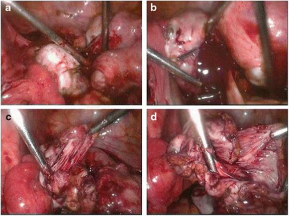
Fig. 9.1
Ovarian endometrioma enucleation, (a) kissing ovaries, (b) spilling of chocolate fluid, (c) and (d) stripping of endometrioma
Molecular genetic findings on the origin of PCOS have not yet led to any practical therapeutic treatment modalities but have provided some insight into the genetic background of the disease. The related genes can be grouped into four categories: those related to insulin resistance; those that interfere with the biosynthesis and action of androgens; those that encode inflammatory cytokines and other candidate genes. Despite intensive investigation, the aetiology and underlying mechanisms of PCOS remain unclear, warranting further investigation. Better understanding of the molecular and genetic basis for PCOS might lead to the invention of novel therapeutic approaches. Long-term interventional studies are needed to address the question of whether lower androgen levels in women with hyperandrogenism might protect against metabolic and cardiovascular co-morbidities.
9.2.5 Premature Ovarian Failure
Premature ovarian failure (POF) is the occurrence of hypergonadotropic hypooestrogenic amenorrhoea in women under 40 years of age. POF is idiopathic in 74–90 % of cases but can be familiar (4–33 %) or sporadic. The known causes are: genetic aberrations; autoimmune ovarian damage; iatrogenic following surgery, radiotherapy or chemotherapy; environmental factors (viruses, toxins, etc.) and metabolic (galactosaemia, 17 hydroxylase (17-OH) deficiency, etc.). Genetic aberrations can involve the x chromosome (monosomy, trisomy or translocations) or be autosomal. Genetic mechanisms include reduced gene dosage and non-specific chromosome effects impairing meiosis, decreasing the pool of primordial follicles and increasing atresia due to apoptosis or failure of follicle maturation [16].
Although it was once thought to be a permanent condition, a substantial number of patients experience spontaneous remissions and even pregnancy. Hormone replacement therapy (HRT) remains the cornerstone of treatment, and the only proven method of achieving pregnancy in POF patients is by ovum donation. New alternatives to HRT and fertility preservation are under development [17].
9.2.6 Contraception
Contraception means the separation of sexuality and reproduction, which allows active family planning.
The contraceptive effect of breastfeeding is the single most important determinant of human population growth rates in traditional societies without access to modern forms of contraception; lactational amenorrhoea is nature’s contraceptive. Even today, in many developing countries breastfeeding still prevents more pregnancies than all modern forms of contraception. Afferent neural inputs from the nipple pass via the spinal cord to the hypothalamus, where they cause a local release of beta-endorphin. This acts to depress GnRH secretion, thereby inhibiting pituitary gonadotrophin secretion, ovarian follicular development, ovulation and menstruation. The hypothalamic beta-endorphin release also inhibits dopamine production, resulting in increased pituitary prolactin secretion. The higher the suckling frequency, the more beta-endorphin is released, and hence the longer the duration of lactational amenorrhoea [18].
Today, there are various products on the market worldwide that make it easy to adjust contraception to an individual patient. Such products include intrauterine devices (IUDs), slow-hormone release-implants or vaginal rings and, of course, contraceptive pills.
The risk–benefit ratio of hormonal contraception [oral contraceptives (OCs)] is positive in adolescents as well as in women over 40 years of age if some essential rules are respected. In adolescents, the acquirement of a normal peak bone mass has to be guaranteed by the use of the OC. The dosage of the OC has to be adapted individually to the basic hormonal situation. Ovulation–inhibition contraception cannot be recommended for young girls who have not yet had their first period or are not yet ovulating regularly. In women over 40 years of age, those with contraindications, such as hypertension, obesity, smoking or dyslipidemia, have to be actively excluded. In both age groups, the risk of a correctly indicated OC is inferior to the risk of an unwanted pregnancy [19]. A non-contraceptive benefit of reducing the number of ovulations with OC’s is a reduced risk of obtaining certain benign as well as malignant tumours, such as benign breast tumours, uterine fibroids and ovarian cysts. Modern low-dose OCs do not increase the risk of liver cell adenomata or carcinomata. OCs do not influence melanoma. Modern data do not suggest a significantly increased risk of breast carcinoma in OC users. Long-term use of OCs leads to a decreased risk of endometrial and colorectal carcinomata. Cervical carcinoma is not influenced directly by OC, but probably indirectly through a change in sexual behaviour. There is no increase of vulvar or vaginal carcinoma, even after long-term use of OCs [20, 21].
9.2.7 Ovarian Surgery by Laparoscopy or Laparotomy (Operative Therapy and Follow-up Studies)
Ovarian cysts are a problem frequently seen in gynaecological practice. They affect women of all ages. The ultrasound diagnosis may imply an abnormal finding that requires systematic assessment and treatment. In the USA, the number of women who are hospitalised for ovarian tumours ranges from 160,000 to 289,000 per year. It should be highlighted that 80–90 % of these women will undergo a surgical procedure [22, 23]. It has been estimated that 5–10 % of all women during their lifespan will eventually have surgery to treat an ovarian cyst [23]. It is well known that this entity is more prevalent during the reproductive years and dominated mainly by endometriomas, serous cystadenomas, follicular cysts, corpus luteum cysts and benign teratomas. This distribution of histopathological diagnosis has been confirmed in a study of 641 adnexal tumours in Kiel, Germany [24, 25].
Laparoscopy is considered the gold standard for treatment of benign ovarian cysts and adhesions and the safety of the procedure has been widely demonstrated [24–27]. Compared with traditional surgery by laparotomy, operative laparoscopy is associated with shorter hospitalisation, faster patient recovery, decreased costs, and a lower incidence of de novo adhesion formation [28, 29].
The surgical technique of ovarian cyst excision by laparoscopy differs from traditional surgery performed at laparotomy. It must be performed under intermittent irrigation. Using laparoscopy, most surgeons perform the so-called stripping technique: two atraumatic grasping forceps are used to pull the cyst wall and the normal ovarian parenchyma in opposite directions, thus developing the cleavage plane (Fig. 9.1). After excision of the cyst wall, hemostasis is achieved by using bipolar forceps, CO2 laser or the harmonic scalpel with ultrasound energy. The residual ovarian tissue is mostly not sutured and the ovarian edges are left to heal by secondary intention [20]. Some authors advocate the placement of sutures to bring together the ovarian edges after enucleation of large cysts to form a normal ovary. It is advisable to use as little bipolar energy or laser as necessary because this may subsequently damage the ovarian parenchyma and the ovarian reserve. This is especially important for patients wishing to become pregnant [24, 25, 30] (Table 9.1, Fig. 9.2). In a recent study, the sizes of the enucleated ovarian cysts (Fig. 9.3) and the histopathological patterns (Fig. 9.4) were evaluated [31]. After menopause we advise to preserve the healthy ovaries till the age of 65; however, there are exceptions. Of course, the patient also presents her views and wishes. Ovariectomy is mostly combined with tube-ectomy.
Table 9.2
Treatment options for benign ovarian cysts
|
1. Laparoscopy |
|
(a) Conservative treatment: enucleation of large cysts after aspiration (b) Drainage and destruction of inner lining using any energy (c) Drainage, GnRH agonists (12 weeks), second look laparoscopy, vaporisation (d) Ovariectomy (e) Adnexectomy |
|
2. Laparotomy |
|
(a) Cyst enucleation (b) Ovariectomy (c) Adnexectomy |
|
3. Ultrasound-guided aspiration |
|
(a) In ovarian hyperstimulation syndrome following ART |

Fig. 9.2
Meta-analysis giving the odds ratio of the recurrence risks of studies comparing ablative versus excisional surgery of ovarian endometrioma. In the excised patients, relapse is substantially decreased ([30], Copyright Cochrane Library, reproduced with permission)

Fig. 9.3
Distribution of sizes of ovarian cyst

Fig. 9.4
Histopathological pattern of ovarian cyst enucleations
It has been suggested that invaginated ovarian coelomic epithelium in endometriomas undergoes metaplasia into typical glandular epithelium and stroma [32]. Others have suggested the role of ovarian follicles in the pathogenesis of chocolate cysts, due to the ability of follicular fluid to induce endometrial cell growth. This theory has been supported by laparoscopic findings of chocolate cysts [33].
The laparoscopic surgical procedure itself has raised concern that cystectomy might not be a tissue-sparing technique and other alternatives should be sought to preserve as much ovarian tissue as possible, especially in women of reproductive age. Women should be offered the best chance of achieving a healthy pregnancy. Several authors have addressed this topic using a wide variety of studies designs, with controversial results. One approach was to compare a previously operated ovary to a non-operated one, regarding the response to hormonal stimulation in ovulation induction or the ovarian volume prior to infertility treatment. Damage to the ovarian reserve has been demonstrated after stripping the capsule in the treatment of ovarian endometriomas [34, 35]. In 38 patients who received hormonal stimulation for IVF and ICSI, a reduced number of dominant follicles, oocytes and embryos was found in a previously operated ovary compared to the non-operated side. However, fertilisation rates and good quality embryos were similar from both ovaries suggesting a quantitative damage to the ovarian reserve more than a qualitative one. In a further series of 188 patients with a history of infertility and previous ovarian cystectomy, a reduced follicular response in natural and clomiphene-stimulated cycles in women younger than 35 years was shown [36]. More reassuring findings were found in IVF patients where it was demonstrated that stripping of the ovarian cyst is an appropriate treatment and did not negatively affect the ovarian response to IVF [37]. Cystectomy was found to be ovarian tissue sparing after histological analysis of ovarian cystectomies in 42 women of reproductive age [28].
Electrical energy as part of the routine surgical technique in the management of ovarian cysts has raised some concern regarding damage to surrounding ovarian tissue, thus decreasing ovarian reserve. In a study of 47 patients with previous ovariectomy, ovarian cysts in the remaining ovary were treated using either bipolar coagulation (n = 21) or sutures (n = 26) to obtain hemostasis. Subsequently, ovarian reserve was studied by measuring basal FSH 3, 6 and 12 months after surgery. The results of the study show that bipolar electrocoagulation of the ovarian parenchyma during laparoscopic removal of endometriotic ovarian cysts adversely affects ovarian function. This can be explained by the destructive effect of bipolar energy on healthy ovarian tissue which is absent when the ovary is sutured [38].
Further consideration was given to the question whether endometriomas should be excised or vaporised/coagulated. A systematic meta-analysis in the Cochrane database shows that excision decreases the risk for relapse by about half (OR 0.41) [39] (Fig. 9.2).
We recently investigated the ovarian reserve biochemically to predict the outcome after assisted reproductive techniques. Ovarian reserve was defined as the quantity and quality of the follicular pool in patients who had previously undergone cystectomy for unilateral or bilateral ovarian cysts. In a systematic literature review, a series of tests was analyzed to assess ovarian reserve. The results of the review show that the basal FSH level is not adequately sensitive to predict poor outcome. The same is true for other parameters, including basal oestradiol, the FSH/luteinising hormone ratio, anti-Müllerian hormone and inhibin-B levels. The clomiphene citrate challenge test (CCCT) has a low sensitivity, but this sensitivity is greater than that of the basal follicle stimulating hormone [40]. Therefore, it was decided to study ovarian reserve as a predictor for outcome in ARTs using the CCCT.
A retrospective analysis was carried out of all patients aged between 18 and 35 years, who underwent previous unilateral or bilateral ovarian cystectomy between January 2004 and October 2005. Enucleation of the ovarian cyst had been performed as previously described [24, 25, 30]. Endocoagulation or bipolar coagulation, incision of the capsule of the cyst using micro scissors and sharp and blunt enucleation without cyst rupture had been carried out. Two atraumatic grasping forceps had been used to pull the cyst wall and the normal ovarian parenchyma in opposite directions. The final pedicle of the cyst, containing most of the vessels, was coagulated with thermal or bipolar energy. The remaining ovarian tissue was coagulated, with borders either sutured or left open. Exclusion criteria were partial or complete oophorectomy and patients currently receiving infertility treatment or ovarian suppression medication.
The CCCT involves the administration of 100 mg clomiphene citrate on days 5–9 and the measurement of FSH concentrations on days 3–5 and 10–11. In patients with normal ovarian reserve, there is a clomiphene citrate dependent rise in FSH. This rise will be suppressed by inhibin-B produced by the follicles. An abnormal test is defined as an abnormally high FSH on day 3 or day 10.
A total of 60 patients were enrolled in the study. All patients were contacted by mail and invited to come to the hospital for an interview. Twenty patients replied and an appointment was scheduled. Two patients refused the test and a further three did not return to have their blood taken. The remaining 15 patients were interviewed at the hospital.
These 15 patients underwent the CCCT. This group was compared to the 60 not taking the CCCT, regarding age, regular/irregular menstrual pattern, parity, infertility, endometriosis, previous ovarian surgery, unilateral/bilateral ovarian cysts and size of the cysts. There were no significant differences in age, menstrual pattern, parity, endometriosis, unilateral/bilateral ovarian cysts and size of cysts between the two groups. Previous infertility was higher in the group taking the CCCT (4/15 26.6 %), compared to the control group (10/60 16.9 %) and previous ovarian surgery was lower in the group taking the CCCT (2/15 13.3 %), compared to the control group (13/60 22.3 %).
In the study group, FSH, LH and oestradiol were measured on day 3–5 and 10–11, as previously described for the CCCT. Basal FSH was 6.36 IU/ml (range 3.7–10.7 IU/ml). After stimulation FSH was 7.32 IU/ml (range 4.9–11.2 IU/ml), indicating a negative CCCT test, because no abnormally high FSH could be observed on day 3 or 10. Therefore, it could be speculated, that these 15 patients had a good prognosis for assisted reproductive technology. In addition, previous ovarian surgery had little or no effect on ovarian reserve in the study group, but previous ovarian surgery was substantially decreased in this group (2/15 13.3 % versus 13/60 22.3 %).
These results may indicate that ovarian cystectomy using microsurgical techniques does not change ovarian reserve or decrease pregnancy rates in subsequent assisted reproductive techniques.
9.2.8 Should the Ovaries Be Conserved at the Time of Surgery After Menopause, Beyond the Age of 45, or Be Taken Out at Hysterectomy?
Ovarian conservation up to the age of 65 benefits long-term survival for women at an average risk of ovarian cancer when undergoing hysterectomy for benign disease. The role of prophylactic oophorectomy at the time of hysterectomy has long been a controversial issue. It is thought that the role of the ovary is for reproduction and hormone production and when each of these functions ceases the ovary serves no purpose other than generating mischief. It is also known that ovaries produce other essential cytokines important for the continuation of life; on the other hand, even unsuspicious-looking ovaries may develop ovarian cancer. Therefore, only an individual decision tailored to the patient’s family history, personal history, previous surgery, present status and wish can be taken [41]. A calculated increase in mortality associated with oophorectomy in the absence of oestrogen therapy derives almost entirely from an enhancement of coronary heart disease; however, the validity of the findings can easily be questioned. Nevertheless, the authors have shown that even if no coronary heart disease increase is seen with oophorectomy, there remains no demonstrable advantage to the procedure in terms of longevity. In the already complex discussion regarding oophorectomy in perimenopausal women, the issue of potential effect upon mortality must be brought up, because it has been shown that patients without oophorectomy live longer. It is our responsibility as physicians and patient advocates to continuously challenge new findings with a critical eye, a reasoned perspective and whenever possible a smattering of imagination. Regarding the decision whether to take out the ovaries or not in females beyond the reproductive age, it seems certain that beyond the age of 65 the ovaries should be taken out in cases of hysterectomy as malignant transformations seem to be further increased from that age onwards. Before the age of 65, the decision whether to leave the ovaries in situ or not remains a decision between the doctor and his patient.
9.3 Factors Likely to Affect the Choice of Therapy
9.3.1 Infertility
If ovarian surgery for cysts, fibromas, adhesions or adnexal tumours is indicated in infertility patients, the major goal of any surgical intervention is to preserve as much undamaged ovarian tissue as possible. Many comparisons of laparoscopic ovarian cyst enucleation versus minilaparotomy or laparotomy have been carried out and the outcome clearly shows that no matter what type of ovarian surgery is performed, it should be with ovarian tissue conservation. For individual patients and their reproductive outcome, the size of abdominal incision, laparoscopy, laparotomy or even vaginal surgery seems to be less important than ovarian functionality. Therefore, every surgeon should apply the method he can best perform with the goal of preserving as much ovarian tissue as possible.
It is generally thought that laparoscopy is superior to laparotomy in the management of benign adnexal cysts as it is associated with shorter hospital stay, less post-operative pain, a better cosmetic outcome and a faster recovery. However, minilaparotomy or the vaginal approach also requires no sophisticated equipment or very specialised training but utilises basic, classical techniques and is a patient-friendly technology. In situations where laparoscopic ovarian cyst enucleation is technically not possible, laparatomy is still a safe technique.
A comparison of the laparoscopic and laparotomic approach regarding outcome, infection, pain and consecutive fertility revealed an advantage of laparoscopy over laparotomy, especially in terms of perioperative morbidity and post-operative pain [42]. Operating time and intraoperative complications were similar for both approaches. At present, laparoscopy should definitely be considered the gold standard for the management of benign adnexal disease. Intraoperative spillage can be avoided in both laparoscopy and laparotomy. The transvaginal resection of ovarian cysts is only performed by some specialists [43].
Tubal pathology in benign cases, such as ectopic pregnancies, sacto or pyosalpinx, should be surgically treated without lacerating or extirpating the ovary in women of reproductive age. Adnexectomy in cases of ectopic pregnancy is usually not indicated and must be left as tubectomy in specific cases [44].
9.3.2 Ovarian Dysfunction and Amenorrhoea
During the reproductive age, the normal cyclic function of the ovary is characterised by recruitment of the follicular cohort, selection of the dominant follicle, ovulation with consecutive corpus luteum formation, corpus luteum function and the final regression of the corpus luteum. In this cycle, the central nervous system, the limbic system, the hypothalamus, the hypophysis and the ovary are partners. Disturbances of these interactions lead to ovarian dysfunction with follicular maturation disturbances, inadequate ovulation and inadequate functioning of the corpus luteum. The resulting bleeding abnormalities (poly-, oligo- or amenorrhoea and also pre, intra and post-ovulatory bleeding) have to be treated separately.
The 1973 WHO classification of ovarian dysfunction still gives the best general idea of diagnostic techniques and therapy [45]. According to this classification, amenorrhoea may be the consequence of an ovarian insufficiency (group 2b), an anatomically caused amenorrhoea (group 4), hyperprolactinemia (groups 5 and 6) or a non-increase in gonadotropins (group 1). The pathophysiology reveals a lack of stimulation of the hypophyseal gonadotropin secretion. Groups 3 and 7 comprise patients with hypergonadotropic ovarian insufficiency.
Anovulation can be defined as absence of ovulation without follicular rupture and no follicles persisting after an initial period of growth. In primary anovulation, ovulation has never commenced. In secondary anovulation, there is an ovulatory period, followed by subsequent anovulation. In anovulation, a peak in LH and FSH is missing and there is a continuous rather low oestradiol and progesterone production. Cyst formation in the ovary frequently prevents the development of ovulatory follicles by compression of the ovarian cortex [25]. In anovulation, the basal body temperature curve is constant, without rise. Using ultrasound, absence of growth in one of the ovarian follicles during the cycle can be observed. Also, there is often an associated atrophic endometrium.
Anatomical problems involved in follicular growth in PCOS can be operated using laparoscopic surgery by a large wedge resection or follicular puncture using a monopolar needle. Ovarian drilling creates large holes which do not allow follicular fluid to continue to accumulate in the unruptured follicles. Alternatively, anovulation can also be treated by endocrine stimulation, such as clomiphene, HMG/HCG or FSH/HCG stimulation, under the continuous observation of oestradiol and progesterone levels and careful ultrasound measurements of follicular growth. In anovulation with single or double-chamber ovarian cysts, the cyst is resected entirely and the ovarian bed is carefully coagulated, if necessary. Ultrasound denaturation or laser coagulation may also be applied. Ovarian cysts and endometriomas have to be totally resected and removed from the abdomen in an endobag. Unilateral or bilateral ovariectomy is advisable in cases of borderline ovarian lesions, bilateral adnexectomy, hysterectomy, omentectomy and lymphadenectomy in cases of ovarian cancer [46].
9.3.3 Benign and Malignant Ovarian Transformations
Benign ovarian transformations are treated by laparoscopy and have been discussed in Sect. 9.2.3. The incidence of ovarian cancer is influenced by country of origin, race and age. The highest percentages are observed in industrialised, the lowest in non-industrialised countries. Multiple factors are discussed for the development of ovarian cancer comprising environmental factors, rubella exposure, coffee, fat, vitamin consumption, number of children, oestrogen replacement therapy, oral contraceptives, tubal ligation, hysterectomy, BRCA1 and 2 mutations and others.
A continuous oral contraceptive uptake may reduce the risk of developing ovarian cancer. The safety of long-term menopausal hormonal replacement therapy is debated. Even tubal ligations and hysterectomies are said to have a protective effect against borderline lesions and ovarian cancer. The most frequent ovarian cancer is the epithelial ovarian cancer compared to the non-epithelial ovarian cancer = germ cell tumours. Radical surgery, including lymphadenectomy, is the treatment of choice for ovarian cancer, followed by chemotherapy, immune therapy and anti-hormonal therapy.
Preoperative investigations include expert ultrasound examination, abdominal and vaginal; CA125 determination, MRI, bone scan or CT scan. Karyotypes should be obtained in all premenarchal girls with ovarian tumours as tumours often arise from dysgenetic gonads.
9.3.4 Polycystic Ovarian Syndrome
The PCO syndrome cannot be defined as a unique entity as various heterogeneous forms of morphology and clinical symptoms are described. The common endocrine increased LH concentrations in serum and a shift of the LH/FSH ratio towards LH are known. Further characteristics are the hyperandrogenism and hyperinsulinemia following an increased peripheral insulin resistance. This disease has been the subject of multiple genetic studies and is fairly well known at the present time. Chronic anovulation, amenorrhoea, hyperinsulinemia and increased insulin resistance are often treated with weight-loss strategies; metabolic treatment with metformin, clomiphene, GnRH agonists and antagonists as well as laparoscopic ovarian drilling. The genetically caused enzyme deficiencies of the adrenal androgen biosynthesis are not yet fully clarified.
With a prevalence of more than 5 %, PCOS is one of the most common diseases in young women. It is defined by the combination of oligo or amenorrhoea, clinical or biochemical hyperandrogenism and the exclusion of pituitary, adrenal or other ovarian disorders.
Polycystic ovaries PCO are associated with the syndrome but are not very specific for the diagnosis. The most common complaints are hirsutism, infertility and obesity although even very slender patients can be affected. Many patients are found to be insulin resistant, associating PCOS with the metabolic syndrome and implicating a risk to develop its sequelae. The consideration of insulin resistance has established insulin sensitisers as a new option for the treatment of PCOS. The older concept of wedge resection or ovarian drilling to remove mechanical compression of normal ovarian tissue by fluid-filled cysts and edematous tissue is still considered a method of treatment.
Laparoscopy offers the possibility of releasing the ovarian capsule and the retended fluid by ovarian drilling which can be performed with monopolar or bipolar electricity, laser or ultrasound. Ovarian drilling is a surgical treatment not accepted by everyone. We personally find it well indicated in ultrasound images with typical “string of pearls” cysts [25]. The number of holes to be drilled (between 5 and 30 on each side) depends on the size of the ovary and the number of follicular cysts. Pure monopolar cutting current with 80 W should be used for the drilling. Careful continuous rinsing with Ringer’s lactate prevents post-surgical adhesion formation.
Metformin increases insulin sensitivity, reduces glucose uptake and inhibits glucose synthesis. In obese women, metformin supports weight loss and improves menstrual cyclicity with increased ovulation rates. With metformin therapy, serum testosterone decreases and symptoms of hirsutism significantly improve. In ovarian stimulation, metformin improves response to clomiphene or FSH with increased pregnancy rates. Some studies suggest a positive effect of metformin, even on early pregnancy, leading to reduced abortion rates. Metformin should be taken for at least 3 months at a dose of 2–3 × 500 mg or 2 × 850 mg daily. This sequence is especially recommended for obese patients with peripheral insulin resistance although even lean patients can benefit from this therapy.
Various combination treatments, including oral contraceptives and metformin as well as ethinyl oestradiol and metformin, have been proposed. A significant effect of the combination of oral contraceptives and metformin together with androgens and SHBG has not been found. However, the combination of metformin with the intermittent application of ethinyl oestradiol and cyproterone acetate seems to improve the symptoms of androgen excess in cases of PCOS. As metformin is currently not yet approved for the treatment of PCOS, it is not given on a wide scale in Germany. The complex and differential diagnosis of PCOS requires a close interdisciplinary cooperation of gynaecologists, endocrinologists and dieticians. Surgical treatment with ovarian drilling and wedge resection stands side by side with endocrine treatment and the necessary weight loss in obese patients. Molecular genetic findings on the origin of PCOS have not yet led to any practical therapeutic treatment modalities but have given some insight into the genetic background of the disease [47].
9.4 Summary and Conclusions
9.4.1 Infertility, Ovarian Stimulation Within ART
It is generally thought that laparoscopy is a leading technique in the management of benign adnexal cysts as it is associated with shorter hospital stay, less post-operative pain and faster patient recovery. Studies comparing conventional laparotomy and laparoscopy for the management of benign adnexal cysts report significant differences in favour of laparoscopy in terms of post-operative pain, length of hospital stay and post-operative recovery of the patient. Ovarian stimulation and follicular puncture increase the ovarian metabolism and thus may lead to an earlier termination of growth of oocytes from immature follicles in comparison to non-treated ovaries. Ovarian stimulation itself is central to any artificial reproductive technology as it allows the possibility of injecting or pairing metaphase II oocytes with the corresponding spermatozoa or single sperm. If contraception and consecutive anovulation have taken place for 20 years, as the patient approaches menopause the number of remaining follicles with the potential for ovulation is increased [41].
9.4.2 Ovarian Dysfunction, Amenorrhoea and Contraception
According to the WHO classification for ovarian insufficiency and amenorrhoea, only groups 2, 4 and 5 are difficult to treat. Patients in groups 2 and 5 are usually responsive to ovarian stimulation; group 5 patients should be given dopamine agonists after exclusion of a hypophyseal tumour. Patients in all other groups can be stimulated for ovulation induction. The patients in group 3 are the real problem.
9.4.3 Ovarian Surgery, Benign and Malignant Transformations
Ovarian surgery in women of reproductive age requires excision only in cases of benign proliferative swellings and cyst formations resistant to endocrine therapy. In cases of malignant transformations, a resection of the gonads is required. In malignant transformations, a rapid and swift spread within the local lymphatic drainage takes place. For this reason any malignant transformation of the ovary requires radical surgical resection including both ovaries, uterus, omentum and the pelvic and paraaortic lymph nodes. The spread of ovarian cancer with small local lesions in the ovary (stage IA) into the paraaortic lymph nodes, even with only one positive lymph node, transforms the stage to a FIGO stage IIIB which requires additional chemotherapy. Stage IA can be cured by performing the necessary radical surgery alone. Laparoscopic ovarian surgery seems to be the method of choice in benign cases, particularly as surgery can be performed under higher magnification. Laparoscopic surgery should only be performed after adequate training, with the necessary care and surgical skill. If possible, adhesion prevention should be carried out at the end of the procedure.
9.4.4 Polycystic Ovary Syndrome and Conservation of Ovaries at the Time of Hysterectomy
Equivalent therapeutic options for the treatment of PCOS are laparoscopic ovarian drilling and hormonal therapy, such as clomiphene or FSH combined with metformin or ethinyl oestradiol plus cyproterone acetate combined with metformin. The main argument in favour of taking out the ovaries at the time of hysterectomy before the age of 65 is absence of the possibility of malignant transformation. The main argument against it is an increased risk for death through coronary heart disease and infarction. While there is an ongoing discussion whether or not the ovaries should be removed together with the uterus in younger postmenopausal women, there seems to be an agreement that in women 65 years and older the ovaries should be taken out because the risk of malignant transformations seems to further increase.
9.4.5 Preservation of Fertility
As ovulation plays a central role in female well-being and reproductive life, much effort is taken to preserve fertility in patients at risk of losing ovarian function due to potentially sterilising therapies. While the cryopreservation of embryos after ART is quite effective, results obtained with different oocytes cryopreservation techniques have been disappointing, particularly those obtained with slow-cooling procedures. A rapid flash-freezing technique known as vitrification has re-kindled interest in oocyte freezing. Certain modifications, especially minimising the volume of storage fluid, have resulted in markedly improved pregnancy rates with vitrified thawed oocytes [21, 48]. An increasing number of well-trained and educated professional women are planning pregnancy at advanced maternal age. The techniques discussed above may be used to postpone pregnancy without increasing the genetic risks associated with advanced maternal age.
9.4.5.1 Fertiprotect
Radiological treatment and chemotherapy often leave females with POFafter a successful cancer treatment. This can be prevented in many haematopoietic and extragenital malignancies by pre-cancer treatment with oocyte, embryo or ovarian tissue freezing, and in genital as well as other cancers sometimes by transposition of ovaries up to the pelvic brim out of the field of radiation. Fertiprotect is a group of European fertility experts working together with oncologists to preserve the fertility of females and males in specific cases.
9.4.5.2 Ovary-Conserving Surgery
In patients of reproductive age with benign ovarian tumours, particular attention should be paid to inflict as little damage as possible to the ovary during the operation in order to preserve as much ovarian fertility and follicular reserve as possible. Therefore, laparoscopic surgery has become a widely accepted standard. However, in cases of malignancy, immediate radical gonadectomy, hysterectomy, omentectomy or lymphadenectomy may have to be performed. There is some consensus that conservation of ovaries at the time of hysterectomy may be preferable for patients up to 65 years of age. However, a family history of malignant ovarian tumours, the patient’s genetic disposition and the patient’s wishes have to be taken into account before a decision can be made [41, 49].
9.5 Future Outlook
Molecular genetic mechanisms of ovarian malignant transformations will allow the diagnosis of ovarian cancer at an early stage. A combination of microarray technology, biochemical investigation, improved ultrasound imaging and computer software will allow a risk calculation and therefore make screening programs possible. Surgery will only be performed laparoscopically. Pelvic and paraaortic lymph nodes can be assessed by laparoscopy. Human ovaries with follicular development and oestrogen/progesterone production will always be in the focus of assisted reproductive techniques. However artificial gamete production derived from either somatic or embryonic pluripotent stem cells will become available a pregnancy will be able to be started without oocytes of the individual patient. Donor oocytes can already be fertilised in vitro and embryos transferred into the uterine cavity of a patient without ovaries under exogenous hormonal stimulation.
9.6 Focus
· All dominant preovulatory follicles are already selected for the pool of germ cells at 24 weeks of gestation.
· Ovulation stands in the centre of female well-being and reproductive life.
· Ovarian stimulation within ART has to be performed knowledgeably, carefully and appropriate to the patient’s endocrine reaction. Damage to the ovary by ovarian stimulation, which can easily be assessed by the hormonal reaction of the patient, has to be prevented. The ovarian overstimulation syndrome should be avoided.
· Contraceptive methods without ovulation inhibition are preferable. If ovulation inhibitory methods in the form of mono-, bi- or triphasic hormonal tablets are applied, a continuous intermediate ovarian hormonal production monitoring should be carried out every 2–3 years. If the patient does not menstruate in the contraceptive pause, she should cease any anti-ovulatory treatment. The cause of amenorrhoea has to be treated according to the final diagnosis. Ovarian surgery for benign alterations has to be performed with the utmost care by laparoscopy; in only a few cases is laparotomy indicated. As much as possible of germ cell production should be preserved. Ovarian cysts have to be carefully enucleated and the remaining regenerative tissue should only be coagulated using monopolar electricity in cases of severe bleeding or when the cyst has not been taken out totally.
· Should an ovarian malipnoma be encountered, immediate radical gonadectomy, hysterectomy, omentectomy and lymphadenectomy have to be performed.
· It is agreed that conservation of ovaries at the time of hysterectomy is preferable for the patient up to the age of 65; however, all signs of malignancy in the family and in the patient have to be carefully considered before such a decision is taken.
· The PCOS is genetically well defined. Equal treatment options include laparoscopic ovarian drilling and metformin combined with various contraceptive regimens.
· In patients of reproductive age with benign ovarian tumours, particular attention should be paid to inflict as little damage as possible to the ovary during the operation in order to preserve as much ovarian fertility and follicular reserve as possible.
· Artificial oocyte production may play an important role within human reproduction. Ethical comments to this statement are not given in this chapter. Pluripotent stem cells can be derived from embryos or somatic cells.
References
1.
Short RV (1998) Difference between a testis and an ovary. J Exp Zool 281:359–361PubMedCrossRef
2.
Short RV (2000) Reinier de Graaf and the discovery of the ovary. In: Te Velde ER, Pearson PL, Broekmanns FJ (eds) Female reproductive aging. Panthenon, New York, pp 1–9
3.
Van Leeuwenhoek A (1678) Observationes D. Antonii Leeuwenhoek, de Natis e semine genitali Animalculis. Philos Trans R Soc Lond 12:1040CrossRef
4.
von Baer CE (1827) De ovi mammalium et Hominis Genesi. Leopld Voss, Lipsiae
5.
Johnson J, Canning J, Kaneko T, Pru JK, Tilly JL (2004) Germline stem cells and follicular renewal in the postnatal mammalian ovary. Nature 428:145–150PubMedCrossRef
6.
Hübner K, Fuhrmann G, Christenson LK, Kehler J, Reinbold R, De La Fuente R, Wood J, Strauss JF 3rd, Boiani M, Scholer HR (2003) Derivation of oocytes from mouse embryonic stem cells. Science 300:1251–1256PubMedCrossRef
7.
Schöler HR (2004) The potential of stem cells. A status update. Bundesgesundheitsblatt Gesundheitsforschung Gesundheitsschutz 47:565–577PubMedCrossRef
8.
Mettler L, Tavmergen EN (1989) Significance of oestradiol values in IVF-ET under a combined GnRH analogue–desensitization and simultaneous gonadotrophin stimulation for the outcome of pregnancies. Hum Reprod 4(8 Suppl):59–64PubMedCrossRef
9.
Al-Inany H, Aboulghar M (2002) GnRH antagonist in assisted reproduction: a Cochrane review. Hum Reprod 17:874–885PubMedCrossRef
10.
Deutsches IVF Register 2000
11.
Deutsches IVF Register 2002
12.
Trounson A, Wood C, Kausche A (1994) In vitro maturation and the fertilization and developmental competence of oocytes recovered from untreated polycystic ovarian patients. Fertil Steril 62:353–362PubMed
13.
Smits G, Olatunbosun O, Delbaere A, Pierson R, Vassart G, Costagliola S (2003) Ovarian hyperstimulation syndrome due to a mutation in the follicle-stimulating hormone receptor. N Engl J Med 21(349):760–766CrossRef
14.
German law for the protection of embryos. http://bundesrecht.juris.de/eschg/
15.
Mettler L, Garzon A, Kühling-von Kaisenberg H, von Kaisenberg CS (2006) Ovarian surgery and follicular reserve. Expert Rev Endocrinol Metab 1:743–752CrossRef
16.
Vujovic S (2009) Aetiology of premature ovarian failure. Menopause Int 15(2):72–75PubMed
17.
Sinha P, Kuruba N (2007) Premature ovarian failure. J Obstet Gynaecol 27(1):16–19PubMedCrossRef
18.
Short RV (1993) Lactational infertility in family planning. Ann Med 25(2):175–180PubMedCrossRef
19.
Neulen J, Thaler C, Birkhäuser M et al (2009) Contraception in adolescence and perimenopause. Ther Umsch 66(2):109–115PubMedCrossRef
20.
Braendle W, Kuhl H, Mueck A et al (2009) Does hormonal contraception increase the risk for tumors? Ther Umsch 66(2):129–135PubMedCrossRef
21.
Braendle W, Kuhl H, Mueck A et al (2008) Vitrification: an effective new approach to oocyte banking and preserving fertility in cancer patients. Clin Transl Oncol 10(5):268–273CrossRef
22.
Seltzer VL, Pearse WH (1995) Women’s primary health care: disorders of the ovary and the fallopian tube, vol 1. International Edition. McGrawhill, pp 211–225
23.
Curtin JP (1994) Management of the adnexal mass. Gynecol Oncol 55(Suppl):42–46CrossRef
24.
Mettler L, Jacobs V, Brandenburg K (2001) Laparoscopic management of 641 adnexal tumors in Kiel, Germany. J Am Assoc Gynecol Laparosc 8:74–82PubMedCrossRef
25.
Mettler L (2001) The cystic adnexal mass: patient selection, surgical techniques and long term follow up. Curr Opin Obstet Gynecol 13:389–397PubMedCrossRef
26.
Mettler L (2003) Pelvic adhesions: laparoscopic approach. Ann N Y Acad Sci 997:255–268PubMedCrossRef
27.
Yuen PM, Yu KM, Yip SK (1997) A randomized prospective study of laparoscopy and laparotomy in the management of benign ovarian masses. Am J Obstet Gynecol 177:109–114PubMedCrossRef
28.
Muzii L, Bianchi A, Croce C, Manci N, Panici PB (2002) Laparoscopic excision of ovarian cysts: Is the stripping technique a tissue-sparing procedure? Fertil Steril 77:609–14PubMedCrossRef
29.
Sagiv R, Golan A, Glezerman M (2005) Laparoscopic management of extremely large ovarian cysts. Obstet Gynecol 105:1319–22PubMedCrossRef
30.
Mettler L (2006) Manual for laparoscopic and hysteroscopic gynecological surgery, vol 3. Jaypee Brothers Inc., New Delhi, pp 74–96, Chapter 6CrossRef
31.
Kühling-von Kaisenberg H, von Kaisenberg CS, Mettler L (2010) Ovarian function and regulation. Eur Obstet Gynaecol 5(1):23–26
32.
Donnez J, Wyns C, Nisolle M (2001) Does ovarian surgery for endometriomas impair the ovarian response to gonadotropin? Fertil Steril 76:662–665PubMedCrossRef
33.
Busacca M, Vignali M (2003) Ovarian endometriosis: from pathogenesis to surgical treatment. Curr Opin Obstet Gynecol 15:321–326PubMed
34.
Exacoustos C, Zupi E, Amadio A (2004) Laparoscopic removal of endometriomas: sonographic evaluation of residual functioning ovarian tissue. Am J Obstet Gynecol 191:68–72PubMedCrossRef
35.
Ragni G, Somigliana E, Benedetti F, Paffoni A (2005) Damage to ovarian reserve associated with laparoscopic excision of endometriomas: a quantitative rather than a qualitative injury. Am J Obstet Gynecol 193:1908–1914PubMedCrossRef
36.
Loh FH, Tan AT, Kumar J (1999) Ovarian response after laparoscopic ovarian cystectomy for endometriotic cysts in 132 monitored cycles. Fertil Steril 72:316–321PubMedCrossRef
37.
Marconi G, Vilela M, Quintana R, Sueldo C (2002) Laparoscopic ovarian cystectomy of endometriomas does not affect the ovarian response to gonadotropin stimulation. Fertil Steril 78:876–878PubMedCrossRef
38.
Fedele L, Bianchi S, Zanconato G, Bergamini V, Berlanda N (2004) Bipolar electrocoagulation versus suture of solitary ovary after laparoscopic excision of ovarian endometriomas. J Am Assoc Gynecol Laparosc 11:344–347PubMedCrossRef
39.
Hart RJ, Hickey M, Maouris P, Buckett W, Garry R (2005) Excisional surgery versus ablative surgery for ovarian endometriomata. Cochrane Database Syst Rev (3): CD004992
40.
Bukulmez O, Arici A (2004) Assessment of ovarian reserve. Curr Opin Obstet Gynecol 16:231–237PubMedCrossRef
41.
Parker WH, Broder MS, Liu Z, Shoupe D, Farquhar C, Berek JS (2005) Ovarian conservation at the time of hysterectomy for benign disease. Obstet Gynecol 106:219–26PubMedCrossRef
42.
Muzii L, Bellati F, Palaia I, Plotti F, Manci N, Zullo MA, Angioli R, Panici PB (2005) Laparoscopic stripping of endometriomas: a randomized trial on different surgical techniques. Part I: clinical results. Hum Reprod 20:1981–1986PubMedCrossRef
43.
Carminati R, Giannice R, Forloni G, Ferdeghini F (2006) Feasibility and safety of anterior or posterior vaginal ovarian cyst repair. EBCOG Torino
44.
Mettler L (1991) Medicosurgical approach to diagnosis and treatment of ectopic pregnancy. Curr Opin Obstet Gynecol 3:427–438PubMedCrossRef
45.
World Health Organization (1973) Agents stimulating gonadal function in the human. Report of a WHO scientific group. WHO Technical Report 514
46.
Schmalfeldt B, du Bois A, Burges A, Emons G, Fink D, Gropp M, Hasenburg A, Jager W, Kimmig R, Kiechle M, Kommoss F, Kreienberg R, Kuhn W, Luck HJ, Meier W, Munstedt K, Ortmann O, Pfisterer J, Richter B, Runnebaum I, Schroder W, Sehouli J, Tanner B, Wagner U, Weis J (2006) [Diagnosis and treatment of malignant ovarian tumors 2005: recommendations of the Kommission Ovar of the AGO]. Zentralbl Gynakol 128:11–17PubMedCrossRef
47.
Strauss JF III (2003) Some new thoughts on the pathophysiology and genetics of polycystic ovary syndrome. Ann N Y Acad Sci 997:42–48PubMedCrossRef
48.
Cobo A, Domingo J, Pérez S et al (2009) Advances in oocyte cryopreservation—part II: rapid cooling using vitrification. Clin Exp Obstet Gynecol 36(1):5–9
49.
Mettler L (2001) The cystic adnexal mass: patient selection, surgical techniques and long term follow up. Curr Opin Obstet Gynaecol 13:289–397CrossRef