STUDY HINTS
Genes are located on chromosomes, and the stable manner in which chromosomes are first replicated and then distributed to daughter cells during cell division is the basis for genetic inheritance. Since much of genetic theory is based on the behavior of chromosomes and the genes they carry, it is very important to understand clearly how nuclear division occurs. In this way you can predict its consequences and understand the effect of errors that might occur in it. Yet the subject of cell division is complex, with many new terms to memorize and numerous things happening simultaneously. It is a continuous process that has been divided into stages somewhat artificially, so that we can describe it conveniently. All of this makes it rather hard to grasp at the beginning. Do not despair! It is really much simpler than it looks at first. The secret is to learn in stages. First one must understand the “strategies” of mitosis and meiosis, and the differences between them.
Mitosis has evolved as a mechanism to distribute accurately a copy of each chromosome present in the original cell to two new cells. The “goal” of meiosis is quite different. Meiosis passes alternate (homologous) copies of each type of chromosome to daughter cells and reduces the total chromosome number by half. These different objectives require slightly different chromosome behaviors. We shall briefly summarize these two processes, keeping in mind the different strategies they represent.
Both processes begin in essentially the same way. The chromosome (and the deoxyribonucleic acid [DNA] molecule it contains) duplicates, forming two identical chromosome strands attached to each other at the centromere. This is accompanied by a physical reorganization (coiling) that greatly reduces the chromosome’s apparent length. The transition between these levels is illustrated in the following figure. One of the earliest signs of coiling is the formation of chromomeres, shown in this “magnified” insert to part A in the figure.

During late interphase, nuclear DNA and the histone proteins in chromosomes (A) duplicate (B), though remaining attached at the centromere. The centromere is really a constriction, but we draw it here as a dark spot so it can be seen easily. The two identical DNA copies are called sister chromatids. During prophase (C and D), these DNA strands coil into discretely identifiable chromosomes.
Each of the identical chromosome strands coils to form one of the two strands (sister chromatids) in a duplicated chromosome. The shape of the chromosome is determined by the position of the centromere. Without such coiling (or condensation), separating chromosomes would be a little like trying to separate a plate of spaghetti into two piles without breaking anything. People sometimes get confused about chromosome number at this point. Just remember that chromosome means “colored body.” It is a single structural unit, no matter how much DNA it holds. So when counting chromosomes, count the centromeres, since whatever is attached to a centromere is a chromosomal unit.
In meiosis, the objective is to reduce the chromosome number so that there is only one copy of each kind in a gamete. The most direct way to do this is to pair the chromosomes carrying the same type of information or the same linear array of genes (homologous chromosomes). This is one purpose of synapsis. The first meiotic division, therefore, simply separates (segregates) homologous copies of each chromosome. The second meiotic division, like mitosis, distributes the identical copies (sister chromatids) that originated from chromosome replication.

These two strategies are contrasted in the preceding figure, in which the original cell has a chromosome number of 2n = 2. In the Important Terms section of the chapter we have listed some of the most important terms you need to know to describe these processes. Definitions are given in the Glossary.
The only additional hint we might add concerns prophase I of meiosis. The divisions of this phase of meiosis are described in your text and summarized in Table 2.1. It is sometimes useful to have a mnemonic (memory-assisting) device to help you remember the sequence of events. For the divisions of prophase I, try this phrase, whose first letters are the same as the letters of the terms you need to remember: “Leaping zebras pound down dunes”: that is, leptotene, zygotene, pachytene, diplotene, diakinesis (see Table 2.1). Also remember these divisions are somewhat artificial, since the process is continuous, and some cell biologists assign events in a slightly different way.
TABLE 2.1 Substages of Prophase I of Meiosis
|
I. Leptotene A. Chromatin begins condensing and becomes visible. 1. Chromomeres form as irregular tightly coiled regions of the chromosome. a. Location, size, and number of chromomeres are chromosome specific. b. G bands on human chromosomes are represented by several chromomeres coiling together. c. Homologous chromosomes have the same banding pattern. B. Homologous search begins. C. Telomere proteins attach the telomeres to nuclear membrane. II. Zygotene A. Pairing of homologous chromosomes occurs. 1. Homologous search and rough pairing take place. 2. Synaptonemal complex is present but homologues are not tightly synapsed. a. The pairs of chromosomes are called bivalents at this stage. b. The number of bivalents is equal to the haploid number. B. Chromosomes continue to coil and shorten. III. Pachytene A. Synapsis tightens so homologues are very close together (about 100 Å between the pairs). The term tetrad is often used here to indicate the presence of 4 chromatids (2 for each chromosome). B. Recombination occurs. C. The telomeres lose contact with the nuclear membrane. IV. Diplotene A. The transition between pachytene and diplotene occurs when the synaptonemal complex breaks down. B. Homologues begin to move apart. C. Chiasmata are present. D. Terminalization begins.a V. Diakinesis A. The homologous chromosomes continue to terminalize.a B. Nuclear membrane breaks down. C. Chromosomes move toward the equatorial plate. |
a As terminalization is completed, the chromosomes line up in the center of the cell as the cell enters metaphase I.
IMPORTANT TERMS
Acrocentric
Bivalent
Centriole
Centromere
Chiasma (pl., chiasmata)
Chromatid
Chromatin
Chromomere
Cytokinesis
Diploid
Dyad
Euchromatin
Gamete
Haploid
Heterochromatin
Homologous chromosomes
Linkage group
Meiosis
Metacentric
Mitosis
Nucleolus
Polar body
Primary oocyte
Primary spermatocyte
Secondary oocyte
Secondary spermatocyte
Spindle
Synapsis
Synaptonemal complex
Telocentric
Terminalization
Tetrad
PROBLEM SET 2
General Study Hint: To help reinforce these ideas, you should sketch the cell and its chromosomes before trying to answer these questions.
1. Suppose that you are studying a new species in which the chromosome number is unknown. You are looking at a spread of cells in some unknown stage of cell division, and you discover that there are seven chromosomes (three metacentric, three telocentric, and one acrocentric) visible in each dividing cell. What type of nuclear division have you found, and what conclusions can be drawn about the chromosome makeup of the species?
link to answer
2. How many chromosomes are there in a human intestinal cell at anaphase?
link to answer
3. How many different kinds of gametes can be produced by an individual who has three linkage groups with each chromosome identifiable by the forms of the genes (alleles) it carries?
link to answer
4. In an organism with a haploid chromosome number of 7, how many sister chromatids are present
(a) in its mitotic metaphase nucleus?
link to answer
(b) in its meiotic metaphase I nucleus?
link to answer
(c) in its meiotic metaphase II nucleus?
link to answer
link to answer
5. A particular beetle species is found to have a chromosome complement of 2n=16 chromosomes. How many linkage groups would the beetle have?
link to answer
6. Recombination can be seen as chiasmata between homologous chromosomes at what stage of cell division? Sketch a bivalent involving metacentric chromosomes having one chiasma.
link to answer
7. Is it possible for meiosis to occur in a haploid cell? Is mitosis possible? Please explain your answer to each question.
link to answer
8. For each of the following figures, identify the specific stage of nuclear division and the diploid, 2n, chromosome number of the organism.

link to answer

link to answer

link to answer

link to answer

link to answer

link to answer
link to answer
9. Draw and label a cell at metaphase I of meiosis, where 2n = 8 metacentric chromosomes.
link to answer
10. Draw and label a cell at late anaphase of mitosis, where 2n = 6 chromosomes (two pairs of acrocentric chromosomes and one pair of telocentric chromosomes).
link to answer
11. Draw and label all cells at telophase II that result from meiosis of a primary spermatocyte (2n = 8 acrocentric chromosomes).
link to answer
12. Draw, label, and compare cells at prophase I and prophase II of meiosis in a species characterized by 2n = 6 acrocentric chromosomes.
link to answer
13. Draw, label, and compare cells at anaphase of mitosis and anaphase I of meiosis in a species having 2n = 4 chromosomes (one homologous pair of metacentric chromosomes and one pair of telocentric chromosomes).
link to answer
14. In humans, atopic allergic disease (ragweed hay fever) is dependent on a dominant gene A, while a neurological disorder called Huntington disease is determined by a dominant gene H. The normal alleles are a and h, respectively. A man who is Aahh married a woman who is aaHh. Assume the A and H are on nonhomologous chromosomes and that the relevant chromosomes are all telocentric.
(a) Diagram the chromosomal constitution of the each parent at metaphase I and label each chromatid with the associated genotype.
link to answer
(b) Diagram the gametes that each parent would produce and label chromosomes in each gamete.
link to answer
link to answer
15. In studying the chromosomes of a new species of moth, you discover that there are eight bivalents formed in meiosis. From this you can conclude that the species has how many linkage groups?
(a) 4;
(b) 8;
(c) 16;
(d) 32;
(e) not enough information is presented to answer the question.
link to answer
16. A mature rat’s sperm cell has an amount of DNA that is the equivalent of a haploid genome, which we will call a “c” amount of DNA. Compared to this sperm cell, how much DNA will a somatic cell have if it is in the G2 phase of interphase?
(a) c;
(b) 2c;
(c) 3c;
(d) 4c;
(e) more than 4c.
link to answer
ANSWERS TO PROBLEM SET 2
1. This cell must be in a postreductional stage of meiosis (that is, in the second meiotic division), since a mitotic cell would have two of each type of chromosome and could not, therefore, total an odd number. The cell you have found must consequently have a haploid chromosome number of n = 7 and a diploid complement of 2n = 14 chromosomes (three pairs of metacentrics, three pairs of telocentric, and one pair of acrocentrics).
2. Answer: 92. At anaphase of mitosis, the centromeres divide, but cytokinesis has not yet been completed. Thus for a short period the cell has twice as many chromosomes as normal. Remember that we have defined whatever is attached to a single centromere as a single chromosome.
3.
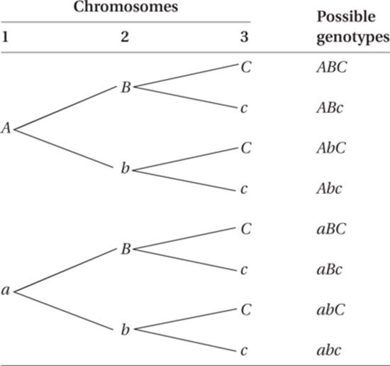
A total of eight different gametes could be produced.
If there are three linkage groups, the species must have a chromosome number of 2n = 6. The number of different types of chromosomes is the same as the haploid number (n = 3 in this case). The haploid number and the number of linkage groups are the same. Let us identify homologous chromosomes in this individual by upper- and lowercase letters. Since each chromosome is individually identifiable, the genotype would be AaBbCc.
Each gamete must contain one of each kind of chromosome. Thus the individual could produce two types of gametes with respect to the first chromosome (an A-containing gamete and an a-containing gamete). Two types of gametes could be produced with respect to each of the three chromosomes, and they could assort in all possible combinations.
4.
(a) At metaphase of mitosis, the chromosomes have duplicated, and there are therefore 2 chromatids present in each chromosome. In a species with a haploid number of 7, the diploid number is 14. Thus there are 28 chromatids present at metaphase of mitosis.
(b) The chromosome makeup of the first meiotic metaphase is the same as that of the mitotic metaphase, although in meiosis the chromosomes are arranged with homologous sets attached to opposing spindle fibers. Thus there is a total of 28 chromatids at metaphase I of meiosis.
(c) In metaphase II, reduction of chromosome number has occurred. Only 14 chromatids remain in the cell.
5. Eight linkage groups. The number of linkage groups is the same as the number of different types of chromosomes, which is the same as the haploid number of the species. If 2n = 16, the haploid number (n) is 8.
6. Recombination is visualized as chiasmata between synapsed homologues in prophase I of meiosis.

7. Meiosis is basically a reduction division from a diploid to a haploid condition. Thus it cannot occur in a haploid cell. Mitosis, on the other hand, is an orderly division of the existing chromosomes to distribute one copy of whatever is present to each daughter cell. Since chromosomes replicate and separate on the spindle independently of each other, mitosis can occur in a haploid cell.
8.
(a) Anaphase I of meiosis; 2n = 6. Note sister chromatids still attached at centromere.
(b) Telophase of mitosis; 2n = 4
(c) Prophase I of meiosis; 2n = 6. Note synapsis of homologous chromosomes.
(d) Metaphase I of meiosis; 2n = 4. Note synapsed bivalents at equator.
(e) Metaphase of mitosis with 2n = 4; or metaphase II of meiosis with 2n = 8.
(f) Anaphase II of meiosis; 2n = 6. Note the number of chromosomes moving to each pole is odd, but it should be even if occurring in a diploid phase like mitosis.
9.

10.

11. Centromeres of the chromosomes are shown as knobs for easy identification (though they are really constrictions). Nuclear membranes are re-forming. Cytokinesis is taking place. Four cells result from the meiotic division of one primary spermatocyte.

12. Prophase I of meiosis is characterized by synapsis of homologous chromosomes. These homologous chromosomes segregate during the first meiotic division, although the centromeres of the chromosomes do not divide until the second meiotic division. Thus in prophase II chromosome number has been reduced to the haploid number (n = 3, in this instance). The dissolution of the nuclear membrane and formation of the meiotic spindle occur in the same way in each.

13. In anaphase of mitosis (left figure), homologous chromosomes attach to spindle fibers independently of each other, and chromatids separate. In anaphase I of meiosis (right figure), segregation of homologous chromosomes occurs, and centromeres do not divide. Division of centromeres in meiosis occurs during the second meiotic division. Also note that the first meiotic division (right figure) reduces the chromosome number by half (in this example, from four chromosomes to two in each resulting cell).

14.
(a)

(b)

15.
(b) 8. Each bivalent is formed by synapsis of all copies of a given linkage group (pair of homologous chromosomes). So there is the same number of linkage groups as there are bivalents. This is also the haploid chromosome number, n.
16.
(d) 4c. The cell is diploid in interphase, but the amount of DNA has doubled during the S phase, in preparation for cell division. There are therefore four DNA strands of each linkage group existing as two homologous chromosomes, each containing two sister chromatids.
CROSSWORD PUZZLE 2
Mitosis and Meiosis

Across
4. Separation of the homologous chromosomes during meiosis I
6. Interphase DNA-protein complex
7. Irregular tightly coiled regions of the chromosome, seen as the chromatin begins condensing during prophase I
13. Stage of mitosis when chromosomes are moving toward the poles
14. Region on chromosome where spindle fibers attach during cell reproduction
16. End of a chromosome
18. Substage of prophase I when the nuclear membrane breaks down and chromosomes move toward the equatorial plate
21. Substage of prophase I when recombination occurs
22. Four associated chromatids of a bivalent
23. One of two replicated chromosome strands held by one centromere
24. Term for a chromosome with the centromere on one end
Down
1. Substage of prophase I when the homologous chromosomes pair
2. Random combination of the genes of non-homologous chromosomes because one pair’s orientation at the equatorial plate does not affect the orientation of another pair during metaphase I
3. Cell with one set of chromosomes
5. Term for a chromosome that has the centromere located near one end
8. Dark staining chromatin that is inactivated or structural regions of the chromosome
9. Lightly staining chromatin where genes are located
10. Stage of mitosis when the chromosomes are lined up in the center of the cell
11. Movement of the chiasmata toward the ends of the bivalents during prophase I of meiosis
12. Term for a chromosome with the centromere located near the center
15. Process takes a diploid nucleus and makes four haploid nuclei
17. Genes that are on the same chromosome
18. Having two sets of chromosomes
19. Pair of homologous chromosomes during synapsis
20. Asexual cloning of the nucleus of a cell to produce two identical nuclei