
Vincent Di Marino1 and Hubert Lepidi1
(1)
UER Médecine, Aix-Marseille Université, Marseille, France
Abstract
These muscles (4 overall) are symmetrical, paired and striated muscles, which are divided into two groups: the group of the ischiocavernosus muscles (connected to the crura of the clitoral corpora cavernosa) and the group of the bulbospongiosus muscles (connected to the spongy bulbs). These muscles also exist at the level of man’s perineum, in which case they are connected to the corpora cavernosa and to the spongy corpus of the penis.
These muscles (4 overall) are symmetrical, paired and striated muscles, which are divided into two groups: the group of the ischiocavernosus muscles (connected to the crura of the clitoral corpora cavernosa) and the group of the bulbospongiosus muscles (connected to the spongy bulbs). These muscles also exist at the level of man’s perineum, in which case they are connected to the corpora cavernosa and to the spongy corpus of the penis.
These 4 muscles are located in the superficial region of the anterior perineum (urogenital perineum), between the superficial fascia of the perineum (fascia of Colles) and the inferior fascia of the urogenital diaphragm, i.e. the perineal membrane. They are innervated by branches from the perineal ramus of the pudendal nerve.
Their role is essential for the mobility of the clitoris and its erectile function.
11.1 Ischiocavernosus Muscles
Over time, they have been given very diverse names: tertius and quartus musculus (Colombo), clitoridis musculus (Fallope), clitoridis musculus tensioni dicatus (Laurent), superior rotundus (Riolan), ischio-clitoral (Dumas and then Testut), erector clitoridis (Cowper, Albinus, Soemmering), ischio-sub-clitoridis, ischio-urethralis (Chaussier) and collateralis sive penem erigens (Spigel). The current terminology is the terminology which had already been adopted by Winslow, Bichat, Portal, Boyden, etc., in their time.
According to traditional descriptions, the ischiocavernosus muscles resemble half-cones enveloping each of the 2 crura of the clitoris at the level of their inferior and medial surfaces. The insertion of each ischiocavernosus muscle is made on either side of the osseous or osteofibrous insertions of the clitoral crus, which it covers.
We have observed that reality appeared to be slightly different.
· Each crus clitoridis is completely surrounded by its ischiocavernosus muscle, which, furthermore, protrudes therefrom by 1 or 3 cm downwards. The clitoral ischiocavernosus muscle is not, such as often described, a small muscle of no importance, with much smaller dimensions than its penile counterpart. We even think that it is the opposite: if a ratio is established between the dimensions of this muscle in women and the dimensions of the thin and short crus, which it covers, it is observed that the clitoral ischiocavernosus muscle represents a powerful and bulky muscle, with major functional capacities. Such as already noted by L. Kobelt, “its length is of 8 cm, sometimes more still, so as to be compatible with the dimensions of the woman’s pubic arcade”.
· Another observation resulting from our experiment: The ischiocavernosus muscle has all of the characteristics of a penniform muscle, i.e. of a muscle “with less shortening possibilities but with the capacity to develop a greater force”: tendon of origin, medial, very long and very solid, aponeurosis originating from this tendon and enclosing the crus, oblique muscular bundles, originating from the tendon and pushed against the inferior and medial surfaces of the aponeurosis, short terminal and tendinous fibres.
· The insertions of the ischiocavernosus muscle are particular (Fig. 11.1):
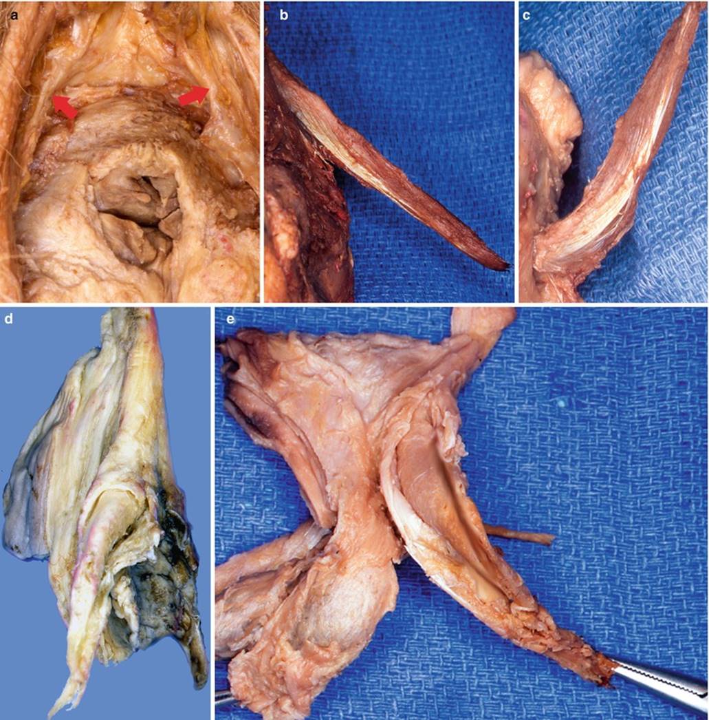
Fig. 11.1
Aspects of the ischiocavernosus muscles. (a) The two ischiocavernosus muscles on a perineal view of the anterior perineum (crura clitoridis dissected and pushed towards the pubis); the red arrows show the tendons of the two muscles. (b) The tendon of the left ischiocavernosus muscle on an anterior view of an anatomic specimen (notice the medial position of this tendon). (c) Inferior view of the same tendon (after raising of the left crus clitoridis). (d) Dorsolateral view of a bulbo-clitoral organ showing the left crus released after longitudinal opening of the ischiocavernosus fibro-muscular layer. (e) Left lateral view of a bulbo-clitoral organ, showing the left crus dissected and released from its fibro-muscular sheath (driven by a clamp)
· It is firstly attached via its tendon of origin, onto the ischial tuberosity. It then takes, via its aponeurosis, osteofibrous attachments, which, on the one hand, are osseous over the ¾ of the ischio-pubic ramus, from the ischial tuberosity, and on the other hand fibrous on the adjacent part of the perineal membrane (thickened as an “attaching lamina of the corpora cavernosa”).1
· From these origins is formed a not very thick, flattened muscle, rolled up like a cone (and not a half-cone, such as described in too many cases) around the crus clitoridis. Thus, the body of the muscle truly encloses the erectile body so that the latter is not visible. In order to observe the crus, it is therefore necessary to longitudinally incise the lower surface of the musculo-aponeurotic envelope and release this crus from this pearly envelope (Fig. 11.1). This operation is delicate due to the fact that very tight adherences connect the albuginea of the crus and the internal surface of the aponeurosis of the ischiocavernosus muscle. When the crus is completely released, the hollow aponeurotic cylinder, inside which it was located, is clearly visible and appears shiny and pearly, completely intact and uninterrupted in its attachment region. There obviously are no muscle fibres on this internal surface. It is thus understood that insertions onto the perineal membrane are made via the external surface of the cone and not via the crus.
· Overall, the ischiocavernosus muscle is such as described by L Kobelt: “The ischiocavernosus muscle is not a lapped muscle, but a hollow muscle, shaped like a cone, which contains, in its cavity, the entire surface of the crus”.
· The muscle fibres are arranged so as to form small superimposed, but irregular, oblique bundles (Fig. 11.2), and it is easy to define the capacity of these bundles to evacuate the blood contained in the sinuses of the crura.2
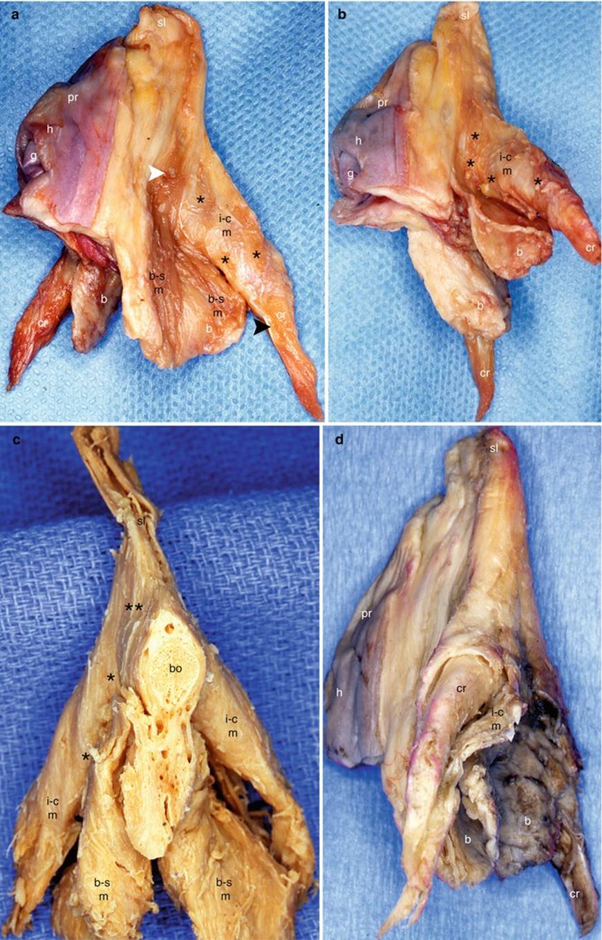
Fig. 11.2
The muscles of the bulbo-clitoral organ. (a) Lateral view; (b) inferior-lateral view; (c) ventral view with frontal section of the body; (d) dorsolateral view (i-c muscular layer opened longitudinally). b bulb, bo body, b-s m bulbospongiosus muscle, cr crus clitoridis, g glans, h hood, i-c m ischiocavernosus muscle, pr prepuce, sl suspensory ligament, black arrowhead it shows the tendon of the i-c muscle, white arrowhead it shows a bundle of the bulbospongiosus muscle reinforcing the i-c muscular layer, *oblique bundles of the i-c muscle, **muscle bundles towards the sl
· The right and left muscles then converge towards the lateral surfaces of the clitoral body, and the muscular bundles give way to short terminal tendinous fibres. As for the tendon of origin, which is generally very long, it borders the muscle up to the point of convergence of the crura, where it is lost and sometimes exchanges tendinous fibres with the contro-lateral tendon.
· The termination of the ischiocavernosus muscles is also complex.
· The superficial bundles, joined by bundles from the homolateral bulbospongiosus muscle, will reach the lateral parts of the body of the clitoris in the angle region and end in the clitoral fascia. Some fibres become lost on the lateral surfaces of the suspensory ligament. Lastly, others are inserted on the superior surface of the clitoral body (a few fibre exchanges with the opposite side).
· The deep bundles are dorsal (they are only visible once the clitoris and its suspensory ligament have been unstuck from the pubic symphysis) and join the bundles on the opposite side, by passing as a bridge behind the crura to provide reinforcement in front of the symphysis, the retro-crural fascia and the subpubic urethral plate (Fig. 11.3).
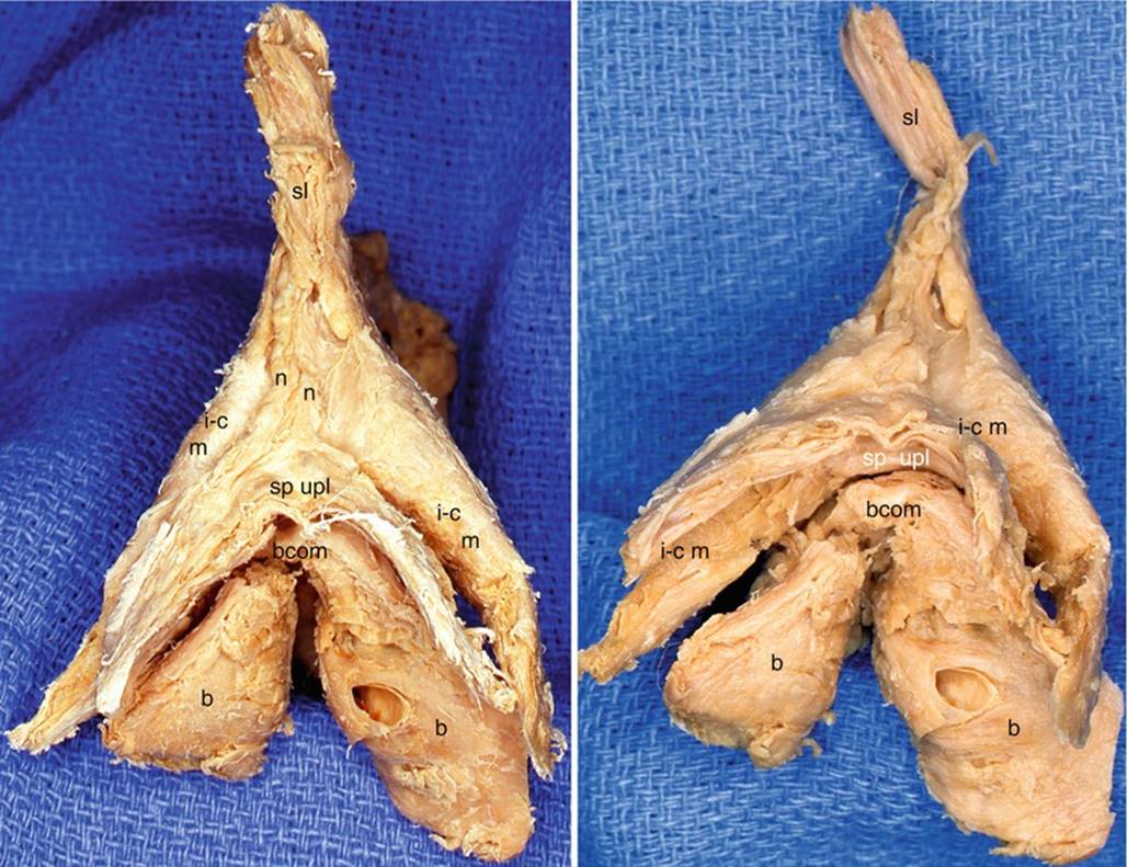
Fig. 11.3
Dorsal views of the bulbo-clitoral organ showing the subpubic urethral plate. b bulb, bcom bulbar commissure, i-c m ischiocavernosus muscle, n dorsal nerve of clitoris, sl suspensory ligament, sp upl subpubic urethral plate (black legend superior surface of the plate, white legend urethral surface)
· The role played by the ischiocavernosus muscles is determined from the anatomical concepts studied previously:
The ischiocavernosus muscles are lowering muscles for the clitoris. During intercourse, they lower the glans clitoridis against the dorsal surface of the penis (Testut).
They play an important part in the erection of the clitoris, as they make the blood from the crura flow back to the body of the clitoris. According to E. Gardner, they can even “contribute to maintaining the erection of the clitoris by compressing the crura and, as a result, delaying the blood returning from this organ”.
11.2 Bulbospongiosus Muscles
Over time, they have also been given many names: primus musculus (Vesalus, Colombo), inferior sive urethram trahens (Spigel), musculus urethrae seu accelerator (Riolan), accelerator urinae (Cowper, Douglas), urethram dilatans (De Graaf), accelerator (Morgagni, Santorini, Albinus, Soemmering), bulbo-urethral (Chaussier) and bulbo-syndesmo-cavernous (Dumas).
· The insertions of the bulbospongiosus muscles are relatively large. They originate at the central tendon of the perineum (former perineal body of Savage or central fibrous core of the perineum according to French authors) and at the perineal membrane (inferior fascia of the urogenital diaphragm). There are also crossed fibres from the external anal sphincter, via the central tendon.
· The muscle fibres from these insertions, spread out from back to front and laterally.
· Large flattened muscular lamina on either side of the vaginal and urethral orifices. This muscular body has two parts (Fig. 11.2):
· A peri-vaginal medial part, referred to as the constrictor muscle of the vagina3 (Sabatier, Portal, Bichat), constrictor cunni (Albinus, Soemmering), clitoridis inferior latus et planus (Arantius), portio musculosa in externâ parte vaginae or vaginae musculi constrictorii (Riolan), sphincter vaginae (Verheyen), perineo-clitoral (Chaussier) and annulo-syndesmo-clitoridien (Dumas), while it only exerts a relatively modest constrictive action on the vaginal wall
· A lateral part, which successively covers the inferior surface of the greater vestibular gland then that of the spongy bulb
· The termination of the bulbospongiosus muscle is even more complex than that of the ischiocavernosus muscle. Two bundles are to be considered (Fig. 13.2):
· The fibres of the superior bundle (often consisting of medial infra-bulbar fibres) meet the fibres of the ischiocavernosus muscle (Figs. 11.2, 11.4 and 8.5) and end on the lateral edges of the body, in the clitoral fascia. Some fibres become lost on the lateral surface of the suspensory ligament. Other fibres of this superior bundle reach the back of the clitoris to merge with the fibres of the counter-lateral homonymous muscle and form a supra-venous strap, capable of compressing the large superficial dorsal vein of the clitoris and its satellite veins. It is this strap which is referred to as the compressing muscle of the dorsal vein of the clitoris(muscle of Houston according to previous authors).
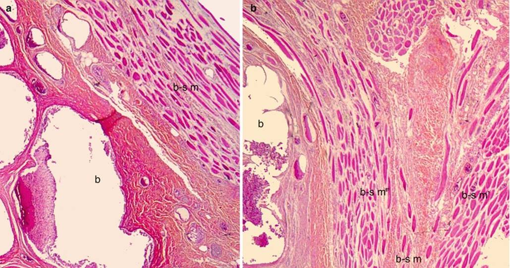
Fig. 11.4
Microscopic aspects of the bulbospongiosus muscle’s bundle. (a) The bulbospongiosus muscle (b-s m) covering the spongy bulb (b). (b) The termination of the bulbospongiosus muscle in two bundles: an inferior bundle b-s m′′, a superior bundle b-s m′
· The fibres of the inferior bundle (often consisting of lateral infra-bulbar fibres) end under the commissure of the bulbs, in the region which separates it from the urethral duct. They have not been studied in depth by the various authors who have examined these muscles. In order to clarify their trajectory, it is necessary to perform a microscopic observation of anatomical sections as we have done in the past. It is then noted that this bundle detaches from the peri-bulbar muscular body and extends medially above the commissure of the bulbs (Fig. 11.4). Then the striated muscle fibres penetrate between the spongy caves to finally be positioned above the urethra, merge with the muscle fibres of the opposite side (Fig. 13.6) and, in fine, will be arranged as a distal sphincter of the female urethra(compressor urethrae muscle of ancient authors).
· The action of the bulbospongiosus muscles is also explained by their anatomy:
1.
2.
3.
Footnotes
1
Due to the existence of 2 osseous insertions, Rouvière describes 2 bundles of origin for this ischiocavernosus muscle: an ischiatic bundle and an ischio-pubic bundle.
2
Thanks to their orientation and their direction, the ischiocavernosus muscle fibres can really squeeze out the entire blood content of the clitoral crura as “a cloth which is being wrung out”!
3
For a long time, it was considered as responsible for vaginismus (it was believed to be due to its spasmodic contraction). Such as we will see further on in this study, the introitus shrinking faculty belongs to the powerful pubo-rectal bundle of the levator ani muscle.