
Vincent Di Marino1 and Hubert Lepidi1
(1)
UER Médecine, Aix-Marseille Université, Marseille, France
Abstract
Significant knowledge of the bulbo-clitoral organ has been acquired thanks to the enormous progress made in relation to new medical imagery techniques, which have been used to explore the pelvis and especially the female pelvis. X-Ray computed tomography and then NMR (nuclear magnetic resonance) are at the forefront of these new exploration techniques and are mutually complementary. At the same time, great progress has been achieved in medical echography, which has not only become a routine and essential process for pregnancy monitoring but also a sophisticated method for studying pelvic organs and more particularly female genitalia.
14.1 General
Significant knowledge of the bulbo-clitoral organ has been acquired thanks to the enormous progress made in relation to new medical imagery techniques, which have been used to explore the pelvis and especially the female pelvis. X-Ray computed tomography and then NMR (nuclear magnetic resonance) are at the forefront of these new exploration techniques and are mutually complementary. At the same time, great progress has been achieved in medical echography, which has not only become a routine and essential process for pregnancy monitoring but also a sophisticated method for studying pelvic organs and more particularly female genitalia.
14.1.1 Computed Tomography (CT Scan Imagery)
The images obtained by X-Ray CT appeared, as soon as this process was developed, to be extremely interesting as they could be compared to (and almost superimposed on) the cadaveric sections prepared by anatomists. The progress made since then, in only 20 years, is considerable and at present the 64-slice scanners, due to their speed of acquisition and to the multiplicity of the cross-sections, can be used to reconstruct (CT image reconstruction) the organs under study in 3D and to generate three-dimensional images.
The bulbo-clitoral organ appears to be extremely complex and complete on the most superficial sections of the pelvis, especially on the sections made in the planes of the anterior perineum. On such sections, which are strictly comparable to anatomical sections (compare Figs. 14.1b and 13.1), all the parts forming the bulbo-clitoral organ are clearly observed. However, this observation varies according to the levels of the sections:

Fig. 14.1
CT scan sections of the bulbo-clitoral organ (scanner of female perineum). (a) Section concerning the spongy parts. (b) Section concerning the cavernous parts. a anus, a (with arrowhead) anal canal, b bulb, bo clitoral body, cr crus clitoridis, fe femur, g glans clitoridis, gm m gluteus maximus muscle, i-a f ischio-anal fossa, i-p r ischio-pubic ramus, p-r pubo-rectalis muscle (levator ani muscle), rspresidual spongy part, u external urethral orifice, u (with arrowhead) urethra, va vaginal orifice, va (with arrowhead) vagina, *ano-coccygeal ligament
· On sections tangent to the pelvic outlet but not concerning this bony part (Fig. 14.1a), it is especially possible to observe 2 spongy bulbs bordering the urethral and vaginal ducts and then further ahead, in the extension of the commissure of the bulbs, the entire IC RSP (infra-clitoral residual spongy part) up to the glans.
· On sections concerning the ischio-pubic rami (Fig. 14.1b), the bulbs are almost no longer visible. It is possible to assume the existence of the commissura bulborum overhanging the urethra, but the IC RSP (infra clitoral residual spongy part) is no longer visible. On the other hand, the crura of the clitoris, applied to the lower rami of the pubis and then backed against the front of the pubic symphysis, are quite visible. On these same sections, the descending portion of the clitoris and the glans are not very visible, which is due to the elbow of the clitoral body and the triplanar architecture of the bulbo-clitoral organ.
· It should finally be recalled that the overlying sections provide a good overview of the pubo-rectal bundles of the levator ani muscles, thus limiting the hiatus of the levators (“urogenital hiatus”).
14.1.2 Nuclear Magnetic Resonance (NMR Imagery)
The images of the female pelvis obtained by means of the NMR technique are extremely interesting: They are generally used to identify more anatomical details than those observed on CT images and to highlight them subject to using sequences, which are the most compatible with the target observation. For the bulbo-clitoral organ, it is incontestably the weighted T2 sequences, which are the most interesting, because they provide very contrasted images, which can be modulated by means of fat saturation techniques (removal of the fat signal). Furthermore, the possibility of obtaining sections of the pelvis in the 3 dimensions of space is particularly useful for studying the bulbo-clitoral organ, whose triplanar architecture is already known.
Anatomical structures of the external genitalia can be highlighted by performing an initial intravenous injection of gadolinium (this process can be combined with the fat saturation technique to obtain striking contrasts) for T1 sequences as well as for T2 sequences.
· The median or paramedian sagittal sections, which are used for exploring the internal genitalia and all of the pelvic organs, provide a good overview of the clitoris (angle, ascending and descending portions of the body) and the glans (Fig. 12.2a). On these median section drawings, the crura of the clitoris and the bulbs, in a more lateral position, are not (or only slightly) visible.
· The coronal sections are used for observing the glans, the vestibule, the labia minora and labia majora.
· The axial transverse-oblique sections are the most interesting because they provide images, which are perfectly similar to the sections obtained in the anatomy laboratory (Fig. 14.2): They show the erectile structures overhanging, in a horseshoe configuration with a posterior concavity, the external superimposed orifices of the urethra and vagina. The 2 crura connected to the ischio-pubic rami are perfectly visible, with their junction area and then their apposition to form the body of the clitoris. Furthermore, NMR can be used to observe the median septum separating the 2 corpora cavernosa merged together as a “clitoral corporeal cylinder”. The bulbs are also very visible on either side of the urethro-vaginal “block”. All these erectile structures are clear, white and luminous, whereas the other perineal structures seem to be grey or black with a low intensity (this is the case of the urethra or the vagina1 or of the greater vestibular glands, observable at the rear of the bulbs). It should be noted that the external urethral orifice and the vaginal orifice are hardly visible. As for ischiocavernosus, bulbo-spongiosus and superficial transverse muscles, they contrast perfectly with the erectile bodies due to their black aspect corresponding to a hyposignal. Lastly, it is possible to observe the pudendal vessels and their division into a perineal branch and dorsal branch of the clitoris, by means of the fat saturation technique (H.E. O’Connel and J.O.L. Delancey). Comparative studies carried out on non-menopaused women and menopaused women (D.D. Suh et al.) did not show significant differences between the 2 groups as regards the bulbo-clitoral organ (a slight reduction in size of the bulbs in menopaused women at the most). The differences, which are all related to the reduction in oestrogen secretions, are observed “at the labia (reduction in size), the vaginal epithelium (thinning), the vaginal canal (narrowing) and the cervix (atrophy)”.
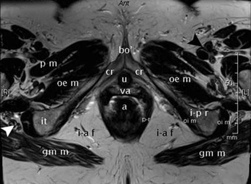
Fig. 14.2
NMR section of female pelvis through the lower third of the ischio-pubic rami (weighted T2 sequence. Very thin section). It shows the two crura clitoridis joining to form the clitoral body. a anal canal, bo′ pre-angular part of the clitoral body (ascending part), cr crus clitoridis, gm m gluteus maximus muscle, i-a f ischio-anal fossa, i-p r ischio-pubic ramus, it ischial tuberosity, oe m obturator externus muscle, oi m obturator internus muscle, pm pectineus muscle, p-r pubo-rectalis muscle (part of the levator ani muscle), u urethra, va vagina, white arrowhead right ischiatic (sciatic) nerve, black arrowhead left femoral artery, left femoral veins and left femoral nerve
Overall, pelvic NMR imagery is used not only to perform a specific study of the bulbo-clitoral organ but also to provide an excellent vision of the entire female genitalia and to perform a complete study of its relationships (sections in the 3 dimensions of space) and a structural study of the various pelvic organs and of the anatomical sites inside which these organs are located. It should be noted that although NMR remains ideal for a topographic anatomical study of the bulbo-clitoral organ, CT provides a better understanding of the descriptive anatomy of this organ, such as can be observed on scanner images (Fig. 14.1).
NMR preceded by the intravenous administration of gadolinium is, on the other hand, an ideal method for studying the bulbo-clitoral organ during a sexual arousal phase. The highlighting achieved at the level of all the erectile bodies makes it possible to explore the modifications occurring in this context: increase in dimensions and, especially, in the diameters of the erectile bodies and intense blood flow congestion with filling of the cavernous and spongy lacuna (K.R. Maravilla et al.).
14.1.3 Ultrasonic Echography (Imagery by Ultrasound)
It is the merit of French echographists (O. Buisson and P. Foldes) to have used ultrasound to explore the bulbo-clitoral organ. They therefore obtained, with this easy-to-use and low-cost technique, very good quality images, which are, especially using the technique of ultrasonic CT, also comparable to anatomy data. They could also easily measure the diameters and lengths of the constituent parts, in particular the dimensions of the clitoris. However, the bulbs remain difficult to observe if there is no stimulation. From a topographic viewpoint, it is the coronal sections, which provide the best anatomical approach: they are especially used to view the commissure of the bulbs, overhanging the urethra (which, itself, is visible), and the anterior wall of the vagina (hypoechoic according to the authors’ experience).
Thereafter, the authors made the same observations by dynamic sonography, while asking the women being examined to voluntarily contract their pelvis: They were thus able to observe movements of the clitoris with a reduction of the inter-crural angle, an increase in the angulation of the clitoral elbow and a forward and downward thrusting movement of the glans. With this process, they were also able to study vaginal penetration by asking the patient under examination to insert a finger into her vagina: this experiment made it possible to show that the pelvic contraction, caused by vaginal penetration, brings the clitoral crura of the anterior vaginal wall closer together.2Thereafter, the authors used dynamic sonography to directly study the anatomy of the bulbo-clitoral organ during intercourse, such as initiated since 1999 by the dynamic genital MRI developers (W. W. Schultz et al.).
Footnotes
1
1 These ducts are highlighted if the fat saturation technique is also used. However, this highlighting remains less significant than that of the components of the bulbo-clitoral organ.
2
2 As a result, the authors have deducted the assumption that during the phase of sexual excitation, the crura of the clitoris, which have become turgescent and congestive and have increased in volume, become stimulation factors for the anterior vaginal wall (one of the assumptions about the “G spot”).