
Vincent Di Marino1 and Hubert Lepidi1
(1)
UER Médecine, Aix-Marseille Université, Marseille, France
Abstract
The frequent lack of knowledge of the anatomy of the clitoris, even among women considered as “experts”, is explained by the fact that the major part of this essential component of the external female genital apparatus is hidden and therefore cannot be observed.
The frequent lack of knowledge of the anatomy of the clitoris, even among women considered as “experts”, is explained by the fact that the major part of this essential component of the external female genital apparatus is hidden and therefore cannot be observed.
Only the distal end of the clitoris can be visualised during a vulvar examination, subject to slightly separating the folds consisting of the labia minora or nymphs, operation which also draws aside the labia majora.1 A median, convex and hemi-cylindrical protrusion is observed at the ventral end of the vulva. It occupies the dip formed by the labia majora before they meet at their ventral commissure. This protrusion corresponds to the body of the clitoris and the skin covering the clitoris is the clitoral prepuce. A cutaneous and nearly circular fold is present at the loose end of this prepuce: the clitoral hood. This hood overhangs and surrounds part of a more or less spherical and central formation, the glans clitoridis. Very often, the glans remains hidden at the bottom of the “preputial cylinder” and is not immediately visible. It can only be observed once the hood has been gently pulled back.
From an anatomical viewpoint, the clitoris belongs to the vulva2 (Fig. 4.2). It is thus located at the level of the anterior perineum,3 behind the ventral commissure of the labia majora (their point of union with the mons pubis).4In order to complete the topographic arrangement, it is essential to recall that the glans clitoridis hangs over the external ostium of the urethra.

Fig. 4.1
Morphological aspect of the female pudendum (vulva) (after shaving). h hood of the glans, lM labium majus (greater lip), lm labium minus (lesser lip), pr female prepuce (praeputium clitoridis) covering the clitoral shaft, vCom ventral commissure (anterior commissure) of the lips, white arrowhead “labio-preputial” sulcus (continuation of the sulcus between majus et minus labia), white curved arrow penetrating into the pudendal cleft in order to accede to the vaginal vestibule, right white arrow genito-femoralis sulcus. NB: The labia minora are visible because the subject observed is a multipare female. Usually, closer young women, the labia minora are concealed within the pudendal cleft and the labia majora are approximated

Fig. 4.2
View of the opened pudendum (vulva) showing the external urethral orifice and the external genitalia. Each labium majus is tracted laterally (white arrow). dCom dorsal commissure of the labia, fclfrenum (frenulum clitoridis), flm frenum of the labia minora (frenulum labiorum pudendi), g glans clitoridis, h hood, lm labium minus (lesser lip), lM labium majus (greater lip), pr female prepuce (praeputium clitoridis), u external urethral orifice, va vaginal orifice (introïtus), Vfo vestibular fossa, Vva vestibule of the vagina, white arrowheadsthey show the Hart’s line (line of junction between labium minus inner skin and vestibule’s epithelium)
The hood of the clitoris is a cutaneous fold, which covers the glans clitoridis over the 2/3 of its periphery (top surface and lateral surfaces). It has various shapes (Fig. 4.3). The most common shape is a rounded hood resembling a roman arch. The hood may also have an ogival shape. It can even be shaped as a dihedron with a top edge (roof-shaped hood) (Fig. 4.9). It extends the skin covering of the body of the clitoris. The clitoral prepuceconsists of this skin covering and the hood. The hood often has several folds. In most cases, the hood is not very thick but it can, even in young women, thicken and adopt a dysmorphic aspect: This hood is called by the French authors a crassiform hood (from the Latin word “crassus” meaning “fat”) (Fig. 4.4). The hood can slide along the surface of the glans, so that the latter can be retracted (such as with the male sexual apparatus). A cavity is therefore formed between the glans and the internal surface of the hood, the preputial chamber (Fig. 4.5). The bottom of this chamber is an arc-shaped groove, which stops, such as the hood, at the level of its inferior surface (due to the presence of the frenula): it is the neck of the glans clitoridis (equivalent to the balano-preputial groove of the male penis). It is at the level of this neck that the external epithelium of the glans is reflected on the internal coating of the hood (Fig. 4.6). The external surface of the prepuce is covered by a keratinised squamous epithelium. In the case of young girls, a mucous layer is generally present on the internal surface of the hood. In the case of women of an age to procreate, this layer is gradually replaced by a non-keratinised squamous epithelium.5 The presence of sebaceous glands (preputial glands) is observed at the neck of the glans, which explains the accumulation of smegma,6especially if the hood is fully covering or if the level of hygiene is not satisfactory. With age, the prepuce tissues tend to undergo a ptosis and distend. This causes the formation of a sort of withered sheath, inside which the glans is often completely hidden (Fig. 4.3). The lateral parts of the hood extend beneath the glans and are gradually incorporated into the lateral surface of the labia minora. The fusion with the labia minora can be precocious (high fusion). It can also occur at a lower level, at the top-third to mid-third junction of the labia minora (low fusion). This case corresponds to a lateral extension of the hood, sliding on the lateral surface of the labium minus, before merging with the latter.7 Generally, there is no symmetry between the right and left levels of the junctions between the lateral extension of the hood and the homolateral labium minus.
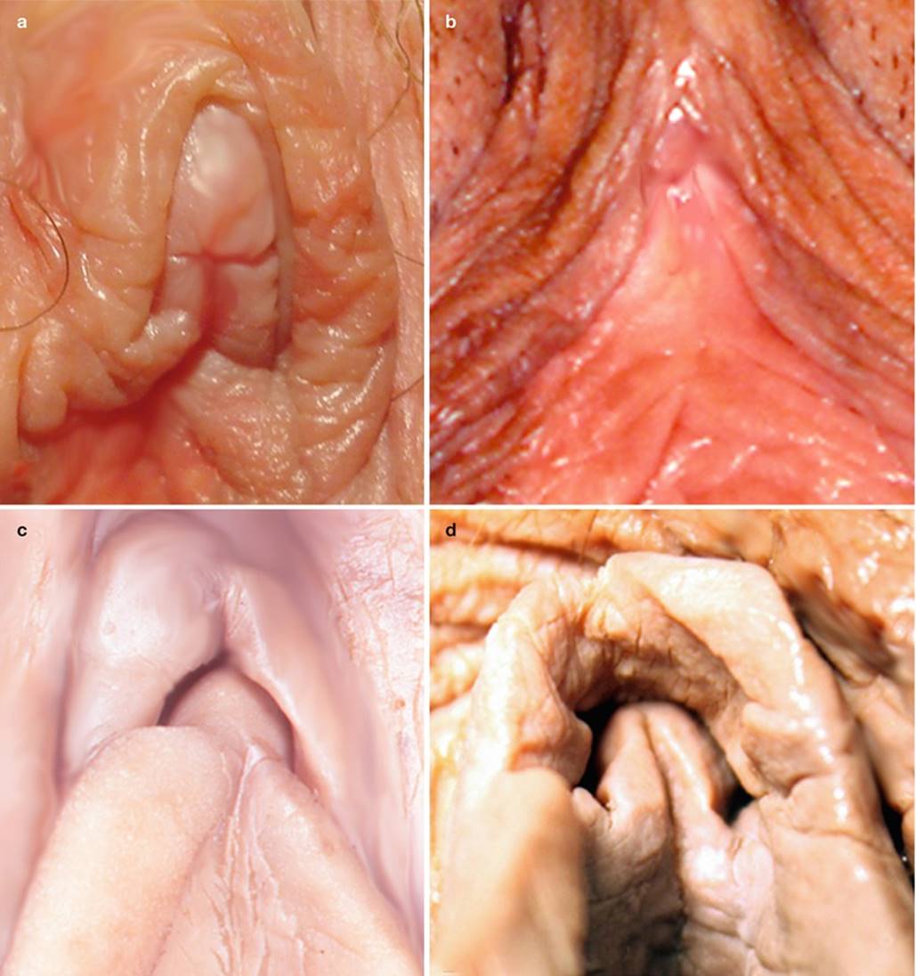
Fig. 4.3
Variations of the hood and their consequences on the cover of glans clitoridis. (a) Protruding glans; (b) barely apparent glans (ptosis of the preputial tissues in an old woman); (c) hidden glans (thick hood and large frenula); (d) hidden glans (only the frenula are visible)

Fig. 4.4
Aspects of two glans (a, b) after sagittal sections of their hoods. These sections allow to see the preputial chamber and the free part of each glans (a, b) better. g glans clitoridis, hclitoral hood (right half), h′ clitoral hood (left half), lm labium minus, ***asterisks locating the preputial chamber on each specimen, V vestibule. NB: On specimen a, the labia minora seem to be born directly from the glans (no frenula!)

Fig. 4.5
Relations of the glans to the hood (end of prepuce) on histological transverse sections, in the axis of the glans. G glans of the clitoris (glans clitoridis), h hood (end of the prepuce), asterisk preputial chamber (clitoro-praeputial sulcus) (The female preputial chamber is analogous to the male preputial sulcus), white curved arrow neck of the glans
The glans is the best known part of the clitoris due to its surface location and accessibility. Moreover, at present, it is rightly considered as the ideal site for female pleasure.
The glans clitoridis challenges neophytes by its resemblance to the glans of the penis, and at first sight, an observer will view the clitoris as a simple miniature penis with a glans whose aspect recalls that of a small-sized male glans! However, an accurate analysis rapidly shows the specific characters and multiplies differences contradicting this point of view.
The glans clitoridis is a protruding tubercle, which is more or less rounded (convex in all directions), and oblique rearwardly. It is often compared to a berry: the bilberry or a tiny acorn (with different colours). However, a slight flatness of the lateral surfaces and a dome-shaped top section are often observed, with a reduction in the diameters from the base to the end, such that the geometrical figure, which best represents the surface of the glans clitoridis, is that of an elliptic paraboloid (ends of a miniature missile or a mini-suppository). It can also have an oblong or cone-type aspect (with a rounded end or, on the other hand, a frankly acuminate end). Finally, it can have a levelled and flat end, shaped as a fish snout (Figs. 4.7, 4.8 and 4.9).

Fig. 4.6
The three principal morphological aspects of the glans clitoridis. (a, a′, b, c) Histological transverse Sections. (a″) Superior view of a dissected clitoris, after resection of the prepuce (skin of the clitoral shaft + hood of the glans). (a, a′, a″) Conoidal appearance; (b) paraboloidal appearance; (c) bulbous appearance. g free part of glans, g′ hidden part of glans, fasfascia of the clitoral body

Fig. 4.7
Some morphological aspects of the glans clitoridis. (a) Cubic glans with asymmetrical tubular frenula. (b) Oblong glans with symmetrical tubular frenula. (c) Glans head fish shaped with large implantation of frenula. (d) Glans with flattened end and angular frenula

Fig. 4.8
Some variations of the glans’ anatomy. 1 rod-shaped frena (rod-shaped frenula clitoridis), 2 glans with several folds of the hood, 3 glans with visible septum, 4 exceptional feature of an accordion-shaped glans, 5 glans with large left frenum arising from its peak
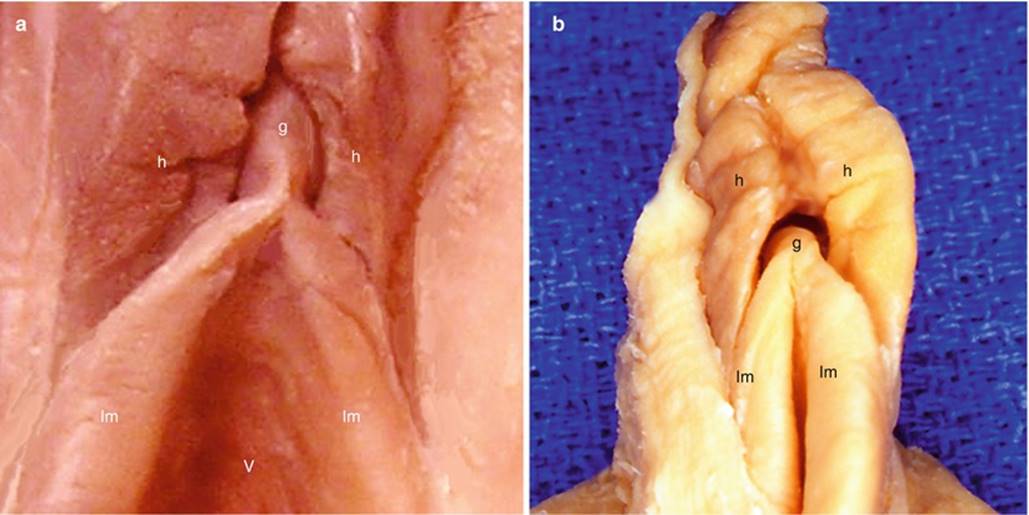
Fig. 4.9
Two examples of “crassiforme” hoods. (a) On the left: the glans, apparent, is flattened laterally. It seems that there is a single frenulum getting lost in a sort of ventral commissure of the labia minora. The top of the vestibule is visible (labia minora moved away). (b) On the right: the gland is spherical and barely visible. The very nearby labia minora close the vulve. These two labia minora seem to be born directly from extremity of the glans (frenula very short, almost non-existent!). g glans clitoridis, h hood, lm labium minus, v vestibule

Fig. 4.10
Examples of “clitoral canal”. (a) Left picture: inferior view of a “perforated” clitoris. (b) Right picture: section of a “clitoral canal”. b bulbe, bco bulbar commissure, bo clitoral body, gglans of the clitoris, h clitoral hood, white arrows they show the one-eyed canal (embryologic residue of the phallic portion of the urogenital sinus), white arrowhead neck of the glans. Remark on the histological section, the epithelium covering the aborted urethra
The prepuce detaches at the neck or base of the glans and becomes the hood. This way it frees the end of this formation. It is rare to observe, at the level of the neck, a basal crown such as is usually observed at the glans of the penis. When this type of crown exists at the glans clitoridis, it is not very marked, except in certain cases (see the glans c on Fig. 5.12). The glans is smooth except on the lower third of its circumference. Its surface is usually uniform; however, a thin white centre line may run along the glans and seem to divide it into two parts. It can, in some cases, lose the regularity of its curves and take on a polyhedral aspect with several facets (“accordion-shaped” glans): this is referred to as the grape currant (Fig. 4.8). The colour of the glans is variable, delicate pink in young women, a more reddish tint (with a tonality identical to that of the medial surface of the labia minora) in women who are not menopaused, and generally a duller colour in menopaused women. It may even have blue-grey aspects in aged women. Its colour actually varies with the intensity of its capillary and arteriolar circulation.
The dimensions of the glans have paradoxically been the subject of in-depth studies performed in vivo by various authors. It is easy to imagine the difficulties involved in such measurements and the discomfort of the patients who had to undergo them!
In his remarkable book on Gynaecology, published in 1931, F.L. Jayle, after having carefully examined and measured the (glans of the) clitoris of 36 successive patients (table attached, Fig. 4.10), found the following average length 5.5 mm (extreme lengths: 2.5 and 10 mm) and as an average width (diameter) 3.5 mm. He concluded that the glans is more long than large! 15 cases are above average including 10, between 7 and 8 mm long. He noted that the width is proportional to the length and classified the large (glans of the) clitorises (7–8 mm long for 4–5 mm large) and small (glans of the) clitorises (4–5 mm long for 2.5–3 mm large).

Fig. 4.11
Measurements of the glans clitoridis carried out by Dr. F.L. Jayle on 36 of his successive patients. Extract from “La Gynecologie”, 1918. Chap: L’anatomie morphologique de la femme
A more recent American study (1992) of B.S. Verkauf et al., based on measurements carried out during 200 gynaecological examinations, provided practically identical results with an average width (diameter) of 3.4 mm ± 1.0 (extreme diameters: 2 and 5 mm) and an average length of 5.1 mm ± 1.4 (extreme lengths: 3 and 8 mm). The authors did not observe any significant difference in the measurements performed on the glans under study, according, on the one hand, to the size and weight of the women subject to examination and according, on the other hand, to the use or not of oral contraceptives. However, all the women who have undergone one or more pregnancies provided significantly greater glans measurements.
There are also significant measurement differences between ethnic groups, such as has been demonstrated by a comparative study performed by Mr. Phillip et al., who measured the external genitals of 570 children, newborn babies born at term, from two different populations (the first included 221 children and the second 349).
Other measurements were used to study the variation of the measurements of the glans clitoridis8 from birth to adulthood.
It is Oberfield et al. who provided the measurements of newborn baby girls born at term (82 cases): the average length is of 4 ± 1.24 mm. The average width (diameter) is of 3.3 ± 0.78 mm.
It is Sane K. and Pescovitz OH. who studied the size of the clitoris from childhood to adolescence (80 studied cases). For an average age of 7 years old, they found a sagittal length average of 5 mm and an average transverse diameter9 of 3.3 mm. According to these authors, there is no significant change until puberty. At the time of puberty, the clitoral growth is moderate and is especially correlated with the increase of body surface. These same authors have also defined a measurement index: the CI or clitoral index, expressed in mm2 and obtained by multiplying the highest sagittal length, expressed in mm, by the transverse diameter, also expressed in mm. Thus, the average CI varies with age: for a newborn baby, it is 13.3 mm2; for an average age of 7 years old, it is 16.7 mm2; from 13 to 18 years old, i.e. for normal postpuberty girls, the average IC is of 20.7 mm2.
GE. Tagatz et al. have studied the clitoral index among adult women, in relation to a bio-essay studying the effects of overdosing with androgens. The results of this study show that for a normal adult woman, the IC does not exceed 35 mm2. On the other hand, beyond 35 mm2, there exists, in half of the cases observed, at least another clinical sign of an excess of androgens. This shows the significance of the IC in endocrinological practice. For our part, it was possible to perform measurements on adult women in the best conditions possible, due to the fact that the glans measurements, carried out on anatomical subjects, during dissection operations, were supplemented by micrometric inspections performed on the histological sections from the dissected parts. Among the 10 cases, in which this double measurement could be carried out, we obtained an average of 5.1 mm for the length (extreme lengths: 2.5 and 6 mm) and of 3.5 mm for the largest diameter (extreme diameters: 3 and 4.5 mm), which corroborates the in vivo measurements carried out by the above-mentioned authors.
On the basis of these various measurements, it is possible to conclude that a normal glans clitoridis is a very small anatomical formation.
What about the rare cases in which dimensions are greater?
Most of the time, these cases are anomalies resulting from severe hormonal disturbances involving the secretion of androgens.10 Clitoridomegaly or clitoromegaly refers to cases in which the dimensions of the glans exceed 10 mm for the length and 7 mm for the diameter.11 Due to their exceptional dimensions, certain hypertrophies have even been the subject of publications or other works: we can mention the treaty of the surgeon, Ambroise Paré, “Des monstres et des prodiges” (Monsters and prodigies) (1573) and that of the not less famous anatomist, Jean Riolan12 “Discours sur les hermaphrodites” (Speech about hermaphrodites) (1614) who mentions clitorises whose length and diameter are that of a finger!
In such cases (pseudo-female hermaphroditism), the clitoris can actually resemble a penis: it is then referred to as a “peniform clitoris”. We can easily imagine the resulting personal, family and social problems and the repercussions on the sexual life of women having this hypertrophy, given that it also involves alterations of the secondary sexual characters and even, in some cases, a coalescence of the labia majora.
The external morphology of the glans varies not only according to its dimensions but also according to its connections with the clitoral hood! Such as has already been proposed by Jayle, four categories of glans clitoridis have been identified:
· Protruding glans, which is immediately visible without needing to separate the labia minora. They are generally very long glans.
· Apparent glans, which is easily visible as soon as the labia minora have been separated.
· Hooded glans, which is easily visible by reclining the hood.
· Hidden glans, which is very difficult to externalise, due to the length of the hood and its many folds.
Regardless of its morphological dimensions and aspects, the glans clitoridis shows two characteristics, which clearly differentiate it from the glans of the penis:
· It does not have an external urethral orifice.
· Its bottom surface is entirely occupied by a double frenulum.
The absence of any orifice at the end of the glans clitoridis is the most common aspect. However, there exist clitorises provided with an orifice opening onto a blind channel, from 4 to 6 mm long, which can be catheterised with the mandrel of a trocar for fluid puncture. We observed this phenomenon three times on the specimen under study. This type of channel is quite visible on histological preparations, where it is bordered by an ordinary squamous epithelium.
Two cases may arise:
· Either the external ostium of such a channel is positioned at the top of the glans clitoridis and a comparison with the external orifice of the male urethra is required. Knowing that in men it is the same epithelial layer, which, while becoming hollow, forms the prepuce of the glans and the terminal urethra with its small fossa navicularis (urethral portion, which will communicate with the phallic urethra from the urogenital sinus), we believe that a similar hollowing process may occur in women. However, this process is abortive since it does not encounter a channel with which it can communicate.
· Or the external ostium of this channel is positioned underneath the neck of the glans (Fig. 4.11), a few mm from the apex of the glans. The abnormal persistence of the terminal segment of the phallic portion of the urogenital sinus may be mentioned as an embryological explanation.
In any event, this may explain certain old texts and the resulting errors, such as the belief according to which “female semen” was released by a clitoral channel (see Chap. 1). It should be remembered that even if a small orifice and a ductal residue are sometimes present at the glans clitoridis, these are not functional urethral structures!
The lower surface of the glans clitoridis is very particular. It may even be said that, in most cases, it does not exist as it is nearly entirely occupied by the double frenulum of the labia minora or nymphs, which we prefer to refer to as frenula clitoridis (Fig. 4.12). The frenula therefore resemble two protrusions of the lower surface of the glans, which each diverge backwards and outwards to join the top edge of the counterpart labium minus. The aspect of each frenulum varies and a right-left symmetry is not very frequent. Each frenulum may be flat (webbed frenulum) or shaped as a cylinder (tubular frenulum) (see Fig. 4.9). There exist short and long frenula exceeding 1 cm in length. The end of each frenulum may vary: In most cases, the frenulum extends with the internal surface of the homolateral labium minus. It may also merge with the extension of the hood and thus form the ventral end of the corresponding labium minus. The inter-frenulum angle is important and plays a considerable role during the coitus. The frenula have a colour similar to that of the upper edges of the labia minora. The frenula may be straight, curved, wrinkled or bent (Fig. 4.8). If the glans has a certain degree of binary constitution (glans with a visible septal line), it may give the impression that each of the two sections extends into the homolateral frenulum. In these cases, the two frenula can be parallel and run alongside each other over ¾ of their length (dual rod frenula) (Fig. 4.9). It will be also noted that in cases where the glans has an orifice (opening onto a short blind channel), the formation of each frenulum starts in contact with this orifice. The two frenula can sometimes delimit a small area between them or a small cavity (“fossa”) in the bottom surface of the clitoris, which is referred to as the infra-glandular cavity. The frenula, such as the medial surface of the labia minora, are covered with a squamous epithelium. This epithelium covers the relatively dense conjunctive tissue, which occupies the internal part of each frenulum. Overall, each of the labia minora (or nymphs) is formed at its juxta clitoral section, by the contribution of two homolateral components: infero-lateral part of the clitoral hood and clitoral frenulum.13 Therefore, there exists a functional nympho-clitoral unit whose physiological importance will be demonstrated in another chapter.
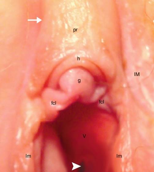
Fig. 4.12
Details of the glans’ external aspect (perineal view). fcl frenum (frenulum clitoridis), g glans clitoridis, h hood, lm labium minus (lesser lip), lM labium majus (greater lip), pr female prepuce (preputium clitoridis) covering the clitoral shaft, V vestibule, white arrowhead external urethral orifice, white arrow labio-preputial sulcus
Upstream of the glans, there is another part of the clitoris, visible via the protrusion it forms in the preputial groove, which separates the two labia majora, immediately behind their ventral commissure: This is the protrusion of the clitoral body,14 covered by the prepuce (M12). On either side of this protrusion, a more or less deep labio-preputial groove extends the interlabial groove15 up to the ventral commissure of the labia minora.
The epithelium of the prepuce is laterally in continuity with that of the labia majora: it is thus also a keratinised squamous epithelium, with high content of sebaceous glands. However, it is less thick and has a lower content in hair follicles, which contrasts with the significant hairiness of the nearby labia majora and especially that of the mons pubis (mons veneris) which is above it. The cutaneous covering of the body of the clitoris plays the same role as the hood at the level of the glans: This consists of a protection role dedicated to the entire clitoral prepuce. For the body of the clitoris, this protection is increased by the presence of a more or less deep groove in which it is located. As for the hood, the protection, which it provides for the glans clitoridis, has been compared by certain anatomists to that of the eyelids for the eye bulb.
From a morphological viewpoint, the body of the clitoris, beyond the glans, is cylindrical. The prepuce covers it like roof tile. As it is necessary to release this clitoral body by dissection in order to study all of its sections, we will describe it in the following chapter with the hidden structures, which are not visible from outside.
Footnotes
1
1If the vulvar lips are not separated, it is impossible to visualise the entire external female genital apparatus: The only visible elements laterally are the labia majora and, in the centre, the ventro-dorsal vulvar cleft (anteroposterior vertical alignment, in a gynaecological position), bordered by the protruding part of the generally irregular edge of the labia minora. Above the vulvar cleft, the mons pubis (mons veneris), the ventral commissure of the labia majora and the clitoral prepuce are only visible if the hairy pubic triangle is shaved or plucked (Fig. 4.1).
2
2The vulva refers to the entire external genital apparatus, i.e. the mons pubis, the labia majora and minora and the vestibule of the vagina.
3
3The perineum consists of all the soft parts, which close at the base of the pelvic excavation, below the levator ani muscles. The external female genital apparatus is located in the anterior part or urogenital part of this perineum (the posterior part of the perineum is called the anal perineum).
4
4Under the teguments, the clitoris extends beyond the ventral bone boundary of the anterior perineum, as its median attachments rise and cover the bottom ¼ of the ventral surface of the pubic symphysis (see Chap. 13).
5
5The epithelia of the prepuce and hood include many immunising cells (dendritic cells, lymphocytes, NK-T cells, etc.). The internal epithelium of the hood, which is not keratinised, does not oppose any effective “mechanical barrier” to the possible penetration of HIV, which the above-mentioned cells will therefore incorporate!
6
6The smegma is a white substance produced in the preputial chamber: it is comprised of desquamed epithelial and sebaceous cells (secretion product of the sebaceous glands, which lubrify the epithelia of the glans and hood and enable the hood to slide). Certain bacteria of the genital flora can generate an unpleasant smegma smell.
7
7In exceptional cases, this protrusion can, on one side, remain independent of the corresponding labium minus (F.L. Jayle).
8
8It is to be noted that in their publications, the authors express their measurements as the “length of the clitoris”, whereas they measure only the glans, i.e. the accessible part while reclining the hood (such as specified).
9
9The authors never specify which transverse diameter is used for their measurements: Is it the diameter of the base of the glans or the largest transverse diameter (the latter is not always the diameter of the base of the glans, according to the clitoral morphology)?
10
10Female pseudo-hermaphroditism occurs following a hypersecretion of androgens by the foetal adrenal gland (endogenous origin). The same virilisation phenomena can occur following the administration of anabolic steroids (exogenous origin)!
11
11If a clitoromegaly is detected in a woman, any sign of androgenic hypersecretion must be researched.
12
12Riolan had already considered that these hermaphrodites had two sexual potentialities. He classified them among what the Greeks called “tribades” and the Romans “fricatrices” (on this topic, read the excellent chapter written by K. Park: The Rediscovery of the Clitoris, French Medicine and the Tribade, 1570–1620).
13
13This contribution is variable:
· The labium minus can be formed through the fusion of the frenulum and the lateral extension of hood.
· The labia minora can be formed from a single frenulum, while the extension of the hood gets lost on its lateral surface.
· More rarely, it is the lateral extension of the hood which forms the labium minus, which will be joined, on its medial surface, by the frenulum.
14
14Still referred to as the “clitoral shaft”.
15
15The interlabial sulcus separates each labium minus from the homolateral labium majus.