
Carlo Alviggi1 , Alessandro Conforti1 and Sandro C. Esteves2
(1)
Department of Neuroscience and Reproductive Medicine, University of Naples Federico II, Naples, Italy
(2)
ANDROFERT, Andrology and Human Reproduction Clinic, Av. Dr. Heitor Penteado, 1464 Campinas, São Paulo, 13075-460, Brazil
Carlo Alviggi
Email: alviggi@unina.it
Abstract
Controlled ovarian stimulation is a mainstay of assisted reproductive technologies and leads to optimal follicular growth and steroidogenesis in the majority of cases. Nonetheless, some women defined as “hyporesponders” require higher amount of exogenous gonadotrophin to achieve an adequate number of oocytes retrieved despite an apparently good prognosis. Clinical observational trials suggest that hyporesponse to exogenous gonadotrophins, including initial poor response, could be a genetically determined trait with specific genotype profile associated with this condition. Specifically, mutation and polymorphisms involving luteinizing hormone (LH) and follicle-stimulating hormone (FSH) and their receptors LH-R and FSH-R have been thoroughly investigated. Among all the mutations discovered, it seems that that carriers of common LH variant and FSH receptor Ser/680 variants require higher doses of exogenous FSH to achieve a normal ovarian response.
In conclusion, the idea of a tailored gonadotrophin administration based on a pharmacogenomic approach may be considered in specific situations and could represent the future research target for a better understanding of the underlying mechanisms that regulate human fertility.
Keywords
PolymorphismControlled ovarian stimulationPharmacogenomicsIVFFollicle-stimulating hormoneLuteinizing hormoneFollicle-stimulating hormone receptors (FSH-R)Luteinizing hormone receptors (LH-R)
14.1 Introduction
Controlled ovarian stimulation (COS) is an essential step in most “in vitro” fertilization (IVF) programs. In this context, GnRH agonist (GnRH-a) long protocol in association with recombinant FSH (r-hFSH) still constitutes the most utilized strategy for normogonadotrophic patients. The use of GnRH antagonists (GnRH-ant) plus r-hFSH does represent a valid alternative. These approaches lead to optimal follicular growth and steroidogenesis in about 85–90 % of women. Conversely, COS results in a very different clinical outcome, from poor ovarian response to the risk of hyperstimulation syndrome (OHSS) in a relevant number of cases. In addition, some women, defined as “hyporesponders,” require higher amounts of r-hFSH to obtain an adequate number (i.e., >4) of oocytes retrieved, despite an apparently good prognosis.
To sort this problem out, several markers have been proposed to predict ovarian response such as age, basal FSH, inhibin-B, anti-Müllerian hormone (AMH), and the count of antral follicles by ultrasonography (AFC). Yet, there is an increasing interest on the possible effect of specific genotype patterns on ovarian response.
In the present chapter, the potential effect of specific mutations/polymorphisms of the gonadotrophins and their receptors on the outcome of COS is explored. Confirmation of these observations would reinforce the idea of a tailored gonadotrophins administration based on a pharmacogenomic approach.
14.2 The Physiology of Gonadotrophins and Their Receptors
The classical “two cells-two gonadotrophins” model is based on the idea that follicle-stimulating hormone (FSH) and luteinizing hormone (LH) exert their roles on two different compartments, granulosaand theca, respectively. According to this model, LH exerts its activity in theca cells, which express enzymatic pathways of androgen synthesis [1, 2]. Theca involucres surround the granulosa cells, whose activities and proliferation are directly regulated by FSH. This hormone induces the expression of the aromatase enzyme, which in turn converts theca-deriving androgens into estradiol (E2).
This theory, reinforcing the notion that granulosa and theca cells are distinct compartments regulated by FSH and LH, respectively, has been revised. More specifically, it has been found that LH receptors are also detected on the granulosa compartment at the intermediate follicular phase [2–5]. Therefore, it appears that LH regulates both granulosa and theca cells.
FSH and LH cooperate in inducing the granulosa cell-specific production of inhibin-B and other TGB-β growth factors. In addition, insulin growth factors (IGF) I and II, which are expressed by both granulosa and theca cells throughout folliculogenesis, are important in promoting follicular maturation [6, 7]. Locally produced peptides, rather than estrogens, are known to be the key factor regulating primate follicle growth and development [8–11]. In light of these finding, we can conclude that (1) both gonadotrophins contribute (via granulosa) to maintain the autocrine-paracrine system governing dominant follicle growth and (2) LH is crucial in sustaining FSH activity in the granulosa during intermediate-late stages of folliculogenesis. On this basis it is possible to argue that high levels of one gonadotrophin can counteract the lack of the other. This hypothesis is consistent with the observation that FSH activity can be totally substituted by LH once granulosa cells express adequate amounts of LH receptors [5, 12]. Conversely, higher exogenous FSH doses during COS are able to compensate GnRH-a-related reduction of LH. It could be argued that if LH concentration and/or activity falls below a hypothetical threshold, an impairment in granulosa paracrine activities will occur, which in turn can lead to higher requirement of FSH.
On the basis of the above information, it could be hypothesized that during COS, different “adaptative” mechanisms may occur. For instance, lack of LH activity in granulosa cells may be counteracted by higher exogenous FSH. Conversely, administration of exogenous LH may optimize FSH activity on the same compartment, which in turn can increase steroidosynthesis and reduce FSH requirement. In the clinical practice, COS protocols are often chosen empirically. As consequence, same protocols are administered in most patients, despite potential biological differences. Adaptation capability of follicles leads ovarian response to an “adequate” profile in almost all women. Nevertheless, this adaptation requires “integrity” of granulosa-theca system. Aging and some genetic characteristics may reduce this capability, leading to “suboptimal” ovarian response. In the following paragraphs, the potential role of some polymorphisms of gonadotrophins and their receptors in conditioning ovarian response to gonadotrophins will be discussed.
14.3 The LH System: A Crucial Variable During COS
14.3.1 LH Polymorphism
Recently, it has been reported that hyporesponders who benefited from LH activity had endogenous levels of LH in the normal range. In addition, endogenous LH concentrations of these patients during early phases of COS was always comparable with those observed in women who had optimal response to FSH and who did not require any change of FSH dose during stimulation. This observation led to the hypothesis that hyporesponse to r-hFSH is associated with a less bioactive LH [13–16].
Among the most valuable β-LH polymorphisms identified (Table 14.1) [17–24], we have recently performed an observational trial [25] aimed to evaluate whether the presence of the most common of them, v-LH, is associated with different profiles of ovarian response to r-hFSH.
Table 14.1
Mutations and polymorphisms of LH
|
Location |
Type |
Amino acid involved |
Effect |
Reference |
|
Exon 3 |
Missense |
Gln54 to Arg |
Absence of spontaneous puberty in male |
Weiss et al. (1992) [17] |
|
Exon 2 |
Missense |
Trp8 Arg Ile15 Thr |
Delayed pubertal progression in male and infertility in female |
Petterson et al. (1991) [18] Furui et al. (1994) [19] Haavisto et al. (1995) [20] |
|
Exon 3 |
Missense |
Ala −3 Thr |
Normal bioactivity |
Jiang et al. (2002) [21] |
|
Exon 3 |
Missense |
Gly102 Ser |
Infertility in male, menstrual disorders in female |
Liao et al. (1998) [22] Ramanujam et al. (1999) [23] |
Adapted from Lamminen and Huhtaniemi [24]
Pettersson and Söderholm [18] were the first to describe this common variant of LH (v-LH) as an immunologically anomalous form of LH. The occurrence of the v-LH varies according to geographic areas (Fig. 14.1) [24]. v-LH is due to two point mutations in the β subunit gene, both altering the amino acid sequence (Trp8Arg and Ile15Thr). v-LH has elevated bioactivity in vitro but significantly shorter (5–9 min) half-life in circulation when compared with the wild type LH (wt-LH) (12–22 min). As the pulse frequency of the v-LH is normal, this results in an overall LH action that is more potent at the receptor site but shorter in duration in vivo.
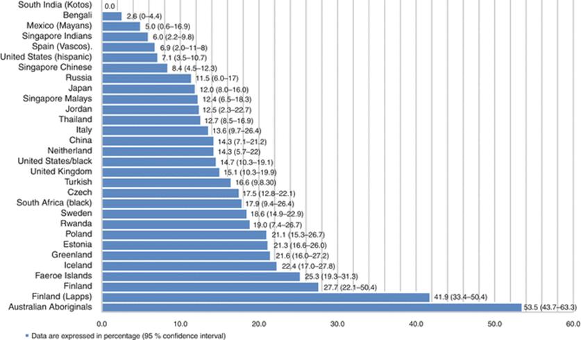
Fig. 14.1
Worldwide occurrence of the common v-LH (From Lamminen and Huhtaniemi [24])
The v-LH is common worldwide, with carrier frequency varying from 0 to 52 % in various ethnic groups. Its incidence in Italy ranges between 12 and 13 %. The v-LH differs functionally from wt-LH, and it seems to predispose its carrier to mild aberrations of reproductive function menstrual irregularities causing infertility [19] and recurrent pregnancy loss [26].
In our observational trial, 60 normogonadotrophic patients undergoing a GnRH-a long downregulation plus r-hFSH for IVF/ICSI, and in whom at least five oocytes were retrieved, were divided into three groups: 22 women requiring a cumulative dose of r-hFSH >3,500 IU constituted group A, 15 patients requiring 2,000–3,500 IU were included in group B, and 23 women requiring <2,000 IU served as control group (group C). The presence of the v-LH was evaluated using immunoassays able to detect both wt-LH and polymorphism. Group A showed a significantly lower (p < 0.05) number of oocytes retrieved when compared with group B and C (7.3 ± 1.5, 11.7 ± 2.4, and 14.7 ± 4.1 in the three groups, respectively). Seven carriers (32 %) of v-LH were found in group A, whereas only one variant (7 %) was observed in group B; no variant was detected in group C. This study suggested, for the first time, an association between a less bioactive LH and a higher FSH requirement. In addition, it supports the idea that hyporesponders represent a specific subgroup of patients. In fact, all women requiring >3,500 IU of FSH had at least five oocytes retrieved and showed peak estradiol >500 pg/ml, which in turn would have lead physicians to classify them as normal responders. Nevertheless, they had a statistically significant reduction of the number of oocytes retrieved and estradiol levels when compared with woman requiring lower FSH doses.
On the basis of these finding we further investigated the relationship between v-LH and ovarian response to FSH [27, 28] in a Danish population. v-LH was present in 11 % of patients, whereas the allelic frequency was 12 %. Patients were divided into two groups according to their LH genotype. Group A included 196 wt/wt women, and group B was constituted by 24 individuals with v-LH (21 heterozygous and 3 homozygous). The mean number of oocytes retrieved, fertilization rate, and pregnancy rate per cycle were similar in the two groups. Group B received a significantly higher cumulative dose of r-hFSH than group A (2,435.86 ± 932.8 IU versus 1,959.8 ± 736.45; P = 0.048). LH genotype had a statistically significant effect (P < 0.01) on the cumulative dose of r-hFSH, showing a progressive increase from wt/wt (1,959.8 ± 736.45 IU) to v-LH heterozygotic (2,267.5 ± 824.3) and homozygotic women (3,558.3 ± 970.9). These results confirmed that carriers of v-LH have hyposensitivity to exogenous FSH during COS.
14.3.2 LH Receptors (Mutations and Polymorphisms)
The luteinizing hormone/choriogonadotrophin receptor (LHCGR) is a member of the superfamily of guanine nucleotide-binding protein-coupled receptors (GPCRs) and belongs to the glycoprotein hormone receptors [29]. LHCGR is expressed in Leydig cells and in ovarian theca, granulosa, as well as luteal cells. These receptors exert a fundamental role in reproductive process since puberty [30]. Several mutations have been identified in LHCGRs, and some of them have been related to reproductive disorders such as male-limited gonadotrophin-dependent precocious puberty, Leydig cell hypoplasia, and anovulation/amenorrhea [30]. In addition, some authors have observed an increased risk for endometrioid adenocarcinoma when rs13405728 mutation in gene LHCGR is associated with SNPs rs2479106 in gene DENND1A [31].
LHCGR mutation can be didactically divided into two categories:
1.
2.
In addition to the LHCGR mutations, more than 200 single nucleotide polymorphisms have been discovered. One of the most widespread polymorphisms is due to the presence of a two-amino acid insertion at position 18 in exon 1 (insLQ) and has been detected in breast cancer patient with lower survival rate [33]. Subsequently, another group have analyzed the same polymorphism in PCOS patients, but found no significant association [34].
A detailed phenotype of novel homozygous inactivating nonsense and missense mutations of the LH-receptor gene (Arg 554 stop codon 554 [TGA] and Ser 616 → Tyr 616, respectively) has been described in a woman with compromised ovulation and luteinization processes but apparent normal pubertal feminization [35]. This aforementioned patient presented with high LH and FSH levels and normal estradiol end progesterone values [36].
Evidence about the relationship between LHCGR and reproductive outcome during COS is scarce. In addition to the previously mentioned Kerkala et al.’s observations [34], some authors have recently observed that a higher expression of LH receptors by human cumulus granulosa cells is associated with lower fertilization rate [37].
14.4 The FSH System: From Physiology to COS
14.4.1 FSH Receptor (Mutations and Polymorphisms)
The FSH receptor (FSH-R), likewise its homologue LH, is a glycoprotein hormone receptor that belongs to subfamily of G protein-coupled receptors (GPCRs). FSH mutations have been extensively studied with more than 1,000 polymorphic variants identified to date [38]. Like LHCGR, FSH-R mutations are categorized in “activating” or “inactivating” mutations.
The first “activating” FSH-R mutation was discovered in a hypophysectomized man who surprisingly showed normal spermatogenesis despite undetectable FSH levels [39].
Other two peculiar cases of constitutively activated FSH-R were characterized by heterozygous Thr449Ile and Asp567Asn mutations. Both affected women had a history of spontaneous OHSS syndrome during pregnancy. The probable explanation for this phenotype is linked to the altered ligand site, which becomes activated in the presence of high hCG levels as normally seen during pregnancy [40, 41].
Carriers of “inactivating” mutations are usually affected by hypergonadotrophic hypogonadism, primary or early-onset secondary amenorrhea, variable sexual development, arrest of follicular maturation between primordial and preantral stage, and poor semen quality. While severe phenotypes have been described in carriers of Ala189Val and Pro348Arg mutations, mild forms have been detected in patients with a compound heterozygous mutation of Ala189Val and Ala419Thr [42–44].
The most investigated variant of the FSH-R consists in the replacement at position 680 of the amino acid asparagine by serine (Fig. 14.2) [32]. This polymorphism has been associated with higher basal FSH levels and an increased number of antral follicles during the early follicular phase [45]. In an observational trial, Perez Mayorga et al. [46] evaluated the relationship between the presence of the Ser/680 FSH-R variant and ovarian response to COS in 161 normo-ovulatory women undergoing IVF. All women were below 40 years. The distribution of genotypes in the study population was 29 % for the Asn/Asn, 45 % for the Asn/Ser, and 26 % for the Ser/Ser FSH-R variant. Both estradiol levels at the day of human chorionic gonadotrophin (hCG) and number of retrieved oocytes were similar in the three groups. Conversely, basal FSH levels were significantly different among the three groups (6.4 ± 0.4 IU/l, 7.9 ± 0.3 IU/l, and 8.3 ± 0.6 IU/l for the Asn/Asn, Asn/Ser, and Ser/Ser groups, respectively, P < 0.05). In addition, the mean number of FSH ampoules required for successful stimulation was significantly different among groups (31.8 ± 2.4, 40.7 ± 2.3, and 46.8 ± 5.0 for the Asn/Asn, Asn/Ser, and Ser/Ser groups, respectively, P < 0.05). These clinical findings demonstrated that ovarian response to FSH stimulation depends on the FSH-R genotype. Following these observations, Behre et al. [47] tested whether the same daily dose of FSH resulted in lower levels of estradiol in women homozygous for the Ser/Ser and whether the difference could be overcome by higher FSH doses. Fifty-nine women undergoing COS for IVF or ICSI and homozygous for the FSH-R polymorphism Ser/680 were randomly allocated in three groups. Group I (Ser/Ser, n = 24) received a daily FSH dose of 150 IU/day, and group II (Ser/Ser, n = 25) received a FSH dose of 225 IU/day. In group III (Asn/Asn, n = 44), FSH dose was 150 IU/day. Age and basal FSH levels were not different between groups. Total FSH doses were comparable in group I (1,631 ± 96 IU) and group III (1,640 ± 57 IU) but significantly higher in group II (2,421 ± 112 IU) (P < 0.001). Peak estradiol levels were significantly lower in group I (5,680 ± 675 pmol/l) compared to group III (8,679 ± 804 pmol/l) (P < 0.05). Increasing the FSH dose from 150 to 225 IU/day overcame the lower estradiol response in women with Ser/Ser (group II, 7,804 ± 983 pmol/l). The authors concluded that patients with the Ser/Ser FSH-R variant have lower FSH receptor sensitivity, which can be overcome by higher FSH doses. This study represented the first case of a pharmacogenomic approach to COS.

Fig. 14.2
Human FSH receptor mutations. The Ser680Asn is in linkage disequilibrium with Thr307Ala (green) (From Huhtaniemi and Themmen [32])
Recently, we have evaluated the occurrence of the Ser/680 FSH-R variant among women classified as “hyporesponders” (Alviggi et al. 2013). Forty-two normogonadotrophic patients in whom at least five oocytes were retrieved after GnRH-a long downregulation protocol followed by stimulation with r-hFSH for IVF/ICSI were retrospectively studied. On the basis of the total r-hFSH consumption, patients were divided into two groups: 17 women requiring a cumulative dose of r-hFSH >2,500 IU constituted group A, whereas 25 patients requiring <2,500 IU served as controls (group B). DNA was analyzed to determine the FSH receptor genotype. Estradiol peak levels were significantly lower in group A (997 ± 385 pg/ml) when compared with group B (1,749 ± 644; P < 0.001). The number of oocytes retrieved was also significantly lower in group A compared with group B (7.1 ± 1.5 versus 9.6 ± 2.4; P < 0.001). Homozygous Ser/Ser receptor variant at codon 680 was observed in 47.0 % of women of group A and in 28.0 % of women of the control group. The homozygous Asn/Asn receptor variant was found in 23.6 and 20.0 % of patients in the two groups, respectively. Heterozygosis Ser/Asn was detected in 29.4 % of patients of group A and in 52.0 % of patients of group B. These results indicated that FSH-R Ser 680/variant is more frequent in women with hyporesponse to r-hFSH.
Although some investigators found a positive association between pregnancy rate and presence of Ser680 genotype [48, 49], a recent meta-analysis confirmed that Ser/Ser genotype carriers have significantly higher basal FSH levels and require higher exogenous FSH doses for COS [50].
Nakayama et al. in 2006 identified another polymorphic variant of FSH-R with possible implication in COS [51]. It consisted of a polymorphism in the 5′-UTR of the FSH-R gene (position 29 A/G; rs1394205), which seems to be associated with a lower luciferase activity compared with G/G 29 allele. Subsequently, Desai and colleagues observed a reduced FSH-R expression in granulosa cells of AA genotype carriers [52].
In women undergoing assisted reproduction, variants A/A have been associated with poor ovarian response with respect to number of oocyte retrieved and doses of exogenous FSH for COS [53].
Lastly, the impact of a new FSH-R polymorphism has been investigated in a female Indian population. Specifically, 50 patients undergoing ART and 100 fertile patients have been recruited. The authors observed that Ala307Ala carriers required lower amount of exogenous FSH for ovulation induction in comparison with Thr307Thr and Thr307Ala subjects. Estradiol levels and incidence of OHSS were higher in the former [54].
FSH-R polymorphisms and the ovarian outcome in women undergoing ovarian stimulation have been widely studied [46, 52–58].
14.4.2 FSH Mutations and Polymorphisms
Several β subunit mutations of FSH have been identified in the literature. Most of them inactivate the FSH effects. In females, primary amenorrhea, impaired fertility, and compromised pubertal development are the most frequent clinical manifestations. In contrast, FSH mutations in males do not affect sexual maturation although they result in azoospermia. Most of FSH mutations interfere with a specific cysteine knot region that is crucial for dimerization with α subunit and biological activity [32].
Unlike LH β, most of FSH polymorphic variants have been found in noncoding regions confirming that FSH β is strongly conserved in the human species [32, 59].
To date, only a single nucleotide polymorphism located into FSH β promoter-211G/T seemed to influence the FSH concentration in males [60, 61]. In addition, it seems that Ser 680 Asn polymorphism may influence serum FSH levels in the male population [62]. The same effects were also reported in the female population. Higher FSH serum levels were observed in women with the FSHB-211 GT + TT/associated with FSHR2039 AA genotype [59]. The impact of FSH polymorphisms and their combination with different FSH-R genotypes is yet to be evaluated.
Conclusion
The unraveling of the mechanisms that regulate the interaction between the gonadotrophins and their receptors is a step forward to a better understanding of why an impaired ovarian response to stimulation occurs in apparent good prognosis patients. There are clinical observational trials suggesting that hyporesponse to exogenous gonadotrophins, including initial poor response, could be a genetically determined trait. This phenomenon has been associated with the presence of at least two common polymorphisms involving LH and FSH-R, respectively. Carriers of v-LH and FSH-R Ser/680 variants, despite normal levels of endogenous gonadotrophins and regular AMH/AFC, require higher doses of exogenous FSH to achieve a normal ovarian response. Thus, the idea of a tailored gonadotrophin administration based on a pharmacogenomic approach may be considered in specific situations. As examples, LH supplementation may be considered in the presence of v-LH, whereas a timely identification of Ser/680 FSH-R variant may represent an indication to administer higher doses of FSH.
References
1.
Fevold H. Synergism of follicle stimulating and luteinizing hormones in producing estrogen secretion. Endocrinology. 1941;28:33–6.CrossRef
2.
Hillier SG, Whitelaw PF, Smyth CD. Follicular oestrogen synthesis: the ‘two-cell, two-gonadotrophin’ model revisited. Mol Cell Endocrinol. 1994;100(1–2):51–4.PubMed
3.
Erickson GF, Wang C, Hsueh AJ. FSH induction of functional LH receptors in granulosa cells cultured in a chemically defined medium. Nature. 1979;279(57):336–8.CrossRefPubMed
4.
Shima K, Kitayama S, Nakano R. Gonadotropin binding sites in human ovarian follicles and corpora lutea during the menstrual cycle. Obstet Gynecol. 1987;69(5):800–6.PubMed
5.
Filicori M, Cognigni GE, Pocognoli P, Ciampaglia W, Bernardi S. Current concepts and novel applications of LH activity in ovarian stimulation. Trends Endocrinol Metab. 2003;14(6):267–73.CrossRefPubMed
6.
Zhou J, Bondy C. Anatomy of the human ovarian insulin-like growth factor system. Biol Reprod. 1993;48(3):467–82.CrossRefPubMed
7.
Huang ZH, Clayton PE, Brady G, Morris ID. Insulin-like growth factor-I gene expression in human granulosa-lutein cells. J Mol Endocrinol. 1994;12(3):283–91.CrossRefPubMed
8.
Rabinovici J, Blankstein J, Goldman B, Rudak E, Dor Y, Pariente C, et al. In vitro fertilization and primary embryonic cleavage are possible in 17 alpha-hydroxylase deficiency despite extremely low intrafollicular 17 beta-estradiol. J Clin Endocrinol Metab. 1989;68(3):693–7.CrossRefPubMed
9.
Pellicer A, Miró F, Sampaio M, Gómez E, Bonilla-Musoles FM. In vitro fertilization as a diagnostic and therapeutic tool in a patient with partial 17,20-desmolase deficiency. Fertil Steril. 1991;55(5):970–5.PubMed
10.
Zelinski-Wooten MB, Hess DL, Baughman WL, Molskness TA, Wolf DP, Stouffer RL. Administration of an aromatase inhibitor during the late follicular phase of gonadotropin-treated cycles in rhesus monkeys: effects on follicle development, oocyte maturation, and subsequent luteal function. J Clin Endocrinol Metab. 1993;76(4):988–95.PubMed
11.
Shetty G, Krishnamurthy H, Krishnamurthy HN, Bhatnagar S, Moudgal RN. Effect of estrogen deprivation on the reproductive physiology of male and female primates. J Steroid Biochem Mol Biol. 1997;61(3–6):157–66.CrossRefPubMed
12.
Zeleznik AJ, Hillier SG. The role of gonadotropins in the selection of the preovulatory follicle. Clin Obstet Gynecol. 1984;27(4):927–40.CrossRefPubMed
13.
De Placido G, Mollo A, Alviggi C, Strina I, Varricchio MT, Ranieri A, et al. Rescue of IVF cycles by HMG in pituitary down-regulated normogonadotrophic young women characterized by a poor initial response to recombinant FSH. Hum Reprod. 2001;16(9):1875–9.CrossRefPubMed
14.
De Placido G, Alviggi C, Mollo A, Strina I, Ranieri A, Alviggi E, Wilding M, et al. Effects of recombinant LH (rLH) supplementation during controlled ovarian hyperstimulation (COH) in normogonadotrophic women with an initial inadequate response to recombinant FSH (rFSH) after pituitary downregulation. Clin Endocrinol (Oxf). 2004;60(5):637–43.CrossRef
15.
De Placido G, Alviggi C, Perino A, Strina I, Lisi F, Fasolino A, et al. Recombinant human LH supplementation versus recombinant human FSH (rFSH) step-up protocol during controlled ovarian stimulation in normogonadotrophic women with initial inadequate ovarian response to rFSH. A multicentre, prospective, randomized controlled trial. Hum Reprod. 2005;20(2):390–6.CrossRefPubMed
16.
Ferraretti AP, Gianaroli L, Magli MC, D’angelo A, Farfalli V, Montanaro N. Exogenous luteinizing hormone in controlled ovarian hyperstimulation for assisted reproduction techniques. Fertil Steril. 2004;82(6):1521–6.CrossRefPubMed
17.
Weiss J, Axelrod L, Whitcomb RW, Harris PE, Crowley WF, Jameson JL. Hypogonadism caused by a single amino acid substitution in the beta subunit of luteinizing hormone. N Engl J Med. 1992;326(3):179–83.CrossRefPubMed
18.
Pettersson K, Söderholm JR. Individual differences in lutropin immunoreactivity revealed by monoclonal antibodies. Clin Chem. 1991;37(3):333–40.PubMed
19.
Furui K, Sagamura N, Tsukahara S, Asada Y, Kikkawa F, Tanaka M, et al. Identification of two point mutations in the gene coding luteinizing hormone (LH) beta-subunit, associated with immunologically anomalous LH variants. J Clin Endocrinol Metab. 1994;78(1):107–13.PubMed
20.
Haavisto AM, Pettersson K, Bergendahl M, Virkamäki A, Huhtaniemi I. Occurrence and biological properties of a common genetic variant of luteinizing hormone. J Clin Endocrinol Metab. 1995;80(4):1257–63.PubMed
21.
Jiang M, Lamminen T, Pakarinen P, Hellman J, Manna P, Herrera RJ, Huhtaniemi I. A novel Ala(−3)Thr mutation in the signal peptide of human luteinizing hormone beta-subunit: potentiation of the inositol phosphate signalling pathway and attenuation of the adenylate cyclase pathway by recombinant variant hormone. Mol Hum Reprod. 2002;8(3):201–12.CrossRefPubMed
22.
Liao WX, Roy AC, Chan C, Arulkumaran S, Ratnam SS. A new molecular variant of luteinizing hormone associated with female infertility. Fertil Steril. 1998;69(1):102–6.CrossRefPubMed
23.
Ramanujam LN, Liao, WX, Roy AC, Ng SC. Screening of a mutation in luteinizing hormone beta-subunit in male infertile patients. The Endocrine Society’s 81st annual meeting. San Diego, June 12–15, 1999. Abstract.
24.
Lamminen T, Huhtaniemi I. A common genetic variant of luteinizing hormone; relation to normal and aberrant pituitary-gonadal function. Eur J Pharmacol. 2001;414(1):1–7.CrossRefPubMed
25.
Alviggi C, Clarizia R, Pettersson K, Mollo A, Humaidan P, Strina I, et al. Suboptimal response to GnRHa long protocol is associated with a common LH polymorphism. Reprod Biomed Online. 2009;18(1):9–14.CrossRefPubMed
26.
Okuda K, Takamatsu J, Okazaki T, Yamada T, Saeki M, Sugimoto O. Hereditary abnormality of luteinizing hormone resulting in discrepant serum concentrations determined by different assays. Endocr J. 1994;41(6):639–44.CrossRefPubMed
27.
Alviggi C, Clarizia R, Pettersson K, Mollo A, Strina I, De Biasio G, et al. A single point mutation in the LH beta subunit is associated with higher exogenous FSH consumption during ovarian stimulation. Hum Reprod. 2009;24 Suppl 1:i3. Abstract O-077.
28.
Alviggi C, Pettersson K, Longobardi S, Andersen CY, Conforti A, De Rosa P, et al. A common polymorphic allele of the LH beta-subunit gene is associated with higher exogenous FSH consumption during controlled ovarian stimulation for assisted reproductive technology. Reprod Biol Endocrinol. 2013;11:51.PubMedCentralCrossRefPubMed
29.
Ascoli M, Fanelli F, Segaloff DL. The lutropin/choriogonadotropin receptor, a 2002 perspective. Endocr Rev. 2002;23(2):141–74.CrossRefPubMed
30.
Troppmann B, Kleinau G, Krause G, Gromoll J. Structural and functional plasticity of the luteinizing hormone/choriogonadotrophin receptor. Hum Reprod Update. 2013;19(5):583–602.CrossRefPubMed
31.
Wang Z, Li T, Zhang W, You L, Zhao Y, Xia M, Zhao H, Chen ZJ. Variants in DENND1A and LHCGR are associated with endometrioid adenocarcinoma. Gynecol Oncol. 2012;127(2):403–5.CrossRefPubMed
32.
Huhtaniemi IT, Themmen AP. Mutations in human gonadotropin and gonadotropin-receptor genes. Endocrine. 2005;26(3):207–17.CrossRefPubMed
33.
Powell BL, Piersma D, Kevenaar ME, van Staveren IL, Themmen AP, Iacopetta BJ, Berns EM. Luteinizing hormone signaling and breast cancer: polymorphisms and age of onset. J Clin Endocrinol Metab. 2003;88(4):1653–7.CrossRefPubMed
34.
Kerkelä E, Skottman H, Friden B, Bjuresten K, Kere J, Hovatta O. Exclusion of coding-region mutations in luteinizing hormone and follicle-stimulating hormone receptor genes as the cause of ovarian hyperstimulation syndrome. Fertil Steril. 2007;87(3):603–6.CrossRefPubMed
35.
Latronico AC, Anasti J, Arnhold IJ, Rapaport R, Mendonca BB, Bloise W, et al. Brief report: testicular and ovarian resistance to luteinizing hormone caused by inactivating mutations of the luteinizing hormone-receptor gene. N Engl J Med. 1996;334(8):507–12.CrossRefPubMed
36.
Arnhold IJ, Lofrano Porto A-Latronico AC. Inactivating mutations of luteinizing hormone subunit or luteinizing hormone receptor cause oligo-amenorrhea and infertility in women. Horm Res. 2009;71(2):75–82.CrossRefPubMed
37.
Maman E, Yung Y, Kedem A, Yerushalmi GM, Konopnicki S, Cohen B, et al. High expression of luteinizing hormone receptors messenger RNA by human cumulus granulosa cells is in correlation with decreased fertilization. Fertil Steril. 2012;97(3):592–8.CrossRefPubMed
38.
Altmäe S, Hovatta O, Stavreus-Evers A, Salumets A. Genetic predictors of controlled ovarian hyperstimulation: where do we stand today? Hum Reprod Update. 2011;17(6):813–28.CrossRefPubMed
39.
Gromoll J, Simoni M, Nieschlag E. An activating mutation of the follicle-stimulating hormone receptor autonomously sustains spermatogenesis in a hypophysectomized man. J Clin Endocrinol Metab. 1996;81(4):1367–70.PubMed
40.
Vasseur C, Rodien P, Beau I, Desroches A, Gérard C, de Poncheville L, et al. A chorionic gonadotropin-sensitive mutation in the follicle-stimulating hormone receptor as a cause of familial gestational spontaneous ovarian hyperstimulation syndrome. N Engl J Med. 2003;349(8):753–9.CrossRefPubMed
41.
Smits G, Olatunbosun O, Delbaere A, Pierson R, Vassart G, Costagliola S. Ovarian hyperstimulation syndrome due to a mutation in the follicle-stimulating hormone receptor. N Engl J Med. 2003;349(8):760–6.CrossRefPubMed
42.
Aittomäki K, Lucena JL, Pakarinen P, Sistonen P, Tapanainen J, Gromoll J, et al. Mutation in the follicle-stimulating hormone receptor gene causes hereditary hypergonadotropic ovarian failure. Cell. 1995;82(6):959–68.CrossRefPubMed
43.
Doherty E, Pakarinen P, Tiitinen A, Kiilavuori A, Huhtaniemi I, Forrest S, Aittomäki K. A novel mutation in the FSH receptor inhibiting signal transduction and causing primary ovarian failure. J Clin Endocrinol Metab. 2002;87(3):1151–5.CrossRefPubMed
44.
Meduri G, Touraine P, Beau I, Lahuna O, Desroches A, Vacher-Lavenu MC, et al. Delayed puberty and primary amenorrhea associated with a novel mutation of the human follicle-stimulating hormone receptor: clinical, histological, and molecular studies. J Clin Endocrinol Metab. 2003;88(8):3491–8.CrossRefPubMed
45.
Greb RR, Grieshaber K, Gromoll J, Sonntag B, Nieschlag E, Kiesel L, Simoni M. A common single nucleotide polymorphism in exon 10 of the human follicle stimulating hormone receptor is a major determinant of length and hormonal dynamics of the menstrual cycle. J Clin Endocrinol Metab. 2005;90(8):4866–72.CrossRefPubMed
46.
Perez Mayorga M, Gromoll J, Behre HM, Gassner C, Nieschlag E, Simoni M. Ovarian response to follicle-stimulating hormone (FSH) stimulation depends on the FSH receptor genotype. J Clin Endocrinol Metab. 2000;85(9):3365–9.PubMed
47.
Behre HM, Greb RR, Mempel A, Sonntag B, Kiesel L, Kaltwasser P, et al. Significance of a common single nucleotide polymorphism in exon 10 of the follicle-stimulating hormone (FSH) receptor gene for the ovarian response to FSH: a pharmacogenetic approach to controlled ovarian hyperstimulation. Pharmacogenet Genomics. 2005;15(7):451–6.CrossRefPubMed
48.
Klinkert ER, te Velde ER, Weima S, van Zandvoort PM, Hanssen RG, Nilsson PR, et al. FSH receptor genotype is associated with pregnancy but not with ovarian response in IVF. Reprod Biomed Online. 2006;13(5):687–95.CrossRefPubMed
49.
Valkenburg O, van Santbrink EJ, Themmen AP, Uitterlinden AG, Fauser BC, Laven JS. FSH receptor polymorphism affects the outcome of ovulation induction in normogonadotrophic anovulatory infertility (WHOII). Reprod Sci. 2009;16:278A.
50.
Yao Y, Ma CH, Tang HL, Hu YF. Influence of follicle-stimulating hormone receptor (FSHR) Ser680Asn polymorphism on ovarian function and in-vitro fertilization outcome: a meta-analysis. Mol Genet Metab. 2011;103(4):388–93.CrossRefPubMed
51.
Nakayama T, Kuroi N, Sano M, Tabara Y, Katsuya T, Ogihara T, et al. Mutation of the follicle-stimulating hormone receptor gene 5′-untranslated region associated with female hypertension. Hypertension. 2006;48(3):512–8.CrossRefPubMed
52.
Desai SS, Roy BS, Mahale SD. Mutations and polymorphisms in FSH receptor: functional implications in human reproduction. Reproduction. 2013;146(6):235–48.CrossRef
53.
Achrekar SK, Modi DN, Desai SK, Mangoli VS, Mangoli RV, Mahale SD. Poor ovarian response to gonadotrophin stimulation is associated with FSH receptor polymorphism. Reprod Biomed Online. 2009;18(4):509–15.CrossRefPubMed
54.
Achrekar SK, Modi DN, Desai SK, Mangoli VS, Mangoli RV, Mahale SD. Follicle-stimulating hormone receptor polymorphism (Thr307Ala) is associated with variable ovarian response and ovarian hyperstimulation syndrome in Indian women. Fertil Steril. 2009;91(2):432–9.CrossRefPubMed
55.
de Castro F, Morón FJ, Montoro L, Galán JJ, Hernández DP, Padilla ES, et al. Human controlled ovarian hyperstimulation outcome is a polygenic trait. Pharmacogenetics. 2004;14(5):285–93.CrossRefPubMed
56.
Laven JS, Mulders AG, Suryandari DA, Gromoll J, Nieschlag E, Fauser BC, Simoni M. Follicle-stimulating hormone receptor polymorphisms in women with normogonadotropic anovulatory infertility. Fertil Steril. 2003;80(4):986–92.CrossRefPubMed
57.
Mohiyiddeen L, Newman WG, McBurney H, Mulugeta B, Roberts SA, Nardo LG. Follicle-stimulating hormone receptor gene polymorphisms are not associated with ovarian reserve markers. Fertil Steril. 2012;97(3):677–81.CrossRefPubMed
58.
Wunsch A, Ahda Y, Banaz-Yaşar F, Sonntag B, Nieschlag E, Simoni M, Gromoll J. Single-nucleotide polymorphisms in the promoter region influence the expression of the human follicle-stimulating hormone receptor. Fertil Steril. 2005;84(2):446–53.CrossRefPubMed
59.
La Marca A, Sighinolfi G, Argento C, Grisendi V, Casarini L, Volpe A, Simoni M. Polymorphisms in gonadotropin and gonadotropin receptor genes as markers of ovarian reserve and response in in vitro fertilization. Fertil Steril. 2013;99(4):970–8.CrossRefPubMed
60.
Grigorova M, Punab M, Zilaitienė B, Erenpreiss J, Ausmees K, Matuleviĉius V, et al. Genetically determined dosage of follicle stimulating hormone (FSH) affects male reproductive parameters. J Clin Endocrinol Metab. 2011;96(9):E1534–41.PubMedCentralCrossRefPubMed
61.
Ferlin A, Vinanzi C, Selice R, Garolla A, Frigo AC, Foresta C. Toward a pharmacogenetic approach to male infertility: polymorphism of follicle stimulating hormone beta-subunit promoter. Fertil Steril. 2011;96(7):1344–9.CrossRefPubMed
62.
Tuttelmann F, Terwort N, Laan M, Gromoll J. FSHB promotor polymorphism influences male reproductive parameters. J Reproduktionsmed Endokrinol. 2011;8:305–45.