
Igal Wolman1, Shiri Shinar2 and Benny Almog3
(1)
Ultrasound Unit, Department of Obstetrics and Gynecology, Lis Maternity Hospital, Tel Aviv Sourasky Medical Center, Sackler Faculty of Medicine, Tel-Aviv University, Tel Aviv, Israel
(2)
Obstetrics and Gynecology, Lis Maternity Hospital, Tel Aviv Sourasky Medical Center, Sackler Faculty of Medicine, Tel Aviv University, Tel Aviv, Israel
(3)
Division of Reproductive Endocrinology, Lis Maternity Hospital, Tel Aviv Sourasky Medical Centre, Sackler Faculty of Medicine, Tel Aviv University, Tel Aviv, Israel
Shiri Shinar
Email: shirishinar1@gmail.com
Abstract
The field of infertility and reproductive endocrinology relies heavily on imaging. Of all imaging modalities, ultrasonography has emerged as the key modality in artificial reproductive technologies. Its role begins with assisting in the diagnosis of the cause of infertility, through deciding on the optimal treatment strategy, execution of personalized fertility treatments and diagnosis and treatment of potential complications arising during treatment. This chapter provides an evidence-based review of the role of ultrasound in COS. Prior to the initiation of treatment, ultrasound assists in pelvic evaluation (predicting treatment success, assuring a dominant follicle has not emerged and diagnosing ovarian abnormalities that should be addressed). After designing the personalized treatment protocol, ultrasound is fundamental in monitoring the response to treatment (endometrial and ovarian follicular response). In in vitro fertilizations it has a key role in guiding embryo transfer. Lastly, its role does not end with achieving pregnancy but is also essential in the diagnosis and treatment of COS complications (abdominal haemorrhage, pelvic infections, adnexal torsion, ectopic pregnancy and ovarian hyperstimulation syndrome). In fact, so indispensible is its use that it would be almost impossible to imagine COS achieved successfully and safely without pelvic ultrasonography.
Keywords
UltrasoundUSControlledOvaryStimulationCOSComplication
20.1 Introduction
Ultrasound, having evolved from a simple two-dimensional B mode picture to a comprehensive three-dimensional real-time image, enables its application in various fields in infertility and reproductive endocrinology. In COS ultrasonography is useful not only during execution of the fertility treatment but also assists in choosing the suitable treatment protocol, performing embryo transfer and diagnosing and treating associated potential complications. This chapter reviews the role of ultrasound in COS, from diagnosis of infertility through monitoring the treatment regimen to treating possible complications.
20.2 Pelvic Evaluation Prior to Treatment
Every patient who is a candidate for COS due to infertility has had a pelvic ultrasound performed during the evaluation of infertility. Moreover, many women have also undergone ovulation induction and intra-uterine insemination and failed to achieve pregnancy, before attempting COS. Nonetheless, a pelvic ultrasound prior to treatment selection and initiation should be performed. The purposes of this are the following.
20.2.1 Predicting Treatment Success
20.2.1.1 Evaluation of Ovarian Reserve by Antral Follicle Count (AFC)
Ovarian reserve can be assessed by various means, including inhibin B, FSH, AMH and AFC. AFC is calculated by adding up the number of antral follicles in both ovaries, optimally performed between 1 and 7 days after the beginning of menstruation. A positive correlation has been shown between AFC and the number of oocytes and the percentage of MII oocytes (metaphase 2) retrieved in ART [1]. The predictive performance of AFC towards poor response in ART is significantly better than that of basal FSH [2]. When comparing different parameters (sonographic and hormonal) of ovarian response, it has been previously shown that AMH and AFC are the two most significant predictors of ovarian response in COS and ART and their predictive accuracy is similar. As a combined test their predictive accuracy is not further improved [3]. While evaluation of AFC and AMH prior to ART is not yet considered routine practice, it is becoming a more popular tool, and it might be considered in women with previous ART failure when advising them regarding further treatment.
20.2.1.2 Ovarian Vascular Indices
Ovarian vascularity indices (vascularization index [VI], flow index [FI] and vascularization-flow index [VFI]) – Various studies performed have not found a correlation between these Doppler values and ovarian response and pregnancy in IVF treatments [4–6].
20.2.1.3 Ovarian Volume
Compared with AFC, ovarian volume is a weak predictor of pregnancy and response to ART. It is not suitable as a routine test for ovarian reserve or response to ART [6].
20.2.2 Assuring a Dominant Follicle Has Not Already Emerged
When the GnRH agonist short protocol is applied an ultrasound prior to the initiation of hormonal treatment can determine if folliculogenesis has not already chosen a dominant follicle.
20.2.3 Diagnosis of Abnormal Ovarian Findings That Should Be Addressed
Ovarian cysts or masses may require treatment before initiation of ART. A controversy exists in the literature regarding aspiration of ovarian cysts versus expectant management before beginning hormonal treatment. Adnexal cysts may interfere in treatment is several ways:
· Disrupted folliculogenesis due to compression of the ovarian parenchyma
· Monitoring of follicular growth
· Complicating ovum pickup
Despite these potential limitations trials have not shown that simple cyst aspiration prior to ART increases the number and quality of retrieved oocytes [7, 8]. As for endometriomas, a Cochrane review in 2010 showed that while surgery (aspiration or cystectomy) versus expectant management does not improve clinical pregnancy rates, aspiration versus expectant management is associated with greater retrieval of MII oocytes and increased ovarian response in ART. Nonetheless, surgical treatment may sometimes be more harmful than the cyst itself to the ovarian reserve. This is especially true in women with endometriosis, in whom oocyte quality is reduced to begin with. Therefore, due to the negative impact ovarian surgery has on the decrease in ovarian reserve, when performing surgery for endometriomas, the conservative laparoscopic approach is more suitable.
20.2.4 Baseline Ultrasound in Between Cycles of Treatment
Baseline ultrasound in between cycles of treatment may be considered between consecutive cycles of stimulation with exogenous gonadotropins. Higher fecundity rates have been observed with consecutive cycles than with alternating cycles [9]. However, when one or more residual ovarian cysts are diagnosed it might be prudent to postpone the treatment briefly, as success rates are lower with the presence of cysts [10].
20.3 Ultrasound in Monitoring Fertility Cycles
The use of ultrasound in fertility treatments, both ovulation induction and IVF, is indispensible and extends from monitoring and timing the interventions to diagnosing and treating complications and predicting success rates.
20.3.1 The Endometrium
20.3.1.1 Endometrial Thickness
Successful implantation depends upon endometrial receptivity. Endometrial thickness is measured as the sum thickness of the two opposing endometrial layers in the mid-sagittal plane (Fig. 20.1). A controversy exists whether endometrial thickness has a positive predictive value on pregnancy rates, or only a negative predictive value. In ovulation induction cycle fecundity increases with endometrial thickness, correlating with serum oestradiol levels. Few pregnancies have been noted in gonadotropin-induced IUI cycles when the endometrium measured <7 mm on the day of hCG (human chorionic gonadotropin)-induced ovulation [11]. Too thick of an endometrium does not seem to negatively impact success [12, 13].

Fig. 20.1
Calculating endometrial thickness – the sum thickness of the two opposing endometrial layers in the mid-sagittal plain
Use of clomiphene citrate for ovulation induction in anovulatory women is common, since it will induce ovulation in approximately 70–80 % of properly selected women [14]. However, in a minority of these women treatment with clomiphene may have anti-oestrogenic effects and inhibit endometrial growth [15]. Contrary to the belief, several studies in women treated with clomiphene and IUI exhibiting a thin endometrial stripe did not find a reduction in pregnancy rates [16, 17].
In ART cycles numerous trials have failed to observe a clear correlation between endometrial thickness or appearance (trilaminar vs. homogenous) on the day of hCG administration and cycle outcomes. Since these endometrial characteristics are not predictive of ongoing pregnancy outcomes, its routine assessment may not be justified.
20.3.1.2 Doppler Studies
Doppler velocimetry is a useful tool for evaluating uterine receptiveness in fertility treatments. Uterine artery Doppler flow indices (vascularization index (VI), flow index (FI) and vascularization flow index (VFI)) of endometrial and sub-endometrial regions have been linked to pregnancy outcome in ART. It has been shown that greater vascularization on the day of oocyte retrieval correlates with higher live birth rates [18]. In COS and IUI treatment a lower pulsatility index (PI) in uterine and intra-ovarian arteries around the time of ovulation was exhibited by women who conceived, as opposed to those who did not [19]. A uterine PI >3 is a poor predictor of pregnancy outcome in ART [20].
20.3.2 The Follicles
20.3.2.1 Measuring Follicular Size
During fertility cycles routine assessment of follicles is mandatory in order to predict ovulation (in IUI cycles or timed coitus) or time hCG administration. In the normal ovulatory cycle the recruited cohort of antral follicles can be identified by cycle day 5–7 and the dominant follicle by day 8–12. The growth rate thereafter is approximately 1–3 mm per day. When the LH surge occurs the dominant follicle measures about 20–24 mm in diameter. Follicles which arrive at maturity ‘naturally’ (without ovulation induction) may not be quite the same as those that reach maturity via ovulation induction. Moreover, different induction protocols achieve oocyte maturity at different time frames, according to the gonadotropin supplied. In gonadotropin-stimulated cycles, dominant follicles reach maturity at a lesser diameter and over a wider range of sizes. While 80 % of the follicles will ovulate when measuring 19–20 mm in diameter, those measuring less than 14 mm ovulate in less than 40 % of the cases [21]. Generally speaking, most agree that follicular maturity is reached at a follicle size ranging between 16 and 22 mm [22]. Follicular growth is monitored differently according to stimulation protocol. We monitor growth as follows:
· In natural cycles (in women with regular menstruation) or IUI cycles without stimulation – ultrasound is performed typically from day 10 of menstrual cycle.
· In gonadotropin-stimulated cycles and IUI – ultrasound is performed typically from day 10, after 5 days of stimulation, except for special cases (poor responders in whom the first ultrasound is performed later than day 10 and hyper-responders, with risk factors for OHSS, in whom the ultrasound is performed earlier than day 10 of stimulation).
· In IVF cycles – ultrasound is performed after 5 days of stimulation.
Follow-up intervals are scheduled according to follicular size, growth rate and blood hormone concentrations of oestradiol. Normally for IUI and natural cycles we perform follow-up every 2–4 days, according to the response rate. For IVF cycles we usually perform follow-up every other day.
20.3.2.2 Timing of Ovulation
Timing ovulation induction by ultrasound only is limited, as it may miss premature LH surges, if given too late, or result in triggering an immature follicle, when given too early. Therefore, while some clinicians monitor ovulation induction by ultrasound only, most coordinate IUI cycles with LH surge detection in urine or blood. A Cochrane review concludes that either way is possible, since both result in similar rates of pregnancy and live birth rates [23].
20.3.2.3 hCG Administration
hCG should be administered according to the size of the dominant follicle and the number of follicles. For ovulation induction a single follicle, and no more than 3–4 dominant follicles, is the goal. The physician may opt to cancel the cycle if more than two follicles are likely to ovulate. For super-ovulation multiple follicular development is the goal. We administer hCG as follows:
· In natural cycles and clomiphene citrate-stimulated cycles – when the dominant follicle is 20–24 mm
· In gonadotropin-stimulated cycles and IUI – when the dominant follicle is larger than 18–20 mm
· In IVF cycles – when three follicles above 17–18 mm are present
Measurement of ovarian follicles can be achieved by two- and three-dimensional ultrasound, as well as by automatic ultrasound counting software. Two-dimensional measuring is performed by measuring the greatest follicular diameter and averaging the two greatest diameters measured (Fig. 20.2). In super-ovulation, when the goal is obtaining multiple large follicles, such a task can be tedious and inaccurate. An emerging technology, Sonographic Automated Follicular Volume Calculation (Sono AVC) using 3D US software, may be preferable in such cases; however, it is still under investigation. A recent review comparing this method to other validated measurement methods found that automated volume measurements are in very good agreement with actual volumes of the assessed structures. This technique seems to provide fast, reliable and highly reproducible results under a variety of conditions, including COS and IVF. It can replace or be used interchangeably with conventional 2D measurements as a method of quality control and may also create opportunities for developing hCG criteria based on follicular volume [24]. Its main disadvantage is in the evaluation of follicles smaller than 10 mm (Fig. 20.3).
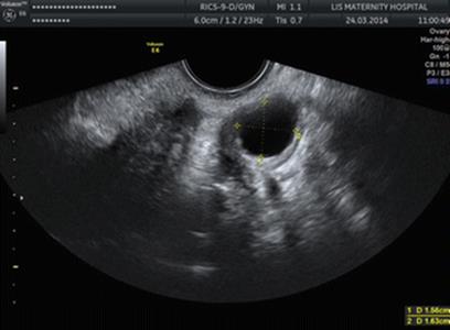
Fig. 20.2
Calculating follicular size. Two dimensional measuring is performed by measuring the greatest follicular diameter and averaging the two greatest diameters measured
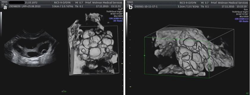
Fig. 20.3
(a, b) Follicle count and volume in a stimulated ovary with SonoAVC
20.4 Monitoring COS with Ultrasonography Only
Fertility cycles are monitored traditionally by both ultrasonography and measurements of oestradiol (E2) serum levels. Current evidence from meta-analysis of RCTs concludes that monitoring COS cycles with ultrasonography only is unlikely to substantially alter the chances of achieving a clinical pregnancy or affect the number of oocytes retrieved per cycle [25]. Moreover, the addition of E2 measurements to ultrasound monitoring of IVF cycles in normal responders seldom changes the timing of hCG administration or the risk of OHSS [26]. According to a Cochrane review, when monitoring IVF and ICSI (intra-cytoplasmic sperm injection) cycles similar pregnancy and live birth rates were achieved with combined ultrasound and E2 as with ultrasound alone. However, until a sufficient RCT to test different ways of monitoring OHSS (a rare but life-threatening complication) is done, cycle monitoring by ultrasound and serum oestradiol is recommended as a precautionary good practice point [27].
20.5 Embryo Transfer
Embryo transfer can be performed under ultrasound guidance or in a tactile manner. With the ultrasound guided transfer technique an abdominal ultrasound transducer is placed supra-pubically to allow clear visualization of the uterus. The transfer catheter loaded with the embryo(s) is gently guided through the cervical opening into the uterine cavity under observation. Once the ultrasound image confirms that the catheter is properly placed, the embryos are injected into the uterus, and the catheter is slowly withdrawn (Fig. 20.4). Several studies have examined the effect of the distance of blastocyst dislodgement, as determined by air bubble position, from the uterine fundus on pregnancy rates. Air bubbles within 10 mm from the fundus are associated with greater pregnancy rates as opposed to placement further away from the fundus [28]. While some believe that the optimal placement of the inner catheter tip is 1.5–2 cm from the fundal endometrial surface [29], others state that pregnancy rates do not differ as long as the catheter tip is placed in the middle to upper third of the uterus [30].

Fig. 20.4
Ultrasound guided embryo transfer
Ultrasound-guided transfer has many potential advantages, such as facilitating the placement of soft catheters, avoiding stimulation of the fundus and endometrial lining and blocking of the catheter tip with blood or mucus and placement of the catheter in cases of an elongated cervical canal. Also, when performed under ultrasound guidance a full urinary bladder is required, which straightens the cervical canal and facilitates access to the uterus [31].
A Cochrane meta-analysis of four trials concluded that the clinical pregnancy rate and ongoing pregnancy rates were significantly greater with ultrasound-guided embryo transfer as opposed to the ‘clinical feel’ approach. The rate of ectopic pregnancies was not affected by the method of transfer [32]. Other trials have found that the advantage of ultrasound-guided transfer is culture day dependent – exists if transfer is performed after 3 or 4 days of culture but not when performed for 5-day-old embryos [33].
20.6 Complications During COS: Sonographic Diagnosis and Treatment
Although generally safe, COS treatments are associated with complications that, albeit rare, can have significant morbidity. The rate of major complications is 0.7 % [34]. All physicians, and not only those performing COS treatments, should be aware of the increased frequency of potential complications and should keep them in mind when treating women undergoing fertility treatments. The role of ultrasonography is not only in the diagnosis of these conditions but may also aid in resolving them.
20.6.1 Follicle Rupture and Bleeding
Bleeding during oocyte aspiration is divided into minor and major episodes. Minor vaginal bleeding is reported to occur in 0.5–8.6 % of oocyte retrievals [35]. In most cases it subsides after brief local compression, and only rarely suturing is required.
According to a recent retrospective study of 973 cycles of COS, cyst rupture and intra-abdominal bleeding following oocyte retrieval are very rare complications, occurring in 0.1 % of the cases [34]. Similar rates were also reported previously [36]. It is caused by puncture of vessels, such as the ovarian capsule vessels, the sacral plexus or the iliac vessels. This life-threatening complication can be avoided by careful ultrasound visualization of the peripheral follicles in a cross-sectional view before puncture and by the use of colour Doppler if available [37] (Fig. 20.5).

Fig. 20.5
Color Doppler aids in visualization of blood vessels during ovum pick up
20.6.2 Pelvic Infections
The risk of infectious complications following oocyte retrieval is low (0.3–0.6 %), even without antibiotic prophylaxis. Therefore, the use of prophylactic antibiotics 30–60 min before retrieval or immediately after the procedure is controversial. Alternatively, some reserve it for women at increased risk of infection (those with a diagnosed endometrioma or a history of pelvic inflammatory disease) [35, 38]. Nearly half of the cases of infection present as tubo-ovarian abscesses, 1–6 weeks after oocyte retrieval [35] (Fig. 20.6). An ultrasound is key to the diagnosis of this important complication. A complex adnexal cystic lesion following trans-vaginal oocyte retrieval that is accompanied by persistent fever and leukocytosis should lead to an early presumptive diagnosis of tubo-ovarian abscess.

Fig. 20.6
Tubo-ovarian abscess with pyosalpinx
20.6.3 Adnexal Torsion
Torsion of the ovary is a serious complication of ovarian stimulation and should be considered in any patient with complaints of sudden abdominal pain accompanied by nausea, appearing during or after ovarian stimulation. Since ovarian cysts are the main risk factor for torsion, the rate is higher among women exhibiting OHSS. It is 11 times more common in ART pregnancies than in non-ART pregnancies [39]. While the absolute incidence of ovarian torsion is 0.8 % in all IVF cycles [36], it can reach 7.5 % in patients with OHSS [40]. The most consistent imaging finding is asymmetric enlargement of the twisted ovary [41], frequently due to an underlying mass. Obstruction of venous outflow in torsion also causes enlargement, as well as stromal oedema and stromal heterogeneity due to haemorrhage and oedema. Peripheral displacement of follicles due to oedema may also be noted, as well as free pelvic fluid. Doppler sonography can aid in the diagnosis and decrease the morbidity associated with this condition. However, studies have found normal Doppler findings in 45–61 % of torsion cases, so they should not be relied on too heavily. Lastly, a sensitive sign is the ‘whirlpool sign’, seen at both grayscale and colour Doppler US as coiled vessels representing a twisted vascular pedicle [42, 43]. In rare cases the fallopian tube can undergo torsion without involving the ipsilateral ovary. In these cases colour Doppler may not facilitate the diagnosis. Due to the limited diagnostic value of sonography in ovarian torsion, management decisions should still be based on clinical grounds.
Distinguishing ovarian torsion from mild OHSS can be challenging, both clinically and radiologically. Clinically, both entities may manifest with abdominal pain, nausea and vomiting. Radiologically, there is overlap in the grayscale US appearance of these conditions, since both may demonstrate ovarian enlargement and heterogeneous ovarian stroma [41]. Peripheral migration of follicles, a finding associated with torsion, may not be evident in the hyperstimulated ovary, due to the increase in follicles. Moreover, free pelvic fluid, a common finding in torsion, is also present in nearly all cases of OHSS. A possible diagnostic hint to differentiate between the two entities is the finding of cortical cysts separated by thickened parenchyma in the twisted ovary, as compared with ovarian follicles separated by thin walls in OHSS [44] (Fig. 20.7).

Fig. 20.7
Torsion of an enlarged (hyperstimulated) ovary. Thickened echogenic parenchyma separates the cortical cysts, distinguishing torsion from OHSS
20.6.4 Ectopic or Heterotopic Pregnancy
While the incidence of ectopic pregnancy in spontaneous gestations is approximately 2 % [45], its incidence following ART ranges from 2.1 to 8.6 % [46]. Many of the risk factors associated with ectopic pregnancies, such as increasing age, prior tubal pregnancy and history of salpingitis, are also characteristic of the ART population. In contrast to the discriminatory threshold of an intra-uterine pregnancy of a beta-hCG level of 1,500, there is no such threshold for the diagnosis of an extra-uterine pregnancy. Moreover, it is important to note that up to 35 % of ectopic pregnancies may not display any extra-uterine mass [47]. The sensitivity of trans-vaginal sonography can be improved by performing 3D imaging in asymptomatic patients [48]. In cases of high clinical suspicion, a normal ultrasound does not exclude the diagnosis. Most ectopic pregnancies resulting from ART are tubal, as in spontaneous pregnancies; however, the frequencies of cervical, interstitial and abdominal pregnancies, though remaining rare, are increased in ART treatments [49, 50]. Interstitial pregnancy is worth noting, due to its potential catastrophic outcome. This type of pregnancy occurs when an embryo implants in the intra-myometrial portion of the fallopian tube. Its sonographic appearance is of an eccentrically located gestational sac surrounded by a thin layer of myometrium measuring less than 5 mm [51] (Fig. 20.8). A specific additional finding is the ‘interstitial line sign’, an echogenic line that likely represents the interstitial portion of the fallopian tube [52]. These ectopic pregnancies tend to manifest later, and rupture may result in the rapid extravasation of blood to the abdominal cavity due to proximity to the uterine artery.

Fig. 20.8
Interstitial pregnancy. A yolk sac within a gestational sac, located in the interstitial portion of the fallopian tube
Heterotopic pregnancy refers to simultaneous intra-uterine and ectopic pregnancies and has an incidence of 1–3 % in ART patients [41]. In order to avoid a late diagnosis due to rupture, as is the case in 50 % of heterotopic pregnancies [53], it is critical to visualize both adnexa when performing an US in women undergoing ART, even when an intra-uterine pregnancy is diagnosed. The most common location of a heterotopic pregnancy is tubal, in 88 % of the cases [52] (Fig. 20.9). Since these pregnancies cannot be followed reliably by the beta-hCG level, the recommended treatment is surgical.

Fig. 20.9
(a, b) Heterotopic gestation (a) An ectopic gestational sac, yolk sac and embryo in the right fallopian tube along with (b) An intra-uterine pregnancy with a gestational sac and yolk sac
20.6.5 OHSS
Ovarian hyperstimulation syndrome is a potentially life-threatening iatrogenic complication of infertility treatments, which can occur in the setting of ovulation induction with exogenous gonadotropin therapy of IVF. Rarely it can also occur in its mild form after treatment with clomiphene citrate. Mild ovarian hyperstimulation probably occurs in 8–23 % of stimulated cycles, moderate forms in <1–7 % and severe forms in ~0.5 % of stimulated cycles [54, 55].
The most frequently used classification for the severity of OHSS, proposed by Golan, is based on the use of ultrasonography to quantify ovarian enlargement and ascites [54, 56] (Table 20.1). Here we will focus on the sonographic appearance of hyperstimulated ovaries. Bilateral symmetrically enlarged ovaries containing multiple variable-sized cystic lesions representing enlarged follicles or corpus luteum cysts, in the presence of ascites, is the hallmark image [41] (Fig. 20.10). The characteristic peripheral location of the follicles surrounding a central core of ovarian stroma has been described as creating a ‘wheel spoke’ appearance, with thin septa separating the cysts [57]. The cysts are usually anechoic but may be complicated by haemorrhage. Associated complications such as deep vein thrombosis or pleural effusion can also be diagnosed with ultrasonography. Follow-up imaging should demonstrate resolution of the ascites/effusion. Ascites drainage should be considered when there is dyspnoea or tense ascites and abdominal pain. It can be performed both vaginally and intra-abdominally, always under ultrasound guidance. Quick symptom relief is usually achieved along with improvement in venous return, cardiac output, diuresis, creatinine clearance and lung ventilation [58].
Table 20.1
Modified Golan Classification scheme for OHSS
|
Category |
Ovarian size (cm) |
Grade |
Signs and symptoms |
|
Mild |
<6 |
I |
Abdominal distention |
|
II |
Abdominal distention, nausea, vomiting, diarrhea |
||
|
Moderate |
6–12 |
III |
Abdominal distention, nausea, vomiting, diarrhea; ascites at US; weight gain |
|
Severe |
>12 |
IV |
Ascites/hydrothorax |
|
V |
Ascites/hydrothorax, hypovolemia, hemo-concentration, coagulation disorder, oliguria, shock |

Fig. 20.10
(a, b) OHSS. (a) enlarged ovary with multiple variable-sized cystic lesions and ascites; (b) ascitic fluid around liver associated with severe OHSS
Conclusions
The role of ultrasound in COS begins with the initial evaluation of the infertile patient through planning and executing the fertility treatment and culminates with treating potential complications. Ultrasound is commonly used to assess the process of folliculogenesis during ovulation induction and to follow the emergence and growth of a dominant follicle in natural cycles. During the early follicular phase ultrasound can assure adequate follicle recruitment, while during the late follicular phase it can assist in timing hCG injection and ovulation according to follicular size and endometrial thickness and velocimentry. In IVF cycles oocyte aspiration is performed under trans-vaginal ultrasound guidance, and in embryo transfer it is used to map the endometrial cavity and determine the optimal location for embryo placement. Complications, including extra-uterine pregnancy, adnexal torsion, OHSS and intra-abdominal bleeding or infection, can also be diagnosed with ultrasonography, while OHSS can sometimes be prevented with careful adjustments of gonadotropin regimens when ultrasound reveals excessive follicular recruitment. In conclusion, in ART trans-vaginal ultrasonography is indispensible, as it has revolutionized both the evaluation and treatment of infertility.
References
1.
Bonilla-Musoles F, Castillo JC, Caballero O, Pérez-Panades J, Bonilla Jr F, Dolz M, Osborne N. Predicting ovarian reserve and reproductive outcome using antimüllerian hormone (AMH) and antral follicle count (AFC) in patients with previous assisted reproduction technique (ART) failure. Clin Exp Obstet Gynecol. 2012;39(1):13–8.PubMed
2.
Hendriks DJ, Broekmans FJ, Bancsi LF, de Jong FH, Looman CW, Te Velde ER. Repeated clomiphene citrate challenge testing in the prediction of outcome in IVF: a comparison with basal markers for ovarian reserve. Hum Reprod. 2005;20(1):163–9.CrossRefPubMed
3.
Jayaprakasan K, Campbell B, Hopkisson J, Johnson I, Raine-Fenning N. A prospective, comparative analysis of anti-Müllerian hormone, inhibin-B, and three-dimensional ultrasound determinants of ovarian reserve in the prediction of poor response to COS. Fertil Steril. 2010;93(3):855–64.CrossRefPubMed
4.
Ng EH, Tang OS, Chan CC, Ho PC. Ovarian stromal blood flow in the prediction of ovarian response during in vitro fertilization treatment. Hum Reprod. 2005;20(11):3147–51.CrossRefPubMed
5.
Ng EH, Chan CC, Tang OS, Yeung WS, Ho PC. Factors affecting endometrial and subendometrial blood flow measured by three-dimensional power Doppler ultrasound during IVF treatment. Hum Reprod. 2006;21(4):1062–9.CrossRefPubMed
6.
Jayaprakasan K, Al-Hasie H, Jayaprakasan R, Campbell B, Hopkisson J, Johnson I, Raine-Fenning N. The three-dimensional ultrasonographic ovarian vascularity of women developing poor ovarian response during assisted reproduction treatment and its predictive value. Fertil Steril. 2009;92(6):1862–9.CrossRefPubMed
7.
Firouzabadi RD, Sekhavat L, Javedani M. The effect of ovarian cyst aspiration on IVF treatment with GnRH. Arch Gynecol Obstet. 2010;281(3):545–9.CrossRefPubMed
8.
Fiszbajn GE, Lipowicz RG, Elberger L, Grabia A, Papier SD, Brugo Olmedo SP, Chillik CF. Conservative management versus aspiration of functional ovarian cysts before ovarian stimulation for assisted reproduction. J Assist Reprod Genet. 2000;17(5):260–3.PubMedCentralCrossRefPubMed
9.
Silverberg KM, Klein NA, Burns WN, Schenken RS, Olive DL. Consecutive versus alternating cycles of ovarian stimulation using human menopausal gonadotrophins. Hum Reprod. 1992;7(7):940–4.PubMed
10.
Akin JW, Shepard MK. The effects of baseline ovarian cysts on cycle fecundity in controlled ovarian hyperstimulation. Fertil Steril. 1993;59(2):453–5.PubMed
11.
Jeon YE, Jung JA, Kim HY, Seo SK, Cho S, Choi YS, Lee BS. Predictive factors for pregnancy during the first four intrauterine insemination cycles using gonadotropin. Gynecol Endocrinol. 2013;29(9):834–8.CrossRefPubMed
12.
Check JH. The importance of sonographic endometrial parameters in influencing success following embryo transfer in the modern era and therapeutic options–part 1: the importance of late proliferative phase endometrial thickness. Clin Exp Obstet Gynecol. 2011;38(3):197–200.PubMed
13.
Yoeli R, Ashkenazi J, Orvieto R, Shelef M, Kaplan B, Bar-Hava I. Significance of increased endometrial thickness in assisted reproduction technology treatments. J Assist Reprod Genet. 2004;21(8):285–9.PubMedCentralCrossRefPubMed
14.
Homburg R. Clomiphene citrate—end of an era? A mini-review. Hum Reprod. 2005;20(8):2043–51.CrossRefPubMed
15.
Dickey RP, Olar TT, Taylor SN, Curole DN, Matulich EM. Relationship of endometrial thickness and pattern to fecundity in ovulation induction cycles: effect of clomiphene citrate alone and with human menopausal gonadotropin. Fertil Steril. 1993;59(4):756–60.PubMed
16.
Asante A, Coddington CC, Schenck L, Stewart EA. Thin endometrial stripe does not affect likelihood of achieving pregnancy in clomiphene citrate/intrauterine insemination cycles. Fertil Steril. 2013;100(6):1610–4.CrossRefPubMed
17.
Kolibianakis EM, Zikopoulos KA, Fatemi HM, Osmanagaoglu K, Evenpoel J, Van Steirteghem A, Devroey P. Endometrial thickness cannot predict ongoing pregnancy achievement in cycles stimulated with clomiphenecitrate for intrauterine insemination. Reprod Biomed Online. 2004;8(1):115–8.CrossRefPubMed
18.
Ng EH, Chan CC, Tang OS, Yeung WS, Ho PC. Endometrial and subendometrial vascularity is higher in pregnant patients with livebirth following ART than in those who suffer a miscarriage. Hum Reprod. 2007;22(4):1134–41.CrossRefPubMed
19.
Tohma H, Hasegawa I, Sekizuka N, Tanaka K. Uterine blood flow. Assessment in an intrauterine insemination program for unexplained infertility. J Reprod Med. 1997;42(8):463–6.PubMed
20.
Ardaens Y, Gougeon A, Lefebvre C, Thomas P, Leroy M, Leroy JL, Dewailly D. Contribution of ovarian and uterine color Doppler in medically assisted reproduction techniques (ART). Gynecol Obstet Fertil. 2002;30(9):663–72.CrossRefPubMed
21.
Silverberg KM, Olive DL, Burns WN, Johnson JV, Groff TR, Schenken RS. Follicular size at the time of human chorionic gonadotropin administration predicts ovulation outcome in human menopausal gonadotropin-stimulated cycles. Fertil Steril. 1991;56(2):296–300.PubMed
22.
Sakhel K, Khedr M, Schwark S, Ashraf M, Fakih MH, Abuzeid M. Comparison of urinary and recombinant human chorionic gonadotropin during ovulation induction in intrauterine insemination cycles: a prospective randomized clinical trial. Fertil Steril. 2007;87(6):1357–62.CrossRefPubMed
23.
Cantineau AE, Janssen MJ, Cohlen BJ. Synchronised approach for intrauterine insemination in subfertile couples. Cochrane Database Syst Rev. 2010;(4):CD006942.
24.
Ata B, Tulandi T. Ultrasound automated volume calculation in reproduction and in pregnancy. Fertil Steril. 2011;95(7):2163–70.CrossRefPubMed
25.
Martins WP, Vieira CV, Teixeira DM, Barbosa MA, Dassunção LA, Nastri CO. Ultrasound for monitoring COS: a systematic review and meta-analysis of randomized controlled trials. Ultrasound Obstet Gynecol. 2014;43(1):25–33.CrossRefPubMed
26.
Lass A. Monitoring of in vitro fertilization-embryo transfer cycles by ultrasound versus by ultrasound and hormonal levels: a prospective, multicenter, randomized study. Fertil Steril. 2003;80(1):80–5.CrossRefPubMed
27.
Kwan I, Bhattacharya S, McNeil A, van Rumste MM. Monitoring of stimulated cycles in assisted reproduction (IVF and ICSI). Cochrane Database Syst Rev. 2008;(2):CD005289.
28.
Friedman BE, Lathi RB, Henne MB, Fisher SL, Milki AA. The effect of air bubble position after blastocyst transfer on pregnancy rates in IVF cycles. Fertil Steril. 2011;95(3):944–7.CrossRefPubMed
29.
Cenksoy PO, Fıcıcıoglu C, Yesiladali M, Akcin OA, Kaspar C. The importance of the length of uterine cavity, the position of the tip of the inner catheter and the distance between the fundal endometrial surface and the air bubbles as determinants of the pregnancy rate in IVF cycles. Eur J Obstet Gynecol Reprod Biol. 2014;172:46–50.CrossRefPubMed
30.
Kovacs P, Sajgo A, Rarosi F, Kaali SG. Does it really matter how far from the fundus embryos are transferred? Eur J Obstet Gynecol Reprod Biol. 2012;162(1):62–6.CrossRefPubMed
31.
Sundström P, Wramsby H, Persson PH, Liedholm P. Filled bladder simplifies human embryo transfer. Br J Obstet Gynaecol. 1984;91(5):506–7.CrossRefPubMed
32.
Sallam HN, Agameya AF, Rahman AF, Ezzeldin F, Sallam AN. Impact of technical difficulties, choice of catheter, and the presence of blood on the success of embryo transfer–experience from a single provider. J Assist Reprod Genet. 2003;20(4):135–42.PubMedCentralCrossRefPubMed
33.
Prapas Y, Prapas N, Hatziparasidou A, Vanderzwalmen P, Nijs M, Prapa S, Vlassis G. Ultrasound-guided embryo transfer maximizes the IVF results on day 3 and day 4 embryo transfer but has no impact on day 5. Hum Reprod. 2001;16(9):1904–8.CrossRefPubMed
34.
Maxwell KN, Cholst IN, Rosenwaks Z. The incidence of both serious and minor complications in young women undergoing oocyte donation. Fertil Steril. 2008;90(6):2165–71.CrossRefPubMed
35.
Bennett SJ, Waterstone JJ, Cheng WC, Parsons J. Complications of transvaginal ultrasound-directed follicle aspiration: a review of 2670 consecutive procedures. J Assist Reprod Genet. 1993;10(1):72–7.CrossRefPubMed
36.
Roest J, Mous HV, Zeilmaker GH, Verhoeff A. The incidence of major clinical complications in a Dutch transport IVF programme. Hum Reprod Update. 1996;2(4):345–53.CrossRefPubMed
37.
Serour GI, Aboulghar M, Mansour R, Sattar MA, Amin Y, Aboulghar H. Complications of medically assisted conception in 3,500 cycles. Fertil Steril. 1998;70(4):638–42.CrossRefPubMed
38.
Dicker D, Ashkenazi J, Feldberg D, Levy T, Dekel A, Ben-Rafael Z. Severe abdominal complications after transvaginal ultrasonographically guided retrieval of oocytes for in vitro fertilization and embryo transfer. Fertil Steril. 1993;59(6):1313–5.PubMed
39.
Gelbaya TA. Short and long-term risks to women who conceive through in vitro fertilization. Hum Fertil (Camb). 2010;13(1):19–27.CrossRef
40.
Mashiach S, Bider D, Moran O, Goldenberg M, Ben-Rafael Z. Adnexal torsion of hyperstimulated ovaries in pregnancies after gonadotropin therapy. Fertil Steril. 1990;53(1):76–80.PubMed
41.
Baron KT, Babagbemi KT, Arleo EK, Asrani AV, Troiano RN. Emergent complications of assisted reproduction: expecting the unexpected. Radiographics. 2013;33(1):229–44.CrossRefPubMed
42.
Lourenco AP, Swenson D, Tubbs RJ, Lazarus E. Ovarian and tubal torsion: imaging findings on US, CT, and MRI. Emerg Radiol. 2014;21(2):179–87.CrossRefPubMed
43.
Valsky DV, Esh-Broder E, Cohen SM, Lipschuetz M, Yagel S. Added value of the gray-scale whirlpool sign in the diagnosis of adnexal torsion. Ultrasound Obstet Gynecol. 2010;36(5):630–4.CrossRefPubMed
44.
Graif M, Shalev J, Strauss S, Engelberg S, Mashiach S, Itzchak Y. Torsion of the ovary: sonographic features. AJR Am J Roentgenol. 1984;143(6):1331–4.CrossRefPubMed
45.
Centers for Disease Control and Prevention (CDC). Ectopic pregnancy–United States, 1990–1992. MMWR Morb Mortal Wkly Rep. 1995;44(3):46–8.
46.
Clayton HB, Schieve LA, Peterson HB, Jamieson DJ, Reynolds MA, Wright VC. Ectopic pregnancy risk with assisted reproductive technology procedures. Obstet Gynecol. 2006;107(3):595–604.CrossRefPubMed
47.
Levine D. Ectopic pregnancy. Radiology. 2007;245(2):385–97.CrossRefPubMed
48.
Harika G, Gabriel R, Carre-Pigeon F, Alemany L, Quereux C, Wahl P. Primary application of three-dimensional ultrasonography to early diagnosis of ectopic pregnancy. Eur J Obstet Gynecol Reprod Biol. 1995;60(2):117–20.CrossRefPubMed
49.
Malinowski A, Bates SK. Semantics and pitfalls in the diagnosis of cornual/interstitial pregnancy. Fertil Steril. 2006;86(6):1764.e11–4.CrossRef
50.
Gun M, Mavrogiorgis M. Cervical ectopic pregnancy: a case report and literature review. Ultrasound Obstet Gynecol. 2002;19(3):297–301.CrossRefPubMed
51.
Lin EP, Bhatt S, Dogra VS. Diagnostic clues to ectopic pregnancy. Radiographics. 2008;28(6):1661–71.CrossRefPubMed
52.
Ackerman TE, Levi CS, Dashefsky SM, Holt SC, Lindsay DJ. Interstitial line: sonographic finding in interstitial (cornual) ectopic pregnancy. Radiology. 1993;189(1):83–7.CrossRefPubMed
53.
Rojansky N, Schenker JG. Heterotopic pregnancy and assisted reproduction–an update. J Assist Reprod Genet. 1996;13(7):594–601. Review.CrossRefPubMed
54.
Golan A, Ron-el R, Herman A, Soffer Y, Weinraub Z, Caspi E. Ovarian hyperstimulation syndrome: an update review. Obstet Gynecol Surv. 1989;44(6):430–40.CrossRefPubMed
55.
Navot D, Bergh PA, Laufer N. Ovarian hyperstimulation syndrome in novel reproductive technologies: prevention and treatment. Fertil Steril. 1992;58(2):249–61.PubMed
56.
Zivi E, Simon A, Laufer N. Ovarian hyperstimulation syndrome: definition, incidence, and classification. Semin Reprod Med. 2010;28(6):441–7.CrossRefPubMed
57.
Jung BG, Kim H. Severe spontaneous ovarian hyperstimulation syndrome with MR findings. J Comput Assist Tomogr. 2001;25(2):215–7.CrossRefPubMed
58.
Delvigne A, Rozenberg S. Preventive attitude of physicians to avoid OHSS in IVF patients. Hum Reprod. 2001;16(12):2491–5.CrossRefPubMed