
Narendra Malhotra1 , Jaideep Malhotra2, Diksha Goswami Sharma2, Shilpi Gupta3, Neharika Malhotra Bora4, Shally Gupta2 and Garima Sharma5
(1)
Department of Obstetrics and Gynaecology, Global Rainbow Healthcare, 84, Mahatma Gandhi Road, Agra, Uttar Pradesh, 282010, India
(2)
IVF & Reproductive Medicine, Department of Obstetrics and Gynecology, Rainbow IVF, Global Rainbow Healthcare, Agra, Uttar Pradesh, India
(3)
IVF & Reproductive Medicine, Department of Obstetrics and Gynecology, Indian College of Obstetricians and Gynaecologists Fellow, Rainbow IVF, Global Rainbow Healthcare, Agra, Uttar Pradesh, India
(4)
Department of Obstetrics and Gynaecology, Bhartiya Vidyapeeth University, Global Rainbow Healthcare, Agra, Uttar Pradesh, India
(5)
Indian College of Obstetricians and Gynaecologists Fellow, Rainbow IVF, Global Rainbow Healthcare, Agra, Uttar Pradesh, India
Narendra Malhotra
Email: n.malhotra@rainbowhospital.org
Abstract
The couple needs to be completely evaluated before stimulation for IVF in order to assess the expected response, check fitness for pregnancy and identify correctable factors for a successful outcome. Main part of this evaluation is by various ovarian reserve tests which give a good measure of the number of expected oocytes and help to individualize the cycle. According to current evidence, antral follicle count and anti-Mullerian hormone are good markers of ovarian response and can help in defining strategy for COH.
Keywords
Ovarian reserve testsAntral follicle countAnti-mullerian hormonePelvic ultrasoundCounseling
2.1 Definition
Controlled ovarian hyperstimulation (COH) is an integral part of assisted reproductive technologies. It traditionally involves stimulation of the ovaries with gonadotropins in combination with GnRH analogues for ovarian suppression to induce development of multiple follicles of the ovaries. Aim of COH is to achieve sufficient number of mature oocytes of good quality enabling selection of two or three good-quality embryos, at the same time avoiding undesirable outcomes like cycle cancellation or hyperstimulation. It is very important to make a complete evaluation of the female before subjecting her to COH.
2.2 Introduction
A detailed evaluation is of high clinical relevance prior to COH in order to assess the expected ovarian response, identify key factors to optimize the cycle outcome as well as do a complete prepregnancy evaluation.
Since there can be considerable variability in an individual response to stimulation with gonadotropins, a proper evaluation helps to tailor the cycle to fit the individual patient. That will enable clinicians to individualize ovulation induction and ovarian stimulation treatment, thereby minimizing complications and the risk of treatment failure while maximizing the chance of ongoing pregnancy.
It also helps in counselling women especially of the possible negative IVF outcomes such as cancellation of cycle, prolonged treatment, increased treatment burden and reduced pregnancy rates and perhaps reduces the number of dropouts particularly among women with an expected poor outcome. At the other end of the spectrum, identifying the expected high responders helps to reduce the burden of OHSS.
2.3 Ovarian Reserve Tests
Evaluation prior to COH is mainly by these tests. Ovarian reserve is defined as the number and quality of the follicles left in the ovary at any given time. The various tests to assess the ovarian reserve usually have good predictive value for the number of oocytes retrieved but have limited value in prediction of ongoing pregnancy.
Before a woman is subjected to any ovarian reserve testing, a complete evaluation of the couple with respect to general health and fitness must be done. This evaluation can be tabulated in simple chart form, a sample of which can be seen in Fig. 2.1. Figure 2.2 shows a sample investigation form for the infertile couple. Tests for evaluation of ovarian reserve include - antral follicle count, FSH, AMH (anti mullerian hormone), and clomiphene challege test.

Fig. 2.1
Pre IVF evaluation and investigation chart
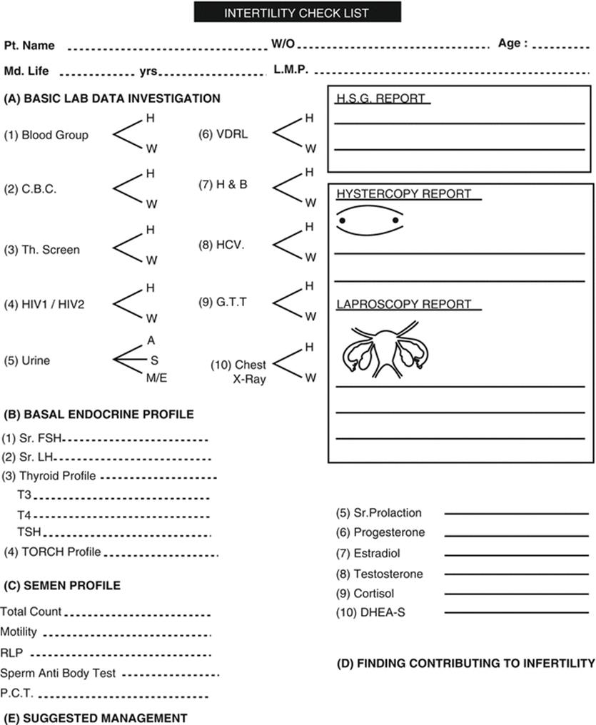
Fig. 2.2
Infertility workup chart
2.3.1 Antral Follicle Count
Antral follicle count (AFC) is the number of antral follicles present in the ovaries and detectable by transvaginal ultrasound scan on day 2 or 3 of the period. It is commonly estimated by counting all identifiable antral follicles of 2–10 mm in diameter in both the ovaries [1]. A major technical improvement in ultrasound has been the development of three-dimensional (3D) automated follicular tracking, which can substantially decrease both intra- and inter-observer variability [2]. Antral follicle count is a good marker to predict the number of oocytes retrieved with false-positive rate of 15–20 % and can identify the expected poor, normal and high responders (Fig. 2.3).
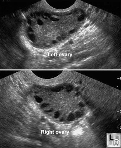
Fig. 2.3
TVS showing ovary with high AFC
However, there is considerable variability in agreed AFC cut-off levels used for predicting poor response. It may vary between AFC of 3 [3] and 12 [4]. A possible reason for such variability is the absence of a standardized measurement of antral follicles with different studies measuring different follicle populations 2–5, 2–9 or 5–9 mm. Most frequently reported cut-off values of AFC for prediction of poor response ranged between 5 and 7 [5]. In order to identify high responders [6], reportedly an AFC value of 16 had apparent sensitivity of 89 % and a specificity of 92 %. Other smaller prospective studies found values ranging between 9 and 14 as the most appropriate cut-off to identify hyper-responders [7, 8].
An easy-to-use algorithm to calculate the gonadotropin dose based on AFC has recently been published [9]. The nomogram calculated the gonadotropin dose based on the age of the woman, Day 3 serum FSH level and AFC. For example, in a woman aged 30 years, with a Day 3 FSH of 4 IU/l and an AFC of 16, the most appropriate gonadotropin dose is 150 IU daily.
2.3.2 AMH (Anti-Mullerian Hormone)
AMH is a dimeric glycoprotein and a member of the transforming growth factor b (TGF-b) family of growth and differentiation factors. AMH is produced by granulosa cells of small growing follicles, is gonadotropin independent and remains relatively consistent in between and within the menstrual cycle. It has been shown to have inhibitory effect on primordial-to-primary follicle transition. AMH also reduces follicle sensitivity to FSH in vivo, and in vitro AMH inhibits FSH-induced pre-antral follicle growth [10]. Thus, there is clear evidence that AMH is involved in the regulation of follicle growth initiation and the threshold for FSH sensitivity. The intrafollicular concentrations of AMH in normal human antral follicles show a gradual reduction as the diameter of the follicle increases, and a sharp decline is observed around 8 mm [11]. The rapid decline in AMH expression corresponds with the selection of follicles for dominance.
Anti-Mullerian hormone was assayed previously using primarily two different assay kits that have now been replaced by a newer assay. Evaluation of AMH levels prior to COS has several clinical utilities. There is substantial evidence in literature that AMH is superior to female age in assessing the quantitative aspects of the ovarian reserve, but its value is much more limited in the prediction of ongoing pregnancy.
Circulating anti-Müllerian hormone (AMH) can predict excessive as well as poor response to ovarian stimulation. A linear relationship exists between AMH and oocyte yield. At one extreme of the response, it helps to identify women at risk of ovarian hyperstimulation syndrome (OHSS) [12]. According to NICE guidelines of in vitro fertilization, an anti-Müllerian hormone level less than or equal to 5.4 pmol/l (0.8 ng/mL) predicts a low response to ovarian hyperstimulation, while a level greater than or equal to 25.0 pmol/l (3.6 ng/mL) predicts a high response [13]. Bologna’s criteria for defining poor responders suggest AMH cut-off of 0.5–1.1 ng/ml [14]. But single value of low AMH especially in young women should not be used to deny treatment as even women with AMH concentrations at the limit of assay sensitivity have a significant chance of conception through IVF. AMH serves as a valuable tool in counselling the patient and may set the patient’s expectations appropriately particularly at the bottom end of the spectrum where only a few oocytes may be retrieved. Nelson et al. have suggested an AMH-based strategy for deciding the protocol and gonadotropin dose for stimulation [15]. Tailoring the dosage of gonadotropin administration to AMH level has been shown to reduce the incidence of excessive response and cancelled cycles.
AMH is also useful in assessing the risk of ovarian damage secondary to chemotherapy, radiotherapy and ovarian surgery. Post-treatment AMH therefore can identify young girls and women receiving cancer therapy likely to have premature menopause or require pubertal induction, distinct from others who may be able to be reassured as to the likelihood of satisfactory ovarian function later in life.
AMH has been found to have sensitivity 44–97 % and specificity 41–100 % in predicting poor response to stimulation. Most of the studies have determined that cut-off level AMH >3.6 ng/ml has sensitivity and specificity of 82 % and 76 %, respectively, for prediction of OHSS (Table 2.1)
Table 2.1
Normal AMH values
|
Ovarian fertility potential |
ng/mL |
|
Optimal fertility |
4.0–6.8 |
|
Satisfactory fertility |
2.2–4.0 |
|
Low fertility |
0.3–2.2 |
|
Very low/undetectable |
<0.3 |
|
High level |
>6.8 |
2.3.3 Basal FSH
Basal FSH levels increase on day 2, 3 or 4 of the menstrual cycle with advancing age. However, assays of FSH have significant inter- and intra-cycle variability which limit their utility. Despite its limitations, FSH is commonly used as a measure of ovarian reserve, and high values have been associated with but not necessarily predict poor response to stimulation and failure to concieve. The sensitivity of FSH in identifying poor responders varies from 10 to 80 % and decreases with increasing cut-offs. Recent study employing efficiency curves demonstrated 100 % specificity for failure to achieve live birth at levels more than 18 IU/l. A single elevated FSH level in women <40 years may not predict poor responders or failure to concieve. But clinical utility lies in being fairly certain that women with abnormally elevated FSH will have diminished reserve [16] (Table 2.2).
Table 2.2
Basal FSH levels and clinical implications
|
FSH levels IU/L |
Clinical implication |
|
<9 |
Reassure |
|
9–10 |
Suboptimal |
|
10–12 |
Decreased ovarian reserve |
|
12–17 |
Markedly reduced ovarian reserve |
|
17–20 |
Poor prognosis |
|
>20 |
No pregnancy |
2.3.4 Female Age
Advanced maternal age causes decreased success rates in ovarian hyperstimulation [17]. However, a younger woman with a raised FSH would respond better than an older woman with raised FSH. Age is one of the most important determinants of ovarian response.
2.3.5 Other Ovarian Reserve Tests
2.3.5.1 Clomiphene Citrate Challenge Test
The test involves measuring baseline FSH after administering clomiphene citrate 100 mg day 5–9 of cycle typically on day 3 and 10. An elevated FSH level after clomiphene stimulation therefore suggests diminished reserve. Cycle day 10 FSH levels have a greater sensitivity but lower specificity compared to cycle day 3 FSH levels [16].
2.3.5.2 Endocrine Challenge Test (Gonadotropin-Releasing Hormone Agonist Stimulation Test)
The purpose of GAST is to evaluate changes in E2 on cycle days 2 and 3 following administration of GnRH agonist (leuprolide acetate).
2.3.5.3 Exogenous FSH Ovarian Reserve Test (EFFORT)
Originally, the test was developed to improve the predictive value of day 3 FSH values in controlled ovarian hyperstimulation for IVF. The E2 level is recorded on cycle day 3 before the administration of 300 IU of purified FSH. Another level of E2 is done 24 h after giving FSH. It was postulated that the dynamic increase in E2 of more than 30 pg/ml would be predictive of a good response in a subsequent IVF cycle.
These dynamic tests for assessing ovarian reserve are considered as too laborious for screening purposes.
2.4 History
All couples presenting with infertility should have had a detailed history and physical examination done, which should be reviewed prior to COS. This should usually include
· Menstrual history: especially in regard to cycle length and duration which might suggest anovulation, PCOS or diminished reserve
· Obstetric history: previous pregnancy outcome
· Past surgeries (procedures, indications and outcomes), serious illnesses or history of pelvic inflammatory disease or sexually transmitted infections
· Any abnormal pap smears and treatment taken
· Symptoms suggestive of other endocrine abnormalities which might be contributing to infertility
· Any medical disease contraindicating pregnancy
· Social history to evaluate for any environmental exposures or social habits (such as smoking, drinking alcohol, drug usage or extreme exercise)
· Family history of birth defects, mental retardation, early menopause or reproductive compromise
· Detailed history of male partner regarding occupational exposures, medical illness, genital surgery or infections, smoking, sexual dysfunction or difficulty in giving semen sample
2.5 Examination
The physical examination of the lady is performed to evaluate the pelvic organs and assess potential hormonal problems. It should include any thyromegaly, breast examination, signs of androgen excess, vaginal or cervical abnormality, secretions or discharge, pelvic or abdominal tenderness enlargement or masses, adenexal masses or cul-de-sac nodularity.
2.5.1 Body Mass Index (BMI)
It is to identify obese or very lean individuals. Obesity is associated with higher miscarriage rates and a higher prevalence of neonatal complications, congenital anomalies and pregnancy-associated complications. Also BMI helps in deciding the starting dose of gonadotropins in association with other variables as suggested by CONSORT study [18]. In assisted reproduction, however, there are conflicting reports on the effect of obesity on oocyte quality, embryo development, lower number of mature oocytes, lower implantation and pregnancy rates. Total amount of gonadotropins used was significantly higher in patients with a BMI ≥25 kg/m [2], when compared to those with a normal BMI.
2.5.2 Male
A complete physical examination should be done including height, gynaecomastia, inguinal area, penile length, curvature and urethral meatus, testes volume and location, varicocele, epdidymal nodularity or tenderness, presence of vas deferens especially in cases of azoospermia and other male factor of infertility.
2.6 Previous Cycle Details
Details of hyperstimulation if done previously must be reviewed including type of protocol, gonadotropin dosage, days of stimulation, number of mature oocytes obtained, fertilization and cleavage rates, details of embryo transfer. This helps in planning of present cycle as well as to identify any anticipated problem.
2.7 Pelvic Ultrasound
A good baseline pelvic scan can provide invaluable information prior to starting COH and can be combined with saline hysterogram SHG examination as discussed earlier.
It helps to determine if there are any problems in the uterine musculature (fibroids or adenomyosis) (Fig. 2.4) or cavity (uterine malformations, adhesions, polyps).
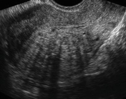
Fig. 2.4
Sagittal view of uterus with adenomyosis
Its good clinical practice to scan the endometrium in the luteal phase of preceeding cycle. A thick homogenous hyperechoic endometrium rules out presence of luteal phase defects. With addition of colour Doppler studies of uterine artery and power angio, the spiral artery and endometrial vascularization can be evaluated (Figs. 2.5 and 2.6).

Fig. 2.5
(a, b) Power Doppler showing well-vascularized endometrium

Fig. 2.6
Sagittal view of uterus showing secretory endometrium
Ovaries must be assessed to rule out any pathology (endometrioma, ovarian tumours) and check for accessibility during oocyte retrieval.
Measuring the volume of ovaries and antral follicle count as described earlier helps decide protocol and dosage for COH.
2.8 Serological Tests
These include HIV, hepatitis B and C, VDRL titre, blood grouping and thalassemia screen of both partners.
2.8.1 HIV
Although the risk of exposure to this virus is very low, it is good clinical practice to check the HIV status of the couple. The concern of a pregnancy in a female who is infected with the virus is that pregnancy increases the death rate dramatically in an HIV-positive woman. The second is the risk of transmitting the HIV virus to a child during childbirth.
2.9 Evaluation of Uterine Cavity
A wide range of endometrial and uterine cavity abnormalities like polyps, fibroids, intrauterine adhesions, and uterine anomalies are reported in women with subfertility, which can affect the implantation rates in an IVF cycle or cause spontaneous abortions. Also, abnormal uterine findings are reported in as many as 50 % of women with recurrent implantation failure [19]. Traditionally, HSG is used to evaluate the uterine cavity. Endometrial polyps or fibroids are shown as filling defects or uterine wall irregularities. HSG can also show intrauterine adhesions and congenital anomalies as it enables clinicians to visualize the general configuration of the cavity. Compared with hysteroscopy, HSG has a high sensitivity (60–98 %) but a low specificity (15–80 %) in detecting uterine abnormalities and is, therefore, associated with relatively high false-positive and false-negative rates [20–22].
The use of TVS in conjunction with saline infusion known as saline hysterography (SHG) or saline infusion sonography improves the delineation of the uterine cavity and is an excellent alternative to hysteroscopy for screening the uterine cavity. It has been reported to have 87.5 % sensitivity, 100 % specificity, 100 % positive predictive value and 91.6 % negative predictive value for the detection of any cavity abnormality as compared with hysteroscopy [23]. Advantages of SHG are that it can be easily performed in the office setting, is non invasive, is well tolerated and can be done as an adjunct to baseline pelvic scan prior to stimulating every IVF patient (Fig. 2.7).
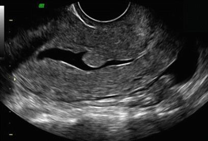
Fig. 2.7
Sonohysterogram showing endometrial polyp
However, hysteroscopy is the gold standard for the investigation of uterine cavity, particularly when pathology is suspected. It permits direct visualization of the uterine cavity, revealing the nature, location, shape, size and vascular pattern of any uterine cavity abnormalities, such as polyps, submucosal fibroids, differences in endometrial thickness and adhesions. It also allows a directed biopsy and therapeutic intervention for the treatment of any pathology. Systematic review by Bosteel et al. [24] found hysteroscopy in the cycle preceding a subsequent IVF attempt nearly doubles the pregnancy rate in patients with at least two failed IVF attempts compared with starting IVF immediately. A study by Makrakis et al. [25] to estimate the effect of hysteroscopy on achieving a pregnancy in women with a history of two implantation failures following IVF 37 % had abnormal findings, 22 % of which were unsuspected, and subsequent IVF treatment showed significantly increased clinical and ongoing pregnancy rates.
2.10 Evaluation of Adnexal Structure: Endometrioma, Hydrosalpinx
2.10.1 Ovarian Cyst
Assessment of any ovarian cyst on day 2 must be made. Large cysts are aspirated before stimulation is started. Smaller ones can be ignored as long as they are not hormone producing. Estradiol and progesterone are raised in case they are hormone producing.
2.10.2 Endometrioma
Endometrioma, if less than 3 cm, can be ignored. Larger endometriomas may cause difficulty in monitoring or oocyte retrieval and hence require removal. Either a surgical removal of cyst wall is done or fulgration may be done if removal is not possible. Medical treatment is not recommended.
2.10.3 Hydrosalpinx
Ultrasound-detected hydrsalpinx is a significant finding. It is recommended that the hydrosalpinx is removed before a patient is taken up for IVF as it impairs implantation. ASRM states that the live birth rate achieved with IVF among women with hydrosalpinges is approximately one half that observed in women without hydrosalpinges. In women with hydrosalpinges, preliminary laparoscopic salpingectomy or proximal tubal occlusion improves subsequent pregnancy and live birth rates achieved with IVF. For every six women with hydrosalpinges, one more ongoing pregnancy will be achieved if salpingectomy or tubal occlusion is performed before IVF [26].
2.11 Male Evaluation
One should not forget the male partner because male factor can contribute up to 50 % to the problem of infertility. Apart from complete history and physical examination as discussed earlier, a complete semen analysis must be performed for the total sperm concentration, motility and morphology (Table 2.3) [27]. Vitality testing should be done if motility is less than 40 %. Some centres assess the teratozoospermic index and sperm deformity index in cases of male infertility. In all cases of severe oligoasthenozoospermia and non-obstructive azoospermia, additional hormonal assay (FSH, LH, testosterone) and karyotyping should be performed. Scrotal ultrasound is indicated in case of atrophic testis, ectopic testis and enlarged testis to rule out neoplasm and to confirm varicocele or epididymal abnormalities. Transrectal ultrasound (TRUS) is an invaluable tool to visualize the distal genital abnormalities like vasal agenesis, absence of seminal vesicle, ejaculatory duct cyst or dilatation. In order to distinguish between obstructive and non-obstructive azoospermia, diagnostic testicular biopsy is indicated and must be combined with cryopreservation. Sperm DNA fragmentation can be assessed by various methods like sperm chromatin structure assay (SCSA), COMET and TUNEL in certain groups of patients.
Table 2.3
World Health Organization semen parameter (2010)
|
Parameter |
WHO 2010 |
|
Volume |
1.5 ml |
|
pH |
>7.2 |
|
Viscosity |
<3 Scale 1–4 |
|
Concentration |
15 million/ml |
|
Total sperm number |
>39 million |
|
Total motility (PR + NP)% |
40 % |
|
Progressive motility |
32 % |
|
Normal forms |
4 % |
|
Normal morphology WBC |
<1 million/ml |
|
MAR test |
<50 % |
|
Immunobead test |
<50 % motile sperms with boundbeads |
|
Vitality % |
>58 % |
|
Seminal zinc (μmol/ejaculate) |
≥2.4 |
|
Seminal fructose (μmol/ejaculate) |
>13 |
From WHO [27] WHO laboratory Manual for examination of human semen 2010
2.12 Counselling
Infertility and its treatment can be a major source of stress for the couple and the family. This stress is usually intensified during treatment, especially throughout an ART cycle. While this is not a mandatory requirement, couples should be encouraged to meet with a counsellor prior to beginning treatment, as well as during and after a cycle. This helps them in pschychological adjustment, reduce stress levels and ultimately contribute towards better pregnancy rates as well as decrease the dropout rates associated with IVF.
Conclusions
As women have a fixed ovarian reserve, which diminishes with age, so complete evaluation of male, female and the couple is mandatory before the start of any controlled ovarian hyperstimulation in order to ensure successful outcome.
References
1.
Broekmans FJ, de Ziegler D, Howles CM, Gougeon A, Trew G, Olivennes F. The antral follicle count: practical recommendations for better standardization. Fertil Steril. 2010;94(3):1044–51.CrossRefPubMed
2.
Deb S, Campbell BK, Clewes JS, Pincott-Allen C, Raine-Fenning NJ. Intracycle variation in number of antral follicles stratified by size and in endocrine markers of ovarian reserve in women with normal ovulatory menstrual cycles. Ultrasound Obstet Gynecol. 2013;41(2):216–22.CrossRefPubMed
3.
Chang MY, Chiang CH, Hsieh TT, Soong YK, Hsu KH. Use of the antral follicle count to predict the outcome of assisted reproductive technologies. Fertil Steril. 1998;69(3):505–10.CrossRefPubMed
4.
Melo MA, Garrido N, Alvarez C, Bellver J, Meseguer M, Pellicer A, Remohí J. Antral follicle count (AFC) can be used in the prediction of ovarian response but cannot predict the oocyte/embryo quality or the in vitro fertilization outcome in an egg donation program. Fertil Steril. 2009;91(1):148–56.CrossRefPubMed
5.
Jayaprakasan K, Campbell B, Hopkisson J, Johnson I, Raine-Fenning N. A prospective, comparative analysis of anti-Müllerian hormone, inhibin-B, and three-dimensional ultrasound determinants of ovarian reserve in the prediction of poor response to controlled ovarian stimulation. Fertil Steril. 2010;93(3):855–64.CrossRefPubMed
6.
Aflatoonian A, Oskouian H, Ahmadi S, Oskouian L. Prediction of high ovarian response to controlled ovarian hyperstimulation: anti-Müllerian hormone versus small antral follicle count (2–6 mm). J Assist Reprod Genet. 2009;26(6):319–25.PubMedCentralCrossRefPubMed
7.
Eldar-Geva T, Ben-Chetrit A, Spitz IM, Rabinowitz R, Markowitz E, Mimoni T, et al. Dynamic assays of inhibin B, anti-Mullerian hormone and estradiol following FSH stimulation and ovarian ultrasonography as predictors of IVF outcome. Hum Reprod. 2005;20(11):3178–83.CrossRefPubMed
8.
Kwee J, Elting ME, Schats R, McDonnell J, Lambalk CB. Ovarian volume and antral follicle count for the prediction of low and hyper responders with in vitro fertilization. Reprod Biol Endocrinol. 2007;5:9.PubMedCentralCrossRefPubMed
9.
La Marca A, Sunkara SK. Individualization of controlled ovarian stimulation in IVF using ovarian reserve markers: from theory to practice. Hum Reprod Update. 2013;20(1):124–40.CrossRefPubMed
10.
Durlinger AL, Gruijters MJ, Kramer P, Karels B, Kumar TR, Matzuk MM, et al. Anti-Müllerian hormone attenuates the effects of FSH on follicle development in the mouse ovary. Endocrinology. 2001;142(11):4891–9.PubMed
11.
Andersen CY, Schmidt KT, Kristensen SG, Rosendahl M, Byskov AG, Ernst E. Concentrations of AMH and inhibin-B in relation to follicular diameter in normal human small antral follicles. Hum Reprod. 2010;25(5):1282–7.CrossRefPubMed
12.
Broer SL, Dólleman M, Opmeer BC, Fauser BC, Mol BW, Broekmans FJ. AMH and AFC as predictors of excessive response in controlled ovarian hyperstimulation: a meta-analysis. Hum Reprod Update. 2011;17(1):46–54.CrossRefPubMed
13.
National Institute for Health and Care Excellence. Fertility: assessment and treatment for people with fertility problems, NICE clinical guideline CG, vol. 156. Manchester: NICE; 2013.
14.
Ferraretti AP, La Marca A, Fauser BCJM, Tarlatzis B, Nargund G, Gianaroli L on behalf of the ESHRE working group on Poor Ovarian Response Definition. ESHRE consensus on the definition of ‘poor response’ to ovarian stimulation for in vitro fertilization: the Bologna criteria. Hum Reprod. 2011;26(7):1616–24.
15.
Nelson SM, Yates RW, Lyall H, Jamieson M, Traynor I, Gaudoin M, et al. Anti-Müllerian hormone-based approach to controlled ovarian stimulation for assisted conception. Hum Reprod. 2009;24(4):867–75.CrossRefPubMed
16.
Practice Committee of the American Society for Reproductive Medicine. Testing and interpreting measures of ovarian reserve: a committee opinion. Fertil Steril. 2012;98(6):1407–15.CrossRef
17.
Harris I, Missmer S, Hornstein M. Poor success of gonadotropin-induced controlled ovarian hyperstimulation and intrauterine insemination for older women. Fertil Steril. 2010;94(1):144–8.CrossRefPubMed
18.
Olivennes F, Howles CM, Borini A, Germond M, Trew G, Wikland M, et al; CONSORT Study Group. Individualizing FSH dose for assisted reproduction using a novel algorithm: the CONSORT study. Reprod Biomed Online. 2009;18(2):195–204.
19.
Pundir J, El Toukhy T. Uterine cavity assessment prior to IVF. Womens Health. 2010;6(6):841–8.
20.
Brown SE, Coddington CC, Schnorr J, Toner JP, Gibbons W, Oehninger S. Evaluation of outpatient hysteroscopy, saline infusion hysterosonography, and hysterosalpingography in infertile women: a prospective, randomized study. Fertil Steril. 2000;74(5):1029–34.CrossRefPubMed
21.
Roma Dalfó A, Ubeda B, Ubeda A, Monzón M, Rotger R, Ramos R, Palacio A. Diagnostic value of hysterosalpingography in the detection of intrauterine abnormalities: a comparison with hysteroscopy. AJR Am J Roentgenol. 2004;183(5):1405–9.CrossRefPubMed
22.
Gaglione R, Valentini AL, Pistilli E, Nuzzi NP. A comparison of hysteroscopy and hysterosalpingography. Int J Gynaecol Obstet. 1996;52(2):151–3.CrossRefPubMed
23.
Ayida G, Chamberlain P, Barlow D, Kennedy S. Uterine cavity assessment prior to in vitro fertilization: comparison of transvaginal scanning, saline contrast hysterosonography and hysteroscopy. Ultrasound Obstet Gynecol. 1997;10(1):59–62.CrossRefPubMed
24.
Bosteels J, Weyers S, Puttemans P, Panayotidis C, Van Herendael B, Gomel V, et al. The effectiveness of hysteroscopy in improving pregnancy rates in subfertile women without other gynaecological symptoms: a systematic review. Hum Reprod Update. 2010;16(1):1–11.CrossRefPubMed
25.
Makrakis E, Hassiakos D, Stathis D, Vaxevanoglou T, Orfanoudaki E, Pantos K. Hysteroscopy in women with implantation failures after in vitro fertilization: findings and effect on subsequent pregnancy rates. J Minim Invasive Gynecol. 2009;16(2):181–7.CrossRefPubMed
26.
Practice Committee of American Society for Reproductive Medicine in collaboration with Society of Reproductive Surgeons. Salpingectomy for hydrosalpinges prior to in vitro fertilization. Fertil Steril. 2008;90(5 Suppl):S66–8.
27.
World Health Organization, Department of Reproductive Health and Research. WHO laboratory manual for the examination and processing of human semen. 5th ed. Geneva: World Health Organization; Cambridge: Cambridge University Press, 2010.