
Umesh Nandani Jindal1 and Sheetal Jindal1
(1)
IVF & Reproductive Medicine, Department of Obstetrics & Gynecology, Jindal IVF and Sant Memorial Nursing Home, 3050, Sector 20 D, Chandigarh, India
Umesh Nandani Jindal (Corresponding author)
Email: unjindal@ivfchandigarh.com
Email: drunjindal@gmail.com
URL: http://www.ivfchandigarh.com
Sheetal Jindal
Email: evangelinesheetal@gmail.com
Abstract
Hypogonadotropic hypogonadism (HH) is a heterogenous disorder in which the testes in males and ovaries in females fail to function because of the lack of gonadotropin drive from the pituitary, despite the presence of complete functional competence. The pituitary by itself may be at fault due to some lesion or there may be deficiency of gonadotropin-releasing hormone pulses from the hypothalamus. A variety of genetic functional and acquired lesions can affect the functioning of the hypothalamus and pituitary. The affected individuals are severely hypogonadic, i.e., the deficiency of estrogens in females and the testosterone in the males.
Despite such severe hypogonadism, normal steroid oogenesis, gametogenesis, and fertility are achievable with appropriate hormone therapy. Pulsatile GnRH therapy can be used in functional and other hypothalamic amenorrheas. In pituitary causes, exogenous gonadotropins are required. Both FSH and LH need to be given. Ovulation and pregnancy rates reach 70–80 % with appropriate therapy.
Keywords
Hypogonadotropic hypogonadismOvulation inductionKallmann syndromeAnorexia nervosaPulsatile GnRHGonadotropinsLeptinHPO axisStress-induced amenorrhea
32.1 Introduction
Hypogonadotropic hypogonadism (HH) is a distinct medical condition. There is hypofunction of the gonads which results because of the absence of gonadotropin drive. The hallmark of the disorder is extremely low serum levels of gonadotropins, i.e., follicle-stimulating hormone (FSH) and luteinizing hormone (LH) which are secreted by the pituitary gland. Pituitary gland may itself have some disease or disorder, or there may be loss of gonadotropin-releasing hormone (GnRH) pulses from the hypothalamus [1]. There is a complete dissociation of hypothalamus-pituitary-ovarian (HPO) axis. The gonads, i.e., the ovaries in the female and the testes in the male, are unable to function despite the presence of full functional competence. There is neither gametogenesis nor steroidogenesis.
The condition of HH has fascinated and intrigued reproductive scientists for years. It has provided a natural experimental model to unravel the mysteries of reproductive physiology. With the administration of purified exogenous gonadotropins (LH and FSH), the individual roles played by every player in the hormonal symphony of HPO axis could be elucidated. The neuroendocrine control of menstruation and ovulation in females and spermatogenesis in males could be better understood. Moreover, pharmacological interventions in HH women formed the basis for superovulation strategies for assisted reproduction techniques (ART) with a special reference to the role of LH and its impact on oocyte and embryo quality [2].
32.2 Etiopathogenesis
The World Health Organization (WHO) classified ovulatory dysfunction into three classes depending upon the level of gonadotropins and estrogens in the blood [3]. Hypogonadotropic hypogonadal anovulation is classified as class I, characterized by decreased levels of gonadotropins (i.e., FSH and LH), resulting in low levels of estrogens (E) and anovulation.
There is an exhaustive list of diseases (genetic, infiltrative, neoplastic, and traumatic) which may affect the pituitary or the hypothalamus (Fig. 32.1). In addition to the organic lesions, the hypothalamic pulse generator is responsive to many systemic influences, e.g., stress, exercise, weight loss, and systemic illnesses. There is functional and reversible HH in these situations [4].
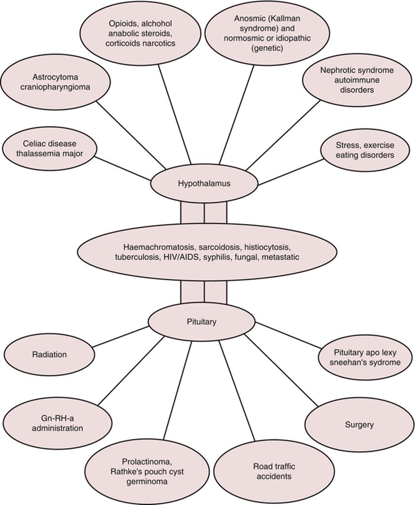
Fig. 32.1
Etiology of hypogonadotropic hypogonadism
The role of leptin and adipokines in the adipocyte and fat metabolism and energy homeostasis and their impact on HPO axis are being increasingly recognized. Leptin, a 167-amino acid hormone, is secreted by adipocytes and its levels are positively correlated with body fat. Earlier, leptin was thought to be a solution for the obesity problem. Slowly, the role of leptin was better defined in energy-deficient states. In response to acute energy deprivation, the reproductive hormones are decreased in order to avoid high metabolic demands and pregnancy. This reduction is mediated through leptin [5]. Kisspeptin is another recently discovered neuromodulator that acts upstream of the GnRH to control pulsatile GnRH release. Kisspeptin is the main mediator which relays the negative and positive steroid feedbacks and information regarding body energy stores to hypothalamus. It has a key role to play in the onset of puberty. Kisspeptin may be the main mediator of metabolic and other factors affecting the hypothalamus [6]. Better elucidation of the role of leptin and kisspeptin in the neuroendocrine control of ovulation is likely to introduce newer therapeutic options in the management of HH and anovulation [6–8].
32.3 Clinical Presentation
The incidence of HH varies according to the population studied. In infertility clinics, the estimates vary from 1 to 3 % of all infertility and approximately 5 % of anovulatory infertility. The prevalence of congenital or idiopathic HH varies from one in 3,000 to 4,000 population and is two to five times more common in males [9].
Women with HH may present with delayed menarche, primary amenorrhea, or secondary amenorrhea. Genetic etiologies, e.g., Kallmann syndrome, present with primary amenorrhea. Sudden onset of secondary amenorrhea may antedate an event, e.g., parturition, irradiation, and surgery. It may have a more gradual onset when amenorrhea is associated with other systemic illnesses. The diagnosis of functional hypothalamic amenorrhea is quite obvious due to the presence of a significant history. History of weight loss and excessive exercise or dietary restriction in an anxious and stressed female presenting with amenorrhea are enough to indicate the diagnosis. The hypogonadotropic hypogonadism related to obesity is only recently recognized. Obesity affects establishment of a healthy HPO axis. In metabolically active obese women, obesity leads to hypogonadism [1] and in women with insulin resistance to PCOS.
The following case history is an illustrative example: a 30-year-old woman presented with progesterone withdrawal negative secondary amenorrhoea for 14 months. Her previous menstrual cycles were regular. She was married for 38 months and gave history of weight gain of (30 kg) since marriage. Her serum hormone levels were FSH 1.2 mIU/ml, LH 0.5 mIU/ml, and E2 15 pg/ml. Her ultrasound examination revealed a small uterus with linear hyperechoic endometrium. Both ovaries were very small but had 5–6 antral follicles of 1–2 mm diameter each. There was negligible stroma around the follicles. Her thyroid and adrenal functions were normal. She responded to ovulation induction with gonadotropins.
32.4 Ovulation Induction
Irrespective of the etiology of HH, the treatment to achieve fertility is very straightforward, i.e., ovulation induction (OI). Despite such profound hypoestrogenism and amenorrhea, these women respond very well to OI and achieve significantly high pregnancy rate. We discuss the topic under the following headings.
32.4.1 Confirmation of Diagnosis and the Underlying Etiology
Before treatment is started, it is mandatory to confirm the diagnosis. Amenorrheic women in whom progesterone (P) withdrawal is negative and combined estrogen (E) and progesterone (E + P) withdrawal is positive can have either hypergonadotropic amenorrhea or hypogonadotropic amenorrhea. Values in very low (i.e.,1 mIU) or low normal range of serum LH and FSH done during amenorrhea are enough to confirm the diagnosis. In case of any doubt, the test may be repeated to rule out a laboratory error. It is counterproductive to induce menstruation with E + P and then do the LH and FSH levels. One must take care to give a gap of at least 1 month between the test and the administration of estrogens or E + P preparations. Exogenous E or E + P preparations downregulate the pituitary and hypothalamus and may lead to false low serum levels of FSH and LH.
In cases of HH, serum E levels are extremely low. The absence of P withdrawal is enough for diagnosis and serum E level testing is not mandatory. Transvaginal ultrasound reveals a very small infantile type of uterus with thin, linear hyperechoic single-layer endometrium. In case the woman has been on E + P for hormone replacement therapy, the uterus may be of normal size and endometrium is better delineated. The ovaries are very small and difficult to locate, sometimes not even seen properly. No assessment of antral follicle count (AFC) is possible in a majority of cases.
A variant of HH with ovaries having PCOS morphology has been described [10, 11]. In such a woman, the ovaries are small but show multiple, small, one to two millimeter-sized follicles. These ovaries also respond to stimulation similar to that in HH but have a high risk of ovarian hyperstimulation syndrome (OHSS) [10, 11].
The case described below is a typical example: a 25-year-old woman presented with history of primary amenorrhea and primary infertility for three years. Her husband had severe oligoasthenozoospermia. She gave history of one abandoned in vitro fertilization (IVF) with intracytoplasmic sperm injection (ICSI) cycle. This was done to avoid OHSS because of the multifollicular development and high risk of OHSS. On reevaluation, her serum FSH level was 0.5mIU/ml and serum LH 0.1 mIU/ml. She gave history of induced menstrual bleeding with E + P, not with progesterone only. On ultrasound examination, her ovaries were medium sized and revealed 10–12 antral follicles of 1–6 mm size on each side and were devoid of any surrounding stroma. She was diagnosed as HH with polycystic ovaries. Second cycle of superovulation for IVF-ICSI was undertaken with recombinant FSH by default. The cycle had to be abandoned. There was follicular development of 8–10 follicles but her E2 levels remained very low. A third cycle with urinary gonadotropins containing both LH and FSH resulted in adequate stimulation for IVF-ICSI and twin live births.
After confirmation of the diagnosis, it is important to diagnose the underlying etiology. History of primary amenorrhea associated with anosmia is a clear pointer toward Kallmann syndrome. In the absence of anosmia, primary HH is labeled as idiopathic HH. A variety of genes have been identified for the idiopathic HH [12].
In case of suspected organic lesion, the history is very important. The diagnosis is easier in cases where there is a suggestive history, e.g., of accident, surgery, drugs, and systemic illness. In case a tumor or an infiltrative lesion of the brain is suspected, magnetic resonance imaging (MRI) of the pituitary region clinches the diagnosis. Despite all the tests, an idiopathic cause remains the most common diagnosis.
32.4.2 Distinguishing Between Hypothalamic and Pituitary Causes
It is important to distinguish the hypothalamic and the pituitary causes in case of HH because of 3 reasons. Firstly, pituitary lesions may have other concomitant endocrinopathies related to adrenal, thyroid, osmoregulation, or somatotropic axis. The importance of diagnosing other deficiencies is quite obvious. Secondly, any lesion in the pituitary may increase during the pregnancy and cause further complications due to the pressure effects. Thirdly, the method of OI may differ. While hypothalamic amenorrhea can be treated with both pulsatile GnRH and exogenous gonadotropins, HH of pituitary origin has only one option, i.e., the exogenous gonadotropins.
Genetic, systemic, functional, and idiopathic categories are generally hypothalamic in origin (see Fig 32.1). In these cases, the pituitary in addition to ovaries also remains responsive to exogenous GnRH. The pituitary origin of HH is mostly pathological. Neoplastic, infiltrative, vascular, or pituitary stalk lesions need to be ruled out (Fig. 32.1).
Appropriate history, blood tests to rule out comittant endocrinopathies, and an MRI of the brain are sufficient to arrive at the diagnosis. Adequacy of thyroid function can be assessed by serum thyroid-stimulating hormone (TSH) and triiodothyronine (T3) and thyroxine (T4) levels. Serum levels of growth hormone (GH), prolactin, cortisol, and dehydroepiandrosterone give reasonable estimate of other functions of pituitary, thyroid, and adrenal glands.
GnRH stimulation test is used to assess the responsiveness of pituitary. Serial samples of LH and FSH are taken after a bolus administration of native GnRH or GnRH analogue. An intact pituitary should respond with spurt of LH and FSH. Although widely used earlier, it does not give much additional information regarding pituitary adrenal axis or pituitary thyroid axis over the baseline hormone testing. An MRI of the sella is more useful. The role of GnRH stimulation test is to be reserved for those cases where basal hormone measurements are not helpful and where there is a strong clinical evidence of pituitary deficiency [1].
32.4.3 Exclusion of Other Infertility Factors
Before starting OI in these women, the couple must be evaluated to rule out other causes of infertility (infertility factors). The minimum required tests include the semen analysis and a hysterosalpingography (HSG) for tubal evaluation. Endometrial biopsy after an E + P-induced menstruation is indicated only if an infective pathology (e.g., genital tuberculosis) is suspected, such as in high-prevalent countries. There is no requirement of diagnostic laparoscopy or hysteroscopy.
Adequacy of the endometrial response may be assessed with a trial cycle of E administration. Estradiol valerate (E2) may be given twice or thrice a day. Transvaginal ultrasound done after 10 days of E2reflects the endometrial response. This step can be omitted if there is history of good menstrual flow with E + P.
32.4.4 Physiological Basis of Ovulation Induction in Hypogonadotropic Hypogonadism
In women who do not desire pregnancy, the goal of therapy is to maintain adequate menstrual function with cyclic replacement of E + P. This will ensure general well-being and bone health. OI has to be undertaken in women desirous of pregnancy.
In HH of the pituitary origin, the only option available is to stimulate the ovary directly with the help of exogenous FSH and LH. This therapy is a substitution therapy, i.e., replacement of the deficient hormones. There is another option in hypothalamic amenorrhea. Pituitary can be stimulated with exogenous, native GnRH given in pulsatile manner mimicking the natural pulses. These GnRH pulses stimulate the pituitary to release LH and FSH, which is a more physiological technique.
FSH is the main stimulator of the ovarian follicles and granulosa cells and is indispensable. The role of LH is however not well understood. HH is one naturally occurring experimental model which has clarified the role of LH in ovulation induction. LH is essential for theca cell function. The synergistic but different effect of FSH and LH on granulosa and theca cells, respectively, is the basis of two cell-two gonadotropin theory. Estrogen is produced by granulosa cells under the influence of FSH from the androgen substrate, produced and supplied by theca cells under the influence of LH. LH is not only essential for steroid production but also for the maturation of oocyte and target tissue responses [13, 14]. It has been estimated that levels of 1–10 IU/L of LH should be sufficient to achieve these effects [13]. Moreover, a higher dose of LH may be detrimental and causes atresia of follicles—the LH ceiling effect [14].
Ovarian stimulation with recombinant FSH or FSH alone in women with HH results in follicular growth but E levels remain low [15, 16]. This is in contrast to successful stimulation with FSH alone in pituitary downregulated, normogonadotropic women. In these women, some residual LH activity remains despite downregulation; LH is essential in HH women who are devoid of any endogenous LH activity.
32.4.5 Pharmacological Agents
1.
2.
32.4.5.1 GnRh: Pharmacology
Native GnRh available in vials is to be given by subcutaneous (SC) or intravenous (IV) routes. A continuous infusion pump has to be used to deliver the precise dose at timely intervals. Local injection site irritation and visibility of the pump are the main disadvantages. Once very popular, pulsatile GnRh is used uncommonly these days. GnRh is not available in India for use.
32.4.5.2 Gonadotropins: Urinary or Recombinant
Either urinary or recombinant gonadotropins are used for ovulation induction. Each ampule of human menopausal gonadotropin (HMG) obtained from the urine of menopausal women contains 75 IU FSH and 75 IU of LH. These are available as HMG or highly purified HMG (HP-HMG) preparations containing 75 or 150 IU of FSH and LH per ampule as lyophilized powder. Urinary HMG can be given only by intramuscular route, while HP-HMG preparations can be given by subcutaneous SC route.
HMG factually contains some LH and mostly LH-like activity which is derived from variable amount of LH and mainly human chorionic gonadotropin (hCG). Urinary LH is highly unstable and has a variable potency. Thus, the LH-like activity is achieved by adding hCG [14].
Recombinant FSH (rFSH) and LH (rLH) are manufactured with genetic engineering technology using Chinese hamster ovary cell lines. Recombinant hormones are highly purified products with consistent batch to batch activity as compared to urinary products. Urinary products are a mixture of various bioisoforms of FSH, while rFSH contains only one isoform and also differs in terminal sialic acid content. Despite extensive purification, urinary FSH preparations retain some LH activity, while rFSH is devoid of any LH activity [16]. Recombinant FSH is available as single- or multiple-dose vials and pens which deliver a very small volume of solution with precision. Two products follitropin alpha and follitropin beta are currently available in the market. In India, many other companies have started marketing recombinant FSH products.
Recombinant LH is available only as 75 IU lyophilized powder. Both rFSH and rLH products are extremely well tolerated and user friendly in administration, although costlier than urinary products. Both products can be used through IM and SC routes. Following single administration, follitropin alpha has terminal half-life of 37 h and 74 % bioavailability [15]. Lutropin alpha has a half-life of about 18 h and bioavailability of 56 % [15].
Conventionally, rLH is given as a single daily injection. Twice daily regimen may have better endocrine profile in the stimulation cycles in HH women [17]. Recently, a mixture of follitropin alpha and lutropin alpha (follitropin alpha/lutropin alpha 150 IU/75 IU) has become available [18]. Early dose finding studies concluded that 75 IU rLH was effective in 94 % of women to achieve adequate follicular maturation and only a few would require a higher dose [19].
The choice between HMG and recombinant preparations depends on the cost and availability. When using high doses, recombinant preparation may have an edge. LH preparation is given separately; dose can be reduced to avoid LH ceiling effect [2]. Urinary products are efficient and cost-effective products. There are no randomized controlled trials comparing the urinary and recombinant products in HH women.
32.4.6 Ovulation Induction Regimens
The objectives of OI in HH are as follows:
1.
2.
3.
4.
32.4.6.1 Pulsatile GnRh
The treatment is suitable for women with intact pituitary, e.g., idiopathic HH or stress-induced amenorrhea [20]). The infusion of GnRh is done with the help of an automated pump at pulse frequency interval between 90 and 120 min [21]. GnRh can be given through IV (5–10 μg/ pulse) and SC (15–20 μg) routes per pulse. IV route is more successful than SC route [21]. Spontaneous LH surge is triggered by rising E levels. An hCG trigger may be given to induce ovulation although it is not mandatory. Intermittent hCG is recommended in the luteal phase since trophic stimulation of corpus luteum from pituitary is lacking in HH cases.
Overall, the treatment results in over 90 % rate of ovulation and a cumulative pregnancy rate of up to 96 % after six cycles [22, 23]. Pulsatile GnRh has also been used successfully for OI in PCOS. In one very large series of 292 anovulatory patients and 900 cycles, there were 130(268) HH women in whom successful ovulation was achieved in 75 % and pregnancy rate in 18 %, per treatment cycle [24]. GnRh was given IV at a dose of 1.25–2.00 micrograms of GnRh every 30–120 min, Maximum cycles required 2.5–5.0 micrograms every 60–90 min. Ovulation and pregnancy rates were higher in all types of HH women as compared to other anovulatory infertilities, like PCOS: only 4 multiple pregnancies occurred (3.8 %) and miscarriage rate was 30 % and even higher in PCOS. No case of OHSS was reported [24].
The main advantage of pulsatile GnRh over gonadotropins is low rate of multiple pregnancy and ovarian hyperstimulation syndrome (OHSS) [22–24]. The main disadvantage is the need to keep the pump connected to the body for quite a long time (2–3 weeks) and the necessity to refill the pump frequently. Once very popular, the treatment is less frequently used in general practice now.
32.4.6.2 Stimulation Regimen with Gonadotropins
Stimulation can be started on any day during amenorrhea or after an E + P withdrawal bleeding. Two to three month priming with sequential E + P treatment may improve response. In author’s personal experience, this priming is not required. A novel concept of LH priming has been suggested recently. Pretreatment with 300 IU SC of rLH for 7 days immediately preceding the rFSH significantly decreased the requirement of FSH [25].
Minimum effective dose of FSH is to be selected and a weekly step-up regimen is to be followed. Most of the ovarian reserve tests, i.e., serum FSH, antral follicle count (AFC), and anti-Müllerian hormone (AMH), do not give a clear idea about the ovarian response. It is best to start with 75 IU and then step up every 5–7 days. In a review article of several studies published from 1966 to 1984, the pregnancy rate varied from 16 to 78 % in different studies [26]. Ovarian response is measured with the help of serum E and transvaginal ultrasound. Triple lining of endometrium on ultrasound is the first sign of ovarian activity and E synthesis. Either of the urinary HMG or recombinant preparations can be used. Coadministration of rLH is mandatory if rFSH used.
The final ovulation trigger is given by hCG injection 10,000 IU when the lead follicle reaches 18 mm in size. This is followed by timed coitus or intrauterine insemination 36–40 h later. Luteal support with hCG decreases the incidence of luteal phase defects and increases the pregnancy rate [27].
There are very few reports in the recent literature on the use of urinary gonadotropins. A combined step-down and step-up approach was also used with the starting dose of 150–225 IU of FSH with 75 IU of LH for 2–3 days, which was then reduced to 75 IU of FSH. Step-up of 75 IU was made every 7 days until follicular growth was achieved [28].
Use of recombinant products gives similar results. Treatment with rFSH and rLH was successful in achieving ovulation in 84 % of cycles, complete luteinization in 80 % of cycles, and pregnancies in 22–24 % of cycles. Cumulative pregnancy rate per patient was 39.5 % [29].
There is no consensus on which is a better regimen—pulsatile GnRh or exogenous gonadotropins. In those cases where pituitary is not functional, the only choice is exogenous gonadotropin. In women with intact pituitary function, the choice depends upon availability, cost, convenience, patient, and physician preference.
32.4.7 Differences Between OI in HH and Polycystic Ovarian Syndrome (PCOS)
The principles of OI are similar in both conditions. PCOS is a very common condition. Most physicians handling OI and infertility get enough experience in managing PCOS. However, one does not see HH cases very often as it is an uncommon condition. There are few important similarities and differences between the OI of PCOS and HH (Table 32.1).
Table 32.1
Comparison of ovulation induction for anovulation due to hypogonadotropic hypogonadism and polycystic ovary syndrome
|
Feature |
Hypogonadotropic hypogonadism |
Polycystic ovary syndrome |
|
Amenorrhea |
Always |
Oligomenorrhea Amenorrhea |
|
Primary/secondary onset |
Both |
Both |
|
Pulsatile GnRH therapy |
Effective in functional HH only |
Effective in all cases |
|
Gonadotropin therapy |
Effective |
Effective |
|
Oral ovulation-inducing agents |
Nil |
Clomiphene, letrozole, metformin, etc. |
|
Treatment start |
Any day during amenorrhea |
After progesterone withdrawal |
|
Starting dose |
75–150 IU |
37.5 rFSH or even lower |
|
Step-up dose |
75 IU |
Ultra low-dose step-up |
|
Step-up interval |
First step-up 7 days |
First step-up 14 days |
|
LH |
Mandatory |
Avoided |
|
Gonadotropin required |
Very high |
Low to medium |
|
Duration of stimulation |
Longer |
Shorter |
|
Multiple pregnancy |
High |
High |
|
Risk of OHSS |
Low |
Very high |
32.4.8 Prognostic Factors
Prognosis of OI and fertility outcome is related to age and the presence or absence of other fertility factors. AMH is generally in the low normal range in HH due to partial gonadotropin-dependent regulation of AMH [30].
The traditional tests, i.e., FSH and serum E levels, remain the preferred choice for the diagnosis of HH However AMH appears to be a promising marker of ovarian reserve and response even in women with HH [31].
The cumulative pregnancy rate in HH with various treatment modalities after 6 cycles reaches up to near 60–70 %. In the remaining women, other incidental infertility factors may be present. In these cases, in vitro fertilization provides excellent results.
32.4.9 Treatment of Functional Disorder: Stress-Induced Amenorrhea
Stress-induced amenorrhea is related to the classic triad of stress, exercise, and dietary restriction. In severe weight loss even without exercise, the hypothalamic pulse generator may be disturbed. The condition is called anorexia nervosa. With increasing participation of women in competitive sports, the effects of strenuous exercise along with dietary restriction, on female reproductive system, are increasingly being stipulated [32]. Even recreational sports have an impact on the highly sensitive HPO axis [33]. Moreover, there is an ever increasing stress of competition with balancing of family and professional lives. In addition to the HPO axis, stress induces disturbance of hypothalamic-pituitary-adrenal axis (HPA axis) and also of thyroid function. Appropriate cognitive behavioral therapy directed toward reducing stress, improving nutrition, and decreasing exercise level may be able to resume ovulation in many such women [34, 35].
Women with functional amenorrhea are hypolepitinemic. Leptin replacement in these women may restore ovulatory menstruation besides correcting metabolic and endocrine derangement [5]. Many women with weight loss-related amenorrhea do not ovulate even after body weight is near normal. In one study, leptin administration resulted in ovulatory menses in three and there was evidence of ovulation in two out of two women [7]. At present, such treatment remains experimental but does hold promise for the future.
32.4.10 Thalassemia Major and Hypogonadotropic Hypogonadism
With the major developments in the field of transfusion medicine and iron chelation therapy, the prognosis for thalassemia major and intermedia has improved significantly, and life expectancy has increased. It is natural to expect the demand for pregnancy from women reaching reproductive years. Besides problems of managing iron overload and need to stop chelation therapy during pregnancy, conception is also a problem. Hypogonadotropic hypogonadism is a common problem in thalassemia major because of the widespread iron deposits. Although fertility can be restored in these women with appropriate OI, it requires a multidisciplinary team to look after these pregnancies. Favorable pregnancy was reported in a large study [36]. Of 46 women of thalassemia major who conceived, 33 did so with gonadotropin OI; others with milder forms of menstrual disturbances conceived spontaneously; 91 % of pregnancies resulted in successful deliveries with appropriate medical management [36].
32.5 Induction of Spermatogenesis in Males with HH
The etiopathogenesis of HH in males is similar to that in females; the congenital form is three to four times more common in males as compared to females [4]. These cases present with delayed puberty erectile dysfunction, loss of libido, and regression of secondary sex characteristics like the testicular size or the facial hair growth. Serum LH, FSH, and testosterone levels are in extremely low range. Fertility is generally not of immediate concern in adolescent age. However, long-term fertility potential is definitely required. Men present late in life or who have been earlier treated with androgens require induction of spermatogenesis for fertility.
Testosterone replacement is the standard therapy for induction of puberty. This is a cost effective and safe and effective way of inducing puberty and secondary sexual characteristics. Long-term therapy maintains libido and erectile function for long time. Testosterone therapy is a replacement but suppressive therapy. hCG and FSH have been used for spermatogenesis. There is some evidence that induction of puberty and spermatogenesis with gonadotropins or GnRH before testosterone therapy may have an edge over testosterone therapy in preserving fertility potential [37, 38].
Both pulsatile GnRh and gonadotropins can be used. Pulsatile GnRh has to be given through the infusion pump; pulses are given at the rate of 100–400 μng/kg every 2 h.
The duration of therapy is at least 4 months. The main disadvantages are the inconvenience and high cost; this treatment is followed by only few centers now [39, 40].
The standard regimen for induction of spermatogenesis is to give hCG injection 1,500–,000 IV twice or thrice a week. With this treatment, Leydig cells are stimulated to produce testosterone. Puberty can be induced with hCG by itself. It can also induce spermatogenesis but is less efficient for this purpose. FSH in the dose of 150 IV twice or thrice a week after 3 months of hCG priming or alternatively started simultaneously restores spermatogenesis early and with more reliability. The combined treatment may have to be given for up to 2 years [41, 42].
Regardless of the hormones used, the total number of sperms remains below the normal threshold, but 50–80 % pregnancy rate achieved even with sperm concentration of 5 million or less [39, 40] The prognostic factors include higher pretreatment testicular volume, the absence of cryptorchidism, and no previous testosterone therapy [37].
Spontaneous pregnancies occur 6–9 months after start of FSH but can require up to 2 years of treatment. If a pregnancy does not occur after a reasonable time after appearance of sperm in the ejaculate, the couple should be advised intrauterine insemination or ICSI as the case may be. The success of ICSI is similar to that in other cases of male infertility [43, 44].
Failure of gonad therapy is almost unknown. In appropriately diagnosed cases of HH, if sperms do not appear in ejaculate after 3–6 months of therapy, then an additional cause for azoospermia need to be ruled out. There may be congenital or acquired obstruction in the vas deferens, seminal vesicles, or ejaculatory ducts. In these cases, the efficacy of gonadotropin therapy can be judged by increase in testicular volume, vascularity, testosterone levels, and fine needle aspiration cytology from testes.
Hypogonadotropic hypogonadism is a challenging disorder in both males and females with appropriate management; the result is very dramatic and rewarding.
References
1.
Santoro N. Update in hyper- and hypogonadotropic amenorrhea. J Clin Endocrinol Metab. 2011;96(11):3281–8.CrossRefPubMed
2.
Krause BT, Ohlinger R, Haase A. Lutropin alpha, recombinant human luteinizing hormone, for the stimulation of follicular development in profoundly LH-deficient hypogonadotropic hypogonadal women: a review. Biologics. 2009;3:337–47.PubMedCentralPubMed
3.
Rowe PJ, Comhaire FH, Hargreave TB, Mellows HJ. WHO manual for the standardized investigation and diagnosis of the infertile couple. Cambridge: Cambridge University Press; 1993.
4.
Silveira LF, Latronico AC. Approach to the patient with hypogonadotropic hypogonadism. J Clin Endocrinol Metab. 2013;98(5):1781–8.CrossRefPubMed
5.
Kelesidis T, Kelesidis I, Chou S, Mantzoros CS. Narrative review: the role of leptin in human physiology: emerging clinical applications. Ann Intern Med. 2010;152(2):93–100.PubMedCentralCrossRefPubMed
6.
Skorupskaite K, George JT, Anderson RA. The kisspeptin-GnRH pathway in human reproductive health and disease. Hum Reprod Update. 2014;20(4):485–500.PubMedCentralCrossRefPubMed
7.
Welt CK, Chan JL, Bullen J, Murphy R, Smith P, DePaoli AM, et al. Recombinant human leptin in women with hypothalamic amenorrhea. N Engl J Med. 2004;351(10):987–97.CrossRefPubMed
8.
Chou SH, Chamberland JP, Liu X, Matarese G, Gao C, Stefanakis R, et al. Leptin is an effective treatment for hypothalamic amenorrhea. Proc Natl Acad Sci U S A. 2011;108(16):6585–90.PubMedCentralCrossRefPubMed
9.
Seminara SB, Hayes FJ, Crowley Jr WF. Gonadotropin-releasing hormone deficiency in the human (idiopathic hypogonadotropic hypogonadism and Kallmann’s syndrome): pathophysiological and genetic considerations. Endocr Rev. 1998;19(5):521–39.PubMed
10.
Shoham Z, Conway GS, Patel A, Jacobs HS. Polycystic ovaries in patients with hypogonadotropic hypogonadism: similarity of ovarian response to gonadotropin stimulation in patients with polycystic ovarian syndrome. Fertil Steril. 1992;58(1):37–45.PubMed
11.
Schachter M, Balen AH, Patel A, Jacobs HS. Hypogonadotropic patients with ultrasonographically detected polycystic ovaries: endocrine response to pulsatile gonadotropin-releasing hormone. Gynecol Endocrinol. 1996;10(5):327–35.CrossRefPubMed
12.
Brioude F, Bouligand J, Trabado S, Francou B, Salenave S, Kamenicky P, et al. Non-syndromic congenital hypogonadotropic hypogonadism: clinical presentation and genotype-phenotype relationships. Eur J Endocrinol. 2010;162(5):835–51.CrossRefPubMed
13.
Chappel SC, Howles C. Reevaluation of the roles of luteinizing hormone and follicle-stimulating hormone in the ovulatory process. Hum Reprod. 1991;6(9):1206–12.PubMed
14.
Filicori M, Cognigni GE, Taraborrelli S, Spettoli D, Ciampaglia W, Tabarelli De Fatis C, et al. Luteinizing hormone activity in menotropins optimizes folliculogenesis and treatment in controlled ovarian stimulation. J Clin Endocrinol Metab. 2001;86(1):337–43.CrossRefPubMed
15.
le Cotonnec JY, Porchet HC, Beltrami V, Munafo A. Clinical pharmacology of recombinant human luteinizing hormone: part I. Pharmacokinetics after intravenous administration to healthy female volunteers and comparison with urinary human luteinizing hormone. Fertil Steril. 1998;69(2):189–94.CrossRefPubMed
16.
Gibreel A, Bhattacharya S. Recombinant follitropin alfa/lutropin alfa in fertility treatment. Biologics. 2010;4:5–17.PubMedCentralPubMed
17.
Awwad JT, Farra C, Mitri F, Abdallah MA, Jaoudeh MA, Ghazeeri G. Split daily recombinant human LH dose in hypogonadotrophic hypogonadism: a nonrandomized controlled pilot study. Reprod Biomed Online. 2013;26(1):88–92.CrossRefPubMed
18.
Agostinetto R. Administration of follitropin alfa and lutropin alfa combined in a single injection: a feasibility assessment. Reprod Biol Endocrinol. 2009;7:48.PubMedCentralCrossRefPubMed
19.
The European Recombinant Human LH Study Group. Recombinant human luteinizing hormone (LH) to support recombinant human follicle-stimulating hormone (FSH)-induced follicular development in LH- and FSH-deficient anovulatory women: a dose-finding study. J Clin Endocrinol Metab. 1998;83(5):1507–14.
20.
Messinis IE. Ovulation induction: a mini review. Hum Reprod. 2005;20(10):2688–97.CrossRefPubMed
21.
Letterie GS, Coddington CC, Collins RL, Merriam GR. Ovulation induction using s.c. pulsatile gonadotrophin-releasing hormone: effectiveness of different pulse frequencies. Hum Reprod. 1996;11(1):19–22.CrossRefPubMed
22.
Shoham Z, Homburg R, Jacobs HS. Induction of ovulation with pulsatile GnRH. Bailliere’s Clin Obstet Gynaecol. 1990;4(3):589–608.CrossRef
23.
Martin KA, Hall JE, Adams JM, Crowley Jr WF. Comparison of exogenous gonadotropins and pulsatile gonadotropin-releasing hormone for induction of ovulation in hypogonadotropic amenorrhea. J Clin Endocrinol Metab. 1993;77(1):125–9.PubMed
24.
Filicori M, Flamigni C, Dellai P, Cognigni G, Michelacci L, Arnone R, et al. Treatment of anovulation with pulsatile gonadotropin-releasing hormone: prognostic factors and clinical results in 600 cycles. J Clin Endocrinol Metab. 1994;79(4):1215–20.PubMed
25.
Balasch J, Fabregues F, Carmona F, Casamitjana R, Tena-Sempere M. Ovarian luteinizing hormone priming preceding follicle-stimulating hormone stimulation: clinical and endocrine effects in women with long-term hypogonadotropic hypogonadism. J Clin Endocrinol Metab. 2009;94(7):2367–73.CrossRefPubMed
26.
Tadokoro N, Vollenhoven B, Clark S, Baker G, Kovacs G, Burger H, Healy D. Cumulative pregnancy rates in couples with anovulatory infertility compared with unexplained infertility in an ovulation induction programme. Hum Reprod. 1997;12(9):1939–44.CrossRefPubMed
27.
Messinis IE, Bergh T, Wide L. The importance of human chorionic gonadotropin support of the corpus luteum during human gonadotropin therapy in women with anovulatory infertility. Fertil Steril. 1988;50(1):31–5.PubMed
28.
Balash J. The role of FSH and LH in ovulation induction: current concepts. In: Gardner DK, Weisman A, Howles CM, Shoham Z, editors. Textbook of assisted reproduction techniques, Clinical perspectives, vol. 2. 4th ed. London: Informa; 2012. p. 85–7.
29.
Burgues S. The effectiveness and safety of recombinant human LH to support follicular development induced by recombinant human FSH in WHO group I anovulation: evidence from a multicentre study in Spain. Hum Reprod. 2001;16(12):2525–32.CrossRefPubMed
30.
Hagen CP, Sorensen K, Anderson RA, Juul A. Serum levels of antimullerian hormone in early maturing girls before, during, and after suppression with GnRH agonist. Fertil Steril. 2012;98(5):1326.CrossRefPubMed
31.
Sonmezer M, Ozmen B, Atabekoglu CS, Papuccu EG, Ozkavukcu S, Berker B, et al. Serum anti-Mullerian hormone levels correlate with ovarian response in idiopathic hypogonadotropic hypogonadism. J Assist Reprod Genet. 2012;29(7):597–602.PubMedCentralCrossRefPubMed
32.
Otis CL, Drinkwater B, Johnson M, Loucks A, Wilmore J. American College of Sports Medicine position stand. The female athlete triad. Med Sci Sports Exerc. 1997;29(5):i–ix.CrossRefPubMed
33.
De Souza MJ, Toombs RJ, Scheid JL, O’Donnell E, West SL, Williams NI. High prevalence of subtle and severe menstrual disturbances in exercising women: confirmation using daily hormone measures. Hum Reprod. 2010;25(2):491–503.CrossRefPubMed
34.
Berga SL, Marcus MD, Loucks TL, Hlastala S, Ringham R, Krohn MA. Recovery of ovarian activity in women with functional hypothalamic amenorrhea who were treated with cognitive behavior therapy. Fertil Steril. 2003;80(4):976–81.CrossRefPubMed
35.
Pauli SA, Berga SL. Athletic amenorrhea: energy deficit or psychogenic challenge? Ann N Y Acad Sci. 2010;1205:33–8.PubMedCentralCrossRefPubMed
36.
Origa R, Piga A, Quarta G, Forni GL, Longo F, Melpignano A, et al. Pregnancy and beta-thalassemia: an Italian multicenter experience. Haematologica. 2010;95(3):376–81.PubMedCentralCrossRefPubMed
37.
Zacharin M, Sabin MA, Nair VV, Dabadghao P. Addition of recombinant follicle-stimulating hormone to human chorionic gonadotropin treatment in adolescents and young adults with hypogonadotropic hypogonadism promotes normal testicular growth and may promote early spermatogenesis. Fertil Steril. 2012;98(4):836–42.CrossRefPubMed
38.
Fraietta R, Zylberstejn DS, Esteves SC. Hypogonadotropic hypogonadism revisited. Clinics (Sao Paulo). 2013;68 Suppl 1:81–8.CrossRef
39.
Zitzmann M, Nieschlag E. Hormone substitution in male hypogonadism. Mol Cell Endocrinol. 2000;161(1–2):73–88.CrossRefPubMed
40.
Han TS, Bouloux PM. What is the optimal therapy for young males with hypogonadotropic hypogonadism? Clin Endocrinol (Oxf). 2010;72(6):731–7.CrossRef
41.
Akarsu C, Caglar G, Vicdan K, Isik AZ, Tuncay G. Pregnancies achieved by testicular sperm recovery in male hypogonadotrophic hypogonadism with persistent azoospermia. Reprod Biomed Online. 2009;18(4):455–9.CrossRefPubMed
42.
De Leo V, Musacchio MC, Di Sabatino A, Tosti C, Morgante G, Petraglia F. Present and future of recombinant gonadotropins in reproductive medicine. Curr Pharm Biotechnol. 2012;13(3):379–91.CrossRefPubMed
43.
Bakircioglu ME, Erden HF, Ciray HN, Bayazit N, Bahceci M. Gonadotrophin therapy in combination with ICSI in men with hypogonadotrophic hypogonadism. Reprod Biomed Online. 2007;15(2):156–60.CrossRefPubMed
44.
Zorn B, Pfeifer M, Virant-Klun I, Meden-Vrtovec H. Intracytoplasmic sperm injection as a complement to gonadotrophin treatment in infertile men with hypogonadotrophic hypogonadism. Int J Androl. 2005;28(4):202–7.CrossRefPubMed