
Charles Kawada, MD
Drorith Hochner-Celnikier, MD
The gynecologist needs to approach each patient not just as a person requiring medical intervention for a specific presenting problem, but also as one who may have a variety of factors possibly affecting her health. The initial approach to the gynecologic patient and the general diagnostic procedures available for the investigation of gynecologic complaints are presented here. Although other aspects of the general medical examination are left to other texts, concern for the patient’s total health and well-being is mandatory.
THE PERIODIC HEALTH SCREENING EXAMINATION
It is now a generally accepted part of the physician’s responsibility to advise patients to have periodic medical evaluations. The frequency of visits varies according to the patient’s age and specific problem.
The periodic health screening examination helps detect the following ailments of women that are especially amenable to early diagnosis and treatment: diabetes mellitus; urinary tract infection or tumor; hypertension; malnutrition or obesity; thyroid dysfunction or tumor; and breast, abdominal, or pelvic tumor. These conditions can be detected by a review of systems, with specific questions regarding recent abnormalities or any variation in function. Determination of weight, blood pressure, and urinalysis may reveal variations from the previous examination. An examination of the thyroid gland, breasts, abdomen, and pelvis, including a Papanicolaou (Pap) smear, should then be performed. A rectal examination also is advisable, and a conveniently packaged test for occult blood (Hemoccult) is recommended for patients older than 40 years. Patients of an advanced age (>50 years) may undergo blood test for lipid profile, bone density scan, pelvic ultrasound examination, and mammogram.
The physician should be concerned about conditions other than purely somatic ones. Unless a patient’s problems require the services of a psychiatrist or some other specialist, the doctor should be prepared to act as a counselor and work with the patient during a mutually agreeable time when it is possible to listen to her problems without being hurried and to give support, counsel, and other kinds of help as required.
HISTORY
To adequately evaluate the gynecologic patient, it is important to establish a rapport during the history taking. The patient needs to tell her story to an interested listener who does not allow body language or facial expressions to imply disinterest or boredom. One should avoid cutting off the patient’s story, because doing so may obscure important clues or other problems that may have contributed to the reasons for the visit.
The following outline varies from the routine medical history because, in evaluating the gynecologic patient, the problem often can be clarified if the history is obtained in the following order.
Identifying Information
A. Age
Knowledge of the patient’s age sets the tone for the complaint and the approach to the patient. Obviously, the problems and the approach to them vary at different stages in a woman’s life (pubescence, adolescence, childbearing years, and premenopausal and postmenopausal years).
B. Last Normal Menstrual Period
The date of onset of the last normal menstrual period (LNMP) is important to define. A missed period, irregularity of periods, erratic bleeding, or other abnormalities may all imply certain events that are more easily diagnosed when the date of onset of the LNMP is established.
C. Gravidity & Parity
The process of taking the patient’s obstetric history is detailed in Chapter 6, but the reproductive history should be recorded as part of the gynecologic evaluation. A convenient symbol for recording the reproductive history is a 4-digit code denoting the number of term pregnancies, premature deliveries, abortions, and living children (TPAL) (eg, 2-1-1-3 means 2 term pregnancies, 1 premature delivery, 1 abortion, and 3 living children).
Chief Complaint
The chief complaint usually is best elicited by asking “What kind of problem are you having?” or “How can I help you?” It is important to listen carefully to the way the patient responds to this question and to allow her to fully explain her complaint. The patient should be interrupted only to clarify certain points that may be unclear.
Present Illness
Each of the problems the patient describes must be obtained in detail by questioning regarding what exactly the problem is, where exactly the problem is occurring, the date and time of onset, whether the symptoms are abating or getting worse, the duration of the symptoms when they do occur, and how these symptoms are related to or influence other events in her life. For example, the site, duration, and intensity of pain must be accurately described. Getting a sense of how the pain affects her life often is helpful in evaluating the intensity of pain: “Does the pain prevent you from standing or walking?”
It is important to maintain eye contact with the patient and to listen to every word. Do not rely on a patient’s sophistication as a measure of her knowledge of anatomy and medical terminology. It is important for the physician to judiciously adjust the level of terminology according to the patient’s knowledge and vocabulary. Communicating with the patient in this manner may help the physician obtain an accurate history and establish rapport.
In addition to physiologic events and the life cycle, symptoms described could be related to starting a new job, the beginning of a new relationship or difficulties in the current relationship, an exercise regimen, new medication, and any emotional changes in the patient’s life.
Past History
After the physician is satisfied that all possible information concerning the present illness and the important corollaries has been obtained, the past history should be elicited.
A. Contraception
Continuing with the history, it is important to elicit whether the patient is using or needs some form of contraception. If she is using contraception, her level of satisfaction with her chosen method should be determined. In patients taking oral contraceptives, the history should reflect the agent and dose, whether there is a great variation in the time of day she takes her pill, and any impact of the pill on other physiologic functions. Other forms of hormonal contraceptives, including vaginal rings, dermal patches, and injectable contraceptives, have become available and have their own unique issues. It is extremely important to ask questions during the remainder of the history and to key the physical examination to ascertain whether there are any contraindications to the patient’s current form of contraception.
B. Medications & Habits
Any medications, prescribed or otherwise, that are being taken or that were being taken when symptoms first occurred should be described. Particular attention must be directed to use of hormones, steroids, and other compounds likely to influence the reproductive tract. Herbal preparations may not be viewed by the patient as medications, so this question should be specifically asked. In addition to medications, the patient should be questioned concerning her use of street drugs. It must be ascertained whether the patient smokes and, if so, how much and for how long. It is important to ascertain the amount of alcohol ingested, if any. This questioning provides an ideal time to indicate the health risks of various habits.
C. Medical
It is important to discover any history of serious medical and psychiatric illnesses and whether hospitalization was required. Particularly important are illnesses in the major organ systems. It is important to know whether there is a major endocrinopathy in the patient’s history. Notable weight gain or loss prior to the onset of the patient’s current symptoms should be detailed. Other important details include when she had her last physical examination, including pelvic examination and Pap smear.
D. Surgical
The surgical history includes all operations, the dates performed, and associated postoperative or anesthetic complications.
E. Allergies
Questioning should continue relating any possible allergic reactions to drugs or specific foods. The reaction produced (eg, rash, gastrointestinal upset) must be elicited and the approximate time when it occurred ascertained. Any testing to confirm or deny the observation must be noted. Latex allergy has become more common and severe and should be considered prior to most medical procedures, such as drawing blood samples, pelvic examination, and taking blood pressures.
F. Bleeding & Thrombotic Diatheses
Determining whether or not the patient bleeds excessively in relation to prior surgery or minor trauma is important. A history of easy bruising or of bleeding from the gums while brushing teeth may be useful in this judgment. The patient should be asked whether she or one of her close relatives experienced venous thromboembolism (VTE). A history of VTE may guide the physician as to which treatment to offer. Suspicion of a bleeding or clotting problem indicates the need for further laboratory evaluation.
G. Obstetrics
The obstetric history includes each of the patient’s pregnancies listed in chronologic order. The date of birth; sex and weight of the offspring; duration of pregnancy; length of labor; type of delivery; type of anesthesia; and any complications should be included.
H. Gynecologic
The first item in the gynecologic past history is the menstrual history: age at menarche, interval between periods, duration of flow, amount and character of flow, degree of discomfort, and age at menopause. The menstrual history often is an important clue in the diagnosis.
A prior history of sexually transmitted disease (STD) needs to be detailed. Although in the past it was more common to note only gonorrhea and syphilis, it is important to also document exposure to human immunodeficiency virus (HIV), hepatitis, herpesvirus, chlamydia, and papillomavirus. Any treatment or admissions to the hospital for treatment of salpingitis, endometritis, or tubo-ovarian abscess must be carefully documented. Attempts to assess the impact of these processes in relation to ectopic pregnancy, infertility, and type of contraception must be elicited.
Although its significance is less than that of the prior stated diseases, the occurrence of episodes of vaginitis should not be dismissed. Their frequency and the medications used to treat them should be discussed. In the case of such infections, it is important to detail whether or not the episode was pathologic or merely a misinterpreted physiologic circumstance.
I. Sexual
The sexual history should be an integral part of any general gynecologic history. In taking a sexual history, the physician must be nonjudgmental and not embarrassed or critical.
Questions that may be covered include the following. Is she currently sexually active? Is the relationship satisfactory to her and, if not, why not? A question regarding whether the patient is heterosexual or lesbian is important but often difficult to ask because the question may be offensive to some patients. It is important, however, not to assume that a relationship is heterosexual because a lesbian woman will lose all rapport with the physician when the physician is insensitive to such issues.
J. Social
A social history can be an extension of earlier questions pertaining to the marital and sexual history. Knowing the type of work the patient does, the type of educational background, and her community activities may assist in ascertaining the patient’s relationship to her entire environment.
The patient’s involvement with her own health care should be carefully elicited, including her attention and knowledge concerning diet, health screening examinations, recreation, and the degree of regular physical exercise.
Family History
The patient’s family history must include the state of health of immediate relatives (parents, siblings, grandparents, and offspring). In addition to listing these relatives, it is useful in cases where genetic illnesses may be apparent to record a 3-generation pedigree.
The incidence of familial heart disease, hypertensive renal or vascular disease, diabetes mellitus (insulin-dependent or non–insulin-dependent), vascular accidents, and hematologic abnormalities should be ascertained. If the patient has a problem with hirsutism or if she perceives excessive hair growth, it is important to elicit whether anyone in her family has the same distribution of hair growth. Familial history of breast, ovarian, and colon cancers is important to elicit because a close familial history may require additional testing and close follow-up. It is important to relate the time of menopause in the mother or grandmother and to ascertain a history of osteoporosis.
American Cancer Society guidelines for breast cancer screening: update 2003. CA Cancer J Clin 2003;53:141–169. PMID: 12809498.
American College of Obstetricians and Gynecologists. Cervical cytology screening. ACOG Practice Bulletin No. 45. Obstet Gynecol 2003;102:417.
Marrazzo JM, Stine K. Reproductive health history of lesbians: implications for care. Am J Obstet Gynecol 2004;190:1298–1304. PMID: 15167833.
Nustaum MR, Hamilton CD. The proactive sexual health history. Am Fam Physician 2002;66:1705–1712. PMID: 12449269.
PHYSICAL EXAMINATION
The physical examination is most useful if it is conducted in an environment that is aesthetically pleasing to the patient. Adequate gowning and draping assist in preventing embarrassment. Often a physician’s assistant escorts the patient to the dressing area and gives explicit instructions about what to take off and how to put on her gown and then may assist in draping the patient.
A physician may have a female assistant remain in the examining room to assist when necessary, but whether or not she remains solely as a chaperone depends on local custom and the preference of the patient and the physician. A chaperone is not legally required, but the physician, male or female, must use good judgment, especially during the breast and pelvic examinations. If the patient wants her partner, relative, or a friend to be present, the request should be honored unless, in the physician’s judgment, such an arrangement would interfere with the examination or with obtaining an accurate history. It is highly recommended that the physician explain the steps and acts that will be taken, especially during the pelvic examination when the patient might lack a direct eye contact with the physician.
General Examination
If the gynecologist is the primary care physician for the patient, a general physical examination should be performed annually or whenever the situation warrants. A complete examination obviously provides more information, demonstrates the physician’s thoroughness, and establishes rapport with the patient.
General Evaluation
A. Vital Signs
As part of every examination—whether for a specific problem, routine annual examination, or a return visit for a previously diagnosed problem—the patient should be weighed and her blood pressure taken. Postmenopausal patients should have their height measured to document any loss of height from osteoporosis and vertebral fractures. Before the patient empties her bladder for the examination, determination should be made as to whether the urine will need to be sent for urinalysis, culture, or pregnancy testing.
The examination of the chest should include visual assessment for any skin lesions and symmetry of movement. Auscultation and percussion of the lungs are important for excluding primary pulmonary problems such as asthma and pneumonia. The examination of the heart includes percussion for size and auscultation for arrhythmias and significant murmurs.
Breast Examination
(See also Chapter 5.)
Breast examination should be a routine part of the physical examination. Breast cancer will occur in 1 in 8 women in the United States during her lifetime. Physicians who treat women should educate patients on the technique of self-examination, because the well-prepared patient is one of the most accurate screening methods for breast disease.
The physical examination provides an ideal time to ascertain the frequency and methodology of breast self-examination. It also is an ideal time to teach the patient how to perform breast self-examination. The patient should be advised to examine herself in the mirror, looking for skin changes or dimpling, and then carefully palpate all quadrants of the breast. Most women prefer to do this with soapy hands while showering or bathing. The examination should be repeated at the same time each month, preferably 1 week after the initiation of the menses, when the breasts are least nodular; postmenopausal women should perform self-examination on the same day each month.
The frequency of mammography or the earlier use of mammography depends on both the individual woman and her family history. Patients with a positive family history of breast cancer should have a mammogram at an earlier age, particularly those whose mother, aunt, or sister developed premenopausal breast cancer. In general, a mammogram should be obtained every 1–2 years from ages 40–50 years and annually thereafter. Ultrasonography now can reliably differentiate solid from cystic lesions; this technique complements but does not supplant mammography. Breast self-examination, physician examination, mammography, and ultrasonography are complementary, and all should be used for the early detection of breast cancer. Annual magnetic resonance imaging (MRI) examination of the breast is indicated only in patients carrying BRCA1/2 mutations or with very strong familial history of breast cancer. However, this examination is complementary to the other techniques for early detection of breast cancer and does not replace them.
The correct technique for breast examination is shown in Figure 35–1. If abnormalities are encountered, a decision should be reached concerning the need for mammography (or other imaging methods) or direct referral to a breast surgeon unless the gynecologist is trained in performing breast biopsies. Skin lesions, particularly eczematous lesions in the area of the nipple, should be closely observed; if they are not easily cured by simple measures, they should be biopsied. An eczematous lesion on the nipple or areola may represent Paget’s carcinoma.

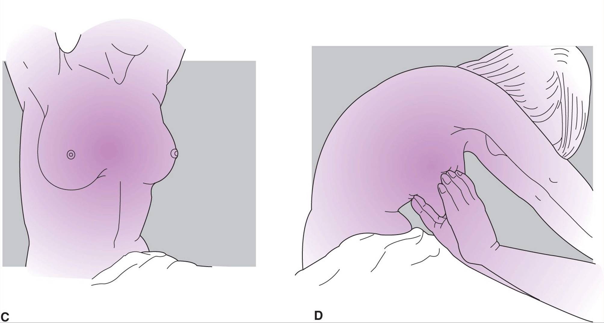
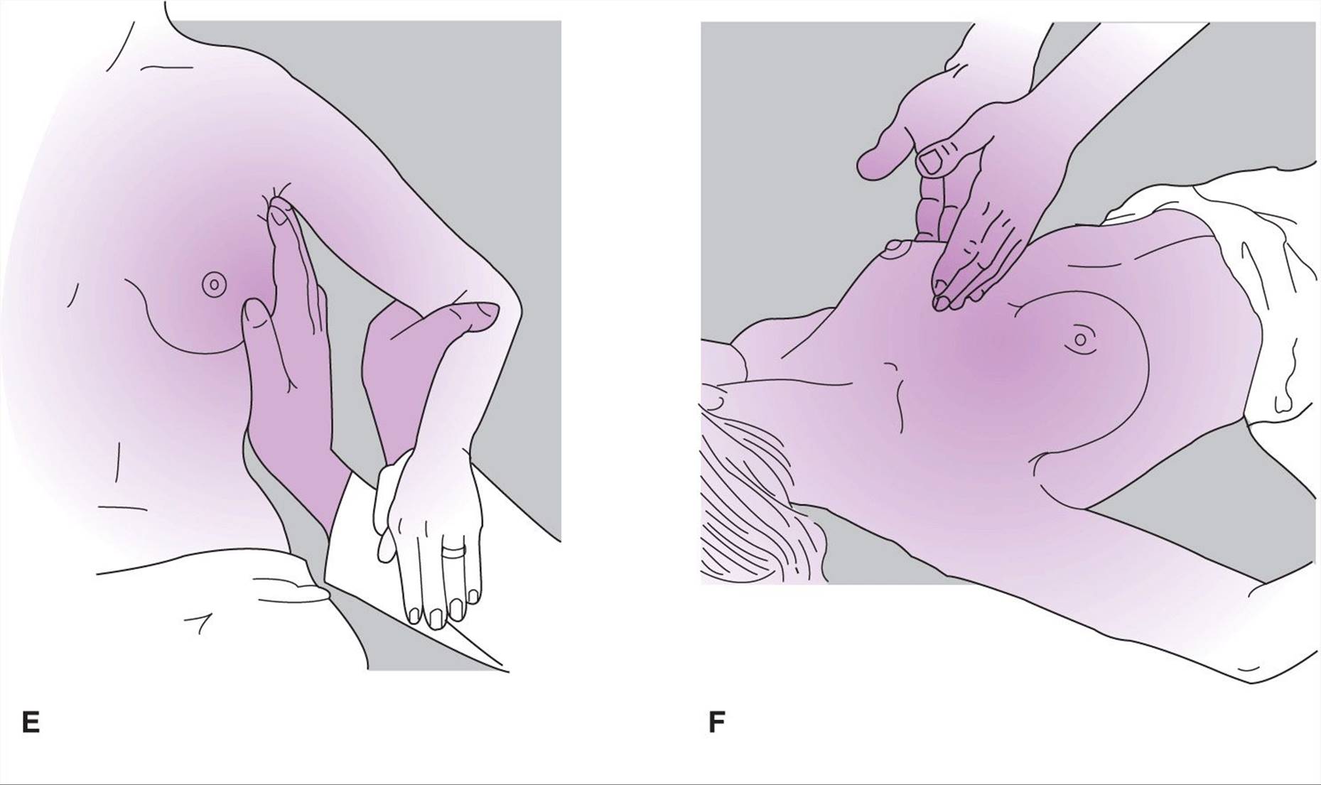

Figure 35–1. Breast examination by the physician. A: Patient is sitting, arms at sides. Perform visual inspection in good light, looking for lumps or for dimpling or wrinkling of skin. B: Patient is sitting, hands pressing on hips so that pectoralis muscles are tensed. Repeat visual inspection. C: Patient is sitting, arms above head. Repeat visual inspection of breasts and perform visual inspection of axillae. D: Patient is sitting and leaning forward, hands on examiner’s shoulders, the stirrups, or her own knees. Perform bimanual palpation, paying particular attention to the base of the glandular portion of the breast. E: Patient is sitting, arms extended 60–90 degrees. Palpate axillae. F:Patient is supine, arms relaxed at sides. Perform bimanual palpation of each portion of breast (usually each quadrant, but smaller sections for unusually large breasts). Repeat examinations C, E, and F with patient supine, arms above head. G: Patient is supine, arms relaxed at sides. Palpate under the areola and nipple with the thumb and forefinger to detect a mass or test for expression of fluid from the nipple. H: Patient is either sitting or supine. Palpate supraclavicular areas.
Abdominal Examination
The patient should be lying completely supine and relaxed; the knees may be slightly flexed and supported as an aid to relaxation of the abdominal muscles. Inspection should detect irregularity of contour or color. Auscultation should follow inspection but precede palpation because the latter may change the character of intestinal activity. Palpation of the entire abdomen—gently at first, then more firmly as indicated—should detect rigidity, voluntary guarding, masses, and tenderness. If the patient complains of abdominal pain or if unexpected tenderness is elicited, the examiner should ask her to indicate the point of maximal pain or tenderness with 1 finger. Suprapubic palpation is designed to detect uterine, ovarian, or urinary bladder enlargements. A painful area should be left until last for deep palpation; otherwise, the entire abdomen can be guarded voluntarily. As a final part of the abdominal examination, the physician should carefully check for any abnormality of the abdominal organs: liver, gallbladder, spleen, kidneys, and intestines. In some instances, the demonstration of an abnormality of the abdominal muscle reflexes may be diagnostically helpful. Percussion of the abdomen should be performed to identify organ enlargement, tumor, or ascites.
Pelvic Examination
The pelvic examination is a procedure feared by many women, so it must be conducted in such a way as to allay her anxieties. A patient’s first pelvic examination may be especially disturbing, so it is important for the physician to attempt to allay fear and to inspire confidence and cooperation. The empathic physician usually finds that by the time the history has been obtained and a painless and nonembarrassing general examination performed, a satisfactory gynecologic examination is not a problem. Relaxing surroundings; a nurse or attendant chaperone if indicated; warm instruments; and a gentle, unhurried manner with continued explanation and reassurance are helpful in securing patient relaxation and cooperation. This is especially true with the woman who has never before undergone a pelvic examination. In these patients, a 1-finger examination and a narrow speculum often are necessary. In some cases, vaginal examination is not possible; palpation of the pelvic structures by rectal examination is then the only recourse. Occasionally an ultrasound examination may be helpful in ascertaining whether the pelvic organs are normal in size and configuration in patients who cannot adequately relax the abdominal muscles. If a more definitive pelvic examination is essential, it can be performed with the patient anesthetized.
A. External Genitalia (Fig. 35–2)
The pubic hair should be inspected for its pattern (masculine or feminine), for the nits of pubic lice, for infected hair follicles, and for any other abnormalities. The skin of the vulva, mons pubis, and perineal area should be examined for evidence of dermatitis or discoloration. The glans clitoridis can be exposed by gently retracting the surrounding skin folds. The clitoris is at the ventral confluence of the 2 labia; it should be no more than 2.5 cm in length, most of which is subcutaneous. The major and minor labia usually are the same size on both sides, but a moderate difference in size is not abnormal. Small protuberances or subcutaneous nodules may be either sebaceous cysts or tumors. External condylomata are often found in this area. The urethra, just below the clitoris, should be the same color as the surrounding tissue and without protuberances. Normally, vestibular (Bartholin’s) glands can be neither seen nor felt, so enlargement may indicate an abnormality of this gland system. The area of vestibular glands should be palpated by placing the index finger in the vagina and the thumb outside and gently feeling for enlargement or tenderness (Fig. 35–3). The perineal skin may be reddened as a result of vulvar or vaginal infection. Scars may indicate obstetric lacerations or surgery. The anus should be inspected at this time for the presence of hemorrhoids, fissures, irritation, or perianal infections (eg, condylomata or herpesvirus lesions).
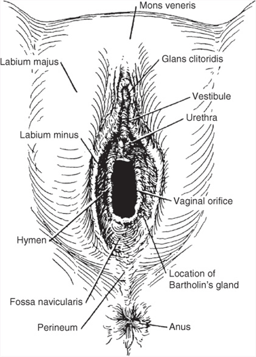
Figure 35–2. Normal external genitalia in a mature woman. (Reproduced, with permission, from Pernoll ML. Benson & Pernoll’s Handbook of Obstetrics and Gynecology. 10th ed. New York, NY: McGraw-Hill; 2001.)
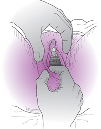
Figure 35–3. Palpation of vestibular glands.
B. Hymen
An unruptured hymen may present in many forms, but only a completely imperforate, cribriform, or septate hymen is pathologic. After rupture, the hymen may be seen in various forms (Fig. 35–4). After the birth of several children, the hymen may disappear almost completely.
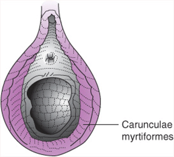
Figure 35–4. Ruptured hymen (parous introitus).
C. Perineal Support
To determine the presence of pelvic relaxation, the physician spreads the labia with 2 fingers and tells the patient to “bear down.” This will demonstrate urethrocele, cystocele, rectocele, or uterine prolapse, although sometimes an upright position may be necessary to demonstrate significant prolapse.
D. Urethra
Redness of the urethra may indicate infection or a urethral caruncle or carcinoma. The paraurethral glands are situated below the urethra and empty into the urethra just inside the meatus. With the labia spread adequately for better vision, the urethra may be “stripped” (ie, pressure exerted by the examining finger as it is moved from the proximal to the distal urethra) to express discharge from the urethra or paraurethral glands.
Vaginal Examination
The vagina should first be inspected with the speculum for abnormalities and to obtain a Pap smear before further examination. A speculum dampened with warm water but not lubricated is gently inserted into the vagina so that the cervix and fornices can be thoroughly visualized (Fig. 35–5). The cervix should be inspected for discharge, color, erosion, and other lesions. At that time, any discharge can be obtained for test of microbiology, virology, or microscopy and a Pap smear performed. After the Pap smear is prepared, the vaginal wall is again carefully inspected as the speculum is withdrawn (Fig. 35–6). The type of speculum used depends on the preference of the physician, but the most satisfactory instrument for the sexually active patient is the Pederson speculum, although the wider Graves speculum may be necessary to afford adequate visualization (Fig. 35–7). For the patient with a small introitus, the narrow-bladed Pederson speculum is preferable. When more than the usual exposure is necessary, an extra large Graves speculum is available. To visualize a child’s vagina, a Huffman or nasal speculum, a large otoscope, or a Kelly air cystoscope is invaluable.
Next, the vagina is palpated; unless the patient’s introitus is too small, the index and middle fingers of either hand are inserted gently and the tissues palpated. The vaginal walls should be smooth, elastic, and nontender.
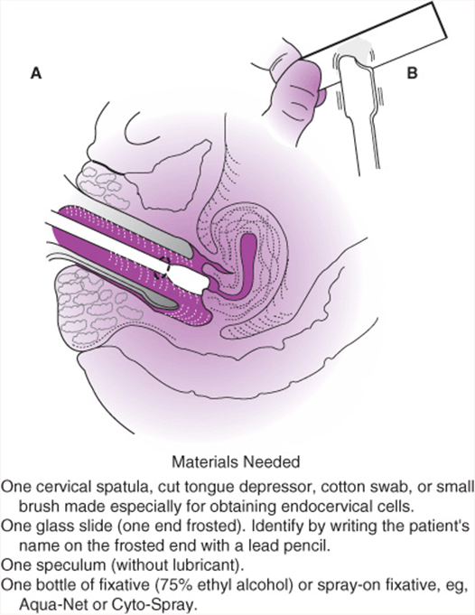
Figure 35–6. Preparation of a Papanicolaou (Pap) smear. A: Obtain cervical scraping from complete squamocolumnar junction by rotating 360 degrees around the external os. B: Place the material 1 in from the end of the slide and smear along the slide to obtain a thin preparation. Place a saline-soaked cotton swab or small endocervical brush into the endocervical canal and rotate 360 degrees. Place this specimen onto the same slide and quickly fix with fixative. (Reproduced, with permission, from Pernoll ML. Benson & Pernoll’s Handbook of Obstetrics and Gynecology. 10th ed. New York, NY: McGraw-Hill; 2001.)

Figure 35–7. Specula. (Reproduced, with permission, from Pernoll ML. Benson & Pernoll’s Handbook of Obstetrics and Gynecology. 10th ed. New York, NY: McGraw-Hill; 2001.)
Bimanual Examination
The uterus and adnexal structures should be outlined between the 2 fingers of the hand in the vagina and the flat of the opposite hand, which is placed on the lower abdominal wall (Fig. 35–8). Gentle palpation and manipulation of the structures will delineate position, size, shape, mobility, consistency, and tenderness of the pelvic structures—except in the obese or uncooperative patient or in a patient whose abdominal muscles are taut as a result of fear or tenderness. Tenderness can be elicited either on direct palpation or on movement or stretching of the pelvic structures.

Figure 35–8. Bimanual pelvic examination.
A. Cervix
The cervix is a firm structure traditionally described as having the consistency of the tip of the nose. Normally it is round and approximately 3–4 cm in diameter. Various appearances of the cervix are shown in Figure 35–5. The external os is round and virtually closed. Multiparous women may have an os that has been lacerated. An irregularity in shape or nodularity may be due to 1 or more nabothian cysts. If the cervix is extremely firm, it may contain a tumor, even cancer. The cervix (along with the body of the uterus) normally is moderately mobile, so it can be moved 2–4 cm in any direction without causing undue discomfort. (When examining a patient, it is helpful to warn her that she will feel the movement of her uterus but that ordinarily this maneuver is not painful.) Restricted mobility of the cervix or corpus often follows inflammation, neoplasia, or surgery.

Figure 35–5. Uterine cervix: normal and pathologic appearance.
B. Corpus of the Uterus
The corpus of the uterus is approximately half the size of the patient’s fist and weighs approximately 70–90 g. It is regular in outline and not tender to pressure or moderate motion. In most women, the uterus is anteverted; in approximately one-third of women, it is retroverted (see Chapter 42). A retroverted uterus usually is not a pathologic finding. In certain cases of endometriosis or previous salpingitis, the “tipped” uterus may be the result of adhesions caused by the disease process. The uterus usually is described in terms of its size, shape, position, consistency, and mobility.
C. Adnexa
Adnexal structures (fallopian tubes and ovaries) cannot be palpated in many overweight women because the normal tube is only approximately 7 mm in diameter and the ovary is no more than 3 cm in its greatest dimension. In very slender women, however, the ovaries nearly always are palpable and, in some instances, the oviducts are as well. Usually no adnexal structures can be palpated in the postmenopausal woman. Unusual tenderness or enlargement of any adnexal structure indicates the need for further diagnostic procedures; an adnexal mass in any woman is an indication for investigation.
Rectovaginal Examination
At the completion of the bimanual pelvic examination, a rectovaginal examination should always be performed especially after age 40 years. The well-lubricated middle finger of the examining hand should be inserted gently into the rectum to feel for tenderness, masses, or irregularities. When the examining finger has been inserted a short distance, the index finger can then be inserted into the vagina until the depth of the vagina is reached (Fig. 35–9). It is much easier to examine some aspects of the posterior portion of the pelvis by rectovaginal examination than by vaginal examination alone. The index finger can now raise the cervix toward the anterior abdominal wall, which stretches the uterosacral ligaments. Usually this process is not painful; if it causes pain—and especially if the finger in the rectum can palpate tender nodules along the uterosacral ligaments—endometriosis may be present.
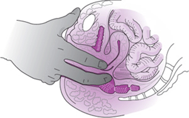
Figure 35–9. Rectovaginal examination.
Occult Bleeding Due to Colorectal Cancer
In the United States, colorectal cancer (CRC) is the third most common cancer diagnosed among men and women and the second leading cause of death from cancer. CRC largely can be prevented by the detection and removal of adenomatous polyps, and survival is significantly better when CRC is diagnosed while still localized. Recent evidence has revealed an unacceptably wide range of sensitivity among strategies aimed at checking the feces for occult blood, with some practices and tests performing so poorly that the large majority of prevalent cancers are missed at the time of screening. Therefore, a single stool sample for fecal occult blood testing obtained by digital rectal examination is not adequate for the detection of CRC and should not be used for CRC screening. Furthermore, it is the physician’s role to encourage patients above 50 years of age or those with familial CRC to undergo procedures aimed at prevention of CRC (such as colonoscopy, sigmoidoscopy, or computed tomography [CT] scan) and to not diagnose CRC using methods detecting occult blood in the stool.
American College of Obstetricians and Gynecologists. Routine Pelvic Examination and Cervical Cytology Screening. ACOG Committee Opinion No 431. Washington, DC: American College of Obstetricians and Gynecologists; 2009.
Levin B, Lieberman DA, McFarland B, et al. Screening and surveillance for the early detection of colorectal cancer and adenomatous polyps, 2008. CA Cancer J Clin 2008;58:130–160. PMID: 18322143.
DIAGNOSTIC OFFICE PROCEDURES
Certain diagnostic procedures can be performed in the office because complicated equipment and general anesthesia are not required. Other office diagnostic procedures useful in specific situations (eg, tests used in infertility evaluation) can be found in appropriate chapters elsewhere in this book.
Tests for Vaginal Infection
If abnormal vaginal discharge is present, a sample of vaginal discharge should be scrutinized. A culture is obtained by applying a sterile cotton-tipped applicator to the suspect area and then transferring the suspect material to an appropriate culture medium. Because this procedure is inconvenient to perform in the physician’s office, most laboratories supply a prepackaged kit that allows the physician to put the cotton-tipped applicator into a sterile container, which is then sent to the laboratory. The vaginal discharge can also be tested for the vaginal pH. An acidic pH of 4–5 is consistent with fungal infection, whereas an alkaline pH of 5.5–7 suggests infections such as bacterial vaginosis and Trichomonas. Often an endocervical infection may be perceived as a vaginal infection. Obtaining a swab for gonorrhea and chlamydia testing from the endocervix is warranted.
A. Saline (Plain Slide)
To demonstrate Trichomonas vaginalis organisms, the physician mixes on a slide 1 drop of vaginal discharge with 1 drop of normal saline warmed to approximately body temperature. The slide should have a coverslip. If the smear is examined while it is still warm, actively motile trichomonads usually can be seen.
The saline slide can also be used to look for the mycelia of the fungus Candida albicans, which appear as segmented and branching filaments. The slide can be useful in looking for bacterial vaginosis by looking for “clue cells,” epithelial cells covered from edge to edge by short coccobacilli-type bacteria.
B. Potassium Hydroxide
One drop of an aqueous 10% potassium hydroxide solution is mixed with 1 drop of vaginal discharge on a clean slide and a coverslip applied. The potassium hydroxide dissolves epithelial cells and debris and facilitates visualization of the mycelia of a fungus causing vaginal infection. The slide can be brought near the nose to determine if the discharge has a “fishy” odor. This odor is strongly suggestive of bacterial vaginosis, a common vaginal infection associated with a mixed anaerobic bacterial flora. In addition, this same slide with a coverslip can be magnified with a microscope to visualize mycelia that may have been hidden by debris with just the saline smear.
C. Bacterial Infection
Bacterial infection may be present, especially if there is an ischemic lesion such as occurs after radiation therapy for cervical carcinoma, or if a patient is suspected of having bacterial vaginosis, gonorrhea, or a Chlamydia trachomatis infection. Material from the cervix, urethra, or vaginal lesion can be smeared, stained, and examined microscopically, or the material can be cultured.
Fern Test for Ovulation
The fern test can determine the presence or absence of ovulation or the time of ovulation. When cervical mucus is spread on a clean, dry slide and allowed to dry in air, it may or may not assume a frondlike pattern when viewed under the microscope (sometimes it can be seen grossly). The fern frond pattern indicates an estrogenic effect on the mucus without the influence of progesterone; thus, a non-frondlike pattern can be interpreted as showing that ovulation has occurred (Fig. 35–10).

Figure 35–10. Patterns formed when cervical mucus is smeared on a slide, permitted to dry, and examined under a microscope. Progesterone makes the mucus thick and cellular. In the smear from a patient who failed to ovulate (bottom), there is no progesterone to inhibit the estrogen-induced fern pattern. (Reproduced, with permission, from Ganong WF. Review of Medical Physiology. 20th ed. New York, NY: McGraw-Hill; 2003.)
Schiller Test for Neoplasia
Although colposcopy is more accurate, the Schiller test can be performed when cancer or precancerous changes of the cervix or vaginal mucosa are suspected. The suspect area is painted with Lugol’s (strong iodine) solution, which interacts and marks the glycogen-rich epithelial cells of the cervix. Any portion of the epithelium that does not accept the dye is abnormal because of the presence of scar tissue, neoplasia and precursors, and columnar epithelium. Biopsy of samples taken from this area should be performed if there is any suspicion of cancer.
Biopsy
A. Vulva & Vagina
For biopsy of the vulva or vagina, a 1–2% aqueous solution of a standard local anesthetic solution can be injected around a small suspicious area and a sample obtained with a skin punch or sharp scalpel. Bleeding usually can be controlled by pressure or by Monsel’s solution, but occasionally suturing is necessary.
B. Cervix
Colposcopically directed biopsy is the method of choice for the diagnosis of cervical lesions, either suspected on visualization or indicated after an abnormal Pap smear. Colposcopy should reveal the full columnar–squamous “transformation zone” (TZ) at the juncture of the exocervix and endocervix. In addition, it may be advisable to sample the endocervix by curettage. Specific instruments have been devised for cervical biopsy and endocervical curettage (Fig. 35–11). The cervix is less sensitive to cutting procedures than is the vagina, so 1 or more small biopsy samples of the cervix can be taken with no or little discomfort to the patient. Bleeding usually is minimal and controlled with light pressure for a few minutes or by use of Monsel’s solution. A “4-quadrant” biopsy sample of the squamocolumnar junction can be taken at 12, 3, 6, and 9 o’clock positions if colposcopy is not available. A Schiller test often may more quickly direct the physician to the area that should be biopsied.

Figure 35–11. Biopsy instruments.
C. Endometrium
Endometrial biopsy can be helpful in the diagnosis of ovarian dysfunction (eg, infertility) or irregular uterine bleeding and as a test for carcinoma of the uterine corpus. Endometrial biopsy can be performed with flexible disposable cannulas, such as the Pipelle, which have replaced most metal curettes previously used (Fig. 35–12). In fact, endometrial biopsies have dramatically reduced the need for formal dilatation and curettage (D&C) because the accuracy of biopsy is nearly the same. Because the procedure causes cramping, the patient should be warned and advised to take a pain medication such as ibuprofen 1 hour prior to the procedure.

Figure 35–12. Sites of endometrial biopsy. (Reproduced, with permission, from Pernoll ML. Benson & Pernoll’s Handbook of Obstetrics and Gynecology. 10th ed. New York, NY: McGraw-Hill; 2001.)
DIAGNOSTIC LABORATORY PROCEDURES
Routine procedures that are not discussed here but should be considered with periodic primary care visits include a complete blood count (including differential white cell count), glucose screening, lipid profile, and thyroid function tests. The frequency with which these tests are performed should be at the discretion of the physician, based on risk factors and presenting complaints.
Urinalysis
Urinalysis should be obtained in symptomatic patients and should include both gross and microscopic examinations. A microscopic examination may reveal crystals or bacteria, but unless the specimen is collected in a manner that will exclude vaginal discharge, the presence of bacteria is meaningless (see below).
Urine Culture
Studies have demonstrated that a significant number of women (approximately 3% of nonpregnant women and 7% of pregnant women) have asymptomatic urinary tract infections. Culture and antibiotic sensitivity testing are required for the diagnosis and as a guide to treatment of urinary tract infections.
Reliable specimens of urine for culture often can be obtained by the “clean-catch” method: The patient is instructed to cleanse the urethral meatus carefully with soap and water, to urinate for a few seconds to dispose of urethral contaminants, and then to catch a “midstream” portion of the urine. It is essential that the urine not dribble over the labia, but this may be difficult for some patients to accomplish.
A more reliable method of collecting urine for culture is by sterile catheterization performed by the physician or nurse. However, care must be exercised in catheterization to minimize the risk of introducing an infection.
Other Cultures
A. Urethral
Urethral cultures are indicated if an STD is suspected.
B. Vaginal
A culture usually is unnecessary for the diagnosis of vaginal infections, because visual inspection or microscopic examination usually will enable the physician to make a diagnosis, eg, curdlike vaginal material that reveals mycelia (candidiasis). However, a culture should be obtained in questionable cases. In cases of vaginal candidiasis refractory to the common treatment, it is advisable to characterize the yeast and examine its specific sensitivity to various antimycotic drugs available, enabling elimination of the troublesome infection.
C. Cervical
As in the case of the urethra, the usual indication for a culture of cervical discharge is the suspected presence of an STD.
Specific Tests
A. Herpesvirus Hominis
Herpesvirus hominis (HSV) (herpes genitalis, both types 1 and 2) is a frequently seen vulvar lesion (see Chapter 43). It can be diagnosed by the cytopathologist, who finds typical cellular changes. Other methods that are more accurate and more often used for the detection of HSV infection include culturing and identifying the virus using polymerase chain reaction (PCR) technique.
B. Human Papillomavirus
Human papillomavirus (HPV) infection is associated with the development of genital warts and the occurrence of vaginal and cervical intraepithelial lesions. Some of these lesions are precancerous or cancerous in origin. Different HPV subtypes are linked to either benign or more aggressive epithelial changes. The different subtypes can be identified by the specific fingerprints obtained from the PCR products.
C. Chlamydia & Gonorrheal Infections
These sexually transmitted infections are the 2 most prevalent infections, with chlamydia being the most common. They are found more often in women who have multiple sexual partners and those who do not use barrier methods of contraception. Nucleic acid amplification testing is the most commonly used method of diagnosis, with a sensitivity >90%.
D. Human Immunodeficiency Virus
Acquired immunodeficiency syndrome (AIDS) has become one of the most difficult issues confronting all kinds of clinicians. The need to screen for HIV in the general population has become more pressing given that the largest increase in incidence is seen in young heterosexually active females with no other risk factors. An accurate blood test is available for diagnosis. Prior to drawing the blood, the physician must discuss with the patient the accuracy of the blood test for diagnosing the presence of HIV. The patient must be made aware that there are infrequent false-positive tests and a “window” during which the test may be falsely negative prior to the development of antibodies. At present, a written consent must be signed by the patient prior to drawing the blood.
Other Specific Tests
Specific diagnostic laboratory procedures may be indicated for some of the less common venereal diseases (eg, lymphogranuloma venereum and hepatitis B and C). A screening test for Streptococcus B carrier is advocated at 35–37 weeks’ gestation. A 1-step culture swab from the lower vagina, followed by the anus, is recommended. These tests are discussed with the specific diseases in other chapters of this book.
Pregnancy Testing
Pregnancy testing is discussed in Chapter 6.
Papanicolaou Smear of Cervix
The Pap smear is an important part of the gynecologic examination. The frequency of the need for this test has been recently revised. Epidemiologic statistics have led the US Preventive Services Task Force to recommend that for the average woman who has had 3 normal Pap smears, a Pap test every 2 or 3 years is adequate. This recommendation is based on the observation that most cervical cancers are slow growing. The American College of Obstetricians and Gynecologists recommends annual Pap smear screenings from 3 years after the start of sexual intercourse but no later than age 21 years. For women aged 30–64 years, the frequency of screening may be reduced to every 2–3 years after 3 consecutive negative Pap smears. Patients at risk, including women with multiple sexual partners, a history of STD, genital condylomata, or prior abnormal Pap smears; women who are receiving immune suppression therapy; women who are infected with HIV; or women who were exposed to diethylstilbestrol (DES) in utero, should continue to be screened annually. Women who are HIV positive should have cervical cytology testing every 6 months after diagnosis, and then annually after 2 consecutive normal test results. HPV vaccination status does not change theses cervical cytology screening recommendations. The physician can consider discontinuing cervical cytology at 65–70 years of age if patient has had 3 or more normal results in a row, no abnormal test results in 10 years, no history of cervical cancer, and no history of DES exposure in utero, is HIV negative, is not immunodepressed, and does not have other risk factors for new acquisition of STDs; if cervical cytology has been discontinued, the physician should review risk factors annually to evaluate the need for re-initiation of screening.
Aside from premalignant and malignant changes, other local conditions often can be suspected by the cytologist. Viral infections, such as HSV, HPV, and condylomata acuminata, can be seen as mucosal changes. Actinomycosis and Trichomonas infections can be detected by a Pap smear.
The Pap smear is a screening test only. Positive tests are an indication for further diagnostic procedures, such as colposcopy, endocervical curettage, cervical biopsy or conization, endometrial biopsy, or D&C. The properly collected Pap smear can accurately lead to the diagnosis of carcinoma of the cervix in approximately 95% of cases. The Pap smear also is helpful in the detection of endometrial abnormalities such as endometrial polyps, hyperplasia, and cancers, but it detects fewer than 50% of cases.
The techniques of collection of a Pap smear may vary, but the following is a common procedure.
The patient should not have douched for at least 24 hours before the examination and should not be menstruating. The speculum is placed in the vagina after it has been lubricated with water only. With the cervix exposed, a specially designed plastic or wooden spatula is applied to the cervix and rotated 360 degrees to abrade the surface slightly and to pick up cells from the squamocolumnar area of the cervical os. Next, a cotton-tipped applicator or a small brush is inserted into the endocervix and rotated 360 degrees. These 2 specimens can be mixed or placed on the slide separately according to the preference of the examiner. A preservative is applied immediately to prevent air drying, which would compromise the interpretation. The slide is sent to the laboratory with an identification sheet containing pertinent history and findings (see Fig. 35–6). Another method called ThinPrep automates the preparation of the Pap smear slide so that the variability introduced by the clinician preparing the slide itself is no longer a factor. With this method, the specimen is placed in a liquid-based medium and sent to the laboratory. In addition, the ThinPrep technique decreases the rate of smears showing atypical squamous cells–undetermined significance (ASCUS), thereby decreasing the need for colposcopic evaluations. For these reasons, in many parts of the country, the ThinPrep technique has replaced the conventional Pap smear. However, any advantages of the liquid-based technique over the conventional method in terms of sensitivity and specificity are unclear
The liquid-based medium allows for testing for high-risk HPV, the most common being subtypes 16, 18, 31, 33, and 35. Testing for high-risk HPV has been proposed by the American Society for Colposcopy and Cervical Pathology as a method of evaluating and sorting out patients with ASCUS Pap smear results. If no high-risk HPV is present in the ASCUS Pap smear, then these individuals can be followed-up with a repeat Pap smear in 1 year, similar to those who have a negative Pap smear. Patients known to have a high-risk HPV subtype would undergo colposcopic evaluation.
The laboratory reports the Pap smear using the Bethesda System, which has advocated a standardized reporting system for cytologic reports. Chapter 48 discusses the recently updated nomenclature.
Alternatives to the traditional Pap smear are being evaluated in an attempt to decrease the false-negative and false-positive Pap smear results. Evidence indicates that computerized screening of Pap smears can decrease the likelihood of missing significant pathologies. Various methods of computerized screening have been developed to aid the human eye in picking up abnormalities, although no system has yet achieved widespread acceptance.
Colposcopy
The colposcope is a binocular microscope used for direct visualization of the cervix (Fig. 35–13). Magnification as high as 60 × is available, but the most popular instrument in clinical use has 13.5 × magnification, which effectively bridges the gap between what can be seen by the naked eye and by the microscope. Some colposcopes are equipped with a camera for single or serial photographic recording of pathologic conditions.
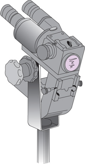
Figure 35–13. Zeiss colposcope.
Colposcopy does not replace other methods of diagnosing abnormalities of the cervix; rather, it is an additional and important tool. The 2 most important groups of patients who can benefit by its use are (1) patients with an abnormal Pap smear and (2) DES-exposed daughters, who may have dysplasia of the vagina or cervix (see Chapter 40).
The colposcopist is able to see areas of cellular dysplasia and vascular or tissue abnormalities not visible otherwise, which makes possible the selection of areas most propitious for biopsy. Stains and other chemical agents are also used to improve visualization. The colposcope has reduced the need to perform blind cervical biopsies for which the rate of finding abnormalities is low. In addition, the necessity for a cone biopsy, a procedure with a high morbidity rate, has been greatly reduced. Thus the experienced colposcopist is able to find focal cervical lesions, obtain directed biopsy at the most appropriate sites, and make decisions about the most appropriate therapy largely based on what is seen through the colposcope.
Hysteroscopy
Hysteroscopy enables the gynecologist to examine the uterine cavity through a fiberoptic instrument, called the hysteroscope. Moreover, surgical interventions such as polypectomy, myomectomy, septectomy, and resection of intrauterine adhesions can be performed via the hysteroscope. In order to inspect the interior of the uterus with the hysteroscope, the uterine cavity is inflated with a solution (usually saline, but other solutions such as glycine or dextran can be used) or by carbon dioxide insufflation. Diagnostic hysteroscopy is usually performed with no sedation; however, intravenous sedation, paracervical block, or general anesthesia is often adequate for operative hysteroscopies.
Hysteroscopic applications include evaluation for abnormal uterine bleeding, resection of uterine synechiae and septa, removal of polyps and intrauterine devices (IUDs), resection of submucous myomas, and endometrial ablation. Most of these therapeutic maneuvers require extensive manipulation, so regional or general anesthesia is required.
Hysteroscopy should be performed only by physicians with proper training. The tip of the instrument should be inserted just beyond the internal cervical os and then advanced slowly, with adequate distention under direct vision. Hysteroscopy is often used in conjunction with other operative procedures, such as curettage and laparoscopy.
Failure of hysteroscopy may be the result of cervical stenosis, inadequate distention of the uterine cavity, bleeding, or excessive mucus secretion. The most common complications include perforation, bleeding, and infection. Perforation of the uterus usually occurs at the fundus. Unless a viscus is damaged or internal bleeding develops, surgical repair may not be required. Bleeding generally subsides, but fulguration following attempts to remove polyps or myomas may be required to stop bleeding in some cases. Parametritis or salpingitis, rarely noted, usually necessitates antibiotic therapy. Intravascular extravasation of fluid or gas from hysteroscopy often does not become clinically significant but has been associated with severe consequences such as hyponatremia, air embolism, cerebral edema, and even death.
Culdocentesis
The passage of a needle into the cul-de-sac—culdocentesis—in order to obtain fluid from the pouch of Douglas is a diagnostic procedure that can be performed in the office or in a hospital treatment room (Fig. 35–14). The type of fluid obtained indicates the type of intraperitoneal lesion (eg, bloody with a ruptured ectopic pregnancy, pus with acute salpingitis, or ascitic fluid with malignant cells in cancer). With refinements in ultrasound technology enabling more definitive evaluation of pelvic pathology, culdocentesis is performed rarely today.

Figure 35–14. Culdocentesis.
Radiographic Diagnostic Procedures
Many common radiologic procedures may be helpful in the diagnosis of pelvic conditions. The “flat film” shows calcified lesions, teeth, or a ring of a dermoid cyst and indicates other pelvic masses by shadows or displaced intestinal loops. Use of contrast media frequently is indicated to help delineate pelvic masses or to rule out metastatic lesions. Barium enema, upper gastrointestinal series, intravenous urogram, and cystogram may be helpful. With the improvement of technologies such as ultrasound, CT scans, and MRI, the use of “flat films” for the diagnosis of gynecologic abnormalities has become less frequent.
Hysterography & Sonohysterography
The uterine cavity and the lumens of the oviducts can be outlined by instillation of contrast medium through the cervix, followed by fluoroscopic observations or film. The technique was first widely used for the diagnosis of tubal disease as part of the investigation of infertile women. Its use now is being extended to the investigation of uterine disease.
To diagnose tubal patency or occlusion, the medium is instilled through a cervical cannula. Filling of the uterine cavity and spreading of the medium through the tubes are watched via a fluoroscope, with the radiologist taking spot films at intervals for subsequent, more definitive, scrutiny. If no occlusion is present, the medium will reach the fimbriated end of the tube and spill into the pelvis—evidence of tubal patency. This procedure can reveal an abnormality of the uterus (eg, congenital malformation, submucous myomas, or endometrial polyps).
Another technique that is gaining acceptance is sonohysterography, in which the uterine cavity is filled with fluid while ultrasound is used to delineate the architecture of the endometrial cavity and detect a spillage through the fallopian tubes. Thus, it becomes easier to diagnose intrauterine abnormalities, such as polyps or fibroids, and tubal patency.
Angiography
Angiography is the use of radiographic contrast medium to visualize the blood vascular system. By demonstrating the vascular pattern of an area, tumors or other abnormalities can be delineated. Angiography also is used to delineate continued bleeding from pelvic vessels postoperatively, to visualize bleeding from infiltration by cancer in cancer patients, to embolize the uterine arteries in order to treat postpartum hemorrhage following vaginal or caesarean deliveries, to decrease acute bleeding in cases of cervical or cornual pregnancies, and/or to reduce the size of uterine myomas. These vessels then can be embolized with synthetic fabrics to stop the bleeding or indicate therapy that can prevent the need for a major abdominal operation in a highly compromised patient.
Computed Tomography
CT scan is a diagnostic imaging technique that provides high-resolution 2-dimensional images. The CT scan takes cross-sectional images through the body at very close intervals so that multiple “slices” of the body are obtained. The beam transmission is measured and calculated through an array of sensors that are approximately 100 times more sensitive than conventional x-rays. The computer is able to translate the densities of different types of tissues into gray-scale pictures that can be read on an x-ray film or a television monitor.
Contrast media can be given orally, intravenously, or rectally. They are used to outline the gastrointestinal and urinary systems, thus helping to differentiate these organ systems from the pelvic reproductive organs. In gynecology, the CT scan is most useful in accurately diagnosing retroperitoneal lymphadenopathy associated with malignancies. It also has been used to determine the depth of myometrial invasion in endometrial carcinoma as well as extrauterine spread. It is an accurate tool for locating pelvic abscesses that cannot be located by ultrasonography. Often a needle can be placed into an abscess pocket to both drain the abscess and determine what organism may be involved. Pelvic thrombophlebitis often can be diagnosed by CT scan as an adjunct to clinical suspicion. Common abnormalities such as ovarian cysts and myomas are easily diagnosed (Fig. 35–15).
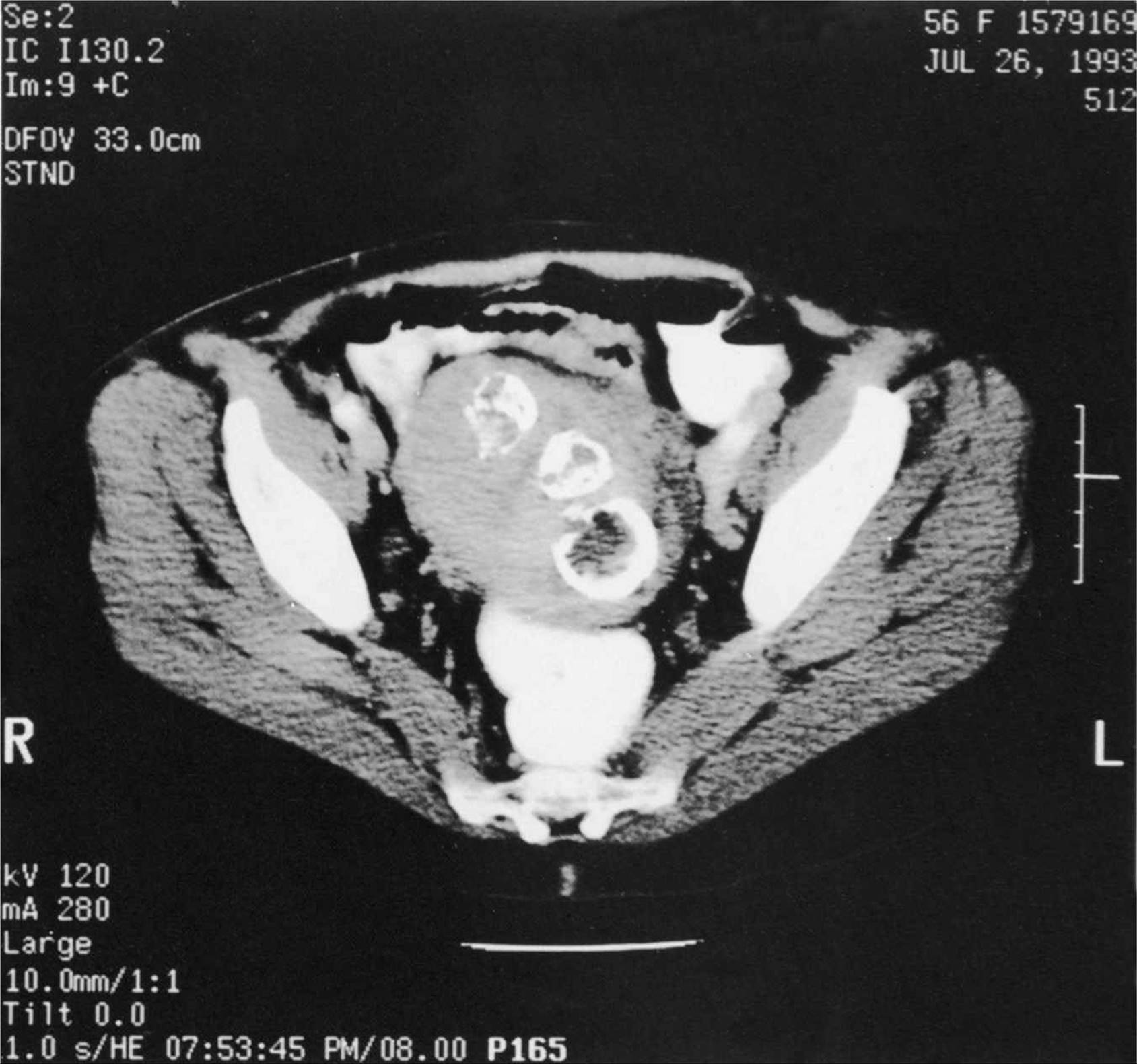
Figure 35–15. Computed tomography scan of the pelvis showing a large fibroid uterus with 3 calcified fibroids in the body of the uterus. (Reproduced, with permission, from Dr. Barbara Carter, New England Medical Center, Boston, MA.)
Magnetic Resonance Imaging
MRI is a diagnostic imaging technique that creates a high-resolution, cross-sectional image of the body like a CT scan. The technique is based on the body absorbing radio waves from the machine. A small amount of this energy is absorbed by the nuclei in the various tissues. These nuclei act like small bar magnets and are influenced by the magnetic field created by the machine. These nuclei then emit some of the radio waves back out of the body. The waves are picked up by sensitive and sophisticated receivers, and these signals are translated into images by computer technology.
The advantages of MRI include the fact that it uses nonionized radiation that has no adverse or harmful effects on the body. MRI is superior to CT in its ability to differentiate among various types of tissue, including inflammatory masses, cancers, and abnormal tissue metabolism. Its disadvantages are mainly its high cost and its poor demonstration of calcifications. Its main use in gynecology appears to be staging and follow-up of pelvic cancers. MRI in obstetrics is used mainly as an adjunct to ultrasonic prenatal diagnosis of fetal anomalies. It allows for multiple image cuts that can help decipher complex anomalies. Other potential uses of MRI include evaluation of placental blood flow and accurate performance of pelvimetry.
Ultrasonography
Ultrasonography records high-frequency sound waves as they are reflected from anatomic structures. As the sounds waves pass through tissues, they encounter variable acoustic densities. Each of the tissues returns a different echo, depending on the amount of energy reflected. This echo signal can be measured and converted into a 2-dimensional image of the area under examination, with the relative densities shown as differing shades of gray.
Ultrasonography is a simple and painless procedure that has the added advantage of freedom from any radiation hazard. It is especially helpful in patients in whom an adequate pelvic examination may be difficult, such as in children, virginal women, and obese and uncooperative patients.
The pelvis and lower abdomen are scanned and recorded at regular intervals of distance, using a sector scanner that provides a better 2-dimensional picture than does the linear array scanner (Fig. 35–16). Generally, the abdominal scan is performed with the bladder full; this condition elevates the uterus out of the pelvis, displaces air-filled loops of bowel, and provides the operator with an index of density—a sonographic “window” differentiating the pelvic organs.
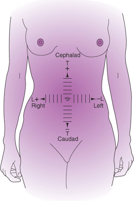
Figure 35–16. Planes of ultrasonograms.
Ultrasonography can be helpful in the diagnosis of almost any pelvic abnormality, as all structures, normal and abnormal, usually can be demonstrated. In most instances, a clinical picture has been developed—by history, physical examination, or both—before ultrasonograms are obtained. Thus, the scan often corroborates the clinical impression, but it also may uncover an unexpected condition of which the clinician should be aware.
There are many indications for ultrasonography. Normal early pregnancy can be diagnosed, as can pathologic pregnancies such as incomplete and missed abortions and hydatidiform moles. Ultrasonography can be extremely helpful in avoiding the placenta and fetus during midtrimester amniocentesis. The uses for ultrasound examination in obstetrics are discussed elsewhere in this book.
Ultrasonography may be used to locate a lost IUD or a foreign body in the vagina of a child. Congenital malformations such as a bicornuate uterus or vaginal agenesis are sometimes, but not always, detected. The development of 3-dimensional CT scan has introduced this technology to the more accurate diagnosis of congenital uterine abnormalities.
Ultrasound examination is useful in the placement of uterine tandems for radiation therapy for endometrial cancer and for guidance during second-trimester abortion procedures.
One of the more common uses for ultrasonography is the diagnosis of pelvic masses. Often because of their location, attachment, and density, myomas can be diagnosed without too much difficulty (Fig. 35–17A).
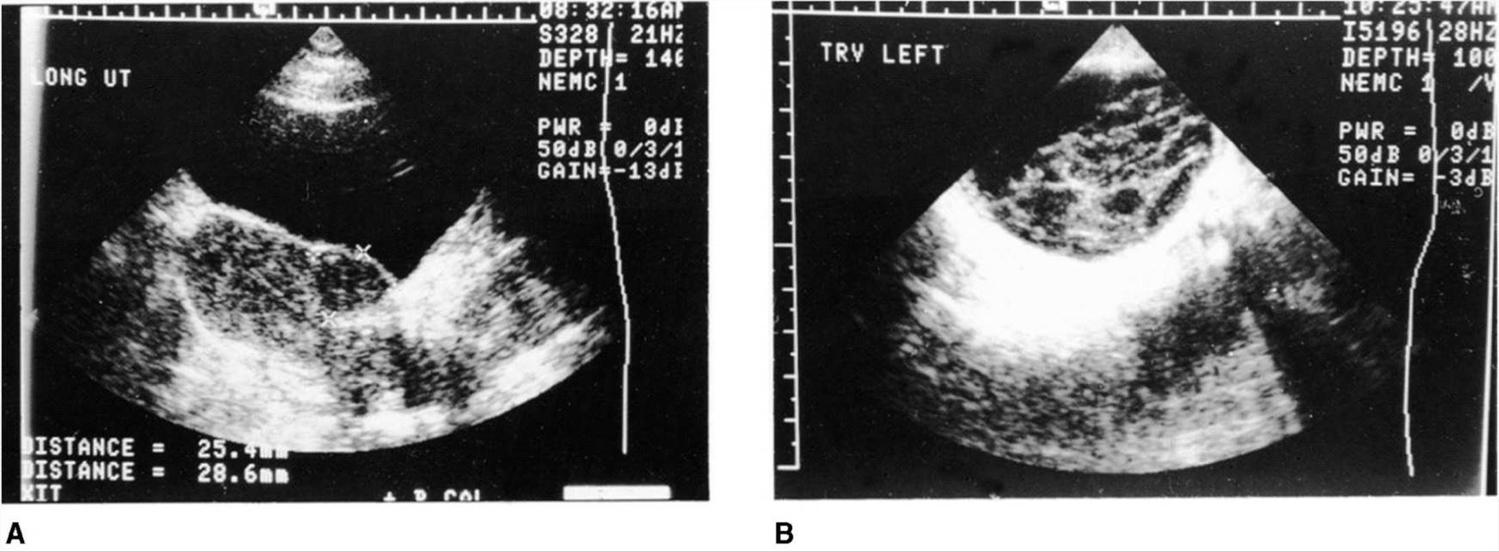

Figure 35–17. A: Longitudinal view of the uterus with anterior fibroid outlined by the x’s; bladder anterior. B: Transverse section through an endometrioma with multiple loculations and debris. C:Longitudinal view of large ovarian cyst outlined by the +’s and x’s with a focal multicystic area. D: Longitudinal view of a dermoid cyst showing areas of fat within the cyst.
Adnexal masses can be found with relative ease by ultrasonography, although an accurate diagnosis is more difficult because of the various types of adnexal masses that can be found (Fig. 35–17B and C).
Ovarian cysts can be described as unilocular or multilocular, totally fluid-filled, or partially solid. A common adnexal mass, the dermoid cyst, can have characteristic ultrasound findings because of fat tissue and bone densities seen in these cysts (Fig. 35–17D). Pelvic abscesses can be diagnosed by ultrasonography, especially if a well-encapsulated large abscess pocket is present.
In addition to the traditional abdominal scan, the vaginal probe scan has become a useful modality. The vaginal probe is used for determining early gestations and can diagnose a pregnancy as early as 5 weeks from the LNMP. The vaginal ultrasonographic probe is capable of visualizing ectopic pregnancies quite accurately.
Ultrasonography is commonly used to diagnose ovarian cysts, especially in obese patients in whom abdominal scans are of limited use. The vaginal scan is used often to determine follicular size with in vitro fertilization and to predict the best time for ovum retrieval.
Innovations in ultrasound probes and computerizing processes of the obtained images enable the development of 3-dimensional ultrasound machines. The 3-dimensional images help to accurately evaluate normal and abnormal findings, such as uterine shape and cavity, pelvic masses, and fetal malformations.
Carbon Dioxide Laser
Controlled tissue vaporization by laser is a modality for treatment of cervical, vaginal, or perineal condylomata and dysplasia. It also can be used for conization of the cervix for diagnosis of dysplasia or carcinoma within the cervical canal.
The vaporization procedure is not difficult, but training is essential, especially in the physics of laser light and the potential risks of laser therapy not only to the patient but to the operator and others in the immediate vicinity. Antiseptic preparation of the vagina should be gentle to avoid trauma to the tissue that is to be examined histologically. Local anesthesia, with or without preliminary intravenous sedation, usually is adequate.
Advantages of the laser method of cervical conization include little or no pain; a low incidence of infection because the beam sterilizes the tissues; decreased blood loss, because the laser instrument—at a decreased energy level—is a hemostatic agent; less tissue necrosis than occurs with electrocautery (but probably the same as with excision by a sharp knife); and a decreased incidence of postoperative cervical stenosis.
Loop Electrosurgical Excision Procedure
Loop electrosurgical excision procedure (LEEP) is another modality of therapy for vulvar and cervical lesions. LEEP uses a low-voltage, high-frequency alternating current that limits thermal damage but at the same time has good hemostatic properties. It is most commonly used for excision of vulvar condylomata and cervical dysplasias and for cone biopsies of the cervix. It has displaced sharp knife and laser cone biopsies for treatment of most cervical dysplasias.
The technique requires the use of local anesthesia followed by the use of a wire loop cautery unit that cauterizes and cuts the desired tissue. Loops of various sizes are used for specimens of different size. The major advantages of LEEP are its usefulness in an office setting with lower equipment cost, minimal damage to the surrounding tissue, and low morbidity.
Vassilakos P, Schwartz D, de Marval F, et al. Biopsy-based comparison of liquid-based, thin-layer preparations to conventional Pap smear. J Reprod Med 2000;45:11–16. PMID: 10664916.
Wright T, Massad LS, Dunton CJ, et al. Interim guidelines for the use of human papillomavirus DNA testing as an adjunct to cervical cytology screening. Obstet Gynecol 2004;103:304–309. PMID: 17917566.