
Richard L. Stouffer1 and Jon D. Hennebold1
(1)
Division of Reproductive & Developmental Sciences, Oregon National Primate Research Center, Oregon Health & Science University, West, Campus, 505 NW 185th Ave, Beaverton, OR 97006, USA
Richard L. Stouffer (Corresponding author)
Email: stouffri@ohsu.edu
Jon D. Hennebold
Email: henneboj@ohsu.edu
Abstract
The primate corpus luteum undergoes a process at the end of a nonfertile menstrual cycle termed luteolysis, which involves considerable structural and functional changes that lead to a loss in the ability to produce the steroid hormone progesterone. Because progesterone is critical for events involved in embryo implantation and sustaining pregnancy, the survival and continued function of the corpus luteum are required throughout the first weeks of pregnancy, after which the placenta becomes responsible for the maintenance of gestation. Extension of the functional lifespan of the primate corpus luteum is achieved through the secretion of chorionic gonadotropin (CG) from the conceptus. CG signals through the luteinizing hormone–chorionic gonadotropin receptor (LHCGR) located on luteal cells to override the cellular and molecular events that are responsible for the demise of the corpus luteum during nonfertile cycles. Thus, in this chapter, the source of various CG forms and the regulation of their production, as well as the mechanisms through which LHCGR signaling regulates cellular activities in the primate corpus luteum during early pregnancy are reviewed. Also, current and possible uses of hCG forms for diagnosis and treatment of infertility and pregnancy disorders are considered.
Keywords
PrimatePregnancyMaternal recognition of pregnancyCorpus luteumLuteolysisLuteal rescueChorionic gonadotropinLuteinizing hormone–chorionic gonadotropin receptorProgesteroneRelaxin
10.1 Introduction
A vital process in the ovary of mammalian species is the differentiation of the corpus luteum from the wall of the ovulatory follicle and its production of hormones, notably progesterone (P4), that are essential for the initiation and maintenance of intrauterine pregnancy. However, species have evolved diverse mechanisms for controlling the functional lifespan of the corpus luteum in adult females during both their ovarian cycle and pregnancy [1]. In primates, including women, the functional lifespan of the corpus luteum during the ovarian (menstrual) cycle is sufficient for oocyte–sperm interaction and fertilization, movement of the early (pre-blastocyst stage) embryo through the oviduct, preparation of the uterus for implantation, plus blastocyst attachment and the early trophoblastic invasion of the endometrium. However, the corpus luteum is a transient gland. The regression of the corpus luteum near the end of the menstrual cycle in primates (approximately 2 weeks after initial differentiation from the luteinizing follicle) typically occurs before the developing placenta acquires the ability to produce progesterone, which keeps the uterus in a quiescent, supportive state for gestation. Thus, the functional lifespan of the corpus luteum must be extended in a fertile cycle for a limited time, until its essential activities (e.g., progesterone production) are usurped by the placenta (i.e., the luteal–placental shift). A critical event for fertility in primates occurs when the early conceptus signals to the mother (i.e., maternal recognition of pregnancy) that intrauterine pregnancy is beginning and extends luteal function until the luteal–placental shift.
Considering the importance of these processes, it is unfortunate that our understanding of the early events in maternal recognition of pregnancy in primates remains so limited (for earlier review, see Stouffer and Hearn [2]). A major factor is the critical differences between nonprimates and primates in the mechanisms employed to extend the luteal lifespan in fertile cycles . In the absence of nonprimate models, investigators have relied on nonhuman primates, but studies on early pregnancy are limited by cost and logistical difficulties. Finally, studies focusing on the regulation of pregnancy initiation in women, and their oocytes and developing embryos, are limited in scope for ethical reasons. Nevertheless, some progress has occurred during the past 15 years in characterizing the structure–function of the embryonic signal for maternal recognition of pregnancy, that is, chorionic gonadotropin (CG), plus CG-receptor signaling and actions promoting the “rescue of the primate corpus luteum” in early pregnancy. This chapter builds on recent reviews [3, 4] while emphasizing the authors’ experience using a nonhuman primate model, the rhesus macaque, for ovarian research.
10.2 Corpus Luteum of the Menstrual Cycle
During the fertile menstrual cycle, the embryonic signal responsible for extending the functional lifespan of the primate corpus luteum is preceded by or acts within a milieu of endocrine and local factors that promote or suppress the structure–function of the corpus luteum [5].
10.2.1 Luteotropic Factors
It is generally accepted that the corpus luteum in many primates (e.g., Old World monkeys to great apes to humans) is dependent upon luteinizing hormone (LH) secreted by the anterior pituitary for its development, maintenance, and steroidogenic function during the menstrual cycle [5]. We recently used gene microarrays to elucidate the transcriptome in the ovulatory luteinizing follicle [6] and in the corpus luteum [7] of rhesus macaques at specific stages of the luteal phase, as well as the dynamics of mRNAs following LH depletion or replacement [8]. These databases, which are publicly available, identify cellular pathways and processes that are promoted (e.g., components in steroid biosynthesis, such as steroidogenic acute regulatory protein, or STAR) or suppressed (e.g., immune factors or response such as interleukin-1 receptor antagonist, or IL1RN) by LH signaling. However, further studies are needed to discern initial (e.g., early-response genes) versus later (e.g., genes maintaining luteal structure) processes regulated by LH, as well as the proteome and protein activity related to the dynamics of the transcriptome.
Global characterization of LH-regulated gene products identified local ligand–receptor systems that mediate, at least in part, the trophic actions of LH. One factor receiving considerable attention during the past two decades is the steroid hormone progesterone [9]. Evidence indicates that locally produced progesterone is not only critical for ovulation, as it is in many mammalian species, but also promotes the development and maintenance of luteal structure–function in primates. The authors employed steroid ablation–progesterone replacement protocols to identify the transcriptome of LH-regulated, steroid/progestin-dependent versus steroid-independent gene products in the rhesus macaque corpus luteum [8]. Similarly, siRNA techniques using adenoviral vectors for transduction successfully “knocked down” the nuclear progesterone receptor (PGR) mRNA/protein in the macaque preovulatory follicle and reaffirmed the function of critical P-nuclear PGR signaling in follicle rupture and luteal development [10]. However, further studies are needed to evaluate the role of nuclear as well as nonnuclear (e.g., progesterone receptor membrane component 1, or PGRMC1 [11]) progesterone receptors and their signaling in the corpus luteum of the cycle. Similarly, LH promotes the synthesis or expression of other ligand–receptor systems that could serve trophic functions [e.g., the prostaglandin-E (PGE)–PGE receptor (PTGER) pathway] [12], while suppressing others [e.g., the corticotrophin-releasing hormone/urocortin (CRH/UCN)-receptor (R)-binding protein (BP) system] [13], and the PGF2α-PTGFR system [12] that could have anti-gonadotropic roles. The expression and actions of various local factors could be interrelated as recent evidence suggests LH-stimulated progesterone suppresses the numbers of immune cells [14, 15] and cytokines [15, 16] in the primate corpus luteum.
10.2.2 Luteolytic Processes
The corpus luteum in primates ceases function and structurally regresses, that is, undergoes functional and structural luteolysis, either (a) at the end of the non-fecund menstrual cycle , or, if pregnancy occurs, (b) after the luteal–placental shift. The processes of functional and structural regression appear temporally distinct, as circulating progesterone levels decline to baseline by 3 days before onset of menstruation, whereas appreciable luteal mass remains into the next follicular phase [14]. A major deficit in our understanding of the regulation of the primate corpus luteum is the mystery surrounding the signal(s) or event(s) that initiate luteolysis [5]. The control of luteal regression in primates (Old World monkeys to humans) is remarkably different from that in many nonprimate mammals. A uterine luteolytic factor (PGF2α) is not released if timely implantation is absent, because hysterectomy does not alter the functional lifespan of the corpus luteum of the menstrual cycle [17]. This realization led to Knobil’s proposal [18] that a “self-destruct” mechanism exists within the primate ovary that controls luteal lifespan. Two factors produced by the primate corpus luteum, estrogen and PGF2α, received initial attention. There are caveats to both factors, but recent evidence that (a) one estrogen receptor isoform ESR2 (also known as ERβ) is abundantly expressed by luteal tissue and downregulated by progesterone [19], and (b) the balance between synthesis/signaling of luteotropic (PGE2) and luteolytic (PGF2α) prostaglandins shifts as the corpus luteum ages [12], supports further evaluation of these factors. However, a number of other factors/processes that can be pro-luteolytic also emerge as the corpus luteum progresses through its lifespan during the menstrual cycle (Fig. 10.1) [5].
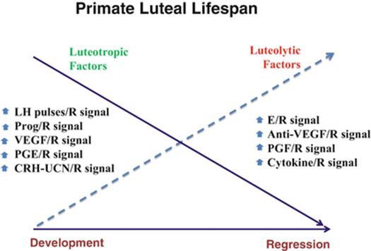
Fig. 10.1
Schematic of proposed changes in the balance between luteotropic and luteolytic signals in the primate corpus luteum during its lifespan in the non-fecund menstrual cycle. VEGFvascular endothelial growth factor, R receptor, Prog progesterone, PGE prostaglandin E, CRH-UCN corticotropin-releasing hormone/urocortin, E estrogen, PGF prostaglandin F. (From Stouffer et al. [5]. Figure reprinted from Reproductive Biology, Vol. 13, Issue 4. Stouffer RL, Bishop CV, Bogan RL, Xu F, Hennebold JD. Endocrine and local control of the primate corpus luteum, p. 256. 2013, with permission from Elsevier)
Recent studies suggest a role for immune cells in regulating the luteal lifespan, but again there may be species differences between primates [14, 15] and other species. The numbers of several types of immune cells (except lymphocytes) increase markedly in the macaque corpus luteum late in the menstrual cycle, but only after progesterone levels decline for 3 days [14], suggesting a major part in structural luteolysis. This information does not rule out earlier roles in controlling luteal development or functional regression, as proposed in domestic animals [20]. But if so, it may involve natural killer (NK) cells , the most abundant immune cell type in the functional corpus luteum in macaques [14], which are also found in the human corpus luteum [21]. The factors that promote immune cell migration and activity in primate luteal tissue remain poorly understood. One factor may be the cytokine C-C motif ligand 2 (CCL2; also known as monocyte chemoattractant 1 or MCP1), which is elevated in the regressing monkey [7] and human [15] corpus luteum. Evidence suggests that luteotropic factors (progesterone, PGE) suppress, whereas luteolytic agents (PGF) promote, CCL2 expression [15].
The authors updated a scenario proposed by Hamberger and colleagues [22] several years ago to summarize the shift from pro-luteotropic to pro-luteolytic factors that may control the lifespan of the primate corpus luteum (Fig. 10.1). Other factors may also be involved, as Duncan and colleagues [23] recently proposed that bone morphogenetic proteins (BMP2, -4, -6) are mediators of luteolysis in women. It is clear, however, that the loss of circulating LH support per se does not control luteolysis. There is a reduction in frequency of LH pulses secreted by the pituitary from early luteal phase to mid-late luteal phase just before the onset of luteal regression. Moreover, by mid-late luteal phase, each pulse of progesterone secretion is entrained to an LH pulse, such that there are intervals replete with and depleted of LH and progesterone [24]. However, sustaining LH levels by either generating endogenous LH pulses [25] or administering LH three times per day [26] did not prolong the luteal lifespan in monkeys. Alternatively, there is a decline in luteal tissue and cell responsiveness to LH as the corpus luteum ages. For example, dispersed cells from the macaque corpus luteum at mid-late luteal phase are less responsive to LH; the dose–response curve for cAMP and progesterone production is shifted, compared to cells from the early luteal phase [27]. The decreased sensitivity appears caused by LH-receptor desensitization, not downregulation, as receptor content does not decline until after progesterone levels decrease [28]. Thus, LH-receptor desensitization may be an early event associated with functional regression, whereas receptor downregulation may be a later event during structural involution. Once again one wonders what process or signal controls the onset of LH-receptor desensitization, and is this a critical signal for luteolysis?
10.3 Corpus Luteum of Early Pregnancy
10.3.1 Rescue by Chorionic Gonadotropin (CG)
Dissimilar from other species, the secretion of an LH-like hormone , chorionic gonadotropin (CG), by the implanting blastocyst and developing placenta “rescues” the corpus luteum in many primates from its impending demise and extends its functional lifespan in early pregnancy (for review of earlier work, see [2]). The steroidogenic function of the corpus luteum during early gestation is similar to that during the menstrual cycle , albeit leading to somewhat higher levels of circulating progesterone and estrogens (Fig. 10.2). Its peptidergic function, as indicated by relaxin and inhibin A production, is also markedly enhanced. It is generally regarded that progesterone is the only luteal product essential for the initiation and maintenance of early pregnancy. However, it now appears that relaxin of luteal origin acts on several tissues, including the embryo, uterus, and cardiovascular system, and may optimize maternal–fetal function and maternal adaptations to pregnancy [29]. Further studies are needed to assess the regulation and roles of luteal hormones during early pregnancy in primates.

Fig. 10.2
Schematic of the changes in circulating levels of progesterone (P4), estrogen (E), relaxin (RLX), and inhibin (I) during the luteal phase of the fertile menstrual cycle in rhesus monkeys (CG). (Adapted from Stouffer and Hearn [2]. Figure reprinted from The Endocrinology of Pregnancy, Vol. 9 of the series. Stouffer RL, Hearn JP. Endocrinology of the Transition from Menstrual Cyclicity to Establishment of Pregnancy in Primates, p. 44. 1998, with permission from Springer)
10.3.2 CG Structure and Production
Chorionic gonadotropin (CG) is a heterodimeric glycoprotein composed of two subunits, termed α and β, that associate noncovalently. It is a member of the glycoprotein hormone family, which includes pituitary-derived follicle-stimulating hormone (FSH) , luteinizing hormone (LH), and thyroid-stimulating hormone (TSH) . Jiang and colleagues [4] summarize the discovery of these hormones and how their clinical application, especially for CG, spurred their purification, synthesis, and structure–activity analyses. Notably, CG shares a common 92-amino-acid α-subunit with all three pituitary glycoprotein hormones. Their specific bioactivity, linked to receptor binding and signaling, relates to differences among their β-subunits. Within the cluster of seven genes encoding β-subunit-like products on human chromosome 19, one encodes the β-subunit of LH (LHβ) , four encode CG β-subunits (CGβ), and two are pseudogenes. The amino acid sequences of human (h) LHβ and CGβ are very similar (82 % homology). The major difference is that CGβ includes the entire 145-amino-acid (aa) protein, whereas the 24-aa leader sequence of LHβ is removed to yield a 121-aa product. It is now clear that circulating CG (as well as LH) exists as a mix of isoforms generated by (a) natural sequence variation, (b) posttranslational modification of the gene products, and (c) metabolism to yield truncated or “nicked” molecules. Not surprisingly, data are accruing that the bioactivity differs between isoforms.
As summarized by Choi and Smitz [3], there are at least four physiologically relevant isoforms of hCG: (a) “regular” hCG, (b) hyperglycosylated or H-hCG, (c) free hCGβ, and (d) pituitary hCG. The hCG molecule is highly glycosylated, a characteristic that also distinguishes it from LH. The α-subunit of both hormones contains two N-linked glycosylated sites, whereas CGβ also contains two N-linked and four O-linked sites. The increased sialic acid content of hCG influences its receptor-binding activity, increases its biological activity, and prolongs its half-life in the circulation compared to LH. The H-hCG isoform has the same amino acid composition as regular hCG, but even larger N- and O-linked oligosaccharides, thus increasing the molecular weight from 36,000–37,000 kDa to 40,000–41,000 kDa. Also, the sources of the various isoforms appear to differ. Fournier and colleagues [30] recently reviewed the literature and proposed a model wherein regular hCG is produced by the forming syncytiotrophoblast, whereas H-hCG is synthesized primarily in the extravillous cytotrophoblast. They also summarize evidence that these isoforms have different biological functions: (a) regular hCG appears to be the primary form for maternal recognition of pregnancy, extending luteal function, as well as other events in the reproductive tract to promote pregnancy initiation and early gestation, whereas (b) H-hCG is synthesized to affect local placental development including trophoblast invasion and increased vascularization. The typically low levels of sulfated hCG, of pituitary origin, increase in women during the perimenopausal interval and then plateau. The role(s), if any, for pituitary hCG are unknown, but its production over time mimics that of hLH.
Little is known regarding the factors or mechanisms controlling CG gene expression and protein production beginning as early as the two-cell embryo [31, 32] within the chronology of pregnancy. Studies of the promoter regions of the CGα and β genes suggest that transcription factors, such as AP2 and SP1 , recognize specific response elements to stimulate gene activity. Also, a number of steroid hormones (e.g., progesterone, estradiol), growth factors, and cytokines (e.g., EGF, IL-6, TGFβ1), and perhaps oxygen-sensitive pathways regulate CG production in model systems, such as cultures of villous trophoblast. Also, a local placental [33]–embryonic [34] loop involving GnRH was proposed as a regulator of CG, analogous to hypothalamic GnRH regulation of pituitary LH/CG production. How or whether any of these factors have key roles in the onset of GG production in the implanting blastocyst and developing syncytiotrophoblast is unknown. However, the mechanisms that sustain, if not initiate, CG production in the placenta must vary somewhat among species, as judged by the differences in patterns and levels of CG circulating during gestation in a variety of primate species (Fig. 10.3). Peak CG levels are highest in women, 10-fold less in great apes (e.g., chimpanzees), and at least 100-fold lower in baboons and Old World and New World monkeys. Although CG is first detected around the time of implantation in all primates, the duration of CG production varies from throughout pregnancy in women and apes to only the first trimester in macaques. Based on emerging evidence for CG action in the human placenta, one can hypothesize that additional “extra-gonadal” roles for CG evolved in higher primates but are limited to maternal recognition of pregnancy in others.

Fig. 10.3
Patterns and quantities of chorionic gonadotropin excreted during gestation by a variety of primate species. The time scale in the upper-right corner denotes the approximate duration of pregnancy and parturition (arrows) for these species. (Adapted from Stouffer and Hearn [2]. Figure reprinted from The Endocrinology of Pregnancy, Vol. 9 of the series. Stouffer RL, Hearn JP. Endocrinology of the Transition from Menstrual Cyclicity to Establishment of Pregnancy in Primates, p. 39. 1998, with permission from Springer)
10.3.3 LHCG-Receptor Binding and Signaling
As reviewed by Choi and Smitz [3], both LH and CG bind to and activate a common receptor, the LHCGR, which is a 675-aa G-protein receptor and a member of the rhodopsin subfamily of glycoprotein hormone receptors that includes FSHR and TSHR. It consists of seven domains spanning the cell membrane and an unusually large extracellular domain containing leucine-rich repeats and glycosylation sites. Jiang, Dias, and He [4] recently reviewed the efforts of structural biologists to understand the interactions of gonadotropin hormones with their receptors at an atomic level. After the crystal structure of hCG was defined [35, 36], an initial view of the hCG–LHCGR complex was predicted [37] in which several aspects of ligand binding and orientation appear confirmed by subsequent studies. Recent research focused on gonadotropin interaction with its receptor ectodomain, so a remaining task is to elucidate the crystal structure of the native LHCGR. Nevertheless, current views support a two-step model: (1) ligand binding to the R high-affinity hormone-binding subdomain induces a conformational change in the hormone to form a “binding pocket,” and (2) subsequent interactions produce a “pull and lift” force that frees tethered extracellular loops, thereby releasing an hypothesized inhibitory influence on the ectodomain leading to the activated conformation of the seven-transmembrane domain [4]. Although this model relates to the monomeric receptor, gonadotropin receptors (including LHCGRs) [38] can exist as dimers or larger number oligomers, which may explain biological phenomena such as negative cooperativity [4].
The expression of LHCGRs in theca cells and granulosa cells of the differentiating antral follicle, as well as luteal cells of the corpus luteum, is well established in primates [3]. LHCGR expression is not constitutive but dynamic, and primarily regulated in the maturing follicle by FSH or LH through both transcriptional and posttranscriptional processes. Similarly, the degradation of mRNA and protein products may be regulated, as well as the production of LHCGR variants by alternate splicing of mRNA [3]. Further studies are needed to understand the regulation of LHCGR expression , including the possible role of splice variants (see following) [39] in the primate corpus luteum. Similarly, mounting evidence supports the “extra-gonadal” expression of LHCGR [3]. These data are particularly relevant in primates, where CG is an endogenous molecule and is increasingly implicated in “extra-gonadal” functions in the female reproductive tract, placenta, and fetus to promote pregnancy and fetal development [40].
Evidence indicates that LHCGR is capable of binding H-hCG , as well as LH and regular hCG, but not the free β-subunit of LH or CG. However, H-hCG may also activate TGFβ-RII in the cytotrophoblast to promote angiogenesis [41], while being a poor stimulator of progesterone production by human luteinized granulosa cells [42]. Thus, different hCG forms may have different receptor affinity, leading to potential differences in signaling activity (see below) and biological function.
Until recently, it was generally considered that, because of their structural similarity, the postbinding effects and actions of hLH and hCG were very similar if not functionally equivalent. It remains clear that the major effect of LH-/CG-receptor binding is the activation of the G protein Gs, which in turn activates adenylate cyclase and thereby increases the production of cyclic AMP [3]. The cAMP/cAMP-dependent protein kinase A pathway is a major, indeed critical, cellular mechanism in LH-/CG-stimulated ovulation and steroid hormone (progesterone) synthesis. However, LH-/CG-receptor binding also activates phospholipase C/inositol phosphate signaling independent of the cAMP/PKA pathway. Given the higher levels of LH or CG required to activate PLC [43], it was hypothesized that this pathway is primarily active during the LH surge in the menstrual cycle or rising CG levels in early pregnancy. Recently, other cellular pathways involving ERK1/2 and AKT were identified in LHCGR signaling that may have a part in “nonsteroidogenic” processes, such as cell proliferation, differentiation, and survival in the follicle [44]. A possible role in the corpus luteum, that includes distinguishing LH versus CG signaling pathways as the mechanism of CG rescue of the corpus luteum in early pregnancy, was recently proposed [44]. Grzesik et al. [45] concluded the differences between LH and CG signaling were determined by interaction of the L2-beta loop of the gonadotropin with the hinge region of the LHCG receptor.
10.3.4 Mechanisms of CG Rescue of the Corpus Luteum
10.3.4.1 Luteal Structure and Remodeling
Luteal rescue serves to maintain the overall cellular integrity and morphology of the primate corpus luteum, similar to that observed during the mid-luteal phase of the menstrual cycle. CG maintains the weight of the primate corpus luteum [46–48], whereas at the cellular level, CG also preserves luteal cell size and morphology [47, 48]. At the subcellular level, electron microscopic evaluation of luteal cells within rhesus monkey corpora lutea obtained at day 13 of pregnancy revealed they are similar in appearance to those at the mid-luteal phase (day 10 post-LH surge); that is, the cells possessed large, round nuclei and abundant lipid droplets ≥1 μm in size [49]. Although the general morphology and cellular organization appear to be similar between corpora lutea at mid-luteal phase and those obtained following CG rescue, there are differences in junctional complexes that are important for forming and maintaining cell-to-cell contacts. In the human corpus luteum at mid-luteal phase, the tight junction proteins claudin 1 (CLDN1) and occludin (OCLN) exhibited significant levels of immunostaining that were evenly distributed among granulosa-lutein cells [50]. After CG rescue, both CLDN1 and OCLN1 staining was significantly reduced. Similarly, expression of endothelial adhesion molecules claudin 5 (CLDN5) and vascular endothelium-cadherin (CDH5) were significantly reduced in the vasculature of corpora lutea obtained following CG rescue relative to the high level of expression observed in corpora lutea collected midway through the luteal phase of the menstrual cycle . From these findings, it was suggested the loss in tight junctions might facilitate the release of key endocrine factors by the corpora lutea (i.e., progesterone, relaxin ) during early pregnancy [50].
During luteolysis, a number of mechanisms, including apoptosis and autophagy, have been proposed to account for the loss of the cellular constituents comprising the primate corpus luteum [51, 52]. At the ultrastructural level , both apoptotic and nonapoptotic death of luteal cells was observed in corpora lutea obtained from women undergoing induced and natural luteal regression. CG rescue of the corpus luteum prevented apoptosis, but not autophagy [52, 53]. Although the importance of apoptosis in primate luteolysis has been challenged [54], several studies reported its existence during the regression of the corpus luteum at the end of the menstrual cycle [55]. Apoptosis is controlled by several intracellular proteins, including those that serve as either (a) antiapoptotic factors such as B-cell lymphoma-2 (BCL2) , myeloid cell leukemia-1 (MCL1), and Bcl-xL, or (b) proapoptotic factors such as Bcl-2-associated X protein (BAX), Bcl-2 interacting killer (BIK), and Bcl-2-associated death protein (BAD) . Cell death by apoptosis is determined by the ratio of pro- and antiapoptotic factors within a given cell. In women, BCL2 mRNA expression was at its lowest point in the regressing corpus luteum and its highest level of expression was in the corpus luteum of early pregnancy [56]. In contrast, BAX mRNA levels were at their highest in the regressing corpus luteum and their lowest in the corpus luteum of early pregnancy. Moreover, BCL2 and BAX protein levels paralleled mRNA levels in the corpus luteum at these stages [56]. These findings differed from an earlier report noting that the expression of BCL2 did not change in the human corpus luteum of the menstrual cycle or after luteal rescue following CG administration [57]. Another study reported that the human corpus luteum also expresses the antiapoptotic protein MCL1 during early pregnancy, although the analysis was performed on a single isolated corpus luteum [58]. Thus, whether luteolysis is mediated solely via apoptosis and, if so, the precise mechanism by which CG prevents it from occurring, remain to be determined. Moreover, the role of autophagy in maintaining primate luteal structure–function is virtually unknown. Only a single paper has reported on this issue in the primate corpus luteum, suggesting that the protein beclin-1 (BECN1) , which is required for the formation of the autophagosome, is expressed in granulosa-lutein cells during early pregnancy and, therefore, may be involved in promoting cell survival rather than cell death [59].
In addition to preserving luteal cells, CG may alter the dynamics of other cell types in the corpus luteum of early pregnancy . For example, CG exposure simulating early pregnancy prevented the rise in CCL-2 expression and immune cells in the human corpus luteum [15]. Also, luteal rescue results in either maintenance or growth of the luteal vasculature, perhaps depending on the species, as well as the markers used to assess angiogenesis. In women treated with CG to simulate early pregnancy, the effect on endothelial cell proliferation was minimal in the rescued corpus luteum [60]. However, in another study, CG treatment of women led to increased luteal endothelial cell area and proliferation as determined by CD34 and Ki-67 immunostaining , respectively [61]. Moreover, human corpora lutea isolated during early pregnancy (6–8 weeks) possessed greater numbers of both blood vessels and the supportive pericytes relative to corpora lutea obtained at mid-luteal phase of the menstrual cycle [62]. In this study, the increased angiogenesis associated with CG rescue was accompanied by increased mRNA levels for vascular endothelial growth factor A (VEGFA) . In the corpus luteum of early pregnancy in women, increased expression of angiopoietin (ANGPT)-1 and decreased expression of ANGPT2 were noted relative to their expression in those at other stages of the luteal phase in a non-fecund cycle [62]. ANGPT1 serves to stabilize pericytes and endothelial cells of blood vessels, whereas ANGPT2 is a natural antagonist of ANGPT1 that serves to destabilize formed blood vessels [63, 64]. To provide a quantitative assessment of luteal vascularity during early pregnancy, three-dimensional power Doppler ultrasonography was employed weekly in a longitudinal study of women from 5 to 11 weeks of pregnancy, with pregnancy being defined based on last menstrual period and fetal crown–rump length [65]. From these studies, it was determined the vascular volume in the ovary containing the corpus luteum was at a maximum at week 5 and declined continuously thereafter. Thus, it appears that increased capacity, if not development, of the vasculature in women occurs early in pregnancy, in the first weeks that CG is produced by the conceptus. In contrast to the corpus luteum of women during early pregnancy , the marmoset and rhesus monkey corpus luteum display minimal or no change in endothelial and pericyte areas or in endothelial cell proliferation relative to the mature corpus luteum obtained in the absence of pregnancy [66, 67], indicating that there may be primate species-specific differences in the degree of vessel development and maturation in the corpus luteum during early pregnancy. Although some studies have investigated VEGFA and ANGPT1/2 expression and action in the corpus luteum of pregnancy in primates, a number of recently discovered factors and pathways involved in angiogenesis and vessel stabilization (e.g., apelin, vasohibin, the ROBO/SLIT pathway, prohibitin-1, prokineticin-1, and the DELTA/JAGGED/NOTCH pathway) [1, 5] await investigation with regard to their role in maintaining the integrity of the vasculature following luteal rescue by CG.
One cellular process that appears regulated by CG during corpus luteum rescue in early pregnancy is tissue or extracellular matrix (ECM) remodeling . During natural luteolysis or following gonadotropin withdrawal, a wave of ECM and cellular remodeling occurs, which is associated with a concomitant increase in luteal protease expression and activity [68–70]. In rhesus monkeys, matrix metalloproteinase-9 (MMP9, gelatinase B) and MMP2 (gelatinase A) mRNA and protein expression are low during luteal formation and the period of peak steroid synthesis, but increase significantly after cessation of progesterone production at luteolysis. In women, MMP2 and MMP9 expression also increased in the corpus luteum during luteal regression [68]. However, administration of exogenous CG maintained luteal function and was associated with a significant reduction in MMP2, but not MMP9, expression and activity. Moreover, connective tissue growth factor (CTGF) , which is associated with tissue and ECM remodeling in wound healing [71], reaches maximal expression in the human corpus luteum at the time of regression [72]. When women received CG to simulate early pregnancy, CTGFmRNA levels decline significantly in luteal fibroblasts and endothelial cells. It was subsequently demonstrated that CG did not downregulate CTGF expression in luteal fibroblasts directly, but required an unidentified paracrine factor produced by granulosa-lutein cells.
10.3.4.2 Luteal Function
In addition to maintaining the structure of the corpus luteum during early pregnancy, CG serves to sustain the level of progesterone production required for supporting pregnancy. The ability of CG to extend the period of luteal function clearly requires continued cholesterol uptake and translocation to the mitochondria where the first step of steroidogenesis occurs, primarily through action of steroidogenic acute regulatory protein (STAR) . Once translocated across the mitochondrial membranes, cholesterol is then converted to pregnenolone by the cytochrome P450 cholesterol side-chain cleavage enzyme (CYP11A1; also known as P450scc). Pregnenolone is subsequently converted to progesterone via 3β-hydroxysteroid dehydrogenase (HSD3B) [73]. In terms of supporting steroidogenesis within the corpus luteum, administration of CG to women during the late luteal phase maintains expression of STAR, CYP11A1, and HSD3B mRNA and protein at levels that are comparable to those in the mid-luteal phase corpus luteum [74]. In rhesus monkeys undergoing a simulated early pregnancy protocol, CG induced the expression of several key steroidogenic enzymes in the corpus luteum relative to those removed from animals not receiving supplemental CG [75, 76]. The mRNA level for HSD3B2 is rapidly upregulated by CG, but it was transient and returned to pretreatment levels after 3 to 5 days of CG exposure. In contrast, mRNAs encoding STAR, CYP11A1, and CYP19A1 (also known as aromatase, critical for estradiol production) exhibited delayed (>3 days of CG treatment) and sustained expression. The expression pattern for these steroidogenic enzymes correlated with the observed level of circulating progesterone and estradiol, with progesterone levels peaking 2 days after initiation of CG treatment and estradiol levels reaching a maximum at 8 days of CG treatment [75].
The primate corpus luteum is not only a site of steroid synthesis; as noted earlier, it also serves as a target of steroid action through the expression of several nuclear hormone receptors including the genomic progesterone receptor (PGR), a plasma membrane-associated progesterone receptor (progesterone membrane component 1, PGRMC1), both estradiol receptors ESR1 (ERα) and ESR2 (ERβ), as well as the androgen receptor (AR) [9, 19, 77–80]. A critical function for steroid action in the primate corpus luteum was demonstrated in rhesus monkeys receiving exogenous CG at the mid-late luteal phase with or without simultaneous treatment of a trilostane, a compound that blocks the synthesis of steroid hormones by inhibiting HSD3B activity [48]. Although CG was capable of maintaining luteal weight and preventing the appearance of luteal cell histology consistent with luteolysis, CG plus trilostane resulted in a significant decrease in luteal weight and luteal cell size [48]. Inhibition of steroid synthesis through the administration of trilostane to rhesus monkeys during either the early or mid-luteal phase leads to luteolysis despite the presence of LH levels sufficient to support luteal function. Administering a synthetic progestin (R5020) along with trilostane maintains luteal weight and prevents the luteal cell loss that occurs following treatment with trilostane alone [70], further supporting the hypothesis that progesterone is a luteotropic agent [9, 81]. It is currently unclear to what extent progesterone action is involved in CG rescue of luteal function and whether any progesterone-dependent effects are mediated through the genomic progesterone receptor (PGR) or the membrane-associated form (PGRMC1), because both are expressed in the primate corpus luteum during simulated early pregnancy [48, 75, 82].
It is possible that the effects of steroid ablation during luteal rescue leading to the demise of the corpus luteum are dependent on the actions of steroids other than progesterone. Duncan and coworkers reported that the estrogen receptor isoform ESR2 was expressed in the late-stage corpus luteum obtained from women and that its levels were unchanged in response to CG rescue [80]. More recently, studies were published that support a role for glucocorticoids (e.g., cortisol) and the glucocorticoid receptor (nuclear receptor subfamily 3, group C, member 1; NR3C1) in maintaining luteal function in response to CG. In women receiving exogenous CG beginning at the mid-luteal phase and continuing for 5–8 days, it was noted that there is a significant increase in luteal expression of the enzyme 11β-hydroxysteroid dehydrogenase-1 (HSD11B1; converts inactive cortisone to biologically active cortisol) relative to stage-matched corpora lutea obtained from women not receiving CG [83]. NR3C1 mRNA and protein were detectable in mid- and late-stage corpora lutea as well as those from women receiving exogenous CG. Immunohistochemistry revealed significant expression of HSD11B1 in the granulosa-lutein cells, whereas NR3C1 immunostaining was localized to numerous cell types, including granulosa-lutein cells, fibroblasts, and endothelial cells, as well as in resident macrophages. Thus, cortisol generated within the corpus luteum via HSD11B1 during early pregnancy may act through NR3C1 to prevent luteolysis in response to CG.
The afore-described pathways and events critical for the extension of luteal lifespan and function controlled by CG and CG-inducible steroids were investigated on a case-by-case basis. With the development of genomic approaches (i.e., DNA microarray technology) that allow for the simultaneous assessment of most, if not all, transcripts in a given rhesus macaque cell type or tissue, it became possible to systematically determine changes in gene expression in the primate corpus luteum in response to CG and steroid hormones. The changes in the levels of mRNAs in corpora lutea obtained from monkeys undergoing simulated early pregnancy were assessed using the Affymetrix GeneChip Rhesus Macaque Genome Array [75]. CG treatment occurred for 1, 3, 6, and 9 days beginning on day 9 after the mid-cycle surge of LH. Following only 1 day of CG treatment, the levels of 419 mRNAs were significantly different (≥2-fold change) compared to control corpora lutea, with 292 mRNAs being upregulated and 127 being downregulated. When comparing subsequent days of treatment, continued CG administration resulted in a limited effect on gene expression, with fewer than 100 mRNAs changing significantly between 3 and 6 days or 6 and 9 days of CG treatment. However, when comparing mRNA levels between stage-matched corpora lutea obtained from animals not receiving CG and those receiving CG, the effects on gene expression were significant. For example, when comparing corpora lutea obtained from days 14 to 16 of the luteal phase of animals not receiving CG (period of declining progesterone synthesis) to those obtained at the same stage of the luteal phase that had received CG for 6 days, there were 2078 mRNAs with increased levels and 452 that decreased. As expected, a number of proteins encoded by differentially expressed mRNAs were related to steroid production (e.g., STAR, CYP11lA1, HSD3B2). CG administration also led to a transient re-expression of the prostaglandin PGE synthesis–receptor signaling system, which is associated with corpus luteum development during the early to mid-luteal phase of the menstrual cycle [12], but suppressed the expression of mRNAs encoding proteins associated with immune function.
To further clarify the direct versus indirect effects (i.e., steroid-mediated processes) of CG that prevent luteal demise and extend the structure–function of the primate corpus luteum in early pregnancy, a DNA microarray study was performed using RNA isolated from the corpus luteum of rhesus macaques receiving CG, CG plus the steroid synthesis inhibitor trilostane, or CG and trilostane plus the synthetic progestin R5020 [84]. Corpora lutea were collected at 1 or 6 days after the initiation of CG treatment, which commenced on day 9 of the luteal phase. The results of this study revealed that the majority of CG-regulated luteal mRNAs are regulated independently of local steroid actions wherein trilostane significantly affected the expression of 50 mRNAs after 1 day and 87 mRNAs after 6 days of CG treatment relative to corpora lutea from animals receiving CG alone. Moreover, the number of genes in the corpus luteum affected by progesterone replacement in CG plus trilostane-treated animals (i.e., CG + trilostane + R5020) relative to those receiving CG plus trilostane alone (i.e., CG + trilostane) was relatively small and included 46 mRNAs after day 1 of CG treatment and 129 mRNAs after 6 days of CG treatment. Although the steroid-regulated genes in the macaque corpus luteum during CG rescue may be few, they are likely essential for sustaining luteal function during early pregnancy based on the fact that trilostane treatment initiates premature structural regression of the corpus luteum during simulated early pregnancy [48]. Also, these data demonstrate that regulation of gene expression in the rescued corpus luteum in early pregnancy differs from the corpus luteum of the menstrual cycle in primates, because in the latter the number of genes whose expression is affected by steroid depletion is considerably greater (>300) [8]. Thus, the availability of high-throughput genomic methods has allowed for a greater understanding and comparison of how different genes are regulated (i.e., gonadotropin- versus steroid dependent) in the primate corpus luteum throughout the menstrual cycle and during pregnancy (e.g., STAR) (Fig. 10.4). Such approaches will also provide the means for investigating the role that individual intraluteal factors (i.e., androgens, glucocorticoids, PGs) play in mediating the effects of CG that are necessary in primates for luteal survival and function during early pregnancy.

Fig. 10.4
Example of the dynamic expression of a specific gene product (STAR mRNA, relative units) during the lifespan of the macaque corpus luteum, plus the effects of LH depletion during the menstrual cycle and hCG administration during simulated early pregnancy. Similar data analyses can be generated for any number of genes of interest from the publicly available NCBI GEO databases (e.g., GSE2276, GSE10367, GSE25335). (From Stouffer et al. [5]. Figure reprinted from Reproductive Biology, Vol. 13, Issue 4. Stouffer RL, Bishop CV, Bogan RL, Xu F, Hennebold JD. Endocrine and local control of the primate corpus luteum, p. 267. 2013, with permission from Elsevier)
10.4 Clinical Applications
Urinary, and more recently recombinant, preparations of hCG are widely used, as a bolus injection, to mimic the endogenous LH surge in controlled ovarian stimulation cycles, for the purpose of collecting meiotically mature oocytes for assisted reproductive technologies (ARTs) in infertility patients [85]. The hCG bolus also promotes luteinization of the antral follicles in COS cycles and, because of the long half-life of hCG, will sustain elevated progesterone levels in the circulation for several days. However, “downregulation” of pituitary LH secretion by either GnRH analogues administered during the follicular phase or elevated steroid levels into the luteal phase causes circulating P levels to decline as CG disappears. To sustain P and its actions, P is often provided as a luteal-phase supplement, which is preferred [86], in part because hCG treatment can cause morbidity from ovarian hyperstimulation syndrome (OHSS) [87] and mask the ability of clinical “early pregnancy test” kits to detect endogenous hCG excreted in the urine.
However, the use of exogenous CG to promote pregnancy initiation at the time or following implantation is controversial. Although rare, two variants of the hCG β-subunit are associated with increased risk of recurrent miscarriage [88]. However, a recent Cochrane review of existing clinical trials [89] indicates that the evidence supporting hCG supplementation to prevent recurrent miscarriage remains equivocal. Because increasing evidence suggests that hCG from the embryo has a critical local role in the uterus to promote pregnancy initiation, as well as extending luteal function , clinical researchers hypothesized that exogenous hCG would facilitate fertility, especially in ART patients in which “lower-quality” embryos are transferred into the uterus. Initial reports suggested that intrauterine hCG administration during transfer of early, cleavage-stage embryos offered some benefit, but recent studies observed no improvement when hCG was administered 2 days before or on the day of blastocyst transfer [90], regardless of embryo quality. Nevertheless, it is now apparent that a number of factors (e.g., BMI, smoking) influence fertility, and that the circulation and maternal–fetal interface includes several different hCG molecules. Further studies with better characterized embryos, including their CG production, and select CG moieties are needed to address this issue. For example, Evans and colleagues [91] propose that H-hCG could prove useful in treating pregnancy disorders.
Similarly, a number of studies are addressing the premise that alterations in a particular type of hCG molecule can help identify dysfunctional pregnancies [92]. There are reports that a low ratio of H-hCG to total hCG (<0.5) is associated with pregnancy loss [93], whereas low levels of hCG β-subunit occur during ectopic pregnancies [92]. However, there is remarkable variation in CG levels between individuals with normal pregnancies , as well as in the ability of clinical assays to reliably detect the various forms of hCG [94]. Our increasing understanding of the types and roles of the various hCG moieties should aid in the development of better assays for diagnosis of early pregnancy and associated disorders, as well as novel treatments for sub- or infertility.
10.5 Final Perspective
Although progress during the past 15 years has increased our knowledge of the types of CG moieties and CG- versus LH-receptor signaling in cells (but not necessarily its specific target cells), it remains unclear how the appearance of CG around the onset of implantation rescues the primate corpus luteum from impending luteal regression and extends its functional lifespan until the later luteal–placental shift in progesterone production. There is evidence that LH- and CG-receptor signaling and activation of intracellular pathways can differ [44], but do these differences actually occur in luteal cells and are they critical for corpus luteum rescue in early pregnancy? Alternatively, is the qualitative and quantitative change in gonadotropin exposure, from low pulses of LH secretion three or four times per day to continuous increasing levels of CG, sufficient for corpus luteum rescue? Notably, Zeleznik [95] reported that either LH or CG could rescue the macaque corpus luteum when given in exponentially increasing doses. Although research on the corpus luteum seems to be losing popularity, perhaps because of recognition that progesterone replacement can replace this endocrine gland in clinical scenarios, there are important unresolved issues, especially in primates, regarding the processes controlling the function and lifespan of the corpus luteum during the menstrual cycle and its rescue in early pregnancy.
References
1.
Stouffer RL, Hennebold JD. Structure, function and regulation of the corpus luteum. In: Plant TM, Zeleznik AJ, editors. Knobil and Neill’s physiology of reproduction, 3rd edn. San Diego: Elsevier Academic Press; 2014, p. 1703–26.
2.
Stouffer RL, Hearn JP. Endocrinology of the transition from menstrual cyclicity to establishment of pregnancy in primates. In: The endocrinology of pregnancy. Totowa: Humana Press; 1998. p. 35–57.
3.
Choi J, Smitz J. Luteinizing hormone and human chorionic gonadotropin: origins of difference. Mol Cell Endocrinol. 2014;383:203–13.CrossRefPubMed
4.
Jiang X, Dias JA, He X. Structural biology of glycoprotein hormones and their receptors: insights to signaling. Mol Cell Endocrinol. 2014;382:424–51.CrossRefPubMed
5.
Stouffer RL, Bishop CV, Bogan RL, Xu F, Hennebold JD. Endocrine and local control of the primate corpus luteum. Reprod Biol. 2013;13:259–71.CrossRefPubMedPubMedCentral
6.
Xu F, Stouffer RL, Muller J, Hennebold JD, Wright JW, Bahar A, et al. Dynamics of the transcriptome in the primate ovulatory follicle. Mol Hum Reprod. 2011;17:152–65.CrossRefPubMed
7.
Bogan RL, Murphy MJ, Stouffer RL, Hennebold JD. Systematic determination of differential gene expression in the primate corpus luteum during the luteal phase of the menstrual cycle. Mol Endocrinol. 2008;22:1260–73.CrossRefPubMedPubMedCentral
8.
Bishop CV, Hennebold JD, Stouffer RL. The effects of luteinizing hormone ablation/replacement versus steroid ablation/replacement on gene expression in the primate corpus luteum. Mol Hum Reprod. 2009;15:181–93.CrossRefPubMedPubMedCentral
9.
Stouffer RL. Progesterone as a mediator of gonadotrophin action in the corpus luteum: beyond steroidogenesis. Hum Reprod Update. 2003;9:99–117.CrossRefPubMed
10.
Bishop C, Hennebold J, Kahl C, Stouffer R. RNAi-knockdown of genomic progesterone (P) receptor in macaque granulosa cells and preovulatory follicles disrupts P production and ovulation. Biol Reprod. 2016;94(5):109.
11.
Peluso JJ, Pru JK. Non-canonical progesterone signaling in granulosa cell function. Reproduction. 2014;147:R169–78.CrossRefPubMedPubMedCentral
12.
Bogan RL, Murphy MJ, Stouffer RL, Hennebold JD. Prostaglandin synthesis, metabolism, and signaling potential in the rhesus macaque corpus luteum throughout the luteal phase of the menstrual cycle. Endocrinology. 2008;149:5861–71.CrossRefPubMedPubMedCentral
13.
Xu J, Xu F, Hennebold JD, Molskness TA, Stouffer RL. Expression and role of the corticotropin-releasing hormone/urocortin-receptor-binding protein system in the primate corpus luteum during the menstrual cycle. Endocrinology. 2007;148:5385–95.CrossRefPubMed
14.
Bishop CV, Xu F, Molskness TA, Stouffer RL, Hennebold JD. Dynamics of immune cell types within the macaque corpus luteum during the menstrual cycle: role of progesterone. Biol Reprod. 2015;93(5):112.CrossRefPubMed
15.
Nio-Kobayashi J, Kudo M, Sakuragi N, Kimura S, Iwanaga T, Duncan WC. Regulated C-C motif ligand 2 (CCL2) in luteal cells contributes to macrophage infiltration into the human corpus luteum during luteolysis. Mol Hum Reprod. 2015;21:645–54.CrossRefPubMed
16.
Peluffo MC, Young KA, Hennebold JD, Stouffer RL. Expression and regulation of tumor necrosis factor (TNF) and TNF-receptor family members in the macaque corpus luteum during the menstrual cycle. Mol Reprod Dev. 2009;76:367–78.CrossRefPubMedPubMedCentral
17.
Stouffer RL. Perspectives on the corpus luteum of the menstrual cycle and early pregnancy. Semin Reprod Endocrinol. 1988;6:103–13.CrossRef
18.
Knobil E. On the regulation of the primate corpus luteum. Biol Reprod. 1973;8:246–58.
19.
Duffy DM, Chaffin CL, Stouffer RL. Expression of estrogen receptor alpha and beta in the rhesus monkey corpus luteum during the menstrual cycle: regulation by luteinizing hormone and progesterone. Endocrinology. 2000;141:1711–7.PubMed
20.
Pate JL, Toyokawa K, Walusimbi S, Brzezicka E. The interface of the immune and reproductive systems in the ovary: lessons learned from the corpus luteum of domestic animal models. Am J Reprod Immunol. 2010;64:275–86.CrossRefPubMed
21.
Wang LJ, Pascoe V, Petrucco OM, Norman RJ. Distribution of leukocyte subpopulations in the human corpus luteum. Hum Reprod. 1992;7:197–202.PubMed
22.
Hamberger L, Hahlin M, Hillensjo T, Johanson C, Sjogren A. Luteotropic and luteolytic factors regulating human corpus luteum function. Ann N Y Acad Sci. 1988;541:485–97.CrossRefPubMed
23.
Nio-Kobayashi J, Trendell J, Giakoumelou S, Boswell L, Nicol L, Kudo M, et al. Bone morphogenetic proteins are mediators of luteolysis in the human corpus luteum. Endocrinology. 2015;156:1494–503.CrossRefPubMed
24.
Ellinwood WE, Norman RL, Spies HG. Changing frequency of pulsatile luteinizing hormone and progesterone secretion during the luteal phase of the menstrual cycle of rhesus monkeys. Biol Reprod. 1984;31:714–22.CrossRefPubMed
25.
Hutchison JS, Nelson PB, Zeleznik AJ. Effects of different gonadotropin pulse frequencies on corpus luteum function during the menstrual cycle of rhesus monkeys. Endocrinology. 1986;119:1964–71.CrossRefPubMed
26.
Duffy DM, Stewart DR, Stouffer RL. Titrating luteinizing hormone replacement to sustain the structure and function of the corpus luteum after gonadotropin-releasing hormone antagonist treatment in rhesus monkeys. J Clin Endocrinol Metab. 1999;84:342–9.PubMed
27.
Eyster KM, Ottobre JS, Stouffer RL. Adenylate cyclase in the corpus luteum of the rhesus monkey. III. Changes in basal and gonadotropin-sensitive activities during the luteal phase of the menstrual cycle. Endocrinology. 1985;117:1571–7.CrossRefPubMed
28.
Cameron JL, Stouffer RL. Gonadotropin receptors of the primate corpus luteum. II. Changes in available luteinizing hormone- and chorionic gonadotropin-binding sites in macaque luteal membranes during the nonfertile menstrual cycle. Endocrinology. 1982;110:2068–73.CrossRefPubMed
29.
Conrad KP. Maternal vasodilation in pregnancy: the emerging role of relaxin. Am J Physiol Regul Integr Comp Physiol. 2011;301:R267–75.CrossRefPubMedPubMedCentral
30.
Fournier T, Guibourdenche J, Evain-Brion D. Review: hCGs: different sources of production, different glycoforms and functions. Placenta. 2015;36(Ssppl 1):S60–5.CrossRefPubMed
31.
Butler SA, Luttoo J, Freire MO, Abban TK, Borrelli PT, Iles RK. Human chorionic gonadotropin (hCG) in the secretome of cultured embryos: hyperglycosylated hCG and hCG-free beta subunit are potential markers for infertility management and treatment. Reprod Sci. 2013;20:1038–45.CrossRefPubMed
32.
Ramu S, Acacio B, Adamowicz M, Parrett S, Jeyendran RS. Human chorionic gonadotropin from day 2 spent embryo culture media and its relationship to embryo development. Fertil Steril. 2011;96:615–7.CrossRefPubMed
33.
Lee HJ, Snegovskikh VV, Park JS, Foyouzi N, Han KT, Hodgson EJ, et al. Role of GnRH-GnRH receptor signaling at the maternal–fetal interface. Fertil Steril. 2010;94:2680–7.CrossRefPubMed
34.
Casan EM, Raga F, Polan ML. GnRH mRNA and protein expression in human preimplantation embryos. Mol Hum Reprod. 1999;5:234–9.CrossRefPubMed
35.
Lapthorn AJ, Harris DC, Littlejohn A, Lustbader JW, Canfield RE, Machin KJ, et al. Crystal structure of human chorionic gonadotropin. Nature. 1994;369:455–61.CrossRefPubMed
36.
Wu H, Lustbader JW, Liu Y, Canfield RE, Hendrickson WA. Structure of human chorionic gonadotropin at 2.6 Å resolution from MAD analysis of the selenomethionyl protein. Structure. 1994;2:545–58.CrossRefPubMed
37.
Jiang X, Dreano M, Buckler DR, Cheng S, Ythier A, Wu H, et al. Structural predictions for the ligand-binding region of glycoprotein hormone receptors and the nature of hormone-receptor interactions. Structure. 1995;3:1341–53.CrossRefPubMed
38.
Roess DA, Smith SM. Self-association and raft localization of functional luteinizing hormone receptors. Biol Reprod. 2003;69:1765–70.CrossRefPubMed
39.
Dickinson RE, Stewart AJ, Myers M, Millar RP, Duncan WC. Differential expression and functional characterization of luteinizing hormone receptor splice variants in human luteal cells: implications for luteolysis. Endocrinology. 2009;150:2873–81.CrossRefPubMed
40.
Banerjee P, Fazleabas AT. Extragonadal actions of chorionic gonadotropin. Rev Endocr Metab Disord. 2011;12:323–32.CrossRefPubMedPubMedCentral
41.
Berndt S, Blacher S, Munaut C, Detilleux J, Perrier d’Hauterive S, Huhtaniemi I, et al. Hyperglycosylated human chorionic gonadotropin stimulates angiogenesis through TGF-beta receptor activation. FASEB J. 2013;27:1309–21.CrossRefPubMed
42.
Crochet JR, Shah AA, Schomberg DW, Price TM. Hyperglycosylated human chorionic gonadotropin does not increase progesterone production by luteinized granulosa cells. J Clin Endocrinol Metab. 2012;97:E1741–4.CrossRefPubMed
43.
Gudermann T, Birnbaumer M, Birnbaumer L. Evidence for dual coupling of the murine luteinizing hormone receptor to adenylyl cyclase and phosphoinositide breakdown and Ca2+ mobilization. Studies with the cloned murine luteinizing hormone receptor expressed in L cells. J Biol Chem. 1992;267:4479–88.PubMed
44.
Casarini L, Lispi M, Longobardi S, Milosa F, La Marca A, Tagliasacchi D, et al. LH and hCG action on the same receptor results in quantitatively and qualitatively different intracellular signalling. PLoS One. 2012;7, e46682.CrossRefPubMedPubMedCentral
45.
Grzesik P, Kreuchwig A, Rutz C, Furkert J, Wiesner B, Schuelein R, et al. Differences in signal activation by LH and hCG are mediated by the LH/CG receptor’s extracellular hinge region. Front Endocrinol (Lausanne). 2015;6:140.
46.
Illingworth PJ, Reddi K, Smith K, Baird DT. Pharmacological ‘rescue’ of the corpus luteum results in increased inhibin production. Clin Endocrinol (Oxf). 1990;33:323–32.
47.
Duffy DM, Hutchison JS, Stewart DR, Stouffer RL. Stimulation of primate luteal function by recombinant human chorionic gonadotropin and modulation of steroid, but not relaxin, production by an inhibitor of 3 beta-hydroxysteroid dehydrogenase during simulated early pregnancy. J Clin Endocrinol Metab. 1996;81:2307–13.PubMed
48.
Duffy DM, Stouffer RL. Gonadotropin versus steroid regulation of the corpus luteum of the rhesus monkey during simulated early pregnancy. Biol Reprod. 1997;57:1451–60.CrossRefPubMed
49.
Koering MJ, Wolf RC, Meyer RK. Morphological changes in the corpus luteum correlated with progestin levels in the rhesus monkey during early pregnancy. Biol Reprod. 1973;9:254–71.PubMed
50.
Groten T, Fraser HM, Duncan WC, Konrad R, Kreienberg R, Wulff C. Cell junctional proteins in the human corpus luteum: changes during the normal cycle and after HCG treatment. Hum Reprod. 2006;21:3096–102.CrossRefPubMed
51.
Smith GW, Meidan R. Ever-changing cell interactions during the life span of the corpus luteum: relevance to luteal regression. Reprod Biol. 2014;14:75–82.CrossRefPubMed
52.
Del Canto F, Sierralta W, Kohen P, Munoz A, Strauss 3rd JF, Devoto L. Features of natural and gonadotropin-releasing hormone antagonist-induced corpus luteum regression and effects of in vivo human chorionic gonadotropin. J Clin Endocrinol Metab. 2007;92:4436–43.CrossRefPubMed
53.
Young FM, Illingworth PJ, Lunn SF, Harrison DJ, Fraser HM. Cell death during luteal regression in the marmoset monkey (Callithrix jacchus). J Reprod Fertil. 1997;111:109–19.CrossRefPubMed
54.
Fraser HM, Lunn SF, Harrison DJ, Kerr JB. Luteal regression in the primate: different forms of cell death during naturaland gonadotropin-releasing hormone antagonist or prostaglandin analogue-induced luteolysis. Biol Reprod. 1999;61:1468–79.CrossRefPubMed
55.
Sugino N, Okuda K. Species-related differences in the mechanism of apoptosis during structural luteolysis. J Reprod Dev. 2007;53:977–86.CrossRefPubMed
56.
Sugino N, Suzuki T, Kashida S, Karube A, Takiguchi S, Kato H. Expression of Bcl-2 and Bax in the human corpus luteum during the menstrual cycle and in early pregnancy: regulation by human chorionic gonadotropin. J Clin Endocrinol Metab. 2000;85:4379–86.PubMed
57.
Rodger FE, Fraser HM, Duncan WC, Illingworth PJ. Immunolocalization of bcl-2 in the human corpus luteum. Hum Reprod. 1995;10:1566–70.CrossRefPubMed
58.
Chen SU, Chen RJ, Shieh JY, Chou CH, Lin CW, Lu HF, et al. Human chorionic gonadotropin up-regulates expression of myeloid cell leukemia-1 protein in human granulosa-lutein cells: implication of corpus luteum rescue and ovarian hyperstimulation syndrome. J Clin Endocrinol Metab. 2010;95:3982–92.CrossRefPubMed
59.
Gaytan M, Morales C, Sanchez-Criado JE, Gaytan F. Immunolocalization of beclin 1, a bcl-2-binding, autophagy-related protein, in the human ovary: possible relation to life span of corpus luteum. Cell Tissue Res. 2008;331:509–17.CrossRefPubMed
60.
Rodger FE, Young FM, Fraser HM, Illingworth PJ. Endothelial cell proliferation follows the mid-cycle luteinizing hormone surge, but not human chorionic gonadotrophin rescue, in the human corpus luteum. Hum Reprod. 1997;12:1723–9.CrossRefPubMed
61.
Wulff C, Dickson SE, Duncan WC, Fraser HM. Angiogenesis in the human corpus luteum: simulated early pregnancy by HCG treatment is associated with both angiogenesis and vessel stabilization. Hum Reprod. 2001;16:2515–24.CrossRefPubMed
62.
Sugino N, Suzuki T, Sakata A, Miwa I, Asada H, Taketani T, et al. Angiogenesis in the human corpus luteum: changes in expression of angiopoietins in the corpus luteum throughout the menstrual cycle and in early pregnancy. J Clin Endocrinol Metab. 2005;90:6141–8.CrossRefPubMed
63.
Xu F, Stouffer RL. Local delivery of angiopoietin-2 into the preovulatory follicle terminates the menstrual cycle in rhesus monkeys. Biol Reprod. 2005;72:1352–8.CrossRefPubMed
64.
Saharinen P, Eklund L, Miettinen J, Wirkkala R, Anisimov A, Winderlich M, et al. Angiopoietins assemble distinct Tie2 signalling complexes in endothelial cell-cell and cell-matrix contacts. Nat Cell Biol. 2008;10:527–37.CrossRefPubMed
65.
Jarvela IY, Ruokonen A, Tekay A. Effect of rising hCG levels on the human corpus luteum during early pregnancy. Hum Reprod. 2008;23:2775–81.CrossRefPubMed
66.
Fraser HM, Duncan WC. Vascular morphogenesis in the primate ovary. Angiogenesis. 2005;8:101–16.CrossRefPubMed
67.
Christenson LK, Stouffer RL. Proliferation of microvascular endothelial cells in the primate corpus luteum during the menstrual cycle and simulated early pregnancy. Endocrinology. 1996;137:367–74.PubMed
68.
Duncan WC, McNeilly AS, Illingworth PJ. The effect of luteal “rescue” on the expression and localization of matrix metalloproteinases and their tissue inhibitors in the human corpus luteum. J Clin Endocrinol Metab. 1998;83:2470–8.PubMed
69.
Priyanka S, Jayaram P, Sridaran R, Medhamurthy R. Genome-wide gene expression analysis reveals a dynamic interplay between luteotropic and luteolytic factors in the regulation of corpus luteum function in the bonnet monkey (Macaca radiata). Endocrinology. 2009;150:1473–84.CrossRefPubMed
70.
Young KA, Stouffer RL. Gonadotropin and steroid regulation of matrix metalloproteinases and their endogenous tissue inhibitors in the developed corpus luteum of the rhesus monkey during the menstrual cycle. Biol Reprod. 2004;70:244–52.CrossRefPubMed
71.
Shi-Wen X, Leask A, Abraham D. Regulation and function of connective tissue growth factor/CCN2 in tissue repair, scarring and fibrosis. Cytokine Growth Factor Rev. 2008;19:133–44.CrossRefPubMed
72.
Duncan WC, Hillier SG, Gay E, Bell J, Fraser HM. Connective tissue growth factor expression in the human corpus luteum: paracrine regulation by human chorionic gonadotropin. J Clin Endocrinol Metab. 2005;90:5366–76.CrossRefPubMed
73.
Miller WL, Auchus RJ. The molecular biology, biochemistry, and physiology of human steroidogenesis and its disorders. Endocr Rev. 2011;32:81–151.CrossRefPubMed
74.
Kohen P, Castro O, Palomino A, Munoz A, Christenson LK, Sierralta W, et al. The steroidogenic response and corpus luteum expression of the steroidogenic acute regulatory protein after human chorionic gonadotropin administration at different times in the human luteal phase. J Clin Endocrinol Metab. 2003;88:3421–30.CrossRefPubMed
75.
Bishop CV, Satterwhite S, Xu L, Hennebold JD, Stouffer RL. Microarray analysis of the primate luteal transcriptome during chorionic gonadotrophin administration simulating early pregnancy. Mol Hum Reprod. 2012;18:216–27.CrossRefPubMed
76.
Duncan WC, Cowen GM, Illingworth PJ. Steroidogenic enzyme expression in human corpora lutea in the presence and absence of exogenous human chorionic gonadotrophin (HCG). Mol Hum Reprod. 1999;5:291–8.CrossRefPubMed
77.
Sasano H, Suzuki T. Localization of steroidogenesis and steroid receptors in human corpus luteum. Classification of human corpus luteum (CL) into estrogen-producing degenerating CL, and nonsteroid-producing degenerating CL. Semin Reprod Endocrinol. 1997;15:345–51.CrossRefPubMed
78.
Duffy DM, Abdelgadir SE, Stott KR, Resko JA, Stouffer RL, Zelinski-Wooten MB. Androgen receptor mRNA expression in the rhesus monkey ovary. Endocrine. 1999;11:23–30.CrossRefPubMed
79.
Peluso JJ. Non-genomic actions of progesterone in the normal and neoplastic mammalian ovary. Semin Reprod Med. 2007;25:198–207.CrossRefPubMed
80.
van den Driesche S, Smith VM, Myers M, Duncan WC. Expression and regulation of oestrogen receptors in the human corpus luteum. Reproduction. 2008;135:509–17.CrossRefPubMed
81.
Rothchild I. The regulation of the mammalian corpus luteum. Recent Prog Horm Res. 1981;37:183–298.PubMed
82.
Duncan WC, Gay E, Maybin JA. The effect of human chorionic gonadotrophin on the expression of progesterone receptors in human luteal cells in vivo and in vitro. Reproduction. 2005;130:83–93.CrossRefPubMed
83.
Myers M, Lamont MC, van den Driesche S, Mary N, Thong KJ, Hillier SG, et al. Role of luteal glucocorticoid metabolism during maternal recognition of pregnancy in women. Endocrinology. 2007;148:5769–79.CrossRefPubMed
84.
Bishop CV, Aazzerah RA, Quennoz LM, Hennebold JD, Stouffer RL. Effects of steroid ablation and progestin replacement on the transcriptome of the primate corpus luteum during simulated early pregnancy. Mol Hum Reprod. 2014;20:222–34.CrossRefPubMed
85.
Andersen CY, Ezcurra D. Human steroidogenesis: implications for controlled ovarian stimulation with exogenous gonadotropins. Reprod Biol Endocrinol. 2014;12:128.CrossRefPubMedPubMedCentral
86.
van der Linden M, Buckingham K, Farquhar C, Kremer JA, Metwally M. Luteal phase support for assisted reproduction cycles. Cochrane Database Syst Rev. 2015;7, CD009154.
87.
Ezcurra D, Humaidan P. A review of luteinising hormone and human chorionic gonadotropin when used in assisted reproductive technology. Reprod Biol Endocrinol. 2014;12:95.CrossRefPubMedPubMedCentral
88.
Nagirnaja L, Venclovas C, Rull K, Jonas KC, Peltoketo H, Christiansen OB, et al. Structural and functional analysis of rare missense mutations in human chorionic gonadotrophin beta-subunit. Mol Hum Reprod. 2012;18:379–90.CrossRefPubMedPubMedCentral
89.
Morley LC, Simpson N, Tang T. Human chorionic gonadotrophin (hCG) for preventing miscarriage. Cochrane Database Syst Rev. 2013;1, CD008611.
90.
Wirleitner B, Schuff M, Vanderzwalmen P, Stecher A, Okhowat J, Hradecky L, et al. Intrauterine administration of human chorionic gonadotropin does not improve pregnancy and life birth rates independently of blastocyst quality: a randomised prospective study. Reprod Biol Endocrinol. 2015;13:70.CrossRefPubMedPubMedCentral
91.
Evans J, Salamonsen LA, Menkhorst E, Dimitriadis E. Dynamic changes in hyperglycosylated human chorionic gonadotrophin throughout the first trimester of pregnancy and its role in early placentation. Hum Reprod. 2015;30:1029–38.CrossRefPubMed
92.
Borrelli PT, Butler SA, Docherty SM, Staite EM, Borrelli AL, Iles RK. Human chorionic gonadotropin isoforms in the diagnosis of ectopic pregnancy. Clin Chem. 2003;49:2045–9.CrossRefPubMed
93.
Sasaki Y, Ladner DG, Cole LA. Hyperglycosylated human chorionic gonadotropin and the source of pregnancy failures. Fertil Steril. 2008;89:1781–6.CrossRefPubMed
94.
Korevaar TI, Steegers EA, de Rijke YB, Schalekamp-Timmermans S, Visser WE, Hofman A, et al. Reference ranges and determinants of total hCG levels during pregnancy: the Generation R Study. Eur J Epidemiol. 2015;30(9):1057–66.CrossRefPubMedPubMedCentral
95.
Zeleznik AJ. In vivo responses of the primate corpus luteum to luteinizing hormone and chorionic gonadotropin. Proc Natl Acad Sci USA. 1998;95:11002–7.CrossRefPubMedPubMedCentral