
Masaki Mandai1 , Ken Yamaguchi2, Noriomi Matsumura2 and Ikuo Konishi2
(1)
Department of Obstetrics and Gynecology, Faculty of Medicine, Kinki University, 377-2 Ono-Higashi, Osaka-Sayama, Osaka 589-5811, Japan
(2)
Department of Gynecology and Obstetrics, Graduate School of Medicine, Kyoto University, Kyoto, Japan
Masaki Mandai
Email: mandai@med.kindai.ac.jp
Abstract
Although it is well known that ovarian cancer, especially clear cell and endometrioid carcinoma, sometimes develops from endometriotic cyst, the precise mechanism of carcinogenesis is not clarified yet. Recently, several molecules, including HNF-1β, AKT/PI3K/Met, and ARID1A, have been shown to be involved in this carcinogenic process. Some of them are included in the “OCCC signature genes” which we identified as a gene group specifically expressed in clear cell carcinoma among ovarian cancers. “OCCC signature genes” contain many stress-related genes and were induced by treatment with the fluid of endometriotic cysts. The fluid of endometriotic cysts contained significantly high concentration of free iron and oxidative stress-related products. These findings suggest that microenvironment within the endometriotic cyst may play an important role in the malignant transformation of endometriosis and development of clear cell carcinoma, a rare histotype among ovarian cancers.
Keywords
CarcinogenesisClear cell carcinomaIronMicroenvironmentOvarian cancer
15.1 Introduction
Endometriosis affects approximately 5–10 % of women of reproductive age and is estimated to be increasing due to late marriage and low birth rates in some countries including Japan. Although the causes of endometriosis have been extensively investigated, as shown in detail in this book, we do not have definite answers yet. Endometriosis is clinically associated with three major disorders, namely, endometriosis-associated pain, endometriosis-associated infertility, and endometriosis-associated ovarian cancer (EAOC). The former two are disorders that occur during reproductive age, but EAOC frequently occurs after menopause when endometriosis itself regresses. This fact indicates two issues: Clinically, it is very important to follow the patient with endometriosis even after menopause, and if there is a sign of malignant transformation, prompt surgery should be considered. From the basic science perspective, it is possible that the menopausal status may somehow contribute to the occurrence of EAOC.
EAOC primarily consists of endometrioid and clear cell subtypes, both of which are relatively rare histotypes among ovarian cancers (Fig. 15.1). However, there is no clear explanation why these particular histologies are associated with endometriosis. As described in detail in another section of this book, the frequency of malignant transformation of endometriotic cysts is apparently higher than that of other benign epithelial ovarian cysts, including serous and mucinous cystadenoma. However, again, the reason is unclear. In this chapter, by reviewing possible mechanisms responsible for the malignant transformation of endometriosis, we will discuss the characteristic carcinogenesis of endometriosis.
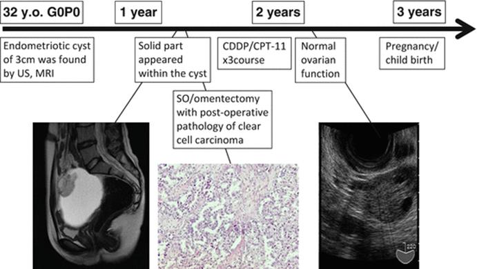
Fig. 15.1
Typical case of malignant transformation of endometriotic cyst
15.2 Endometriosis as a Precursor of Ovarian Cancer
15.2.1 Molecular Evidence
Endometriosis is currently classified as a tumorlike lesion according to the WHO classification, although it is not fully described. However, a variety of genetic analyses have demonstrated that endometriosis, especially the endometriotic cyst of the ovary, is a monoclonal lesion. In the late 1990s, X chromosome-linked polymorphism analysis revealed that an endometriotic cyst consisted of monoclonal epithelium [1–3]. Later, fluorescence in situ hybridization (FISH) analysis demonstrated the possibility that specific chromosomal loss or gain plays a role in the development and/or progression of endometriosis [4]. Similarly, another FISH study indicated that perturbations of chromosome 17 and the p53 locus occur frequently in severe, late-stage endometriosis [5]. In an analysis of DNA from 40 cases of endometriosis, 11 cases (28 %) demonstrated LOH at one or more loci, although no mutations were detected in the p53 or K-ras genes [6]. Most other similar analyses [1–3, 7, 8] revealed the monoclonal nature of endometriotic epithelium, except for one paper [9] in which the interpretation of the data may be inadequate. These findings clearly demonstrated that a majority of the endometriotic cysts are monoclonal and neoplastic. Moreover, genetic events may accumulate in endometriotic epithelia in parallel with the development of endometriosis.
15.2.2 Pathological View
Given the fact that endometriosis is a neoplastic disorder, it may have precancerous potential. Most endometrioses show a benign character, but malignant transformation may accompany some morphologic and genetic alterations. In pathology, we sometimes encounter the so-called atypical endometriosis, a putative intermediate between benign endometriosis and endometriosis-associated ovarian cancer (EAOC). Sampson first described ovarian cancer in endometriosis and defined a criterion for EAOC [10]. In a review of 194 cases of ovarian endometriosis, Czernobilsky and Morris found severe cytological atypia in 3.6 % of the cases [11]. Moreover, in their study, 2.0 % of the cases showed adenomatous, hyperplasia-like lesions. LaGrenade and Silverberg presented four cases of ovarian carcinomas contiguous with atypical glandular epithelial changes in endometriosis [12]. Subsequently, Fukunaga et al. reported that as many as 61 % of the cases of endometriosis coexisting with ovarian cancer had atypical endometriosis foci, while only 1.7 % of the cases of endometriosis without ovarian cancer exhibited atypical lesions [13]. Ogawa et al. reported that atypical endometriosis was found in 78 % of the cases with EAOC [14]. In their investigations, the transition from typical to atypical endometriosis was detected in 22 of 37 cases, and the transition from atypical endometriosis to carcinoma was found in 23 cases. These pathological observations suggest that benign endometriosis develops into ovarian cancer in some cases via atypical endometriosis, a premalignant stage.
15.2.3 Links Between the Pathology and Molecular Findings
Several studies have demonstrated that atypical endometriosis, which is a putative pathological transition between benign endometriosis and EAOC, has actually shown an intermediate nature by molecular analyses. Sáinz de la Cuesta et al. evaluated the immunohistochemical expression of p53 in normal endometrium, endometriosis, atypical endometriosis, and ovarian cancer associated with endometriosis. They found that 82.4 % of cancers associated with endometriosis and all the atypical endometriosis samples showed P53 overexpression, whereas only 11.8 % of the endometriosis samples, and none of the endometrium samples, showed P53 overexpression [15]. In the report by Obata et al., frequent LOH was observed on chromosome 6q (60.0 %) and chromosome 10q (40.0 %) in ovarian atypical endometriosis [16]. However, not all the endometriosis cases that are coincident with ovarian cancer have atypical endometriosis lesions, and the biological significance of atypical endometriosis is still unclear.
15.3 Molecular Events Associated with Malignant Transformation of Endometriosis
15.3.1 Loss of Heterozygosity (LOH)
There are several ways of analyzing LOH. LOH is usually used to estimate the locus of a tumor suppressor gene associated with a corresponding event, the development and malignant transformation of endometriosis in this case. Using DNA from 40 cases of endometriosis, Jiang et al. analyzed candidate ovarian tumor suppressor loci on chromosome arms 6q, 9p, 11q, 17p, l7q, and 22q [17]. LOH was detected on chromosomes 9p (18 %), 1lq (18 %), and 22q (15 %), and, in total, 11 of 40 (28 %) cases demonstrated LOH at one or more of these loci. The same investigators subsequently examined 14 cases of endometriosis synchronous with ovarian cancer for LOH on 12 chromosome arms and for X chromosome inactivation. In all four of the cases in which the carcinoma had arisen within the endometriosis and in five of the seven cases in which the carcinoma was adjacent to the endometriosis, common genetic lesions were detected to be consistent with a common lineage [17]. Prowse et al. analyzed LOH in 10 EAOCs with coexisting endometriosis using 82 microsatellite markers and found that, of 63 LOH events detected in the carcinoma samples, 22 were also detected in the corresponding endometriosis samples [18]. Goumenou et al. reported that LOH in p16, GALT, and p53, as well as APOA2, a region frequently lost in ovarian cancer, occurred in endometriosis, even in stage II of the disease [19]. Sato et al. examined 20 ovarian endometrioid carcinomas, 24 clear cell carcinomas, and 34 solitary endometrial cysts of the ovary for LOH at 10q23.3 [20]. In five endometrioid carcinomas synchronous with endometriosis, three cases displayed LOH events common to both the carcinoma and the endometriosis. In seven clear cell carcinomas that are synchronous with endometriosis, three displayed LOH events common to both the carcinoma and the endometriosis. No LOH events were found in solitary endometriosis. These findings indicate that attenuation of tumor suppressor genes is associated with the malignant transformation of endometriosis.
15.3.2 Mutation and Altered Expression of Oncogenes and Tumor Suppressor Genes
Mutations and altered expression of several genes have been implicated in the malignant transformation of endometriosis.
15.3.2.1 Augmented Expression of HNF-1β
Hepatocyte nuclear factor-1β is a transcription factor that is expressed specifically in clear cell carcinoma. Kato et al. examined expression of HNF-1β in 30 clear cell tumors (26 malignant, three borderline, and one benign) and in 40 endometriotic cysts [21]. All of the 30 clear cell tumors expressed HNF-1β. In 9 of 12 cases with the endometriotic epithelium, expression of HNF-1β was detected in the endometriotic epithelium as well as in the clear cell tumor. Furthermore, 16 of 40 (40 %) endometriotic cysts without neoplasms also expressed HNF-1β. They concluded that early differentiation into the clear cell lineage takes place in ovarian endometriosis, not only in atypical endometriosis but also in endometriosis with degenerative and regenerative changes.
15.3.2.2 The AKT/PI3K/Met Pathway
An array-based comparative genomic hybridization (CGH) analysis by Yamashita et al. revealed Met gene amplification in 4/13 ovarian clear cell carcinomas and 2/8 cell lines [22]. Amplification of the AKT2 gene was also observed in 5/21 samples. In 73 ovarian clear cell cases, 37.0 % demonstrated Met gene amplification (>4 copies), and 8.2 % had AKT2 amplification, suggesting that the Met signaling pathway plays an important role in clear cell carcinogenesis.
According to Yamamoto et al., exons 9 and 20 of the PIK3CA gene were analyzed in 23 clear cell carcinomas with synchronous putative precursor lesions [23]. Somatic mutations of the PIK3CA gene were detected in 10/23 (43 %) carcinomas and in the coexisting endometriotic epithelium adjacent to the carcinoma in 9/10 (90 %) cases. Moreover, in six of the nine lesions, the mutation was identified even in the endometrioses lacking cytological atypia. The authors suggested that mutations of the PIK3CA gene are early events in tumorigenesis, most likely initiating the malignant transformation of endometriosis.
15.3.2.3 ARID1A Mutations
ARID1A encodes BAF250a, one of the components of the SW1/SNF chromatin remodeling complex. In 2010, two groups identified mutations of the ARID1A gene in EAOC and in ovarian clear cell carcinoma. Genome-wide mutational analysis exhibited ARID1A mutations in almost one half of the clear cell carcinomas, one third of the endometrioid carcinomas, and none of the serous carcinomas of the ovary [24]. BAF250a protein expression was also lost in these cases. In some cases, loss of BAF1a expression was also found in atypical endometriosis adjacent to the cancer lesions. In another study, ARID1A mutations were detected in 57 % of the cases as well as PI3CA mutations in 40 % of the cases [25]. Although ARID1A is thought to play a role as a tumor suppressor gene [26], the precise effects of mutations in this gene are not fully understood.
Clinically, loss of ARID1A expression was significantly correlated with advanced FIGO stage, high CA125 levels, and with shorter progression-free survival of patients with clear cell carcinomas treated with platinum-based chemotherapy [27]. Although loss of ARID1A protein expression is thought to be an early event in the development of ovarian clear cell adenocarcinoma [28], its biological effect on the malignant transformation of endometriosis should be further clarified.
15.4 Environmental Factors That Affect Malignant Transformation of Endometriosis
15.4.1 Microenvironments in Endometriotic Cysts
15.4.1.1 Oxidative Stress and Carcinogenesis
In our bodies, live cells are continuously exposed to oxidative stresses, which are produced internally or externally. Internal stress consists of mitochondrial respiratory stress, cytochrome P450 metabolism, and inflammatory responses. External stresses are generated by various agents, including chemicals, xenobiotics, irradiation, and metal ions, including Fe ions. One of the well-known mechanisms by which metal ions produce intracellular ROS is called the Fenton reaction [29]. During this reaction, highly toxic dOH and anoxidized metal ions are generated from H2O2. Metal-induced ROS causes DNA damage, including single- or double-strand breaks, base modifications, deoxyribose modifications, and DNA cross-linking, which ultimately contributes to carcinogenesis.
In normal conditions, ROS overproduction is avoided by endogenous antioxidants, including superoxide dismutase (SOD), catalase (CAT), and glutathione peroxidase (GPx). If the balance between the cellular antioxidant defense and ROS generation is impaired, excessive ROS can cause oxidative stress. Prolonged and excessive oxidative stress mediates a variety of chronic and degenerative diseases, including cancers, inflammation, aging, and neuronal disorders [29].
15.4.1.2 Oxidative Stress in Endometriotic Cysts
The content of endometriotic cysts consists of old blood containing a high concentration of iron derived from hemoglobin. We compared the concentrations of free iron in endometriotic cysts with those in other ovarian cysts, including serous and mucinous cystadenoma, and found that the concentrations were significantly higher in endometriotic cysts [30] (Fig. 15.2). Likewise, lactose dehydrogenase (LDH) (a marker of tissue damage), potential antioxidant (PAI) (an antioxidant marker), lipid peroxidase (LPO) (Fig. 15.2) (a marker of oxidative stress), and 8-hydroxy-2-deoxyguanosine (a marker of DNA damage) were all significantly elevated in endometriotic cysts compared with other ovarian cysts. These data strongly suggest that the epithelial cells of the endometriotic cyst are constantly exposed to excessive oxidative stress and are subjected to cellular and DNA damage (Fig. 15.3).
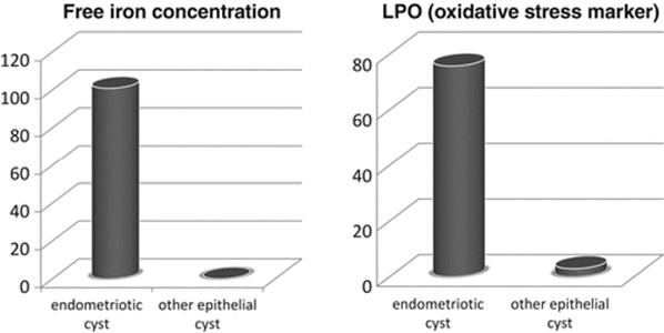
Fig. 15.2
Differences in Fe concentration/oxidative stress between endometriotic cysts and other benign epithelial cysts
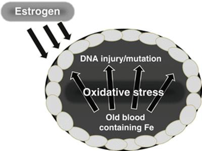
Fig. 15.3
Intrinsic and extrinsic factors affecting malignant transformation of endometriotic cyst
To elucidate the mutagenic property of the fluid in chocolate cysts, we conducted an experiment in vitro, which demonstrated that the fluid in chocolate cysts is more mutagenic than that in other cysts [30]. There are several reports suggesting a link between a stressful microenvironment and cancer development. (1) Chromosomal aberrations are more frequent in ovarian endometriotic cysts than in extra-gonadal endometriosis [31]. (2) Malignant transformation of endometriotic cysts increases with the duration of endometriosis [32].
15.4.2 Clear Cell Carcinoma and the Stress Response
If the microenvironment in endometriotic cysts affects cancer development, is it also associated with the phenotype of the ovarian cancer that arises as a result of malignant transformation? To assess this possibility, we began by identifying the gene signature that distinguishes clear cell carcinoma from other types of ovarian cancer using microarray datasets [33]. The signature composed of 437 genes was designated as the ovarian clear cell carcinoma (OCCC) signature. By using a categorical analysis, we demonstrated that the OCCC signature contains genes in three major categories, that is, stress response, sugar metabolism, and coagulation. Stress-related genes were estimated to play a central role, suggesting that the stress response is the fundamental feature of clear cell carcinoma. Actually, the OCCC signature was shown to involve a signal network consisting of many stress-related genes, including HIF1-α, IL-6, and SOD2. Moreover, when ovarian surface epithelial cells were treated with the contents of endometriotic cysts, the OCCC signature was increased in a time-dependent manner. The constitutive expression of the OCCC signature in clear cell carcinoma may reflect gene induction in response to the microenvironment in endometriotic cysts.
These data raise a novel and important concept that the specific microenvironment induces unique gene expression and leads to a cancer of a special phenotype. The local microenvironment, including ROS or inflammation, is likely not only to be implicated in cancer development [34], but it is also likely to be related to the occurrence of a specific cancer phenotype.
15.5 The Carcinogenic Scheme of Endometriotic Cysts and Future Applications
15.5.1 The Natural History of Endometriosis and Malignant Transformation
As mentioned earlier, endometriosis itself is largely a monoclonal, neoplastic disorder and has a potential for malignant transformation. Because endometriotic epithelium is a mullerian-type epithelium, it may be subject to mullerian organ carcinogenesis, leading to the development of various types of cancers, including serous carcinoma, mucinous carcinoma, and borderline tumors such as mucinous mullerian borderline tumors [35]. However, these types of cancer are relatively rare. A major type of cancer that develops from endometriosis is endometrioid carcinoma. The carcinogenic process of development of endometrioid carcinoma from endometriosis may resemble that in the endometrium under the influence of (unopposed) estrogen. In this case, progestin may play a prophylactic role, although no definite data are available yet.
The third mechanism, which is associated with the occurrence of clear cell carcinomas, is strongly influenced by the unique microenvironment within the chocolate cyst [35]. Continuous exposure to oxidative stress, partly caused by iron, results in the expression of stress-responsive genes, the OCCC signature. Constitutive expression of the OCCC signature genes is closely associated with the stress-resistant and slow-growing phenotype of malignancy, that is, clear cell carcinoma [36] (Fig. 15.4). Regarded as the prevention of clear cell carcinoma, surgical treatment to improve the microenvironment may be the best option. Thus, clarification of the precise natural course of malignant transformation of endometriotic cysts should lead to the proper management of endometriosis in terms of malignant transformation.
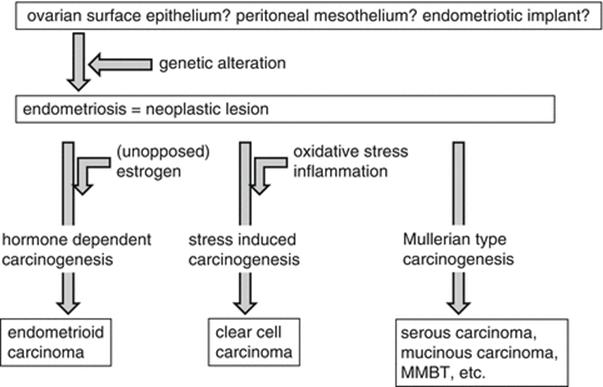
Fig. 15.4
Hypothetical mechanism of malignant transformation of endometriotic cyst
15.5.2 Future Treatment Strategy of EAOC, Especially Ovarian Clear Cell Carcinoma
Recent advances in understanding the molecular and biological features of clear cell carcinoma enable us to consider several new treatment strategies. Frequent activation of the AKT/PI3K/Met pathway in EAOC suggests that mTOR inhibitors may be effective in the treatment of these cancers. Mabuchi et al. showed that the mTOR inhibitor RAD001 is effective in the treatment of clear cell carcinoma of the ovary [37]. We have shown that sorafenib, a multikinase inhibitor, showed therapeutic effects in the RMG-2 clear cell cancer cell line, which is resistant to cisplatin [38]. In the future, the exploration of more precise mechanisms of malignant transformation of endometriosis may lead not only to various molecular targeted therapies but also to therapies targeting neovascularization or cancer metabolism [36].
References
1.
Jimbo H, Hitomi Y, Yoshikawa H, Yano T, Momoeda M, Sakamoto A, Tsutsumi O, Taketani Y, Esumi H. Evidence for monoclonal expansion of epithelial cells in ovarian endometrial cysts. Am J Pathol. 1997;150(4):1173–8.PubMedCentralPubMed
2.
Nilbert M, Pejovic T, Mandahl N, Iosif S, Willén H, Mitelman F. Monoclonal origin of endometriotic cysts. Int J Gynecol Cancer. 1995;5(1):61–3.PubMedCrossRef
3.
Tamura M, Fukaya T, Murakami T, Uehara S, Yajima A. Analysis of clonality in human endometriotic cysts based on evaluation of X chromosome inactivation in archival formalin-fixed, paraffin-embedded tissue. Lab Invest. 1998;78(2):213–8.PubMed
4.
Shin JC, Ross HL, Elias S, Nguyen DD, Mitchell-Leef D, Simpson JL, Bischoff FZ. Detection of chromosomal aneuploidy in endometriosis by multi-color fluorescence in situ hybridization (FISH). Hum Genet. 1997;100(3–4):401–6.PubMedCrossRef
5.
Bischoff FZ, Heard M, Simpson JL. Somatic DNA alterations in endometriosis: high frequency of chromosome 17 and p53 loss in late-stage endometriosis. J Reprod Immunol. 2002;55(1–2):49–64.PubMedCrossRef
6.
Jiang X, Hitchcock A, Bryan EJ, Watson RH, Englefield P, Thomas EJ, Campbell IG. Microsatellite analysis of endometriosis reveals loss of heterozygosity at candidate ovarian tumor suppressor gene loci. Cancer Res. 1996;56(15):3534–9.PubMed
7.
Nabeshima H, Murakami T, Yoshinaga K, Sato K, Terada Y, Okamura K. Analysis of the clonality of ectopic glands in peritoneal endometriosis using laser microdissection. Fertil Steril. 2003;80(5):1144–50.PubMedCrossRef
8.
Wu Y, Basir Z, Kajdacsy-Balla A, Strawn E, Macias V, Montgomery K, Guo SW. Resolution of clonal origins for endometriotic lesions using laser capture microdissection and the human androgen receptor (HUMARA) assay. Fertil Steril. 2003;79 Suppl 1:710–7.PubMedCrossRef
9.
Mayr D, Amann G, Siefert C, Diebold J, Anderegg B. Does endometriosis really have premalignant potential? A clonal analysis of laser-microdissected tissue. FASEB J. 2003;17(6):693–5.PubMed
10.
Sampson J. Endometrial carcinoma of the ovary, arising in endometrial tissue in that organ. Arch Surg. 1925;10:1–72.CrossRef
11.
Czernobilsky B, Morris WJ. A histologic study of ovarian endometriosis with emphasis on hyperplastic and atypical changes. Obstet Gynecol. 1979;53(3):318–23.PubMed
12.
LaGrenade A, Silverberg SG. Ovarian tumors associated with atypical endometriosis. Hum Pathol. 1988;19(9):1080–4.PubMedCrossRef
13.
Fukunaga M, Nomura K, Ishikawa E, Ushigome S. Ovarian atypical endometriosis: its close association with malignant epithelial tumours. Histopathology. 1997;30(3):249–55.PubMedCrossRef
14.
Ogawa S, Kaku T, Amada S, Kobayashi H, Hirakawa T, Ariyoshi K, Kamura T, Nakano H. Ovarian endometriosis associated with ovarian carcinoma: a clinicopathological and immunohistochemical study. Gynecol Oncol. 2000;77(2):298–304.PubMedCrossRef
15.
Sáinz de la Cuesta R, Izquierdo M, Cañamero M, Granizo JJ, Manzarbeitia F. Increased prevalence of p53 overexpression from typical endometriosis to atypical endometriosis and ovarian cancer associated with endometriosis. Eur J Obstet Gynecol Reprod Biol. 2004;113(1):87–93.PubMedCrossRef
16.
Obata K, Hoshiai H. Common genetic changes between endometriosis and ovarian cancer. Gynecol Obstet Invest. 2000;50 Suppl 1:39–43.PubMedCrossRef
17.
Jiang X, Morland SJ, Hitchcock A, Thomas EJ, Campbell IG. Allelotyping of endometriosis with adjacent ovarian carcinoma reveals evidence of a common lineage. Cancer Res. 1998;58(8):1707–12.PubMed
18.
Prowse AH, Manek S, Varma R, Liu J, Godwin AK, Maher ER, Tomlinson IP, Kennedy SH. Molecular genetic evidence that endometriosis is a precursor of ovarian cancer. Int J Cancer. 2006;119(3):556–62.PubMedCrossRef
19.
Goumenou AG, Arvanitis DA, Matalliotakis IM, Koumantakis EE, Spandidos DA. Microsatellite DNA assays reveal an allelic imbalance in p16(Ink4), GALT, p53, and APOA2 loci in patients with endometriosis. Fertil Steril. 2001;75(1):160–5.PubMedCrossRef
20.
Sato N, Tsunoda H, Nishida M, Morishita Y, Takimoto Y, Kubo T, Noguchi M. Loss of heterozygosity on 10q23.3 and mutation of the tumor suppressor gene PTEN in benign endometrial cyst of the ovary: possible sequence progression from benign endometrial cyst to endometrioid carcinoma and clear cell carcinoma of the ovary. Cancer Res. 2000;60(24):7052–6.PubMed
21.
Kato N, Sasou S, Motoyama T. Expression of hepatocyte nuclear factor-1beta (HNF-1beta) in clear cell tumors and endometriosis of the ovary. Mod Pathol. 2006;19(1):83–9.PubMedCrossRef
22.
Yamashita Y, Akatsuka S, Shinjo K, Yatabe Y, Kobayashi H, Seko H, Kajiyama H, Kikkawa F, Takahashi T, Toyokuni S. Met is the most frequently amplified gene in endometriosis-associated ovarian clear cell adenocarcinoma and correlates with worsened prognosis. PLoS One. 2013;8(3):e57724.PubMedCentralPubMedCrossRef
23.
Yamamoto S, Tsuda H, Takano M, Iwaya K, Tamai S, Matsubara O. PIK3CA mutation is an early event in the development of endometriosis-associated ovarian clear cell adenocarcinoma. J Pathol. 2011;225(2):189–94.PubMedCrossRef
24.
Jones S, Wang TL, Shih IM, Mao TL, Nakayama K, Roden R, Glas R, Slamon D, Diaz Jr LA, Vogelstein B, Kinzler KW, Velculescu VE, Papadopoulos N. Frequent mutations of chromatin remodeling gene ARID1A in ovarian clear cell carcinoma. Science. 2010;330(6001):228–31.PubMedCentralPubMedCrossRef
25.
Wiegand KC, Shah SP, Al-Agha OM, Zhao Y, Tse K, Zeng T, Senz J, McConechy MK, Anglesio MS, Kalloger SE, Yang W, Heravi-Moussavi A, Giuliany R, Chow C, Fee J, Zayed A, Prentice L, Melnyk N, Turashvili G, Delaney AD, Madore J, Yip S, McPherson AW, Ha G, Bell L, Fereday S, Tam A, Galletta L, Tonin PN, Provencher D, Miller D, Jones SJ, Moore RA, Morin GB, Oloumi A, Boyd N, Aparicio SA, Shih IM, Mes-Masson AM, Bowtell DD, Hirst M, Gilks B, Marra MA, Huntsman DG. ARID1A mutations in endometriosis-associated ovarian carcinomas. N Engl J Med. 2010;363(16):1532–43.PubMedCentralPubMedCrossRef
26.
Guan B, Wang TL, Shih IM. ARID1A, a factor that promotes formation of SWI/SNF-mediated chromatin remodeling, is a tumor suppressor in gynecologic cancers. Cancer Res. 2011;71(21):6718–27.PubMedCentralPubMedCrossRef
27.
Katagiri A, Nakayama K, Rahman MT, Rahman M, Katagiri H, Nakayama N, Ishikawa M, Ishibashi T, Iida K, Kobayashi H, Otsuki Y, Nakayama S, Miyazaki K. Loss of ARID1A expression is related to shorter progression-free survival and chemoresistance in ovarian clear cell carcinoma. Mod Pathol. 2012;25(2):282–8.PubMed
28.
Yamamoto S, Tsuda H, Takano M, Tamai S, Matsubara O. PIK3CA mutations and loss of ARID1A protein expression are early events in the development of cystic ovarian clear cell adenocarcinoma. Virchows Arch. 2012;460(1):77–87.PubMedCrossRef
29.
Lee JC, Son YO, Pratheeshkumar P, Shi X. Oxidative stress and metal carcinogenesis. Free Radic Biol Med. 2012;53(4):742–57.PubMedCrossRef
30.
Yamaguchi K, Mandai M, Toyokuni S, Hamanishi J, Higuchi T, Takakura K, Fujii S. Contents of endometriotic cysts, especially the high concentration of free iron, are a possible cause of carcinogenesis in the cysts through the iron-induced persistent oxidative stress. Clin Cancer Res. 2008;14(1):32–40.PubMedCrossRef
31.
Körner M, Burckhardt E, Mazzucchelli L. Higher frequency of chromosomal aberrations in ovarian endometriosis compared to extragonadal endometriosis: a possible link to endometrioid adenocarcinoma. Mod Pathol. 2006;19(12):1615–23.PubMedCrossRef
32.
Melin A, Sparén P, Persson I, Bergqvist A. Endometriosis and the risk of cancer with special emphasis on ovarian cancer. Hum Reprod. 2006;21(5):1237–42.PubMedCrossRef
33.
Yamaguchi K, Mandai M, Oura T, Matsumura N, Hamanishi J, Baba T, Matsui S, Murphy SK, Konishi I. Identification of an ovarian clear cell carcinoma gene signature that reflects inherent disease biology and the carcinogenic processes. Oncogene. 2010;29(12):1741–52.PubMedCrossRef
34.
Gonda TA, Tu S, Wang TC. Chronic inflammation, the tumor microenvironment and carcinogenesis. Cell Cycle. 2009;8(13):2005–13.PubMedCrossRef
35.
Mandai M, Yamaguchi K, Matsumura N, Baba T, Konishi I. Ovarian cancer in endometriosis: molecular biology, pathology, and clinical management. Int J Clin Oncol. 2009;14(5):383–91.PubMed
36.
Mandai M, Matsumura N, Baba T, Yamaguchi K, Hamanishi J, Konishi I. Ovarian clear cell carcinoma as a stress-responsive cancer: influence of the microenvironment on the carcinogenesis and cancer phenotype. Cancer Lett. 2011;310(2):129–33.PubMedCrossRef
37.
Mabuchi S, Kawase C, Altomare DA, Morishige K, Sawada K, Hayashi M, Tsujimoto M, Yamoto M, Klein-Szanto AJ, Schilder RJ, Ohmichi M, Testa JR, Kimura T. mTOR is a promising therapeutic target both in cisplatin-sensitive and cisplatin-resistant clear cell carcinoma of the ovary. Clin Cancer Res. 2009;15(17):5404–13.PubMedCentralPubMedCrossRef
38.
Matsumura N, Mandai M, Okamoto T, Yamaguchi K, Yamamura S, Oura T, Baba T, Hamanishi J, Kang HS, Matsui S, Mori S, Murphy SK, Konishi I. Sorafenib efficacy in ovarian clear cell carcinoma revealed by transcriptome profiling. Cancer Sci. 2010;101(12):2658–63.PubMedCrossRef