
Merete Kolberg Tennfjord, Marie Ellstrom Engh, and Kari B0
|
Abbreviations |
||
|
B-FLUTS |
Bristol Female Lower Urinary Tract Symptoms |
|
|
FSD |
Female sexual dysfunction |
|
|
FSFI |
Female Sexual Function Index |
|
|
ICIQ-FLUTSsex |
International Consultation on Incontinence Modular Questionnaire sexual matters module |
|
|
ICIQ-VS |
International Consultation on Incontinence Modular Questionnaire—Vaginal symptoms questionnaire |
|
|
I-QOL |
Incontinence Quality of Life Questionnaire |
|
|
LA muscle |
Levator ani muscle |
|
|
PEDro |
Physiotherapy Evidence Database |
|
|
PFD |
Pelvic floor dysfunctions |
|
|
PFM |
Pelvic floor muscles |
|
|
PFMT |
Pelvic floor muscle training |
|
|
PISQ-12 |
Pelvic Organ Prolapse/Urinary Incontinence Sexual Questionnaire |
|
|
POP |
Pelvic organ prolapse |
|
|
PT |
Physical therapist |
|
|
QoLS-N |
Quality of life scale |
|
|
RCT |
Randomized controlled trial |
|
|
SAI |
Sexual arousal inventory |
|
|
SAI-E |
Sexual arousal inventory-expanded form |
|
|
SII |
Sexual interaction inventory |
|
|
SUI |
Stress urinary incontinence |
|
|
UI |
Urinary incontinence |
|
|
WSQ |
Women’s Sexuality Questionnaire |
|
17.1 Introduction
Female sexual dysfunction (FSD) has been classified as sexual interest/arousal disorder, orgasmic disorder, and genitopelvic pain/penetration disorder [1]. It is commonly found among women of all ages [2]. Despite increasing demand for clinical services and the potential impact of these disorders on relationship satisfaction and quality of life [3], there is a lack of knowledge in how to approach women with FSD. It is an interlinked and complex disorder rooted in biological, social/relation- ship, and psychological aspects [4].
It has been postulated that the pelvic floor muscles’ (PFM) tone, strength, and ability to contract are important factors for vaginal receptivity and responsiveness, for the pleasure during intercourse for both partners, and for the orgasmic muscular response [4]. Some studies have shown that strong PFM may be associated with better orgasmic and arousal potentials, desire, excitement, and vaginal lubrication [5, 6], in addition to improved vaginal sensation and tightness [7, 8]. However, studies have also shown contradictory results [9, 10]. On the other hand, sexual pain disorders have been linked to hypertonicity (overactivity) of the PFM [4, 11], and different treatment strategies including a psychosocial approach, relaxation, and stretching techniques of the PFM and perineal tissue especially in postpartum women have been postulated [12].
The PFM comprise a superficial layer: ischiocavernosus, bulbospongiosus, and the transverse perineal muscles and a deep layer including the levator ani muscles: (the puboviseralis (pubococcygeus), the puborectalis, and the iliococcygeus muscles) [13, 14]. It is assumed that in addition to the changes in muscle tone set by the emotional motor system, it is especially the levator ani muscles and the bulbocaver- nosus muscle that are thought to play an active part in the sexual response in human beings. Upon clitoral and cervical stimulation, a contraction of the levator ani muscle is seen, together with a simultaneous contraction of the bulbocavernosus muscle. This activity is mainly mediated through reflex contractions that may contribute to the achievement of the orgasmic stage as well as physical changes in the vagina preparing for penetration [15-17].
The theory behind the rationale for strength training of the PFM is that strength training may build up the structural support of the pelvic floor by elevating the levator plate inside the pelvis to a higher location and by enhancing hypertrophy and stiffness of the PFM and adjacent connective tissue [18]. Braekken et al. [19] also showed that pelvic floor muscle training (PFMT) significantly reduced the levator hiatus area. Further, an increase in vascularization and neurogenic and musculo- genic factors may potentially affect lubrication and sensation and enhancing orgasm [18, 20, 21]. However, FSD is multifactorial [4], and a positive effect of different physical therapy interventions, including PFMT in the treatment of sexual dysfunction may have several explanations, for example, psychosocial aspects such as improved self-esteem and self-acceptance, body awareness, and satisfaction.
The aim of this chapter is to give an overview of randomized controlled trials (RCTs) investigating the effect of different physical therapy interventions in the treatment of FSD [11, 22].
17.2 Methods
A systematic literature search was performed in the PubMed electronic database, the Cochrane systematic reviews, the PEDro database, and the AMED (Ovid) database in December 2014 (updated in January 2016) containing all relevant RCTs investigating the effect of different physical therapy interventions on FSD. Physical therapy was defined according to the Physiotherapy Evidence Database (PEDro) and the following treatments were included as relevant: acupuncture, education, electrotherapy/heat/cold, strength training, and stretching/ mobilization/manipulation/massage. A broad search was performed to be able to find studies including relevant treatment methods and where FSD was not the primary aim of the study. The following combinations were used in the different databases: sexual function and pelvic floor, pelvic floor muscle training and postpartum, pelvic floor muscle training and female or sexual function, pelvic floor muscle and perineum or genitourinary system and pelvic and clinical trials, pelvic floor and female, female sexual dysfunction, and randomized controlled trial. Reference tracking of the eligible studies was also performed and included where appropriate. Chronic pain conditions of the pelvic floor were not included in this chapter. The methodological quality of the included trials was assessed using the PEDro quality score [23].
17.3 Results
The search revealed 640 articles, including duplicates. After an elimination process, we identified 14 RCTs on the effect of physical therapy interventions on FSD in either postpartum women [24-27], in women with pelvic organ prolapse (POP) [7, 28-30], in women with stress urinary incontinence (SUI) [31-33], in women with orgasmic disorders [34, 35], and in gynecological cancer patients [37]. Results from those trials are summarized in Table 17.1. There was a considerable variety in study
Table 17.1 Randomized controlled trials evaluating the effect of pelvic floor muscle training on female sexual function

(continued)
Table 17.1 (continued)


(continued)
Table 17.1 (continued)

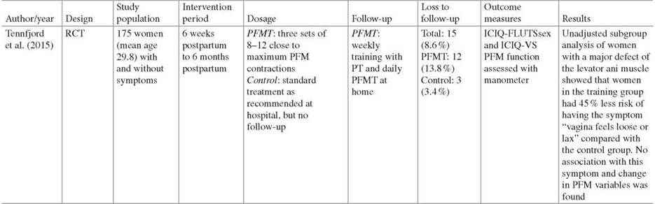
SUI stress urinary incontinence. POP pelvic organ prolapse. PT physical therapist. UI urinary incontinence. RCT randomized controlled trials. FSFI Female Sexual Function Index. QoLS-N Quality of life scale. B-FLUTS Bristol Female Lower Urinary Tract Symptoms. I-QOL Incontinence Quality of Life Questionnaire. PISQ-12 Pelvic Organ Prolapse/Urinary Incontinence Sexual Questionnaire. SAFE sexual arousal inventory-expanded form. WSQ Women’s Sexuality Questionnaire. SAI sexual arousal inventory. SII sexual interaction inventory. ICIQ-FLUTSsex International Consultation on Incontinence Modular Questionnaire sexual matters module. ICIQ-VS International Consultation on Incontinence Modular Questionnaire—Vaginal symptoms questionnaire population, type of intervention, training dosage, length and type of follow-up, outcome measures, and assessment methods of PFM. In a primary prevention study, Citak et al. [24] was the only study investigating effect of PFMT in a healthy population, making it difficult to compare and to generalize the findings from the published studies. The search strategy also revealed two nonrandomized trials in women with urinary incontinence (UI) [38, 39]. Since we aimed for randomized trials only in this chapter, the two latter studies are not included. The PEDro score of the included studies is summarized in Table 17.2.
Nine studies reported an overall positive effect of physical therapy for different aspects of FSD: improved arousal, lubrication and orgasm, satisfaction with sexual life, increased control and stronger PFM, “tighter vagina,” increased libido, less pain, increased sensibility and awareness around the pelvic floor, improved confidence, and partner’s sexual satisfaction [7, 24, 26-29, 31, 33, 37]. Of these, six studies included PFMT as the only treatment option [7, 24, 26-28, 31]. The other three studies combined PFMT with biofeedback [37]; PFMT with biofeedback, infrared, reinforcement exercises, and relaxation [29]; and PFMT with continence strategies [33]. However, five studies showed contradictory results [25, 30, 32, 34, 35].
Table 17.2 PEDro quality score of randomized controlled trials evaluating effect of pelvic floor muscle training on female sexual function
|
Study |
E |
1 |
2 |
3 |
4 |
5 |
6 |
7 |
8 |
9 |
10 |
Total score |
|
Citak et al. (2010) |
+ |
+ |
+ |
— |
— |
+ |
— |
— |
+ |
+ |
5 |
|
|
Wilson and Herbison (1998) |
+ |
+ |
— |
+ |
— |
— |
+ |
— |
— |
+ |
+ |
5 |
|
B0 et al. (2000) |
+ |
+ |
+ |
+ |
— |
— |
+ |
+ |
— |
+ |
+ |
7 |
|
Brækken et al. (2014) |
+ |
+ |
+ |
+ |
— |
— |
— |
+ |
+ |
+ |
+ |
7 |
|
Liebergall—Wischnitzer et al. (2012) |
+ |
+ |
+ |
+ |
— |
— |
+ |
— |
— |
+ |
+ |
6 |
|
M0rkved et al. (2007) Abstract |
+ |
+ |
+ |
+ |
— |
— |
+ |
— |
— |
+ |
+ |
6 |
|
Yang et al. (2012) Pilot |
+ |
+ |
— |
+ |
— |
— |
+ |
+ |
— |
+ |
+ |
6 |
|
Hagen et al. (2014) |
+ |
+ |
+ |
+ |
— |
— |
+ |
— |
+ |
+ |
+ |
7 |
|
Eftekhar et al. (2014) |
+ |
+ |
— |
+ |
— |
— |
— |
+ |
+ |
+ |
+ |
6 |
|
Wiegersma et al. (2014) |
+ |
+ |
+ |
+ |
— |
— |
+ |
+ |
+ |
+ |
+ |
8 |
|
Chambless et al. (1984) |
+ |
+ |
— |
— |
— |
— |
+ |
— |
— |
+ |
+ |
4 |
|
Trudel and Saint-Laurent (1983) |
+ |
+ |
— |
+ |
— |
— |
— |
+ |
— |
+ |
+ |
5 |
|
Handa et al. (2008) [36] |
+ |
+ |
+ |
+ |
— |
— |
+ |
— |
— |
+ |
+ |
6 |
|
Tennfjord et al. (2015) |
+ |
+ |
+ |
+ |
— |
— |
+ |
+ |
+ |
+ |
+ |
8 |
E eligibility criteria specified, 1 subjects randomly allocated to groups, 2 allocation concealed, 3 groups similar at baseline, 4 subjects blinded, 5 therapist administering treatment blinded, 6 assessors blinded, 7 measures of key outcomes obtained from >85 % of subjects, 8 data analyzed by intention to treat, 9 statistical comparison between groups conducted, 10 point measures and measures of group variability provided + criterion clearly satisfied, — criterion is not satisfied. Total score is determined by counting the number of criteria satisfied, except “eligibility criteria satisfied” score is not used to generate the total score. Total scores are out of 10 Points 4 and 5 are unable to meet satisfactory criteria in all studies as subjects allocated to PFMT and therapists administering the treatment are aware of the intervention
What characterizes these studies are a high dropout rate [25, 32, 34] and low adherence to the treatment protocol [30]. In addition, the studies reporting no effect of the intervention did not carry out an assessment (or provided information about doing so) of a correct PFM contraction by observation and palpation of the PFM before the intervention (Fig. 17.1) [40, 41]. Further, the studies by Liebergall-Wischnitzer et al. [32] and Chambless et al. [34] found a positive effect in both groups, but the results did not reach a significant between-group difference. Possible explanations could be differences in training dosage and follow-up between the two intervention groups [32], whereas the study by Chambless et al. [34] provided both intervention groups with the same information and encouragement to discuss their concerns which may have led to both groups being more aware and responsive.
Seven studies reported improved PFM strength after intervention compared with the control group [7, 24, 26, 27, 31, 33, 37], but only the studies by Braekken et al. [7] and Yang et al. [37] reported the improvement in PFM strength to be associated with improvement in sexual function. It is therefore difficult to conclude whether there is a link between improved sexual function and improved PFM function. Given the multidimensional etiology of FSD, it might be that the PFM indirectly play a part in female sexual function and dysfunction. Eight studies included questions related to sexual pain disorders (pelvic floor pain and dyspareunia) [7, 24, 25, 27, 29, 31-33], and two studies reported effect of PFMT (PFMT alone or combined) in women with POP [9, 29]. However, the comparison group with surgery may not have been appropriate in the study by Eftekhar et al. [29], as the surgery group reported worsening of dyspareunia following rectocele repair. Due to the nature and complexity of this symptom [4], more studies are needed and factors related to success of physical therapy interventions must be identified. Long-term follow-up was performed in two studies, showing contradictory results [26, 28].

Fig. 17.1 Through observation and vaginal palpation, the physical therapist instructs the woman in how to contract the pelvic floor muscles correctly, defined as an inward movement of the perineum and a squeeze around the pelvic openings. This contraction is palpated as a squeeze around the examiners’ finger and an inward lift (Photo: private with permission from patient)
17.4 Summary
This chapter reflects some positive results of physical therapy in the treatment of FSD. However, it must be kept in mind the complexity of FSD and that a multidisciplinary approach often is necessary. Physical therapy may therefore be an important part in the treatment of these women. Furthermore, an individual approach is crucial in the evaluation and treatment of each woman. It is an urgent need of more high- quality studies in this field of women’s health and a standardization of study populations, interventions, outcome measures, and assessment methods of PFM function. Factors related to success of physical therapy interventions (Table 17.3) must be identified before we can make conclusions upon which approach is most effective.
Table 17.3 Suggestions for physical therapy in women with sexual dysfunction
|
A thorough anamnesis including relevant background factors: other types of pelvic floor dysfunctions and treatment of those, natural functions, hormonal contraceptives, parity, delivery mode, level of physical activity, sleep disturbances, depression, sexual activity, experience and function, social factors, partner, medication, history of illness/disease, abuse. What caused the problem, onset, duration, location, frequency, intensity, factors that trigger/ relieve the problem. Keep in mind red and yellow flags. Own expectations for treatment. Use of pain body mapping and pain scale (NRS or VAS). Note that specific conditions requires additional attention. These questions are more general for all conditions |
|
Referral to other professions and multidisciplinary team |
|
Assessment of external musculature and joints, use of pain body mapping and pain scale (NRS or VAS) including lumbar spine, pelvis, and hips. Assess for posture, mobility, tension, strength, temperature, sweat, relevant scars pain |
|
Observation of skin color (redness), scar tissue, ability to contract and relax. Palpation with assessment of superficial and deep pelvic floor musculature, obturator internus, sacrotuberale ligament. Assess for ability to contract, relax, tender points, muscle tone, including hypo -and hypertonicity. Assessment of strength, local muscular endurance and vaginal resting tone using appropriate devices (ultrasound, electromyography, manometer, dynamometer) |
|
Summarize findings and set up a treatment plan related to the findings including suggested physical therapy interventions: life style advice, body awareness, education, electrotherapy/ heat/cold, strength training, stretching/mobilization/manipulation/trigger point treatment/ massage/acupuncture may be tried. Consider follow-up and home exercise |
References
1. Ishak WW, Tobia G. DSM-5 changes in diagnostic criteria of sexual dysfunctions. Reprod Sys Sex Disord. 2013;2(2):122-4.
2. Laumann EO, Paik A, Rosen RC. Sexual dysfunction in the United States: prevalence and predictors. JAMA. 1999;281(6):537-44.
3. Skinner EM, Dietz HP. Psychological and somatic sequelae of traumatic vaginal delivery: a literature review. Aust N Z J Obstet Gynaecol. 2015;55(4):309-14.
4. Graziottin A. Female pelvic floor dysfunction and evidence-based physical therapy. In: B0 K, Berghmans B, M0rkved S, Van Kampen M, editors. Evidence based physical therapy for the pelvic floor: bridging science and clinical practice. 2nd ed. Philadelphia: Churchill Livingstone Elsevier; 2015. p. 243-70.
5. Martinez CS, Ferreira FV, Castro AAM, Gomide LB. Women with greater pelvic floor muscle strength have better sexual function. Acta Obstet Gynaecol Scand. 2014;93:457-502.
6. Lowenstein L, Gruenwald I, Gartman I, Vardi Y. Can stronger pelvic muscle floor improve sexual function? Int Urogynecol J. 2010;21(5):553-6.
7. Braekken IH, Majida M, Ellstrom Engh M, Bo K. Can pelvic floor muscle training improve sexual function in women with pelvic organ prolapse? A randomized controlled trial. J Sex Med. 2015;12(2):470-80.
8. Tennfjord MK, Hilde G, Staer-Jensen J, Siafarikas F, Engh ME, Bo K. Coital incontinence and vaginal symptoms and the relationship to pelvic floor muscle function in primiparous women at 12 months postpartum: a cross-sectional study. J Sex Med. 2015;12(4): 994-1003.
9. Chambless DL, Stern T, Sultan FE, Williams AJ, Goldstein AJ, Lineberger MH, et al. The pubococcygens and female orgasm: a correlational study with normal subjects. Arch Sex Behav. 1982;11(6):479-90.
10. Baytur YB, Deveci A, Uyar Y, Ozcakir HT, Kizilkaya S, Caglar H. Mode of delivery and pelvic floor muscle strength and sexual function after childbirth. Int J Gynaecol Obstet. 2005;88(3):276-80.
11. Messelink B, Benson T, Berghmans B, Bo K, Corcos J, Fowler C, et al. Standardization of terminology of pelvic floor muscle function and dysfunction: report from the pelvic floor clinical assessment group of the International Continence Society. Neurourol Urodyn. 2005;24(4): 374-80.
12. Rosenbaum TY. An integrated mindfulness-based approach to the treatment of women with sexual pain and anxiety: promoting autonomy and mind/body connection. Sex Relation Ther. 2013;28(1-2):20-8.
13. Ashton-Miller JA, Howard D, DeLancey JO. The functional anatomy of the female pelvic floor and stress continence control system. Scand J Urol Nephrol Suppl. 2001;207:1-7; discussion 106-25.
14. Ashton-Miller JA, DeLancey JO. Functional anatomy of the female pelvic floor. In: B0 K, Berghmans B, M0rkved S, van Kampen M, editors. Evidence-based physical therapy for the pelvic floor: bridging science and clinical practice. 1st ed. Philadelphia: Churchill Livingstone Elsevier; 2007. p. 19-33.
15. Vodusek DB. Neuroanatomy and neurophysiology of the pelvic floor muscles. In: B0 K, Berghmans B, M0rkved S, van Kampen M, editors. Evidence-based physical therapy for the pelvic floor: bridging science and clinical practice. 2nd ed. Philadelphia: Churchill Livingstone Elsevier; 2015. p. 35-41.
16. Achtari C, Dwyer PL. Sexual function and pelvic floor disorders. Best Pract Res Clin Obstet Gynaecol. 2005;19(6):993-1008; quiz A1-8.
17. Shafik A. The role of the levator ani muscle in evacuation, sexual performance and pelvic floor disorders. Int Urogynecol J Pelvic Floor Dysfunct. 2000;11(6):361-76.
18. B0 K. Pelvic floor muscle training is effective in treatment of female stress urinary incontinence, but how does it work? Int Urogynecol J. 2004;15:76-84.
19. Braekken IH, Majida M, Engh ME, Bo K. Morphological changes after pelvic floor muscle training measured by 3-dimensional ultrasonography: a randomized controlled trial. Obstet Gynecol. 2010;115(2 Pt 1):317-24.
20. Berman J, Berman L, Kanaly K. Female sexual dysfunction: new perspectives on anatomy, physiology, evaluation and treatment. EAU Updat Ser. 2003;1(3):166-77.
21. B0 K, Aschehoug A. Strength training. In: B0 K, Berghmans B, M0rkved S, van Kampen M, editors. Evidence-based physical therapy for the pelvic floor: bridging science and clinical practice. 1st ed. Philadelphia: Churchill Livingstone Elsevier; 2007. p. 119-32.
22. Bo K, Frawley HC, Haylen BT, Abramov Y, Almeida FG, Berghmans B et al. An International Urogynecological Association (IUGA)/International Continence Society (ICS) joint report on the terminology for the conservative and nonpharmacological management of female pelvic floor dysfunction. Int Urogynecol J. 2016. doi: 10.1007/s00192-016-3123-4.
23. Maher CG, Sherrington C, Herbert RD, Moseley AM, Elkins M. Reliability of the PEDro scale for rating quality of randomized controlled trials. Phys Ther. 2003;83(8):713-21.
24. Citak N, Cam C, Arslan H, Karateke A, Tug N, Ayaz R, et al. Postpartum sexual function of women and the effects of early pelvic floor muscle exercises. Acta Obstet Gynecol Scand. 2010;89(6):817-22.
25. Wilson PD, Herbison GP. A randomized controlled trial of pelvic floor muscle exercises to treat postnatal urinary incontinence. Int Urogynecol J Pelvic Floor Dysfunct. 1998;9(5):257-64.
26. Morkved S, Rommen K, Schei B, Salvesen KA, Bo K. No difference in urinary incontinence between training and control group six years after cessation of a randomized controlled trial, but improved sexual satisfaction in the training group. Neurourol Urodyn. 2007;26(5):667.
27. Tennfjord MK, Hilde G, Stær-Jensen J, Siafarikas F, Ellstrom Engh M, B0 K. Effect of postpartum pelvic floor muscle training on vaginal symptoms and sexual dysfunction—secondary analysis of a randomised trial. BJOG. 2016;123(4):634-42.
28. Hagen S, Stark D, Glazener C, Dickson S, Barry S, Elders A, et al. Individualised pelvic floor muscle training in women with pelvic organ prolapse (POPPY): a multicentre randomised controlled trial. Lancet. 2014;383(9919):796-806.
29. Eftekhar T, Sohrabi M, Haghollahi F, Shariat M, Miri E. Comparison effect of physiotherapy with surgery on sexual function in patients with pelvic floor disorder: a randomized clinical trial. Iran J Reprod Med. 2014;12(1):7-14.
30. Wiegersma M, Panman CM, Kollen BJ, Berger MY, Lisman-Van Leeuwen Y, Dekker JH. Effect of pelvic floor muscle training compared with watchful waiting in older women with symptomatic mild pelvic organ prolapse: randomised controlled trial in primary care. BMJ. 2014;349:g7378.
31. Bo K, Talseth T, Vinsnes A. Randomized controlled trial on the effect of pelvic floor muscle training on quality of life and sexual problems in genuine stress incontinent women. Acta Obstet Gynecol Scand. 2000;79(7):598-603.
32. Liebergall-Wischnitzer M, Paltiel O, Hochner Celnikier D, Lavy Y, Manor O, Woloski Wruble AC. Sexual function and quality of life of women with stress urinary incontinence: a randomized controlled trial comparing the Paula method (circular muscle exercises) to pelvic floor muscle training (PFMT) exercises. J Sex Med. 2012;9(6):1613-23.
33. Handa VL, Whitcomb E, Weidner AC, Nygaard I, Brubaker L, Bradley CS, et al. Sexual function before and after non-surgical treatment for stress urinary incontinence. Female Pelvic Med Reconstr Surg. 2011;17(1):30-5.
34. Chambless DL, Sultan FE, Stern TE, O’Neill C, Garrison S, Jackson A. Effect of pubococcygeal exercise on coital orgasm in women. J Consult Clin Psychol. 1984;52(1):114-8.
35. Trudel G, Saint-Laurent S. A comparison between the effects of Kegel’s exercises and a combination of sexual awareness relaxation and breathing on situational orgasmic dysfunction in women. J Sex Marital Ther. 1983;9(3):204-9.
36. Handa VL, Cundiff G, Chang HH, Helzlsouer KJ. Female sexual function and pelvic floor disorders. Obstet Gynecol. 2008;111(5):1045-52.
37. Yang EJ, Lim JY, Rah UW, Kim YB. Effect of a pelvic floor muscle training program on gynecologic cancer survivors with pelvic floor dysfunction: a randomized controlled trial. Gynecol Oncol. 2012;125(3):705-11.
38. Beji NK, Yalcin O, Erkan HA. The effect of pelvic floor training on sexual function of treated patients. Int Urogynecol J Pelvic Floor Dysfunct. 2003;14(4):234-8; discussion 8.
39. Zahariou AG, Karamouti MV, Papaioannou PD. Pelvic floor muscle training improves sexual function of women with stress urinary incontinence. Int Urogynecol J Pelvic Floor Dysfunct. 2008;19(3):401-6.
40. Kegel AH. Stress incontinence and genital relaxation; a nonsurgical method of increasing the tone of sphincters and their supporting structures. Ciba Clin Symp. 1952;4(2):35-51.
41. Bo K, Hagen RR, Kvarstein B, Larsen S. Pelvic floor muscle exercise for the treatment of female stress urinary incontinence. II. Validity of vaginal pressure measurements of pelvic muscle strength and the necessity of supplementary methods for control of correct contraction. Neurourol Urodyn. 1990;9(5):479-87.