
Jean Calleja-Agius1 and Mark Brincat1
(1)
Department of Obstetrics and Gynaecology, Mater Dei Hospital, Birkirkara, Malta
Jean Calleja-Agius
Email: jean.calleja-agius@um.edu.mt
Mark Brincat (Corresponding author)
Email: brincatm@maltanet.net
17.1 Introduction: Endocrinology and Immunology of Parturition
Hormonal interactions are indispensable to control the establishment of pregnancy, foetal development, and even the process of parturition [1]. The placental progesterone, along with other hormones, act as allocrine factors, being produced by one organ and used by another, to modify the maternal environment and satisfy the needs of the growing foetus.
Parturition is divided into different phases depending on the contractile activity of the myometrium. The initial phase is termed “Phase 0” (quiescence), wherein the myometrium is relaxed and relatively insensitive to stimulatory uterotonics (substances that regulate myometrial tone and contractility) such as prostaglandins and oxytocin. Instead relaxatory uterotropins and uterotonics, such as progesterone, β-adrenergic agents, prostacyclin (PGI2), relaxin, CRH, nitric oxide, work to maintain pregnancy. Generally, all these regulatory factors activate adenylyl cyclase and increase intracellular cAMP [1].
Parturition begins at the transition from “Phase 0” to “Phase 1”, when the myometrium gains response to uterotonics and starts to contract forcibly and rhythmically. This is made possible by progesterone withdrawal and an increase in oestrogen drive [2]. Also, the myometrium is transformed by the expression of genes known as contraction-associated proteins (CAPs), which upregulate gap junctions between cells, ion channels, uterotonin receptors and enzymes [3]. The progesterone withdrawal and oestrogen activation are not mediated by changes in the levels of these hormones but rather by the myometrial responsiveness to these hormones [4]. Progesterone responsiveness depends on the activity of nuclear prostaglandin receptors (nPRs). PR-B is a principal ligand-dependant transcriptional modulator of progesterone responsive genes, which mediates relaxatory actions. Conversely, PR-A represses the transcriptional activity mediated by PR-B. In fact, it is the PR-A/PR-B expression ratio, which determines progesterone responsiveness, and it is hypothesised that progesterone withdrawal is mediated by an increase in this ratio [5]. With regard to oestrogen, the two major subtypes of receptors are ERα and ERβ [6]. ERα is drastically increased at term with the onset of labour, and it is directly associated with Cx43 expression (oestrogen-responsive CAP gene), thereby causing the functional oestrogen activation. Conversely, ERβ is not influenced by the onset of labour [7].
“Phase 2” (active labour) is then initiated by the increase in the levels of prostaglandins and the increased sensitivity of the myometrium to the prostaglandins and oxytocin. This is characterised by rhythmic contractions, which become progressively more forceful. These contractions propel the foetus forward toward the birth canal, and eventually dilate the softened cervix so that the foetus and the placenta are both delivered [1]. The main prostaglandins involved are PGE2 and PGF2α, which are produced by intrauterine tissues, mainly the amnion, chorion, decidua and myometrium [8]. The rate limiting step of prostaglandins production is catalysed by cyclooxygenase, COX-1 and COX-2, enzymes [9]. The prostaglandins are metabolised by prostaglandin dehydrogenase (PGDH), which irreversibly converts PGE2 and PGF2α to inactive forms. The actions of prostaglandins are mediated by specific prostanoid receptors [10]. The other important mediator, oxytocin, is the most potent and specific stimulant of uterine contraction and is used to induce labour and treat postpartum haemorrhage. It is produced during pregnancy by the amnion, chorion and decidua [11]. Despite its potency, it is generally not regarded as being involved in the initiation of labour [12].
“Phase 3” begins after the placenta is expelled. The myometrial contractions are sustained in order to constrict the spiral arteries and minimise post-partum haemorrhage. Finally, the uterus returns to menstrual state by myometrial cell apoptosis and atrophy. The cervix also returns to its closed and rigid state [1].
In pregnancy, immunological suppressor activity is carried out by T regulatory (Treg) cells that increase in number when progesterone levels are high. Treg cells influence other cells through cytokines, which may influence the survival and growth of the foetoplacental unit [13]. Cytokines are also produced by the reproductive tract such as the uterine epithelium and trophoblast. Suppression may also be mediated by a soluble factor that is related to transforming growth factor-β (TGF-β) and by Prostaglandin (PGE2) by decidual cells and macrophages. All these cytokines and factors may inactivate lymphocytes [14, 15].
During pregnancy, the maternal immune system must protect the mother against any infections and tumours but at the same time must protect the foetus from any harmful immunological effects. This occurs by an altered T-helper 1 and T-helper 2 (TH1/TH2) cytokine balance [16].
The establishment of pregnancy, successful foetal development and parturition is achieved by hormonal interactions between the mother, placenta and foetus [1]. In addition, it is essential that there must be well-coordinated interactions between the maternal innate immune system and the trophoblast. The trophoblast and maternal immune system protect the foetus and mother against infectious microorganisms by acting in synchrony. Potentially dangerous molecular signatures are first identified by the trophoblast and then the maternal immune system responds in a coordinated way. The success of a pregnancy depends on good communication between the trophoblast and maternal immune system [17]. Cytokines, which can be elaborated with or without the involvement of the immune system, are involved in this communication [18]. The type of cytokines is very important in the success of pregnancy and it varies between implantation, early and late pregnancy and parturition [19]. There are mutual interactions between the closely related endocrine and immune systems. The steroid hormones and cytokines act as mediators and messengers to achieve a successful pregnancy [20]. An imbalance in hormones or cytokines can cause problems in pregnancies such as preterm birth, as well as miscarriage, pre-eclampsia and intrauterine growth retardation (IUGR) [21–24].
17.2 Preterm Labour
Preterm delivery refers to birth occurring before 37 completed weeks or 259 days of gestation [25]. In cases of uncertainty about the gestational age only, foetal weight becomes valuable in determining preterm birth, with a threshold of 500 g. However, this method is rather inaccurate since viable neonates born after 24 weeks may be less than 500 g due to IUGR and some unviable neonates may weigh more than 500 g [17]. Preterm births may be classified as “spontaneous”, which may occur either with intact membranes or with PPROM. Otherwise, preterm births are classified as “indicated/iatrogenic”, meaning that preterm labour is induced or elective caesarean section is performed due to obstetric complications such as pre-eclampsia, IUGR, multiple pregnancies or chorioamnionitis [17]. A significant proportion of infants born preterm develop one or more related complications, some of which require life-long care [26].
17.2.1 Common Pathway of Parturition
Preterm labour results from pathological processes which trigger some or the entire components characteristic of the normal physiologically activated pathway seen in term labour, with the critical difference being a smaller gestational age [27]. These characteristics include increased uterine contractility, cervical changes and membrane rapture [28]. Labour is characterized by a drastic change from “contractures”, which refer to several minutes of myometrial activity with moderate increase in intrauterine pressure, to “contractions” which are short episodes of dramatic increase in intrauterine pressure [29]. Various cervical changes which have been described include softening which begins in early pregnancy, ripening which involves a decrease in the concentration of collagen and dispersion of collagen fibrils and dilation which may be described as an inflammatory process with influx of macrophages and neutrophils accompanied by matrix degradation [30, 31]. Membrane rupture normally occurs in preparation for delivery and involves the degradation of foetal fibronectin (fFn) at the chorion–decidual interface. However, PROM may also occur due to amnion epithelial apoptosis and localized inflammation [32].
The common pathway is a term defined as the anatomical, physiological, biochemical, endocrinological, immunological and clinical events occurring at term or preterm within the mother and/or the foetus [33–35]. These events are mostly notable in the uterine component, but extrauterine components are also involved in this pathway. Many scientific research led prostaglandins, a lipid compound, to be considered the primary mediators of the most important changes associated with the onset of labour, including myometrial contractility, cervical ripening and membrane activation [36]. Prostaglandins production is stimulated by an increasingly high oestrogen/progesterone ratio at parturition [2]. This then leads to an increase in the intracellular calcium concentration of uterine smooth muscle cells by upregulating sarcoplasmic and transmembrane calcium fluxes. Prostaglandins also increase contractility by upregulating transcription of oxytocin receptors, gap junctions and prostaglandin receptors (EP and FP) [37]. A second effect of this lipid compound is to stimulate the foetal membrane and the cervix to synthesis MMPs, which are involved in membrane rapture and cervical ripening [38]. Thirdly, increased expression of progesterone receptor isoforms (PR-A/PR-B) is made possible by prostaglandin (PGE2 and PGF2α) to induce a functional progesterone withdrawal [39].
17.3 Aetiological Mechanisms of Spontaneous Preterm Birth
Preterm birth is regarded to be a “syndrome”, implying that it has multiple aetiologies with a combination of signs and symptoms creating the characteristic clinical presentation of the mother [40]. It is believed to be of multifactorial origin, since certain causes of preterm birth, such as micro-organisms, are attributed to an environmental influence and cause inflammation under genetic control [17]. The main mechanical processes that have been implicated in preterm birth syndrome are intrauterine infection, inflammation, uteroplacental ischaemia and haemorrhage, maternal and foetal stress, uterine over distension, cervical disease, endocrine disorders, allergic phenomena and abnormal allogenic recognition [17].
The main hormone implicated as having an influence on the process of preterm parturition is progesterone. This hormone performs a number of critical functions in order to maintain pregnancy, including gap junction downregulation, cervical ripening inhibition and downregulation in chemokine production. Therefore, any abnormalities in its function may lead to preterm birth. Evidence has shown that a “functional progesterone withdrawal” occurs in intrauterine tissue, in both term and preterm parturition, creating a change in the ratio of oestrogen and progesterone that triggers components of the common pathway of parturition [41].
17.4 Prevention and Management
Many methods have been employed in an attempt to predict preterm delivery, but to date, they are of limited clinical use. Without this knowledge, preventative measures would be useless. However, different techniques have been and are still being developed to prevent the problem in those females at high risk of experiencing preterm birth [42]. The fact that the exact mechanism of the disease is not yet understood makes it difficult to provide successful diagnostic tests and successful prophylactic and/or treatment methods [17]. Obstetricians in clinical practice often face the dilemma of how to manage an established preterm labour. The physician should always balance the risks to both mother and foetus of delivering the baby prematurely against the risk of trying to prolong the pregnancy. Treatment is aimed either at preventing the initiation of myometrial contractility or at preventing cervical dilatation.
17.4.1 17-a-Hydroxy Progesterone
The corpus luteum serves as a source of steroid hormones, mainly progesterone, for the first trimester, corresponding to approximately the first 13 weeks of gestation. However, the placenta then takes over in the second and third trimesters and remains the major source during most of the pregnancy by acting in concert with the mother and the foetus [2]. Progesterone is an important hormone produced by the placenta, which plays the indispensable role of maintaining pregnancy. Progesterone production involves the transport of low-density lipoprotein (LDL) cholesterol from the maternal compartment into the placental compartment where it is converted first to pregnenolone and then to progesterone in a rapid and efficient manner [43]. As shown in Fig. 17.1, the process requires the cholesterol desmolase P450scc (cholesterol side-chain cleavage) and 3β-hydroxysteroid dehydrogenase (3β-HSD), which are expressed in the human placenta. This process of steroidogenesis terminates with progesterone because the fact that the human placenta lacks 17α-hydroxylase and 17,20-lyase (whose activity is mediated by a single enzyme called P450c17), which allows the formation of glucocorticoids and androgens in the adrenal cortex [1, 2].
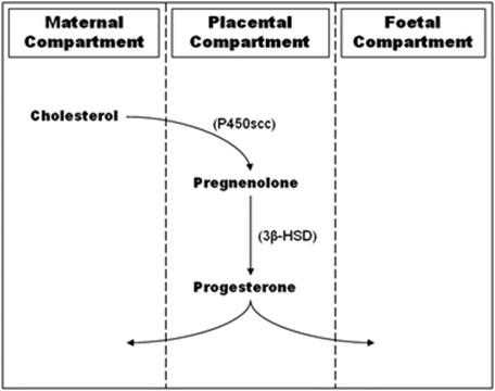
Fig. 17.1
Biosynthetic pathway of placental progesterone
This “progestational” hormone prevents rejection of the conceptus by the immune system and is responsible for the maintenance of pregnancy. This is possible since progesterone blocks the maternal immune responses to foreign antigens through suppression of T-lymphocyte cell-mediated responses [44]. Table 17.1 shows the effect of progesterone on the myometrium.
Table 17.1
Actions of progesterone on the myometrium
|
Decreases conduction of contractions |
|
Increases threshold for stimulation |
|
Decreases spontaneous activity |
|
Decreases number of oxytocin receptors |
|
Suppresses the inflammatory cascade |
|
Acts as a calcium antagonist |
|
Inhibits T lymphocyte development |
|
Promotes expression of prostaglandin EP2 receptor |
|
Prevents formation of gap junctions |
|
Administration of progesterone antagonists stimulates onset of labor in women at term |
17-a-Hydroxy progesterone has also been documented to be a successful drug in the prevention and in decreasing the rate of recurrent preterm labour [45]. Progesterone increases cAMP which maintains quiescence by promoting the uptake of calcium into the sarcoplasmic reticulum [46]. In a randomized study from Brazil, a daily 100 mg progesterone vaginal suppository decreased the incidence of preterm delivery from 28.5 % (placebo group) to 13.8 %. Delivery prior to 34-week gestation was reduced from 18.5 % to 2.7 % [47].
A systematic review and meta-analysis of individual patient data investigated the use of vaginal progesterone in women with an asymptomatic sonographic short cervix in the midtrimester decreases preterm delivery and neonatal morbidity [48].
In total, 5 trials were included, which involved 775 women and 827 infants. Vaginal Progesterone (dose range 100–200 mg daily) showed significant reduction in the rate of preterm birth < 33weeks and was also of benefit <35 weeks and <28 weeks. This study shows that vaginal progesterone used prophylactically reduces the risk of preterm birth and the neonatal morbidity and mortality in women with a short cervix (<25 mm).
Another recent systematic review and indirect comparison meta-analysis has been carried out on the use of vaginal progesterone versus cervical cerclage for the prevention of preterm birth in women with a sonographic short cervix, singleton gestation and previous preterm birth. Four studies were included where treatment using vaginal progesterone versus placebo was compared in 158 patients. Five studies were also included where cerclage was compared versus no cerclage in 504 patients. Both interventions were associated with a statistically significant reduction in the risk of preterm birth less than 32 weeks of gestation and composite perinatal morbidity and mortality compared with placebo/no cerclage. Adjusted indirect meta-analyses did not show statistically significant differences between vaginal progesterone and cerclage in reducing preterm birth or adverse perinatal outcomes. In conclusion, both vaginal progesterone and cerclage are equally efficacious in the prevention of preterm birth. However, selection should be on an individual basis.
A UK-based randomised controlled trial (OPPTIMUM) is being carried out to provide further evidence on the effectiveness of vaginal progesterone for prevention of preterm birth and improvement of neonatal outcomes in selected groups of women with singleton pregnancy at high risk of preterm birth [49]. Additionally, it will determine whether any reduction in the incidence of preterm birth is accompanied by improved childhood outcome.
Dydrogesterone in high-risk pregnancies showed comparable preterm delivery rates 7.9 % vs. 7.1 % as the control population (Muscat Baron et al., unpublished data). Seventy-six women with bad obstetric history and/or problematic current high-risk pregnancies taking 10 mg TDS–QDS dydrogesterone for the first 34 weeks of pregnancy. 26 women were on low-dose aspirin and 6 were on heparin 5,000 iu bd. These were compared to 140 normal pregnant controls.
Initial data suggests that in high-risk pregnancies dydrogesterone may be a useful adjunct to prevent preterm delivery.
17.5 Conclusion
Preterm birth is a very important health problem since it is a leading cause of perinatal mortality and morbidity. Perinatal mortalities and morbidities may be significantly decreased if preterm labour can be predicted early enough and treated accordingly. Many aetiologies and mechanisms that lead to preterm birth are better understood. This understanding is leading to the identification and evaluation of a number of novel markers as predictors of SPB. Since SPB is multifactorial, it is highly unlikely that a single test can be found that is a reliable and accurate predictor of SPB. Currently, fFn, ultrasonographic measurement of cervical length and obstetric history are used to predict SPB in women at highest risk. However, there is no biological marker that is recommended as a universal test among asymptomatic women for the prediction of SPB. The right direction for future research may be to try to combine a number of biological markers and produce a multi-marker test for the prediction of SPB.
References
1.
Strauss JF, Barbieri RL (2009) Yen & Jaffe’s reproductive endocrinology, 6th edn. Saunders Elsevier, Philadelphia, pp 249–281
2.
Costanzo LS (2010) Physiology, 4th edn. Saunders. ISBN: 978-1-4160-6216-5.
3.
Challis JRG, Lye SJ (1994) Parturition. In: Knobil E, Neil JD (eds) The physiology of reproduction. Raven Press, New York, NY, p 985
4.
Conneely OM, Lydon JP (2000) Progesterone receptors in reproduction: Functional impact of the A and B isoforms. Steroids 65:571PubMedCrossRef
5.
Merlino AA, Welsh TN, Tan H, Yi LJ, Cannon V, Mercer BM, Mesiano S (2007) Nuclear progesterone receptors in the human pregnancy myometrium: Evidence that parturition involves functional progesterone withdrawal mediated by increased expression of progesterone receptor-A. J Clin Endocrinol Metabol 92:1927CrossRef
6.
Gustafsson JA (2000) An update on estrogen receptors. Semin Perinatol 24:66PubMedCrossRef
7.
Mesiano S, Chan EC, Fitter JT, Kwek K, Yeo G, Smith R (2002) Progesterone withdrawal and estrogen activation in human parturition are coordinated by progesterone receptor A expression in the myometrium. J Clin Endocrinol Metab 87:2924–2930PubMedCrossRef
8.
Mitchell MD, Romero RJ, Edwin SS, Trautman MS (1995) Prostaglandins and parturition. Reprod Fertil Dev 7:623–32PubMedCrossRef
9.
Slater D, Dennes W, Sawdy R, Allport V, Bennett P (1999) Expression of cyclo-oxygenase types-1 and -2 in human fetal membranes throughout pregnancy. J Mol Endocrinol 22:125PubMedCrossRef
10.
Coleman RA, Smith WL, Narumiya S (1994) International Union of Pharmacology classification of prostanoid receptors: properties, distribution, and structure of the receptors and their subtypes. Pharmacol Rev 46:205PubMed
11.
Chibbar R, Miller FD, Mitchell BF (1993) Synthesis of oxytocin in amnion, chorion and decidua may influence the timing of human parturition. J Clin Invest 91:185PubMedCentralPubMedCrossRef
12.
Fuchs AR, Romero R, Keefe D, Parra M, Oyarzun E, Behnke E (1991) Oxytocin secretion and human parturition: Pulse frequency and duration increase during spontaneous labor in women. Am J Obstet Gynaecol 165:1515–1523CrossRef
13.
Toder V, Carp H, Fein A, Torchinsky A (2002) The role of pro- and anti-apoptotic molecular interactions in embryonic maldevelopment. Am J Reprod Immunol 48:235–44PubMedCrossRef
14.
Lee JH, Ulrich B, Cho J, Park J, Kim CH (2011) Progesterone promotes differentiation of human cord blood fetal T cells into T regulatory cells but suppresses their differentiation into Th17 cells. J Immunol 187:1778–87PubMedCentralPubMedCrossRef
15.
Guerin LR, Prins JR, Robertson SA (2009) Regulatory T-cells and immune tolerance in pregnancy: a new target for infertility treatment? Hum Reprod Update 15:517–35PubMedCentralPubMedCrossRef
16.
Southcombe J, Tannetta D, Redman C, Sargent I (2011) The immunomodulatory role of syncytiotrophoblast microvesicles. PLoSONE 6:e20245, Retrieved 19 Jan 2012, from doi:10.1371/journal.pone.0020245CrossRef
17.
Creasy RK, Resnik R, Iams JD, Moore TR, Lockwood CJ (2009) Creasy and Resnik’s maternal-fetal medicine: principles and practice, 6th edn. Saunders. ISBN-13: 978-1416042242.
18.
House RV, Descotes J (2007) Cytokines in human health: immunotoxicology, pathology, and therapeutic applications. Humana Press. ISBN: 1597453501, 9781597453509.
19.
Udo MR (2005) Immunology of gametes and embryo implantation. Karger, Basel. ISBN 3-8055-7951-9
20.
Zen M, Ghirardello A, Iaccarino L, Tonon M, Campana C, Arienti S, Rampudda M, Canova M, Doria A (2010) Hormones, immune response, and pregnancy in healthy women and SLE patients. Swiss Med Wkly 140:187–201PubMed
21.
Saini V, Arora S, Yadav A, Bhattacharjee J (2011) Cytokines in recurrent pregnancy loss. Clin Chim Acta 412:702–8PubMedCrossRef
22.
Molvarec A, Szarka A, Walentin S, Beko G, Karádi I, Prohászka Z, Rigó J Jr (2011) Serum leptin levels in relation to circulating cytokines, chemokines, adhesion molecules and angiogenic factors in normal pregnancy and preeclampsia. Reprod Biol Endocrinol 9:124PubMedCentralPubMedCrossRef
23.
Xu DX, Wang H, Ning H, Zhao L, Chen YH (2007) Maternally administered melatonin differentially regulates lipopolysaccharide-induced proinflammatory and anti-inflammatory cytokines in maternal serum, amniotic fluid, fetal liver, and fetal brain. J Pineal Res 43:74–9PubMedCrossRef
24.
Golightly E, Jabbour HN, Norman JE (2011) Endocrine immune interactions in human parturition. Mol Cell Endocrinol 335:52–9PubMedCrossRef
25.
World Health Organization (1992) International classification of diseases and related health problems, 10th rev. World Health Organization, Geneva
26.
Kumar P, Suresh G (2008) Complications after preterm birth: an overview for emergency physicians. Clin Pediatr Emerg Med 9:191–199CrossRef
27.
Romero R, Gomez R, Mazor M, Ghezzi F, Yoon BH (1997) The preterm labor syndrome. In: Elder MG, Romero R, Lamont RF (eds) Preterm labor. Churchill Livingstone, New York, NY, pp 29–49
28.
Romero R, Mazor M, Munoz H, Gomez R, Galasso M, Sherer DM (1994) The preterm labor syndrome. Ann N Y Acad Sci 734:414–429PubMedCrossRef
29.
Hsu HW, Figueroa JP, Honnebier MB, Wentworth R, Nathanielsz PW (1989) Power spectrum analysis of myometrial electromyogram and intrauterine pressure changes in the pregnant rhesus monkey in late gestation. Am J Obstet Gynecol 161:467–473PubMedCrossRef
30.
Winkler M, Rath W (1999) Changes in the cervical extracellular matrix during pregnancy and parturition. J Perinat Med 27:45–60PubMedCrossRef
31.
Sakamoto Y, Moran P, Bulmer JN, Searle RF, Robson SC (2005) Macrophages and not granulocytes are involved in cervical ripening. J Reprod Immunol 66:161–173PubMedCrossRef
32.
Bell SC, Meade EA (2004) Fetal membrane rupture. In: Critchley H, Bennett P, Thornton S (eds) Preterm birth. RCOG Press, London, pp 195–212
33.
Romero R, Mazor M, Gomez R (1993) Cervix, incompetence and premature labor. Fetus 3:1
34.
Romero R, Sepulveda W, Baumann P, Yoon BH, Brandt F, Gomez R, Mazor M, Sorokin Y, Cotton DB (1993) The preterm labor syndrome: biochemical, cytologic, immunologic, pathologic, microbiologic, and clinical evidence that preterm labor is a heterogeneous disease. Am J Obstet Gynecol 168:288
35.
Romero R, Yoon BH, Mazor M, Gomez R, Gonzalez R, Diamond MP, Baumann P, Araneda H, Kenney JS, Cotton DB (1993) A comparative study of the diagnostic performance of amniotic fluid glucose, white blood cell count, interleukin-6, and Gram stain in the detection of microbial invasion in patients with preterm premature rupture of membranes. Am J Obstet Gynecol 169:839–851PubMedCrossRef
36.
Olson DM (2003) The role of prostaglandins in the initiation of parturition. Best Pract Res Clin Obstet Gynaecol 17:717–73PubMedCrossRef
37.
Myatt L, Lye SJ (2004) Expression, localization and function of prostaglandin receptors in myometrium. Prostaglandins Leukot Essent Fatty Acids 70:137–148PubMedCrossRef
38.
Yoshida M, Sagawa N, Itoh H, Yura S, Takemura M, Wada Y, Sato T, Ito A, Fujii S (2002) Prostaglandin F(2alpha), cytokines and cyclic mechanical stretch augment matrix metalloproteinase-1 secretion from cultured human uterine cervical fibroblast cells. Mol Hum Reprod 8:681–687PubMedCrossRef
39.
Madsen G, Zakar T, Ku CY, Sanborn BM, Smith R, Mesiano S (2004) Prostaglandins differentially modulate progesterone receptor-A and -B expression in human myometrial cells: evidence for prostaglandin-induced functional progesterone withdrawal. J Clin Endocrinol Metab 89:1010–1013PubMedCrossRef
40.
Romero R, Espinoza J, Goncalves LF, Gomez R, Medina L, Silva M, Chaiworapongsa T, Yoon BH, Ghezzi F, Lee W, Treadwell M, Berry SM, Maymon E, Mazor M, DeVore G (2004) Fetal cardiac dysfunction in preterm premature rupture of membranes. J Matern Fetal Neonatal Med 16:146–157PubMedCrossRef
41.
Zakar T, Hertelendy F (2007) Progesterone withdrawal: key to parturition. Am J Obstet Gynecol 196:289–296PubMedCrossRef
42.
Chandraharan E, Arulkumaran S (2005) Recent advances in management of preterm labor. J Obstet Gynecol India 55:118–124
43.
Simpson GF, Harbert GM Jr (1985) Use of β-methasone in management of preterm gestation with premature rupture of membranes. Obstet Gynecol 66:168–175PubMed
44.
Siiteri PK, Febres F, Clemens LE, Chang RJ, Gondos B, Stites D (1977) Progesterone and maintenance of pregnancy: is progesterone nature’s immunosuppressant? Ann N Y Acad Sci 286:384PubMedCrossRef
45.
Ibrahim M, Mohamed Ramy AR, Younis MAF (2009) Progesterone supplementation for prevention of preterm labor: a randomized controlled trial. Middle East Fertil Soc J 15:39–41CrossRef
46.
Thornton S, Gillespie JI (1992) Biochemistry of uterine contractions. Contemp Rev Obstet Gynaecol 4:121–6
47.
da Fonseca EB, Bittar RE, Carvalho MH, Zugaub M (2003) Prophylactic administration of progesterone by vaginal suppository to reduce the incidence of spontaneous preterm birth in women at increased risk: A randomized placebo-controlled double-blind study. Am J Obstet Gynecol 188:419–24PubMedCrossRef
48.
Romero R, Nicolaides K, Conde-Agudelo A, Tabor A, O’Brien JM, Cetingoz E, Da Fonseca E, Creasy GW, Klein K, Rode L, Soma-Pillay P, Fusey S, Cam C, Alfirevic Z, Hassan SS (2012) Vaginal progesterone in women with an asymptomatic sonographic short cervix in the midtrimester decreases preterm delivery and neonatal morbidity: a systematic review and metaanalysis of individual patient data. Am J Obstet Gynecol 206(2):124.e1–19. doi:10.1016/j.ajog.2011.12.003
49.
Norman JE, Shennan A, Bennett P, Thornton S, Robson S, Marlow N, Norrie J, Petrou S, Sebire N, Lavender T, Whyte S (2012) Trial protocol OPPTIMUM– does progesterone prophylaxis for the prevention of preterm labour improve outcome? BMC Pregnancy Childbirth 12:79. doi:10.1186/1471-2393-12-79PubMedCentralPubMedCrossRef