
Liselotte Mettler1 , Wael Sammur1 and Ibrahim Alkatout1
(1)
Department of Obstetrics and Gynecology, University Clinics of Schleswig-Holstein, Arnold-Heller-Str. 3, House 24, 24105 Kiel, Germany
Liselotte Mettler
Email: lmettler@email.uni-kiel.de
5.1 Background
As a disease affecting an estimated 176 million females of reproductive age worldwide endometriosis is considered the second most common benign female genital disease after uterine myomas [1]. It has been defined as the presence of endometrial glands and stroma outside the internal epithelial lining of the uterine cavity. Endometrial implants are typically situated in the pelvis (genital endometriosis) but can occur anywhere (extragenital endometriosis) (Fig. 5.1). Figure 5.2 reveals the histopathological picture of an endoscopic lesion.

Fig. 5.1
Overview of the typical locations of endometriosis genitalis externa. Implants can be raised flame-like patches, whitish opacifications, yellow-brown discolorations, translucent blebs, or reddish irregularly shaped spots

Fig. 5.2
Hematoxylin Eosin (HE staining, EEC stage II) section of an endometriotic lesion in a 32-years-old patient on cycle day 8 showing endometriosis in the fibro-muscular stroma and an unspecific chronic fibrotic infectious reaction with some blood residuals. The subepithelial stroma tissue resembles endometrial glands and stroma. Neutrophil granulocytes or lymphocytes can hardly be found
Clinical manifestations of endometriosis can be separated into pelvic pain, infertility, and pelvic mass. The range of symptoms includes chronic pelvic pain, dysmenorrhea, deep dyspareunia, cyclical bowel or bladder symptoms (e.g., dyschezia, bloating, constipation, rectal bleeding, diarrhea and hematuria), subfertility, abnormal menstrual bleeding, chronic fatigue, or low back pain. In an overview of the literature, 77 % of endometriosis patients suffer from dysmenorrhea, 50 % from abdominal pain, 37 % from dyspareunia, and 40 % from pain during gynecological examinations. Other symptoms, such as dysuria or pain during bowel movements, are less common and dependent on the localization of endometriotic spots. Nevertheless, about 20 % of women without endometriosis report similar symptoms and many women with even severe endometriosis are completely asymptomatic [2–6]. Nevertheless, about 50 % of teenagers and up to 32 % of women of reproductive age operated on for chronic pelvic pain or dysmenorrhea suffer from endometriosis [7]. The percentage of women treated for infertility with a confirmed endometriosis ranges between 9 and 50 % [8, 9]. These figures indicate that the prevalence of endometriosis in the general population is unclear as symptoms are diverse and nonspecific.
The time interval between the first unspecific symptoms and the medical diagnosis of endometriosis is about 7 years. The condition is usually diagnosed for the first time between the ages of 20 and 40. In cases of secondary sterility, the frequency increases parallel to the time elapsed since the last pregnancy: <5 years = 7 %, 5–10 years = 19 %, and longer than 10 years = 26 % [10, 11]. Earlier this disease was only assessed morphologically, considering selectively the mechanical spreading. Today we deal with an integrated concept of symptoms that require understanding and treatment.
As the pathogenesis of endometriosis is not clearly understood, a causal treatment is still impossible. Treatment options include expectant management, analgesia, hormonal medical therapy, surgical intervention, and the combination of medical treatment before and/or after surgery. Therapeutic methods can be classified into three groups: medical, surgical, and combined treatment. As it has been demonstrated that endometriosis growth is promoted by estrogen, various medical treatments can be applied [12–14]. A structured therapeutical pathway was introduced by Mettler and Semm in 1983 [15]. It involves diagnostic laparoscopy, removing all visible endometriosis foci as far as possible, a 3- to 6-month endocrine therapy and a subsequent second-look laparoscopy with resection of residual foci, adhesiolysis, and reconstruction of organs. This concept renders also the possibility to evaluate treatment strategies.
The recently established international consensus statement on the current management of endometriosis [1] with the engagement of 56 representatives of 34 national and international medical and nonmedical organizations and persons led to the assumption of endometriosis being a chronic disease with multifaceted appearances and treatment options.
Medical Treatment
In the past the main strategy was the induction of a pseudopregnancy and the application of gestagens and later danazol and GnRH analogues [14]. Up to now this theory has been regarded as the “gold standard,” but it is now supplemented by a simple progesterone (dienogest—mg per day) treatment or a GnRh analogue treatment with add-back therapy [16]. To prevent side effects of the GnRH agonist, such as bone demineralization, vasomotor symptoms, and mood swings, a serum estradiol concentration of approximately 60 pg/ml is required [14, 17–19]. Every medical treatment today is well tolerable but should only be used as long as necessary. In case it is used as long-time treatment it should reduce the number of surgical interventions and improve the quality of life.
Targeted Treatment
Research work has focused on inhibiting the interaction of various mediators which maintain the illness by way of inflammatory processes, vascularization, and cell proliferation. Specific aromatase inhibitors (such as Letrozole, Anastrozole, or Exemestane) or selective COX-2 inhibitors (e.g., Celecoxib, Rofecoxib) are of great interest and have been studied in clinical trials [20–22]. There is no proven evidence that one medical therapy is superior to another in the treatment of the clinical symptoms of endometriosis or infertility.
Surgical Treatment
As endometriosis is a progredient disease, which can cause the anatomic destruction of the reproductive organs, surgical therapy plays an important role. Laparoscopy provides the only possibility to ascertain the expected diagnosis of endometriosis. Endometriosis has a varying phenotype and can appear as raised flame-like patches, whitish opacifications, yellow-brown discolorations, translucent blebs, or reddish irregularly shaped spots (Fig. 5.1) [2, 6]. In advanced stages, pain and sterility are predominantly caused by organ damage, fibrosis, and adhesions, thus constituting a clear indication for surgical intervention. Early laparoscopy can prevent any delays in diagnosis of the disease or symptom progression. The importance of laparoscopy with biopsy and/or resection is reinforced as visual diagnosis alone can often lead to a misdiagnosis [23, 24]. Risk factors and disadvantages of laparoscopy include damage of organs adjacent to the affected areas and postoperative complications, such as adhesion formation or infection [10, 12, 25–27]. Symptom relief is achieved in most patients after successful ablation/resection of endometriosis and adhesiolysis. Nevertheless, the recurrence rate is as high as 40 % after a 10-year follow-up [26, 28–30].
Combined Treatment
The combined treatment involves diagnostic laparoscopy, removing all visible endometriosis foci as far as possible, a 3- to 6-month endocrine therapy, and a subsequent second-look laparoscopy with resection of residual foci, adhesiolysis, and reconstruction of organs [22, 29–33]. Despite maximal efforts, the therapy of first choice in the management of endometriosis is still unclear [11, 12].
In the following we focus on current treatment possibilities and the obstetrical outcome in endometriosis patients.
5.2 Three-Step Therapy of Endometriosis
In a recent study 450 endometriosis patients underwent one of three different therapeutic strategies (medication, surgical, or combined treatment) at the Kiel University Department of Obstetrics and Gynecology [34]. The evaluation aims at determining the most successful of the available endometriosis therapies.
5.2.1 Patients
Patients were selected among those treated at the Kiel University Department of Obstetrics and Gynecology. Informed consent forms were completed by all patients. This study, which included operation, medical treatment, and a selected second-look operation, was approved by the Ethical Committee of the Christian-Albrechts-University Kiel, Germany (D 426/10). Each patient signed an informed consent form for the use of his specimen and clinical data.
The study comprised 450 symptomatic endometriosis patients for whom two consecutive laparoscopic interventions were to be assessed. Each of the three groups consisted of 150 patients, 40 of whom did not return for the second-look pelviscopy. The symptoms of the 450 pain and/or infertility patients, aged between 18 and 44 years, were analyzed within the framework of pelviscopic treatment at the Kiel University Department of Obstetrics and Gynecology. Four hundred and ten patients from the original collective returned for a second-look laparoscopy.
Endometriosis was diagnosed or confirmed by laparoscopy and rated according to the Endoscopic Endometriosis Classification (EEC) introduced by Kurt Semm and Liselotte Mettler (Fig. 5.3) [35] which compares well to the rAFS classification.
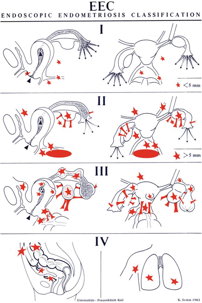
Fig. 5.3
The EEC system is used to classify endometriotic lesions. In contrast to the rASRM classification, the EEC classification includes extragenital endometriosis and is divided into four stages
Figure 5.4 differentiates stage I, II, and III in the laparoscopic appearance.
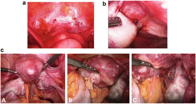
Fig. 5.4
Endoscopic image of endometriosis EEC stage I (a), EEC stage II (b), EEC stage III (c: A–C)
5.2.2 Tissue Samples
Samples of ectopic endometrium (n = 450) were obtained from patients undergoing diagnostic hysteroscopy and laparoscopy for the treatment of endometrioma.
The patients ranged in age from 18 to 44 years and received no hormonal treatment prior to surgery. Cryostat sections were prepared and stained with hematoxylin–eosin. Histopathological assessment confirmed the site of origin, i.e., proliferative endometrium or endometrioma cyst wall, respectively (Fig. 5.2).
5.2.3 Interventions
The 450 patients were randomly distributed to the following three treatment groups, 150 per group. Of the original 450 patients, 410 returned for the second-look pelviscopy and their findings were assessed:
· Group 1 (n = 125) underwent hormonal treatment after diagnostic laparoscopy with 3.75 mg of leuprorelin acetate depot which was injected subcutaneously in monthly intervals over 3 months. Leuprorelin acetate depot is a GnRH agonist and is commercially available in Germany as Enantone Gyn Depot.
· Group 2 (n = 137) underwent surgical laparoscopy without any subsequent medical treatment. Endometriosis foci were totally excised, adhesions removed, and the normal anatomy of the reproductive organs was restored. Ureter and superficial bowel lesions were removed. For infertility patients tubal patency was checked and chromoperturbation was performed at the second-look laparoscopy. Patients with deep infiltrating endometriosis with bladder or rectum resection were not included in the study.
· Group 3 (n = 148) underwent the same hormonal therapy as group 1 over the same time period after surgical laparoscopy. The combined or three-step therapy comprised diagnostic laparoscopy, removal of all visible endometriosis foci, a 3-month endocrine therapy with GnRH agonists (e.g., 3.75 mg of leuprorelin acetate depot), and a subsequent second-look laparoscopy 1–2 months after conclusion of the hormonal therapy with resection of residual foci and reconstructive surgery of organs.
The same team of physicians performed the primary and secondary intervention as well as the primary and secondary endometriosis staging according to the EEC [15, 35]. For groups 1 and 3, a second-look laparoscopy was performed 1–2 months after hormonal therapy, and for group 2, 5 to 6 months after surgical endometriosis treatment. After the second-look laparoscopy patients were monitored over a period of 2 years and completed an extensive questionnaire to determine recurrence of symptoms and pregnancy rates.
5.2.4 Maine Outcome Measures
The central issue for this study was: Which endometriosis therapy is currently the most successful technique? The success of each therapeutic strategy was assessed—independent of the original EEC stage—according to the following criteria after the second-look laparoscopy:
1.
2.
3.
Within the framework of this study, the endometriosis therapy that fulfilled all of the criteria or at least two of them was regarded as the most successful therapy.
5.2.5 Statistical Evaluation and IRB Approval
Our results were statistically evaluated with the chi-squared test and analyzed with a significance level of p < 0.05 and a confidence interval of 95 %. Institutional review board approval was obtained at the beginning of the study.
5.3 Results
Primarily, the results of the three different treatment groups were analyzed to assess the new endometriosis staging or EEC down-staging, respectively (Table 5.1 and Fig. 5.5).
Table 5.1
Distribution of patients to EEC stages after therapy
|
Therapy methods |
EEC stage (in %) |
|||
|
EEC 0 |
EEC I |
EEC II |
EEC III |
|
|
Group 1: hormonal treatment (n = 125) |
50 |
32 |
13 |
5 |
|
Group 2: surgical treatment (n = 137) |
55 |
13 |
23 |
9 |
|
Group 3: combined treatment (n = 148) |
60 |
18 |
17 |
5 |

Fig. 5.5
Distribution of patients to EEC stages after therapy
In group I (hormonal treatment), 40 % of the 125 patients presented at the outset of the study with EEC stage 1, 38 % with ECC II, and 22 % with EEC III. After the hormonal therapy, and independent of the previous EEC stage, 32 % of group 1 had EEC stage I, 13 % EEC II, and 5 % EEC III. In 50 % of the cases, the second-look laparoscopy showed no signs of endometriosis (EEC 0). These patients appeared to be cured (cure rate = 50 %).
In group 2 (surgical treatment) 50 % of the 137 patients originally presented with EEC I, 32 % with EEC II, and 18 % with EEC III. As a result of the second-look laparoscopy the patients could be down-staged. We then found 13 % with EEC I, 23 % with EEC II, and 9 % with EEC III. The total cure rate for the exclusively surgically treated group was 55 % (EEC O).
In group 3 (combined therapy), the 148 patients were classified as 53 % with EEC I, 24 % with EEC II, and 23 % with EEC III. After the combined surgical and hormonal treatment, we found 18 % of the patients with EEC I, 17 % with EEC II, and 5 % with EEC III. With this treatment form we achieved a cure rate of 60 % (EEC 0).
The three treatment options achieved—independent of the initial EEC stage–an overall cure rate of 50 % or higher. The combined treatment reached a cure rate of 60 %, the exclusively hormonal therapy 55 %, and the exclusively surgical treatment 50 %. Within the framework of our study, cure has been defined as a reduction to EEC O. This new endometriosis down-staging was confirmed by the second-look laparoscopy. Regardless of the fact that the combined therapy obtained the best total cure rate, there is no statistically significant difference between the three percentages (p > 0.05).
In the second step, we distinguished between light, intermediate, and advanced cases of endometriosis and evaluated therapeutic strategies leading to an improvement of at least 75 % to EEC stages 0–1 as highly efficient. These conditions were met by the combined therapy (three-step therapy) with a response rate of 78 % and the exclusively hormonal treatment with a rate of 82 %.
As endometriosis is generally a disease which causes recurrent pain, we asked our study patients to complete an extensive questionnaire and report on recurrent symptoms, 1 year after the end of all therapeutic activities. Patients in the combined therapy group achieved the lowest general recurrence rate and the lowest recurrence rates per symptom: 16 % of the patients reported dysmenorrhea, 8 % dyspareunia, and 17 % abdominal pain. In contrast to this, the exclusively hormonal therapy showed the highest recurrence rates: 28 % of the patients complained of dysmenorrhea, 26 % of abdominal pain, and 22 % of dyspareunia (Table 5.2 and Fig. 5.6).
Table 5.2
Comparison of recurrence rates for the three therapy methods after 1 year
|
Therapy methods |
Recurrent symptoms |
||
|
Dysmenorrhea |
Dyspareunia |
Abdominal pain |
|
|
Group 1: hormonal treatment (n = 125) |
28 |
22 |
26 |
|
Group 2: surgical treatment (n = 137) |
20 |
15 |
24 |
|
Group 3: combined treatment (n = 148) |
16 |
8 |
17 |
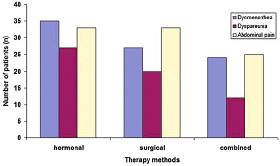
Fig. 5.6
Comparison of recurrence rates for the three therapy groups after 1 year
The overall recurrence rate for the combined therapy after 1 year was 41 %, whereas surgical treatment alone resulted in a recurrence rate of 59 % (p < 0.01). In comparison with the exclusively hormonal therapy strategy with a recurrence rate of 76 %, the combined therapy had a significantly better success rate (p < 0.01).
Since active endometriosis as a functional disorder or endometriosis-related secondary damage can constitute anatomical reasons for infertility, we also assessed the pregnancy rate and the birth and miscarriage rates, with the help of a questionnaire 2 years after the end of each therapeutic strategy.
We determined an overall pregnancy rate of 55–65 % for the three treatment options, independent of the ECC stage (Table 5.3 and Fig. 5.7). The pregnancy rate after the exclusively surgical restoration was 55 %, after the combined therapy 60 %, and after the exclusively hormonal treatment 65 %. There was no statistical significance (p > 0.05) between these results.
Table 5.3
Comparison of pregnancy rates for the three therapy methods after 2 years
|
Therapy methods |
Pregnancies |
Abortions |
Extrauterine pregnancies |
Live births |
|
Group 1: hormonal (n = 125) |
81 (65 %) |
10 (8 %) |
2 (2 %) |
69 (55 %) |
|
Group 2: surgical (n = 137) |
75 (55 %) |
12 (9 %) |
1 (1 %) |
62 (45 %) |
|
Group 3: combined (n = 148) |
89 (60 %) |
13 (9 %) |
3 (2 %) |
74 (50 %) |

Fig. 5.7
Comparison of pregnancy rates for the three therapy groups after 2 years
Of these 245 pregnancies, 41 (17 %) were not carried to term (6 extrauterine pregnancies, 35 abortions); on the other hand 205 children were born (including one set of twins). There was no statistically significant difference between the three therapeutic strategies regarding the pregnancies and their course.
Within the framework of our study, the combined therapy turned out to be the most successful treatment for endometriosis. Even though there was no significant difference between the different therapeutic strategies as far as cure and pregnancy rates were concerned, the three-step therapy resulted in the highest response rate or cure for endometriosis. Sixty percent of the cases had down-staged to EEC stage 0 at the second-look laparoscopy. The combined or three-step therapy group also had the lowest recurrence rate of 41 % (p < 0.01), 1 year after the end of the therapy.
5.4 Endometriosis and Obstetrical Outcome
The largest study to date of endometriosis in pregnant women has found that the condition is a major risk factor for premature birth [36]. This study was presented at the 25th Annual Conference of the European Society of Human Reproduction and Embryology (ESHRE) in 2009 by Henrik Falconer of the Department of Woman and Child Health, Karolinska Institute, Stockholm, Sweden. His team found that women with endometriosis had a higher risk of pregnancy complications and premature deliveries and were more likely to deliver by Cesarean section.
The researchers investigated the association between adverse pregnancy outcome, assisted reproduction technology (ART), and a previous diagnosis of endometriosis in 1,442,675 single births to Swedish women. They were 13,090 singleton births among 8,922 women diagnosed with endometriosis. Compared with women without endometriosis, they had a 33 % greater risk of preterm birth.
Women with endometriosis were also more likely to have difficulty in conceiving and the use of ART was more prevalent, which in itself may be considered a risk factor for adverse pregnancy outcome, although we strongly disagree with this concept. Once conception takes place the embryo has to prove itself and fight for survival. That certainly promotes a healthy embryo but, of course, we agree that it is tiring to start the fight for life against possible cytokines or antibodies at that early stage.
Among women with endometriosis, 11.9 % conceived after ART compared with the 1.4 % of women without endometriosis who used the technique. This means that patients with endometriosis have a better chance of becoming pregnant with ART than without it.
The risk of preterm birth associated with endometriosis among women with ART was 1.24 and among women without ART 1.37.
Dr. Falconer said that endometriosis appears to be a risk factor for preterm birth, irrespective of ART. The findings of the study indicate that women with endometriosis may be considered a high-risk group and need special care during pregnancy.
Endometriosis is a chronic inflammatory disease, affecting up to 15 % of all women of reproductive age, in which the endometrial cells that line the uterus are deposited in other areas. Such displacement of endometrial cells can lead to anatomical distortion and also the release of anti-inflammatory cytokines, signaling molecules used in communication between cells. Known symptoms of endometriosis include severe pelvic pain, heavy menstrual periods, and nausea.
One explanation for the interaction of endometriosis with preterm birth has been given by the group of Ivo Brosens with the enigmatic uterine junctional zone [37].
In addition to an increased risk of preterm birth, the researchers also found other differences in the pregnancies of women with endometriosis. Dr. Falconer reported that nearly twice as many women in this group were delivered by Cesarean section. The study group observed that among these women the risk of induced preterm birth was higher than for spontaneous preterm birth. They believe that women with endometriosis are more frequently scheduled for preterm Cesarean section, possibly due to placental complications.
Women with endometriosis were also more likely to suffer from pre-eclampsia, a condition that develops in the second or third trimester of pregnancy and involves the development of high blood pressure and the presence of protein in the urine. However, this is strongly contradicted by others [38].
Antepartal bleeding was also found to be more common among women with endometriosis.
As endometriosis is so strongly associated with infertility, we know that women suffering from endometriosis are of higher maternal age and have fewer children. Even after adjusting for maternal age and other confounding factors, the strong association between endometriosis and risky pregnancies still remains.
Given that endometriosis is relatively common in women of childbearing age, we advise pregnant patients with a history of previous endometriosis to observe extra care, to enable them to have a normal pregnancy and give birth to a healthy baby.
Concerning pain in pregnancy in patients with endometriosis, there are several reports on intensified pain in pregnancy, although others report that pain totally disappears during pregnancy and breast feeding. Pregnancy appears to be a cure for these patients. However, it is a misconception to believe that pregnancy cures endometriosis. The symptoms usually recur after each pregnancy [39].
5.5 Discussion
Our team did basic research in endometriosis as for the distribution pattern of the Macrophage Colony-Stimulating Factor Receptor (M-CSFR) producing cells in endometrial and endometriotic tissue. Membrane-bound M-CSF or soluble M-CSF of peritoneal macrophages may cause the survival of dystopic endometrium by direct interaction, giving rise to endometriosis [40].
We also compared in a c-DNA microarray analysis a set of 940 genes expressed in endometrium and endometriosis and identified 38 genes which were differentially expressed in endometriotic implants compared to uterine endometrium [41]. Based on older extensive studies a lot of further research is necessary [42–44]. Concerning endometriosis and fertility we advise to use the fertility index of David Adamson [45].
The presented clinical studies comparing medical, surgical, and combined therapy and the assessment of how endometriosis can affect pregnancy and deliveries show the current needs for the treatment of endometriosis and point out some advice for future therapeutic modalities.
5.5.1 Three-Step Therapy of Endometriosis
In the presented study 450 endometriosis patients, aged 18–44 years, were randomly assigned to one of three different therapeutic strategies (medical, surgical, or combined treatment) at the Kiel University Department of Obstetrics and Gynecology, Germany. The success of each therapeutic strategy was assessed—independent of the original EEC stage [35]—according to the following criteria:
1.
2.
3.
4.
Within the framework of this study, the endometriosis therapy that fulfilled the majority of the criteria, or at least two of them, was regarded as the most successful therapy. The three treatment options reached an overall cure rate of 50 % or higher. There was no statistically significant difference (p > 0.05), but with a cure rate of 60 % the combined therapy ranks first. The combined (three-step) and the exclusively hormonal therapy managed to surpass the 75 % response rate with 78 and 82 %, respectively. Nevertheless, the combined treatment reached the lowest recurrence rate per symptom at a statistically significant level. No statistically significant difference was recorded for the pregnancy rate which ranged between 55 and 65 %, independent of the therapeutic strategy. As an overall result, we have been able to confirm the high efficacy of the combined endometriosis therapy in this study.
Medical therapy can be applied prior to surgery to decrease the size of endometriotic implants and the extent of the operation [46]. However, so far there is no clear evidence that perioperative hormonal treatment decreases the extent of operation necessary to remove endometriotic implants, delays or prevents recurrence, or increases pregnancy rates. In contrast, several trials were able to report an increased duration of pain relief and delayed recurrence rates using postoperative medical therapy [6, 46, 47]. Schweppe concluded that in all cases of active endometriosis, pelviscopic treatment alone is not sufficient [48]. Schindler demonstrated that the primary surgical intervention reduced the total r-AFS score (revised American Fertility Society) by 34 %, whereas the combined therapy brought about a reduction of 66 % [49].
Our study showed only a weak and statistically nonsignificant difference between the combined treatment (decrease of EEC stage by 60 %) and the solitary surgical treatment (decrease of EEC stage by 55 %). Regidor found a significant improvement of the r-AFS score after treatment with triptorelin (GnRH analogue). Sixty-three percent of these patients were no longer diagnosed with endometriosis, 30 % presented with stage I residual endometriosis according to the AFS classification, and only 7 % had stage II endometriosis [4]. It could be demonstrated that after administration of buserelin (GnRH analogue) the average AFS score went down from 17.4 ± 12.9 before therapy to 7.2 ± 8.2 after a 6-month therapy [49, 50]. Although up to 90 % of patients experience some symptom relief with medical therapy, medical treatment alone neither enhances fertility, nor diminishes pelvic mass, nor removes adhesions [6, 14].
Similar to Schweppe and Römer [48, 51], we also determined a significantly lower recurrence rate after application of the three-step therapy. Römer reported that retrospective analyses 12–48 months after endometriosis therapy presented a recurrence rate for hormonal and surgical treatment (three-step concept) of only 16.7 %, whereas the recurrence rate for exclusively surgically treated patients was 47 %.
Regidor showed in a long-term follow-up study that 70 of 112 patients (62.5 %) again reported ailments and that the recurrence-free interval amounted to an average of 11 months after finishing the three-step therapy (with the GnRH analogue leuprorelin acetate) [52]. In another long-term follow-up study, Schindler established recurrent endometriosis in 62 of 112 patients (55 %) after a combined surgical–hormonal therapy [14, 41]. Our recurrence rate (41 %) was lower than Regidor’s rate (62.5 %) and Schindler’s rate (55 %) for the combined therapy. Zupi et al. were able to show that patients treated with GnRH agonists had a significantly higher rate of symptom reduction (pelvic pain, dysmenorrhea, and dyspareunia) than women treated with continuous estrogen–progestin oral contraceptives. Quality of life was increased by extending the GnRH treatment to include add-back therapy [17]. Other investigations comparing oral contraceptives to GnRH agonists found an equal reduction of pain [53]. Sutton 1994 and Abbott 2004 performed a second-look laparoscopy 6–12 months after the primary operation and found that 29–45 % of the patients had disease progression, 22–29 % disease regression, and 33–42 % the disease remained static [28, 54].
Endometriosis can reduce the fecundability rate without completely preventing conception. Impaired fertility might be due to anatomic variations after adhesion formation and endometriomas [55]. An enhancement of fertility rates through ovulation suppression has not yet been proven [14]. In our study after the combined therapy, we had 89 (60 %) pregnancies in 148 patients and 13 abortions and 3 extrauterine pregnancies. Sixteen (18 %) of the 89 pregnancies did not lead to a live birth (13 abortions and 3 extrauterine pregnancies). Regidor reported 55 (60 %) pregnancies for 91 patients for the same therapeutic strategy [52]. Nineteen (34.5 %) of the 55 pregnancies were not carried to term (5 extrauterine pregnancies, 14 abortions). Our pregnancy rate was comparable to Regidor’s rate, but our abortion rate was significantly lower than his. After the exclusively surgical treatment we registered a pregnancy rate of 55 %. In comparison, Marcoux et al. presented a pregnancy rate of 29 % [56].
All research focusing on macroscopic or microscopic markers as well as biochemical criteria for assessing the degree of activity of endometriosis are not convincing. Essential factors for deciding the optimal endometriosis therapy are clinical symptoms, the patient’s age, localization, severity, duration of the disease, recurrence rate, and activity [57, 58]. Active endometriosis foci are characterized by hyper vascularisation, edema, and infiltration of inflammatory cells [40]. It still needs to be determined how endometriosis activity can best be characterized using macroscopic, microscopic, and biochemical criteria [25]. Laparoscopy currently constitutes one of the most accurate methods of diagnosing endometriosis.
5.5.2 The Influence of Endometriosis on Obstetrical Outcome
Concerning this constellation let us pose two critical questions:
1.
2.
The effect on the obstetrical outcome seems to be premature delivery.
The question whether endometriosis triggers recurrent spontaneous abortions was investigated following the observation that “Natural Killer (NK) cell activity” is low in endometriosis patients and high in unexplained recurrent abortions. There is good evidence that endometriosis is associated with an opposite regulation of NK cell behavior [59]. From 1991 to 1995 at least nine independent groups reported a functional defect of peripheral NK cells in patients with endometriosis. However, Somigliana et al. in 1999 concluded that the relationship between NK cell activity, endometriosis, and infertility seems to be “more puzzling” than considered so far [59]. These fine implied mechanisms may still reveal interesting biological and clinical possibilities for treatment.
Several case reports deal with obstetrical emergencies arising during delivery through endometriosis. Let us discuss a few of them:
1.
2.
3.
4.
5.6 Summary and Conclusions
5.6.1 The Three-Step Therapy
Since the identification of endometriosis as a progredient estrogen-related disease, various substances have been used to suppress ovarian steroid biosynthesis. Currently all modern therapeutic strategies aim at ovarian downregulation with GnRH agonists or gestagens. In most cases therapeutic approaches take into consideration not only medical but also laparoscopic and, if required, laparotomic surgical treatment of endometriosis and the combined therapy. The three-step therapy comprises surgical laparoscopy with removal of all visible endometriosis foci, a 3- to 6-month endocrine therapy and a subsequent second-look laparoscopy with resection of residual foci, adhesiolysis, and reconstructive surgery of the organs [64].
5.6.2 Endometriosis and Obstetrical Outcome
As endometriosis remains an enigmatic disease, there is a growing realization that the origin of major obstetrical complications and problems during pregnancy may lie in very early pregnancy events. Recent studies have focused on the implantation window, particularly in endometriosis patients. The implantation window may not only be responsible for delayed implantation but also for defective deep placentation leading to preterm labor, fetal growth restriction, and pre-eclampsia.
It is a myth that pregnancy can heal endometriosis as severe pain attacks due to endometriosis can also occur during pregnancy. A relation between endometriosis and abortions remains questionable.
Obstetrical emergencies based on endometriosis, such as bleeding endometriotic lesions, uterine arteries, or veins; interruption of ureter; and postpartum hemorrhage due to decidualized endometriosis, have occurred and have to be considered.
References
1.
Johnson NP, Hummelshoj L (2013) Consensus on current management of endometriosis. Hum Reprod 28(6):1552–1568. doi:10.1093/humrep/det 050PubMedCrossRef
2.
Olive DL, Schwartz LB (1993) Endometriosis. N Engl J Med 328(24):1759–1769PubMedCrossRef
3.
Clement PB (2007) The pathology of endometriosis: a survey of the many faces of a common disease emphasizing diagnostic pitfalls and unusual and newly appreciated aspects. Adv Anat Pathol 14(4):241–260PubMedCrossRef
4.
Regidor PA, Imberg C, Bühler K, Reiners C, Schindler AE (1993) Treatment of endometriosis with the GnRH analog triptorelin with special reference to the effect on bone density. Zentralbl Gynakol 115(11):473–477PubMed
5.
Sinaii N, Plumb K, Cotton L, Lambert A, Kennedy S, Zondervan K, Stratton P (2008) Differences in characteristics among 1,000 women with endometriosis based on extent of disease. Fertil Steril 89(3):538–545PubMedCentralPubMedCrossRef
6.
Kennedy S, Berggvist A, Chapron C, D‘Hooghe T, Dunselman G, Greb R, Hummelshoj L, Prentice A, Saridogan E, ESHRE Special Interest Group for Endometriosis and Endometrium Guideline Development Group (2005) ESHRE guideline for the diagnosis and treatment of endometriosis. Hum Reprod 20(10):2698–704PubMedCrossRef
7.
Chatman DL, Ward AB (1982) Endometriosis in adolescents. J Reprod Med 27(3):156–160PubMed
8.
Sangi-Haghpeykar H, Poindexter AN 3rd (1995) Epidemiology of endometriosis among parous women. Obstet Gynecol 85(6):983–992PubMedCrossRef
9.
Missmer SA, Hankinson SE, Spiegelman D, Barbieri RL, Marshall LM, Hunter DJ (2004) Incidence of laparoscopically confirmed endometriosis by demographic, anthropometric, and lifestyle factors. Am J Epidemiol 160(8):784–796PubMedCrossRef
10.
Christensen B, Freie HM, Schindler AE (1995) Endometriosis–diagnosis and therapy. Results of a current survey of 6,700 gynecologists. Geburtshilfe Frauenheilkd 55(12):674–679PubMedCrossRef
11.
Engemise S, Gordon C, Konje JC (2010) Endometriosis. BMJ 340:c2168PubMedCrossRef
12.
Practice Committee of American Society for Reproductive Medicine (2008) Treatment of pelvic pain associated with endometriosis. Fertil Steril 90(5 Suppl):S260–S269
13.
Mahmood TA, Templeton A (1990) The impact of treatment on the natural history of endometriosis. Hum Reprod 5(8):965–970PubMed
14.
Hughes E, Brown J, Collins JJ, Farguhar C, Fedorkow DM, Vandekerckhove P (2007) Ovulation suppression for endometriosis. Cochrane Database Syst Rev (3):CD000155
15.
Mettler L, Semm K (1983) Three step medical and surgical treatment of endometriosis. Ir J Med Sci 152(Suppl 2):26–28PubMedCrossRef
16.
Strowitzki T, Marr J, Gerlinger C, Faustmann T, Seitz C (2010) Dienogest is as effective as leuproreline acetate in treating the painful symptoms of endometriosis. Hum Reprod 25:633–641PubMedCrossRef
17.
Zupi E, Marconi D, Sbracia M, Zullo F, De Vivo B, Exacustos C, Sorrenti G (2004) Add-back therapy in the treatment of endometriosis-associated pain. Fertil Steril 82(5):1303–1308PubMedCrossRef
18.
Wong CL, Farguhar C, Roberts H, Proctor M (2009) Oral contraceptive pill as treatment for primary dysmenorrhoea. Cochrane Database Syst Rev (2):CD002120
19.
Davis L, Kennedy SS, Moore J, Prentice A (2007) Modern combined oral contraceptives for pain associated with endometriosis. Cochrane Database Syst Rev (3):CD001019
20.
Mousa NA, Bedaiwy MA, Casper RF (2007) Aromatase inhibitors in the treatment of severe endometriosis. Obstet Gynecol 109(6):1421–1423PubMedCrossRef
21.
Ferrero S, Camerini G, Seracchioli R, Ragui N, Venturini PL, Remorgida V (2009) Letrozole combined with norethisterone acetate compared with norethisterone acetate alone in the treatment of pain symptoms caused by endometriosis. Hum Reprod 24(12):3033–3041PubMedCrossRef
22.
Mettler L, Taylor RN, Missmer SA, Shah DK, Pistofidis G, Tuttlies F, Keckstein J, Ulrich U, Chapron C, Borghese B, de Ziegler D (2011) Challenges of endometriosis. Eur J Obstet Gynecol 6(2):83–90
23.
Stegmann BJ, Sinnaii N, Liu S, Segars J, Merino M, Niemann LK, Stratton P (2008) Using location, color, size, and depth to characterize and identify endometriosis lesions in a cohort of 133 women. Fertil Steril 89(6):1632–1636PubMedCentralPubMedCrossRef
24.
Wykes CB, Clark TJ, Khan KS (2004) Accuracy of laparoscopy in the diagnosis of endometriosis: a systematic quantitative review. BJOG 111(11):1204–1212PubMedCrossRef
25.
Buchweitz O, Poel T, Diedrich K, Malik E (2003) The diagnostic dilemma of minimal and mild endometriosis under routine conditions. J Am Assoc Gynecol Laparosc 10(1):85–89PubMedCrossRef
26.
Mettler L, Schollmeyer T, Alkatout I (2012) Adhesions during and after surgical procedures, their prevention and impact on women’s health. Womens Health (Lond Engl) 8(5):495–498CrossRef
27.
Alkatout I, Schollmeyer T, Doniec M, Egberts JH, Jonat W, Mettler L (2013) Endometriose: Medikamentöse und operative Therapieoptionen. gynäkologie+geburtshilfe 18 (1):38–45
28.
Abbott J, Hawe J, Hunter D, Holmes M, Finn P, Garry R (2004) Laparoscopic excision of endometriosis: a randomized, placebo-controlled trial. Fertil Steril 82(4):878–884PubMedCrossRef
29.
Healey M, Ang WC, Cheng C (2010) Surgical treatment of endometriosis: a prospective randomized double-blinded trial comparing excision and ablation. Fertil Steril 94(7):2536–2540PubMedCrossRef
30.
Jacobson TZ, Barlow DH, Garry R, Koninckx P (2001) Laparoscopic surgery for pelvic pain associated with endometriosis. Cochrane Database Syst Rev (4):CD001300
31.
Crosignani PG, Vercellini P, Biffignandi F, Costantini W, Cortesi I, Imparato E (1996) Laparoscopy versus laparotomy in conservative surgical treatment for severe endometriosis. Fertil Steril 66(5):706–711PubMed
32.
Taylor E, Williams C (2010) Surgical treatment of endometriosis: location and patterns of disease at reoperation. Fertil Steril 93(1):57–61PubMedCrossRef
33.
Yeung PP Jr, Shwayder J, Pasic RP (2009) Laparoscopic management of endometriosis: comprehensive review of best evidence. J Minim Invasive Gynecol 16(3):269–81PubMedCrossRef
34.
Alkatout I, Mettler L, Beteta C, Hedderich J, Jonat W, Schollmeyer T, Salmassi A (2013) Combinded surgical and hormonal therapy of endometriosis results in the most effective treatment: a prospective randomized controlled trial. J Minim Invasive Gynecol 20(4):473–481PubMedCrossRef
35.
Mettler L (1984) Diagnostik und Therapie der Endometriose. Euromed 5:234–240
36.
Stephansson O, Kieler H, Granath F, Falconer H (2009) Endometriosis, assisted reproduction technology and risk of adverse pregnancy outcome. Hum Reprod 24:2341–2347PubMedCrossRef
37.
Brosens I, Derwig I, Brosens J, Fusi L, Benagiono G, Pijneborg R (2010) The enigmatic uterine junctional zone: the missing link between reproductive disorders and major obstetrical disorders. Hum Reprod 25:569–574PubMedCrossRef
38.
Hadfield RM, Lain SJ, Raynes-Greenow CH, Morris JM, Roberts CL (2009) Is there an association between endometriosis and the risk of pre-eclampsia? A population based study. Hum Reprod 24:2348–2352PubMedCrossRef
39.
Wood R. Myths and misconceptions in endometriosis. http://www.endometriosis.org
40.
Mettler L, Schmutzler AG, Koch K, Schollmeyer T, Salmassi A (2004) Identification of the M-CSF receptor in endometriosis by immunohistochemistry and RT-PCR. Am J Reprod Immunol 52:298–305PubMedCrossRef
41.
Mettler L, Salmassi A, Schollmeyer T, Schmutzler AG, Püngel F, Jonat W (2007) Comparison of c-DNA microarray analysis of gene expression between eutopic endometrium and ectopic endometrium (endometriosis). J Assist Reprod Genet 24(6):249–258. doi:10.1007/s%10815-007-9116-yPubMedCentralPubMedCrossRef
42.
Sampson JA (1927) Peritoneal endometriosis due to menstrual dissemination of endometrial tissue into peritoneal cavity. Am J Obstet Gynecol 14:422–469
43.
Ebert AD (2003) Endometriose. Walther de Gruyter Verlag, Berlin
44.
Schweppe KW, Rabe T, Langhardt M, Woziwodzki J, Petraglia F, Kiesel L (2012) Endometriose: Entstehung, Diagnostik, Behandlungsmölichkeiten und Probleme in Klinik und Praxis. In: Rabe T (ed) Seminar in Gynäkologischer Endokrinologie, Heidelberg, S.516–534
45.
Adamson GD, Pasta DJ (2010) Endometriosis fertility index: the new validated endometriosis staging system. Fertil Steril 94:1609–1615PubMedCrossRef
46.
Yap C, Furness S, Farquhar C (2004) Pre and post operative medical therapy for endometriosis surgery. Cochrane Database Syst Rev (3):CD003678
47.
Seracchioli R, Mabrouk M, Manuzzi L, Vicenzi C, Frasca C, Elmakky A, Venturoli S (2009) Post-operative use of oral contraceptive pills for prevention of anatomical relapse or symptom-recurrence after conservative surgery for endometriosis. Hum Reprod 24(11):2729–2735PubMedCrossRef
48.
Schweppe KW (2002) Therapie der Endometriose unter Berücksichtigung der Aktivitätsgrade. Gynakologe 35:255–261CrossRef
49.
Schindler AE, Bühler K, Mettler L, Fuchs U, Cirkel U, Ochs H, Schweppe KW, Koch R (1994) Treatment of endometriosis with the GnRH agonist buserelin (Suprecur): a multicenter study]. Geburtshilfe Frauenheilkd 54(10):569–573PubMedCrossRef
50.
Raitz von Frentz M, Schweppe KW (1990) Ovarian suppression by the GnRH analog buserelin in the treatment of endometriosis. Clinical, biochemical and pelviscopic studies. Geburtshilfe Frauenheilkd 50(8):634–639
51.
Romer T, Schwesinger G (1999) Wie durchbricht man den Teufelskreis der aktiven Endometriose? Neue Aspekte der Therapieentscheidung bei aktiver Endometriose. Zentralbl Gynakol 121(7):336–340PubMed
52.
Regidor PA, Regidor M, Schmidt M, Ruwe B, Lübben G, Förtig P, Kienle E, Schindler AE (2001) Prospective randomized study comparing the GnRH-agonist leuprorelin acetate and the gestagen lynestrenol in the treatment of severe endometriosis. Gynecol Endocrinol 15(3):202–209PubMedCrossRef
53.
Guzick DS et al (2011) Randomized trial of leuprolide versus continuous oral contraceptives in the treatment of endometriosis-associated pelvic pain. Fertil Steril 95(5):1568–1573PubMedCentralPubMedCrossRef
54.
Sutton CJ, Ewen SP, Whitelaw N, Haines P (1994) Prospective, randomized, double-blind, controlled trial of laser laparoscopy in the treatment of pelvic pain associated with minimal, mild, and moderate endometriosis. Fertil Steril 62(4):696–700PubMed
55.
Cahill DJ (2002) What is the optimal medical management of infertility and minor endometriosis? Analysis and future prospects. Hum Reprod 17(5):1135–1140PubMedCrossRef
56.
Marcoux S, Maheux R, Berube S (1997) Laparoscopic surgery in infertile women with minimal or mild endometriosis. Canadian Collaborative Group on Endometriosis. N Engl J Med 337(4):217–222PubMedCrossRef
57.
Barcena de Arellano ML, Arold J, Vercellino F, Chiantera V, Schneider A, Mechsner S (2011) Overexpression of nerve growth factor in peritoneal fluid from women with endometriosis may promote new outgrowth in endometriotic lesions. Fertil Steril 95(3):1123–6PubMedCrossRef
58.
Keichel S, Barcena de Arellano ML, Reichelt U, Riedlinger WF, Schneider A, Köhler C, Mechsner L (2011) Lymphangiogenesis in deep infiltrating endometriosis. Hum Reprod 26(10):2713–2720PubMedCrossRef
59.
Somigliana E, Vigano P, Vignali M (1999) Endometriosis and unexplained recurrent spontaneous abortion. Pathological status resulting from aberrant modulations of natural killer cell functions. Hum Reprod 5:40–51CrossRef
60.
Chiodo I, Somigliana E, Dousset B, Chapron C (2008) Urohemoperitoneum during pregnancy with consequent fetal death in a patient with deep endometriosis. J Minim Invasive Gynecol 15(2):202–204PubMedCrossRef
61.
Wada S, Yoshiyuki F, Fujino T, Sato C (2009) Uterine vein rupture at delivery as a delayed consequence of laparoscopic surgery for endometriosis: a case report. J Minim Invasive Gynecol 16(4):510–512PubMedCrossRef
62.
Falcão JODA Jr, Silva WE Jr, Rezende ACDS (2011) Endometriosis in obstetric emergency. Free Communications and Posters, P1-108, at the 11th World Congress of Endometriosis, Montpellier, France, 4–7 September 2011
63.
Mabrouk M, Alboni C, Gibertini G, Neri S, Tondi C, Algeri I, Pazzoni F, Malmusi S, Ghirardini G (2011) Spontaneous postpartum hemoperitoneum in primipara with decidualized pelvic endometriosis. Free Communications and Posters, P1-112, at the 11th World Congress of Endometriosis, Montpellier, France, 4–7 September 2011
64.
Mettler L, Schollmeyer T, Lehmann-Willenbrock E, Schüppler U, Schmutzler A, Shuklar D, Zavala A, Lewin A (2003) Accuracy of laparoscopic diagnosis of endometriosis. JSLS 7(1):15–18PubMedCentralPubMed