David A. Eschenbach
A large number of microbes are present in the female reproductive tract, and pelvic infections are common. Many pelvic infections are sexually transmitted (Table 32.1). This chapter provides data on usual presentations and updated treatment of pelvic infections.
|
|
|
TABLE 32.1. Sexually transmitted infections |
The impact of pelvic infections on women ranges from minor annoyance to serious illness and, rarely, even death. The cost of treating pelvic infections is enormous from both direct medical costs and indirect costs, including time lost from work. Using pelvic inflammatory disease (PID) as an example, previous estimates were that, by 2000, one of every four women who reached reproductive age in the 1970s had an episode of PID. Of women with PID, 25% will be hospitalized, 25% will have major surgery, and 20% will have tubal sterility.
Upper genital tract sites (endometrium, fallopian tubes, ovaries) formerly considered sterile are subject to ascending microbial traffic and occasionally infection from lower genital tract microbes. Some microbes preferentially infect certain sites and give rise to characteristic symptoms while other microbes cause few symptoms until major pathologic changes occur or until congenital neonatal infection or male-partner infection ensues. Clinicians should have special knowledge of the infections caused by Neisseria gonorrhoeae, Chlamydia trachomatis, group B streptococci, Treponema pallidum, anaerobic bacteria, bacteria associated with bacterial vaginosis, and Mycobacterium tuberculosis; these infections either are common or potentially produce severe sequelae. It is now appreciated that most viral infections of the genital tract are asymptomatic. Several viruses are common and produce severe disease in both adults and neonates, including herpesvirus, cytomegalovirus, hepatitis B virus, human papillomavirus, and human immunodeficiency virus (HIV).
VULVA
Herpes
Type-specific serologic assays indicate that one third of women 20 to 45 years of age have been exposed to herpes simplex virus type 2 (HSV-2). Between 60% and 85% of women with HSV-2 antibodies never have a recognized genital infection. Despite the frequency of asymptomatic infection, HSV infection is a common cause of vulvar ulcers. Genital ulcers also are caused by syphilis and chancroid. Ulcers from HSV usually occur 3 to 7 days after exposure. Symptomatic primary (first) genital infections typically consist of multiple vesicles that rapidly produce ulcerations of the vulva that can be exceedingly painful. The cervix and vagina may also be involved, producing a gray, necrotic cervix and profuse leukorrhea. External dysuria is common, and bilateral inguinal lymphadenopathy is usual. Vulvar lesions may last for 3 or more weeks before complete healing. Constitutional symptoms of fever, malaise, headache (i.e., aseptic meningitis), and urinary retention (i.e., myelitis) may persist for a week.
After primary infection, latent HSV usually localizes in the sacral ganglion and perhaps the dermis. Periodic asymptomatic viral shedding occurs, particularly in the first 6 months after primary infection. HSV is isolated on 1% of the days with no symptoms or physical evidence of infection. Most patients develop a secondary (recurrent) infection from latent virus weeks to months after the primary infection. Secondary lesions are less painful and more localized and last for a shorter time (3 to 7 days) than the lesions of primary infection. Systemic manifestations are unusual with secondary infection.
From 75% to 85% of genital herpes infections are caused by HSV-2, with the remainder caused by HSV-1, the primary cause of oral herpes. The two types of herpes infections are clinically indistinguishable except that genital recurrence is unusual from HSV-1. Vesicles and ulcers contain many highly infectious virus particles, and viral shedding occurs until the lesions disappear. Thus, direct contact with either genital or oral HSV lesions leads to a high rate of infection. Transmission usually occurs by direct contact with ulcerative lesions. Transmission is greatest during a primary infection, intermediate during a secondary infection, and probably least with asymptomatic shedding.
The diagnosis of herpes can be made clinically if typical, painful, shallow multiple vulvar ulcers are present. However, many HSV lesions are atypical. Laboratory confirmation of atypical lesions and lesions that appear during pregnancy is best attained by virus isolation (which can usually be achieved within 48 hours) or by polymerase chain reaction (PCR) identification. Other direct HSV identification methods, including Pap smear, fluorescein tagging, and immunoperoxidase staining, are insensitive. Accordingly, a negative direct Pap smear does not exclude HSV infection. Complement fixing and neutralizing antibodies appear within 1 week of the onset of infection; failure of an experienced laboratory to identify antibodies within 3 weeks is evidence against HSV infection. However, even high antibody levels do not protect against recurrent HSV infection, although antibody passively transferred to the fetus offers considerable protection against neonatal infection.
The rising incidence of herpes infection and the potentially serious fetal infection caused by HSV make this an important infection in pregnancy. New guidelines for herpes are discussed in Chapter 19.
Oral acyclovir (Zovirax), 400 mg three times daily, famciclovir (Famvir), 250 mg three times daily, or valacyclovir (Valtrex), 1 g twice daily all for 7 to 10 days, shortens the ulcerative phase. Antiviral therapy does not eradicate HSV or prevent recurrence. Patients with episodic recurrent herpes should be provided a supply of drugs to take for 5 days beginning with prodromal symptoms or within a day of the lesion appearance. Patients with six or more yearly recurrences may benefit from suppressive therapy of acyclovir, 400 mg twice daily, famciclovir 250 mg twice daily, valacyclovir, 500 mg daily, or valacyclovir, 1 g daily for up to 1 year. The expense of this drug limits routine or prolonged use. Local therapy of genital herpes is limited to pain relief. Local treatment neither penetrates into virus-containing cells nor influences epithelial damage from HSV. Corticosteroids and antimicrobial ointments offer no benefit as they prevent drying and, thus, delay healing. Wet-to-dry therapy is often helpful (i.e., 10-minute sitz bath three or four times daily followed by drying with hair dryer).
Human Papillomavirus
Genital human papillomaviruses (HPV) are DNA viruses that are distinct from papovaviruses that cause the common wart. HPV thrives in the moist genital area and usually is sexually transmitted. HPV infection is common and typically subclinical. HPV DNA is found in the genitalia in 30% to 45% of women by PCR DNA amplification. The vulva is positive for HPV DNA in more than 40%, and the cervix in over 30%. Only 1% of the women have visible warts, and only 9% have a history of genital warts. The average incubation period for visible warts is 3 months. Genital warts most commonly occur on the labia and posterior fourchette (Fig. 32.1). They originally appear as individual lesions, although large confluent growths can occur if neglected. Vaginal and cervical warts are even more common than labial warts, although most of these are flat lesions visible only by colposcopy. Over 30 HPV types infect the genital tract. Visible genital warts are usually caused by HPV types 6 and 11; 3% of college women had these types. The flat-wart variant is caused by HPV types 16, 18, 31, 33, and 35 (found in 22% of college women tested) and is visible only by colposcopy. A biopsy of flat or atypical-appearing cervical warts is required to exclude cervical neoplasia. Biopsies of warts also should be performed for pigmented, unresponsive, or fixed lesions, or in immunocompromised patients. HPV types 16, 18, 31, 33, and 35 are associated with high-grade cervical dysplasia and cervical cancer where the HPV DNA is integrated into the cancer cell. Women with flat warts should have frequent Pap smears. At present, routine typing of HPV is not recommended to aid PAP smear interpretation or predict cervical dysplasia.
|
|
|
FIG. 32.1. Condylomata acuminata of the vulva. |
Vulvar warts must be differentiated from the less verrucous, flatter growths of syphilitic condyloma latum (Fig. 32.2) and from carcinoma in situ of the vulva; dark field examination or punch biopsies may be required to differentiate these lesions. Small to medium-sized verrucous lesions can usually be treated with patient-applied podofilox (Condylox), imiquimod (Aldara), or by providers (cryotherapy, podophyllin, or trichloroacetic acid). Intralesional interferon and laser surgery represent alternative regimens. Small amounts of podophyllin (0.25 mL) should be used to avoid severe burns. Podophyllin, imiquimod, and podofilox are contraindicated during pregnancy. Large amounts of podophyllin have produced coma in adults and fetal death in pregnancy. A biopsy should be done on atypical lesions before therapy is initiated because podophyllin causes bizarre histologic changes that persist for months. Cryotherapy, trichloroacetic acid, or laser ablation can be used on vaginal warts during pregnancy. Recurrence rates of 50% probably relate to the failure of these methods to kill the virus in adjacent untreated areas. Severe burns have occurred from the use of 5-fluorouracil (5-FU) to treat warts; as a result its use is not recommended. Large warts may not respond to surgical or laser removal alone but also may require regional interferon therapy. Examination of sexual partners is unnecessary because most are already infected.
|
|
|
FIG. 32.2. Condylomata lata of the vulva and perineum. (From Curtis AH, Huffman JW. A textbook of gynecology, sixth ed. Philadelphia: WB Saunders, 1950, with permission.) |
Vestibulitis
Patients with vestibulitis characteristically have pain with vaginal penetration (i.e., intercourse or tampon insertion) and, in extreme cases, have difficulty sitting or wearing tight clothing. This condition is frequently treated as vaginitis because acidic vaginal discharge increases local irritation. Patients typically have an erythematous area, most commonly at the 4-o'clock and 8-o'clock positions just outside the hymenal ring. There is no clear evidence that HPV or bacteria cause the inflammation, but an accelerated inflammatory response to Candida is suspected in many patients. Treatment is often not effective, but regimens include topical corticosteroids (i.e., without an alcohol base), local corticosteroid injection, oral tricyclic antidepressants, pelvic floor muscle physical therapy, and, in severe cases, skinning vulvectomy.
Furunculosis
Hair follicles or areas of hidradenitis in the vulva may become infected by staphylococci or other bacteria, giving rise to pustules. This condition must be distinguished from herpetic and syphilitic lesions. The diagnosis can be made by culture or by the finding of Gram-positive cocci in Gram stains of pus. If only a few small lesions are present, treatment with hot, wet compresses or hexachlorophene scrubbing helps. If a larger area is involved, administration of antistaphylococcal antibiotics is required until infection subsides, which may take weeks. Daily low-dose suppressive antibiotic therapy (e.g., erythromycin, 250 mg) can reduce frequent recurrences.
Bartholinitis
Two stages of Bartholin gland infection occur. The first is an acute infection of the duct and gland, usually caused by either N. gonorrhoeae or C. trachomatis. If infection causes obstruction of the duct, an abscess stage can result. Anaerobic bacteria can be isolated from most abscesses. Rarely, synergistic vulvar gangrene results from bartholinitis.
Cultures and a Gram stain of material expressed from the duct may identify gonococci. Cervical gonococcal and chlamydial cultures should be obtained, and treated, if present. Patients with an abscess usually require abscess marsupialization or incision with placement of a catheter in the abscess cavity for 3 to 6 weeks to establish a new duct. Simple incision and drainage should be avoided, since it does not address the drainage of mucus from a functioning gland. Recurrent infection from vaginal flora and mucus cyst formation are common sequelae of bartholinitis.
Chancroid
The soft chancre of chancroid is a painful ulcer with a ragged, undermined edge and a raised border. In contrast, the syphilitic chancre is painless and indurated. “Kissing ulcers” on opposing surfaces of the vulva occur. Tender, unilateral adenopathy is common, and node suppuration occurs in about 50% of patients with lymphadenopathy. The incubation period of this sexually transmitted disease (STD) is 2 to 5 days. The infection is caused by Haemophilus ducreyi, a Gram-negative bacterium that forms a school-of-fish pattern on Gram-stain preparation. The organism is fastidious, and it is best identified by culture of material from aspirated lymph nodes or from the chancre using special selective media or PCR tests. The differential diagnosis includes syphilis, genital herpes, and lymphogranuloma venereum.
Preferred treatment is azithromycin (Zithromax), 1 g orally, or ceftriaxone sodium (Rocephin), 250 mg intramuscularly, in single doses, or ciprofloxacin, 500 mg twice daily for 3 days or erythromycin, 500 mg three times daily for 7 days. Sexual partners should be examined and treated.
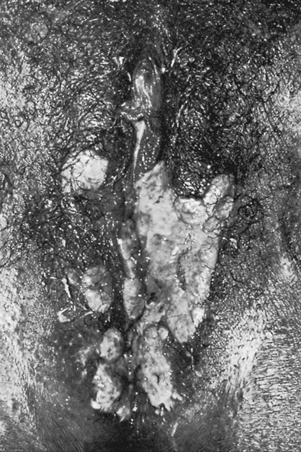
Granuloma Inguinale
Granuloma inguinale is rare in temperate climates and is usually considered an STD, although gastrointestinal transmission can occur. The initial papular lesion typically ulcerates and develops into a soft, painless, progressive granuloma that may be covered by a thin, gray membrane. The granuloma may spread over the course of many months to involve the anus and rectum (Fig. 32.3). Lymph nodes are moderately enlarged and painless, but they do not suppurate. The infection can become chronic, and long-standing disease may cause genital scarring and depigmentation, as well as lymphatic fibrosis with consequent genital edema.
|
|
|
FIG. 32.3. Vulval granuloma inguinale of relatively recent origin. Some lesions are separate, others confluent. The margin of lesion is raised and scrolled; the base is granular and covered imperfectly by thin, gray slough. (From Demis DJ, Crounse RG, Dobson RL, et al, eds. Clinical dermatology, Vol 3. Hagerstown, MD: Harper & Row, 1972, with permission.) |
Infection is caused by a gram-negative bacillus, Calymmatobacterium granulomatis, which is difficult to culture because it is an intracellular parasite. The identification is usually made from scraped material or a biopsy specimen obtained from the periphery of the lesion. Bipolar-staining bacteria are best identified within mononuclear cells (i.e., Donovan bodies) by Wright or Giemsa staining.
Therapy of choice is a 3-week course of doxycycline, 100 mg, or trimethoprim/sulfamethoxazole (double strength) or ciprofloxacin, twice daily. Erythromycin and azithromycin offer alternative therapies.
Lymphogranuloma Venereum
The incubation period for lymphogranuloma venereum (LGV) is 2 to 5 days. Thereafter, a transient, primary, painless genital or anorectal ulcer develops. Multiple, large, confluent inguinal nodes develop 2 to 3 weeks later and eventually suppurate. Acute infection may cause generalized systemic symptoms. If untreated, the infection enters a tertiary phase that can lead to extensive lymphatic obstruction. This development, together with continued infection, causes fistulae or strictures of the anal, urethral, or genital area. Women with LGV are particularly susceptible to rectal stricture. Edema and elephantiasis of the external genitalia and lower extremities are other serious sequelae.
The infection is caused by the sexually transmitted organism C. trachomatis, an intracellular bacterium. Only L1-3 Chlamydia serovars, which produce accelerated in vitro tissue destruction, typically cause LGV. The diagnosis can be made by culturing chlamydiae from genital lesions or lymph nodes. The most specific and sensitive serologic test is the microimmunofluorescent antibody test, in which the specific L immunotypes are identified. The results of complement fixation (CF) tests are positive in 95% of patients with LGV, but the CF test lacks specificity; test results are often falsely positive in patients who do not have LGV but have previously been exposed to Chlamydia. High (≥1:64 CF) titers offer some specificity.
LGV responds to 3-week regimens of doxycycline or erythromycin in the usual doses. Large lymph nodes should be aspirated to avoid chronic drainage. Surgical excision of scarred areas may be necessary.
ACUTE URETHRAL SYNDROME
Acute cystitis is present in approximately 50% of women with symptoms of dysuria and urinary frequency. Cystitis is defined by pyuria and midstream urine cultures that contain more than 105 organisms per milliliter of coliform or staphylococcal organisms. It is now apparent that about one-half of the remaining symptomatic women also have cystitis, but with less than 105 coliforms or Staphylococcus saprophyticus organisms per milliliter of urine obtained by suprapubic aspiration or urethral catheterization. Virtually all of these women have pyuria of eight or more leukocytes per high-power field of urine. The pyuria that occurs among another 25% of women with recent onset of internal dysuria and urinary frequency and negative urine cultures is termed acute urethral syndrome. These patients usually have C. trachomatis. The remaining 25% of patients with these symptoms have no pyuria, bacteriuria, or chlamydial infection and a variety of diseases are involved, including candidal or herpetic vulvitis, or no infection origin. Treatment of acute urethritis consists of therapy for the infectious agent, whether it is coliform or S. saprophyticus cystitis, or C. trachomatis urethritis.
VAGINITIS
Vaginitis is the most common reason for a gynecologic visit. Symptoms of vaginitis include increased vaginal discharge, vulvar irritation and pruritus, external dysuria, a foul discharge odor, and a yellow discharge color. However, symptoms are very poor indicators of the specific cause of vaginitis. Women with infectious vaginitis have either an STD (i.e., trichomonads) or a quantitative increase in normal flora (i.e., Candida, Gardnerella vaginalis, anaerobes). At least four types of infectious vaginitis are found: candidal, trichomonal, bacterial vaginosis, and, in children, gonococcal. Every effort should be made to establish the diagnosis of one of these specific infections and to avoid the diagnosis of a nonspecific vaginitis. A specific diagnosis is mandatory to select effective therapy. Treatment of nonspecific vaginitis inevitably fails.
Other conditions that may cause excessive vaginal discharge include cervicitis, normal cervical mucus from cervical ectopy, vaginal foreign bodies (most commonly, retained tampons), and allergic reactions to douching or vaginal contraceptive agents. Atrophic vaginitis among postmenopausal women can produce burning and dyspareunia, but an infectious cause is not established.
A small amount of vaginal discharge may be normal, particularly midcycle, when large amounts of cervical mucus production produce a clear vaginal discharge. A normal vaginal discharge should not have a foul odor or produce irritation or pruritus.
Examination
External genitalia may be normal or edematous, erythematous, excoriated, or fissured. Local vulvar disease, especially vestibulitis, must be excluded from a secondary effect of vaginitis.
On speculum examination, the vaginal mucosa may be erythematous. Discharge characteristics that are important to observe are viscosity, floccular appearance, color, and odor. Vaginal pH status must be determined. A pH less than 4.5 indicates Candida or a normal vaginal discharge. A potassium hydroxide (KOH) odor test and a microscopic examination consisting of a normal saline and 10% KOH wet mount should be done. A drop of each solution is mixed with discharge. Before placing a cover glass over the two separate drops, the KOH portion is tested for the presence of a fishy amine odor. Microscopic examination of the KOH portion is made for hyphae under the 100× objective, and examination of the saline portion is made for trichomonads and clue cells under the 400× objective. Multiple causes of vaginitis are frequent.
Vaginal cultures are not particularly helpful except when used selectively to identify Candida. Microscopy is specific, but only 80% sensitive in identifying various types of vaginitis. When infectious vaginitis is suspected in patients in whom a specific diagnosis cannot be established, a repeat examination should be performed 2 weeks later.
Candidiasis
The most prominent symptom of candidiasis is vulvar and vaginal pruritus. Increased vaginal discharge is infrequent. Vulvar signs of edema, geographic erythema, and fissures may occur. Classically, the vaginal walls are red and contain adherent, white, curdy plaques. However, most women with candidiasis have atypical symptoms, little discharge, and no erythema.
Candida albicans causes about 90% of vaginal yeast infections. Noncandidal species cause the remaining infections. These saprophytic fungi are isolated from the vagina in 15% to 25% of asymptomatic women. Thus, the mere presence of vaginal Candida does not always identify an infection, but large numbers of organisms lead to symptomatic vaginitis. However, severe symptoms develop in some women with only a few organisms but with an accelerated immune response to Candida. Candidiasis occurs because changes in host resistance or immune response allow organism detection or cause inflammation. The most widely accepted risk factors for candidiasis include pregnancy, diabetes, and use of immunosuppressive drugs and broad-spectrum antibiotics. Frequent vaginal intercourse and vaginal douching also are risk factors. Because cellular, not humoral, immunity is required to resist candidal infections, pregnant women and patients receiving immunosuppressive drugs that decrease cellular immunity are predisposed to candidiasis. Candidal overgrowth is also favored by high urine glucose levels that can occur in diabetes or pregnancy. Broad-spectrum antibiotics cause suppression of the normal vaginal and gastrointestinal bacterial flora, allowing fungal overgrowth. The role of oral contraceptives in candidal infection remains controversial.
Most women have uncomplicated infection defined as a sporadic, mild, C. albicans infection in a host with normal immunity. About 5% of women have complicated infection defined as recurrent, severe, or non-albicans infection. Many of these occur in immunosuppressed women (pregnant, diabetic, or debilitated or immunosuppressed).
Candidiasis is best diagnosed in KOH wet-mounts. Vaginal plaques, vaginal discharge, or vulvar scrapings from the edge of the erythematous border are mixed with 10% KOH (Fig. 32.4). The mycelial form is usually found only during an infection; mycelia can be identified by KOH wet mount in 80% of cases. The pH of vaginal discharge is normal (i.e., 4.7 or less). Fungi can readily be isolated on various media. However, Candida is part of normal vaginal flora, and a positive culture does not necessarily indicate infection. Cultures for Candida should be limited to KOH wet-mount–negative patients with symptoms or signs of candidiasis. In fact, 50% of women with candidiasis have a negative wet mount, but a positive Candida culture.
|
|
|
FIG. 32.4. Candida albicans growing as hyphae and pseudohyphae within infected tissue (×320). (From Monif GRG. Infectious diseases in obstetrics and gynecology. Hagerstown, MD: Harper & Row, 1974, with permission.) |
Local vaginal therapy is used because most antifungal preparations are not absorbed from the intestinal tract. For uncomplicated infection, various intravaginal azole agents used for 3 to 5 days are equally effective for women with primary and infrequent candidal vaginitis. These agents include miconazole, clotrimazole, butoconazole (Femstat), tioconazole (Vagistat-1), and terconazole (Terazol). Azole drugs are not absorbed to any degree from the vagina, and the same local regimens can be used safely in pregnancy. The insertion of boric acid powder in capsules into the vagina is also effective. A one-time dose of fluconazole (Diflucan), 150 mg orally, or itraconazole (Sporanox), 400 mg initially then 200 mg for 2 days, is also effective for those with uncomplicated infection. Patients with complicated infection from severe symptoms need 7 to 14 days of intravaginal azole therapy or fluconazole 150 mg with the dose repeated in 4 days. Oral nystatin administration to decrease gastrointestinal colonization does not markedly improve therapeutic cure rates or diminish recurrence rates. About 15% of male sexual contacts of women with candidiasis have symptomatic balanitis; symptomatic males should be identified and treated to prevent recurrent female infection.
The number of non-albicans candidal infections appears to have increased. These infections respond poorly to azole therapy, including fluconazole. Boric acid or nystatin therapy works best. Even gentian violet 1% aqueous solution has a limited place in such patients.
Patients with frequently recurrent candidiasis represent the most difficult problem in treatment. Extended 2- to 3-week vaginal therapy, male therapy, and reduction of sugar intake are usually ineffective. A glucose tolerance test and HIV testing should be performed in recurrent or resistant cases to exclude unrecognized diabetes and HIV infection. In addition, some women with candidiasis have other concurrent vaginal infections; a repeat physical and wet-mount examination may clarify the problem.
Complicated infection from frequently recurring candidiasis (four or more times a year) should be treated first with standard anticandidal therapy for 2 weeks, followed by suppressive therapy with an intravaginal azole or boric acid twice weekly or an intravaginal azole daily for 5 days once a month, oral fluconazole, 150 mg weekly, or oral itraconazole, 400 mg monthly. Suppressive treatment should continue for at least 1 year; recurrent candidiasis is usually reduced to no or one infection yearly, but recurs in 30% to 40% of patients upon cessation of suppression.
Trichomoniasis
Characteristic symptoms of trichomoniasis include a profuse, yellow, malodorous, often uncomfortable vaginal discharge with vulvar irritation. Trichomonas vaginalis is a common sexually transmitted organism, present in 3% to 15% of asymptomatic women and in up to 20% of women who attend clinics for STDs. T. vaginalis is most likely identified in symptomatic women with recent acquisition. However, about 50% of all women with trichomoniasis are asymptomatic. Most male contacts of women with trichomoniasis asymptomatically carry the organism in the urethra and prostate.
A classic profuse, frothy, yellow vaginal discharge is present in only about one-third of women. The vulva may be edematous and inflamed by the discharge. Sometimes, subepithelial redness of the cervix (i.e., strawberry cervix) is seen with the naked eye; smaller red areas are more commonly identified colposcopically. The discharge in women with symptomatic trichomoniasis often has a pH greater than 4.5 and forms an amine odor with 10% KOH. Motile trichomonads are demonstrated in the saline wet-mount smear (Fig. 32.5). Trichomonads are larger than white blood cells (WBCs) and have a jerky motility. The wet mount usually also contains many polymorphonuclear leukocytes. Although the wet mount can identify trichomonads with 80% sensitivity among symptomatic women, less than 50% of all women with trichomoniasis by culture have positive wet mounts. Trichomonads also can be seen occasionally on a Pap smear.
|
|
|
FIG. 32.5. Characteristic configuration of a trichomonad seen in wet smear at high-power magnification. (From Monif GRG. Infectious diseases in obstetrics and gynecology. Hagerstown, MD: Harper & Row, 1974, with permission.) |
T. vaginalis is an anaerobic protozoan. A culture of this organism is easy to perform but not readily available, and culture should be limited to cases where the diagnosis is suspected, but cannot be confirmed by wet mount. Screening cultures in asymptomatic women are not recommended, except for certain high-risk populations. Women with trichomoniasis should also be cultured for N. gonorrhoeae because of a close association between the microbes.
T. vaginalis resides not only in the vagina but also in the urethra, bladder, and Skene glands, so systemic, rather than local, therapy is needed. Metronidazole is effective in treating trichomoniasis; the preferred regimen is 2 g in one dose because of complete patient compliance and high effectiveness. Extended 7-day metronidazole therapy, 500 mg twice daily, does not increase the 95% cure rate of a single dose. Simultaneous treatment of the male sexual partner is recommended. Recurrent trichomoniasis is usually attributable to either a lack of compliance or reexposure to an untreated sexual partner. A single recurrence should be treated with the recommended regimens. However, increasing in vitro and in vivo resistance of T. vaginalis to metronidazole exists, and repeated treatment failure should be treated with metronidazole, 2 g daily for 3 to 5 days. The Centers for Disease Control and Prevention (CDC) has consultation available for patients who fail this regimen (see Sexually Transmitted Disease Treatment Guidelines, 2002).
Metronidazole therapy is controversial because of its tumor-causing potential in humans. In animals, large doses (equivalent to 350 to 1,000 human doses) cause tumors. The drug also causes mutation of salmonellae associated with carcinogenic potential. No increased tumor rates were found in a small series of women evaluated for up to 10 years after metronidazole therapy for trichomoniasis. However, these data are only slightly reassuring that the drug does not cause cancer because longer, larger studies are needed to exclude this possibility. A meta-analysis and other studies show no consistent evidence of teratogenesis from use of metronidazole in pregnancy. The CDC Sexually Transmitted Disease Treatment Guidelines, 2002 no longer advise avoiding metronidazole in pregnancy. Treatment of asymptomatic T. vaginalis has not reduced prematurity and should be limited to symptomatic women. Persistent discharge after adequate treatment for trichomoniasis should lead to repeat examination for Trichomonas, candidiasis, and gonorrhea.
Bacterial Vaginosis
Bacterial vaginosis describes the vaginal condition resulting from overgrowth of both anaerobic bacteria and G. vaginalis. Both anaerobes and G. vaginalis are normal inhabitants of the vagina, but overgrowth of the normal Lactobacillus-dominant flora by these bacteria results in a thin, homogeneous, fishy-smelling, gray vaginal discharge that adheres to the vaginal walls and often is present at the introitus. In contrast to findings in other causes of vaginitis, the vaginal epithelium appears normal, and WBCs are usually not present. The fishy amine odor produced by anaerobes is accentuated when 10% KOH is added to the discharge.
The diagnosis of bacterial vaginosis is based on the presence of three of the following four characteristics of the discharge: pH greater than 4.5, a homogeneous thin appearance, a fishy amine odor with the addition of 10% KOH, and clue cells. Clue cells are vaginal epithelial cells with many organisms attached. The cell border of clue cells is so obscured by adherent bacteria that it is not identifiable. In bacterial vaginosis, 20% to 50% of the epithelial cells are clue cells. Polymorphonuclear leukocytes and lactobacilli are notably absent. Gram stains used for diagnosis rely on a reduction in Lactobacillusmorphotypes and an increase in small Gram-negative rods and Gram-positive cocci. Commercially available card tests for high concentrations of G. vaginalis(Affirm), pH and trimethylamine (FemExam), and proline aminopeptidase (PIP Activity Test Card) are available. Cultures are not helpful because anaerobes and G. vaginalis can be recovered from women without bacterial vaginosis. In fact, up to 40% of asymptomatic women without vaginitis carry G. vaginalis. The distinguishing feature of bacterial vaginosis is the 10- to 1,000-fold increased concentration of anaerobic bacteria and G. vaginalis.
Factors leading to the overgrowth of anaerobes and G. vaginalis are poorly established but include the absence of Lactobacillus and a new sexual partner. Sexual transmission of the infection is considered a risk factor, but is unproven, especially since the treatment of sexual contacts does not prevent recurrence.
Treatment is not advocated for most asymptomatic women with bacterial vaginosis because it can spontaneously disappear. However, the 10- to 1,000-fold increased concentration of potentially virulent bacteria in the vagina is related to upper genital tract infection after surgery. A significant, increased relative risk (RR) of postoperative infection has been reported in patients with bacterial vaginosis following cesarean section (RR = 6), hysterectomy (RR = 3 to 4), and induced abortion (RR = 3). Bacterial vaginitis has also been associated with spontaneous PID and postpartum endometritis after vaginal delivery. Treatment of bacterial vaginosis is particularly beneficial for those undergoing elective surgery. Bacterial vaginosis in pregnancy has also been related to premature delivery (RR = 2 to 4), amniotic fluid infection (RR = 2 to 3), and chorioamnionitis (RR = 2 to 3); treatment of women with prior preterm delivery has reduced the incidence of preterm delivery, but treatment of low-risk asymptomatic women had no effect on preterm delivery.
Metronidazole, 500 mg orally twice daily for 7 days, metronidazole gel, intravaginally once daily for 5 days, and clindamycin cream, intravaginally at bedtime for 7 days are the recommended regimens for bacterial vaginosis. Alternative regimens include 2 g oral metronidazole in a single dose, 300 mg oral clindamycin twice daily for 7 days, and clindamycin ovules intravaginally at bedtime for 3 days. Metronidazole and clindamycin are particularly effective against the anaerobes. Fluoroquinolones, tetracycline, sulfonamides, and erythromycin are ineffective. Treatment of the male sexual contact with metronidazole does not prevent recurrent bacterial vaginosis and is not recommended.
Toxic Shock Syndrome
Toxic shock syndrome is an acute illness caused by toxin-producing Staphylococcus aureus. About 6% of women carry S. aureus in the vagina, but only 2% of women have S. aureus capable of producing the toxic shock toxin. The syndrome is highly associated with menstruation and probably with tampon use, but it has also occurred from S. aureus infection of the breast and endometrium after delivery and from abdominal surgical wounds. Characteristic features include a high fever (>102°F [38.9°C]), a diffuse rash, hypotension, skin desquamation (usually 1–2 weeks later), and a wide variety of systemic effects, including gastrointestinal (vomiting, diarrhea), muscular (myalgia), mucous membrane (hyperemia), renal (elevated blood urea nitrogen or creatinine level), hepatic (enzyme abnormalities), hematologic (thrombocytopenia), and neurologic (disorientation, coma). Vaginal or specific-site cultures recover S. aureus. Blood, throat, and cerebrospinal fluid cultures, together with serologic tests for Rocky Mountain spotted fever, leptospirosis, and measles, are usually indicated to exclude diseases with similar clinical presentations.
A vaginal tampon, if present, should be removed. Patients should be hospitalized and, when indicated, given large fluid volumes for blood pressure maintenance. Beta-lactamase–resistant antibiotics are recommended, and if other causes of bacterial sepsis such as meningococcemia cannot be excluded, additional antibiotics are necessary. Other life-supporting measures such as intubation, vasopressor administration, and dialysis are often necessary. The case:fatality ratio has been reduced from 15% to 3% with supportive therapy. Antibiotics are of no proven benefit in the acute stage, but they do reduce recurrence rates from 30% to 5%.
Although the effectiveness is uncertain, it is prudent for all women to avoid the prolonged and overnight use of tampons. It is recommended that postpartum women not use tampons for 6 to 8 weeks after delivery. Women with toxic shock syndrome should be warned of recurrent episodes and advised against resuming tampon use.
SYPHILIS
Physicians must constantly be aware of possible syphilitic infection, particularly in populations with high rates of HIV infection. Most women with syphilis are asymptomatic and have only serologic evidence of infection. T. pallidum rapidly enters lymphatics after exposure, but a primary chancre lesion usually takes about 3 weeks to develop. The classic chancre ulcer is painless and firm with sharply defined, raised edges; however, most syphilitic ulcers are atypical. Any suspicious genital ulcer should be studied by dark field examination. Serous material expressed from the ulcer base is mixed with saline solution, and because T. pallidum is an anaerobe, this mixture must be immediately placed under a cover slip with the edges occluded by petroleum jelly. Identification of typical spirochetes by dark field microscopy establishes a diagnosis of primary syphilis. Dark field examination of ulcer material from possible syphilitic ulcers should be done on 3 consecutive days. VDRL test or rapid plasma reagin (RPR) and fluorescent treponemal antibody (FTA) serology should be performed for a patient with a suspicious lesion. If the serologic results are nonreactive and spirochetes cannot be demonstrated by dark field examination, serologic tests should be repeated in 1 month.
Secondary syphilis appears 6 or more weeks later and is characterized by a symmetric, macular, papular, or papulosquamous rash and generalized, nontender lymphadenopathy. Condylomata lata (see Fig. 32.2) are highly infectious, hypertrophied, wartlike lesions of secondary syphilis that usually occur in moist areas such as the vulva or perineum; they must be distinguished from other vulvar lesions. Superficial, painless mucosal erosions of the mouth or vagina, called mucous patches, develop in one-third of patients. Systemic symptoms of fever, weight loss, and malaise may occur. Serologic tests are positive in the secondary stage.
Untreated patients will enter a latent phase of syphilis during which clinical and physical manifestations are absent. Diagnosis in the latent phase is established by serologic tests. Intermittent spirochetal bloodstream invasion may occur in the early latent phase (first 4 years). In pregnancy, the risk of congenital fetal infection in the primary and secondary phases of syphilis is 80% to 95%; the risk during the early latent phase is 70%. During the late latent phase, immunity develops, which reduces blood invasion, and the risk of congenital syphilis decreases to 10%. Congenital syphilis is reported to be on the rise, affecting 1 in 10,000 live-born infants. Fetal or perinatal death occurs in 40% of those with congenital syphilis. About one-third of adult patients with untreated late syphilis manifest central nervous system or cardiovascular symptoms of tertiary syphilis.
VDRL and RPR tests detect a nontreponemal, nonspecific reagin antibody. The tests can be titrated, and the titer either falls or disappears after therapy for early or secondary syphilis. Thus, the VDRL test can be used to judge the activity of either a first episode or reacquired infection in a patient with documented syphilis. Treated patients with latent syphilis may retain high, stable VDRL titers. Acute bacterial or viral infections can give rise to acute false-positive serologic reactions that last for up to 6 months. Several conditions, such as aging, addiction to drugs, autoimmune disease, and pregnancy, can lead to chronic, nonspecific, false-positive VDRL reactions. False-positive VDRL titers usually are 1:8 or less. By contrast, the FTA test involves a specific antitreponemal antibody, and false-positive FTA reactions are rare. Patients with a positive VDRL reaction must have a confirmatory FTA test to exclude a false-positive VDRL reaction. Patients with a false-positive VDRL reaction will have a negative FTA reaction. In patients with syphilis, the FTA test remains positive indefinitely, and because the test is not titrated, repeat FTA testing should not be done in a known positive patient.
The treatment schedules for syphilis currently recommended by the U.S. Public Health Service's CDC are as follows:
· Early syphilis: Early syphilis is defined as primary, secondary, or latent syphilis of less than 1 year's duration. The drug of choice is penicillin G benzathine, 2.4 million U intramuscularly in a single dose. Alternative choices for penicillin-allergic patients include 2-week regimens of doxycycline, 100 mg twice daily, or tetracycline, 500 mg four times daily. Ceftriaxone, 1 g daily intramuscularly for 8 to 10 days, may be used if close follow-up can be ensured.
· Syphilis of longer than 1 year's, or of unknown, duration: Penicillin G benzathine, 2.4 million U intramuscularly each week for 3 successive weeks (7.2 million U total), is the drug of choice in this situation. Alternative choices for the penicillin-allergic include doxycycline and tetracycline for 4 weeks. Intravenous aqueous penicillin G or penicillin G procaine is recommended for neurosyphilis. Erythromycin is not recommended for neurosyphilis. Spinal tap to exclude asymptomatic neurosyphilis is recommended for those with neurologic or ophthalmologic signs, other evidence of active disease (aortitis, gummas), HIV infection, treatment failure, and infection for more than 1 year with a titer of 1:32 or greater. Neurosyphilis and syphilis in HIV-positive patients should be treated by infectious disease specialists.
· Syphilis in pregnancy: Parenteral penicillin is the only documented efficacious treatment in pregnancy. Treatment with penicillin is the same as for the corresponding stage of syphilis among nonpregnant women. For pregnant patients who are allergic to penicillin, tetracycline is not used because of toxicity and erythromycin is not used because of high failure rates to cure the fetus. Penicillin is so superior to other antibiotics for treating syphilis in pregnancy that pregnant, penicillin-allergic patients should be desensitized. The Jarisch-Herxheimer reaction commonly occurs in early syphilis, and pregnant women should be hospitalized in anticipation of this possibility for monitoring of themselves and the fetus. The reaction is ascribed to the sudden massive destruction of spirochetes by antibiotics; it is marked by fever, myalgia, tachycardia, and occasionally hypotension. The reaction usually begins within 24 hours and subsides spontaneously in the next 24 hours. All patients need to be followed with quantitative serologic tests to monitor treatment results and offered HIV testing. All sexual partners need to be contacted and tested for syphilis.
CERVICITIS
Acute cervicitis is defined as the presence of yellow cervical mucopus or an increased number of WBCs in cervical mucus. Symptoms are usually limited to a purulent vaginal discharge. Physical findings include mucopus in the endocervical canal or bleeding after swabbing of the cervix. Organisms that infect the cervical columnar epithelium, C. trachomatis or N. gonorrhoeae, can be isolated separately or in combination from about one-half of women with purulent cervicitis. Unknown microbes cause the other cases. The diagnosis can also be established by the finding of more than 10 WBCs per 1,000× microscopic field.
Infectious ulcers of the cervix caused by herpesvirus, syphilis, and chancroid must be distinguished from erosion and the other conditions described in Chapter 33. Depending on the nature of the lesion, Gram stain, Pap smear, culture, dark field examination, colposcopy, and, in some cases, biopsy may be required.
If N. gonorrhoeae is found by culture or DNA technique, treatment should be the same as for gonorrhea, including treatment for coexisting C. trachomatis. If N. gonorrhoeae is not found, azithromycin or doxycycline regimens used for C. trachomatis are recommended. Ofloxacin or levofloxacin can also be used.
ENDOMETRITIS
Lymphocytes and neutrophils normally appear in the endometrium in the second half of the menstrual cycle; their presence does not necessarily constitute endometritis. However, plasma cells are not normally in the endometrium, as they represent an immune response, usually to a bacterial antigen.
Endometritis produces nonspecific symptoms, and it should not be diagnosed unless plasma cells, neutrophils within glands, or a specific causative infection is found. Endometritis may occur in the following situations:
· puerperal endometritis (see Chapter 19)
· chlamydial or gonococcal endometritis, often occurring among patients with salpingitis
· endometritis after endometrial instrumentation or surgery
· tuberculous endometritis
· purulent endometritis, occurring in pyometra caused by a cervical stricture or after radium insertion
· endometritis occurring characteristically in the presence of an intrauterine device (IUD).
The fact that chronic endometritis is associated with the use of IUDs is well documented; it results from organisms attached to the IUD surface rather than from the foreign body per se. Transfundal endometrial cultures of hysterectomy specimens from women who had used tailed IUDs for more than a few weeks uniformly recovered bacteria, while cultures from women who did not use an IUD were negative. Bacteria that can be recovered are usually of low pathogenicity, but more virulent intrauterine bacteria occasionally cause malodorous discharge and salpingitis. In addition, an anaerobe, Actinomyces israelii, has been found in Pap smears from about 5% of women using IUDs, but not in those who do not use IUDs. This organism appears to colonize the IUD, and when it is found on a Pap smear, asymptomatic patients should be warned of abnormal discharge or abdominal pain, representing infection. If symptomatic infection occurs, the IUD should be removed, and patients treated with ampicillin for 2 to 4 weeks. Asymptomatic patients should not have the IUD removed because of A. israelii.
Chronic plasma cell endometritis in nonpregnant women who do not use an IUD is often related to endometrial infection with C. trachomatis and, to a lesser extent, N. gonorrhoeae. Plasma cell endometritis occurs in up to 50% of women with acute cervicitis and over 80% of women with acute salpingitis. Symptoms are abnormal uterine bleeding and mild uterine tenderness. Untreated endometritis can progress to clinically evident salpingitis.
GONORRHEA
Gonorrhea is caused by the Gram-negative diplococcus N. gonorrhoeae which attach only to columnar or transitional cells by pili and are rapidly brought intracellularly by pinocytosis. They attract leukocytes, commonly giving rise to purulent discharge. Gonorrhea is usually sexually transmitted, although organisms can be acquired by neonates passing through an infected cervix, causing gonorrheal ophthalmia.
Course of the Disease
N. gonorrhoeae in the lower genital tract infects the urethra, Bartholin glands, and endocervix. The anus and rectum can also be infected either from cervical infection or during anal coitus. Urinary frequency, dysuria, and a purulent vaginal discharge are the first symptoms to appear 2 to 5 days after exposure. At least 50% of women with N. gonorrhoeae infection have no symptoms. Many women do not seek medical attention if these symptoms are mild. The discharge occasionally is locally irritating and causes vulvar edema and soreness. Pharyngitis may result from gonorrheal pharyngeal infection. In 2% of infected women, disseminated gonococcal infection occurs, causing fever, septicemia, dermatitis, arthritis, endocarditis, or meningitis, in various combinations. Untreated gonorrhea is associated with premature delivery and premature rupture of membranes.
In 10% to 17% of women with untreated gonorrhea, the organisms ascend to produce upper genital tract infection or acute PID (Fig. 32.6). Acute PID is the most common serious sequela of gonorrhea. The mechanical and antibacterial properties of cervical mucus probably provide a barrier against upward extension, but during menstruation the mucus barrier is lost, and gonococci can disseminate to the uterus and fallopian tubes. A transient endometritis occurs as the organisms pass through the uterine cavity and reach the fallopian tubes, where they produce an acute and usually bilateral inflammatory reaction of tubal mucosa. The tubes characteristically become swollen and reddened when the muscularis and serosa become inflamed. If exudate drips from the fimbriated ends of the tubes, pelvic peritonitis occurs that ultimately can cause peritoneal adhesions. The swollen and congested fimbriae may adhere and produce tubal occlusion.
|
|
|
FIG. 32.6. Mode of transmission of gonococcal pelvic infection. Portal of entry is external genitalia. The organism enters cervix, following mucous membrane, passes up through the uterine cavity, and attacks the fallopian tube. Pelvic peritonitis results from escape of pus from tubal fimbria. (From Wharton LR. Gynecology and female urology. Philadelphia: WB Saunders, 1943, with permission.) |
The process can take any of the following courses. With prompt, appropriate antibacterial therapy, the infection may subside with little permanent damage to the reproductive tract. The fimbriae may occlude, producing permanent tubal infertility. The swollen and congested fimbriae may adhere to one another or to the ovary, trapping the exudate in the tube and giving rise to pyosalpinx or, if the ovary becomes infected, a tuboovarian abscess. The mucosal folds may adhere to one another, forming glandlike spaces that are filled at first with exudate and later, as the process becomes chronic, with watery secretion in follicular salpingitis (Fig. 32.7). If the infection subsides after agglutination of the fimbriae and closure of the distal tube, watery secretion accumulates and distends the tube, forming a hydrosalpinx (Fig. 32.8).
|
|
|
FIG. 32.7. Follicular salpingitis, an end stage of gonorrheal salpingitis. Mucosal folds are adherent, giving rise to innumerable round or irregular cystlike cavities lined by cuboidal epithelium. (From Kelly HA. Operative gynecology, Vol 2. New York: Appleton, 1898: plate XI; drawing by Max Brodel, with permission.) |
|
|
|
FIG. 32.8. Hydrosalpinx. (From Curtis AH, Huffman JW. A textbook of gynecology, sixth ed. Philadelphia: WB Saunders, 1950, with permission.) |
Symptoms and Signs
Except for the discharge, which can be milked from the urethra or is present in the vagina or cervix, there are few signs of acute gonococcal infection in women. Bilateral, mild to severe lower abdominal pain may occur with acute salpingitis. Pelvic peritonitis can cause pain on movement of the cervix, direct and rebound abdominal tenderness, muscle guarding that prevents abdominal palpation, and tender adnexa to various degrees on bimanual examination. However, a sizable proportion of women with salpingitis experience either mild or no symptoms. In subacute salpingitis, infection continues with signs and symptoms that are even less overt than those of the acute stage. In the end stage of salpingitis, the uterus and the adnexa can be fixed by pelvic peritoneal adhesions. The adnexa may be either adherent to the posterior aspect of the uterus or prolapsed in the cul-de-sac, which may pull the uterus into a retroverted position. Notable features are dyspareunia, sterility, and chronic, aching pelvic pain that increases before menstruation.
Diagnosis
The diagnosis of gonorrhea depends on a culture of N. gonorrhoeae or identification of N. gonorrhoeae by DNA tests. The finding of intracellular Gram-negative diplococci in the Gram stain of cervical or urethral exudate points to gonorrheal infection, but Gram stains for gonorrhea are insensitive. Gonococcal testing should be performed on women with positive Gram stains, symptoms or signs suggestive of gonorrhea (e.g., cervicitis, undiagnosed vaginitis, dysuria), other STDs, bartholinitis or skenitis, acute lower abdominal pain suggestive of acute salpingitis, or suspected disseminated gonococcal infection, as well as on women with male sexual contacts with gonorrhea.
Sites to be tested in order of importance are: cervix, anal canal, pharynx, and urethra. Vaginal discharge should be wiped away from the cervix before mucus is obtained for testing. Genital samples must be cultured on Thayer-Martin or similar media containing antimicrobial agents that inhibit growth of the normal bacterial and fungal flora. Patients with gonorrhea should also receive a test for chlamydial infection because 10% to 30% of women with N. gonorrhoeae also have C. trachomatis.
Because gonorrhea is an STD, it usually is present in the male partner. The fact that more than 40% of the male contacts of women with gonorrhea are asymptomatic carriers who otherwise do not seek treatment underscores the importance to identify and treat male sexual contacts.
Drug Therapy of Uncomplicated Lower Genital Tract Gonorrhea
The CDC-recommended treatment schedules for gonorrhea reflect increased resistance to penicillin and tetracycline and in Asia, the Pacific rim, and the West Coast to quinolones, frequent coexistence of chlamydial infection with gonorrhea, potentially serious complications, and frequently no testing for C. trachomatis. Penicillin is no longer recommended to treat gonorrhea because β-lactamase production by extrachromosomal plasmids destroys β-lactam antibiotic activity in a large number of gonococcal strains. Resistance to quinolones will likely increase.
The recommended drug therapy regimens are given in Table 32.2. A loading dose of cephalosporin or quinolones is used to inhibit N. gonorrhoeae. Patients allergic to penicillin should receive spectinomycin (Trobicin) or other cephalosporins or quinolones. If C. trachomatis is not excluded, azithromycin or doxycycline is indicated. Quinolones and doxycycline should not be given to pregnant women. Special antibiotic regimens are recommended for patients with complicated gonococcal infections. These regimens are published in the widely circulated CDC Sexually Transmitted Disease Treatment Guidelines 2002. Prophylactic regimens to prevent ophthalmia neonatorum include silver nitrate, erythromycin, and tetracycline applications.
|
|
|
TABLE 32.2. Treatment regimens for gonorrhea and chlamydia |
The first-choice regimens are so effective that test-of-cure cultures are not recommended. A rescreening culture in 1 to 2 months is reasonable. Patients treated with alternative regimens should have a test-of-cure culture in 4 to 7 days after therapy. All male sexual contacts need referral for evaluation and treatment.
Patients with gonorrhea should have serologic tests for syphilis. Those with incubating syphilis (i.e., seronegative, without clinical signs of syphilis) are likely to be cured by all the regimens mentioned, except for quinolones and spectinomycin. Patients treated with these regimens need a follow-up serologic test for syphilis.
CHLAMYDIAL INFECTION
C. trachomatis is a sexually transmitted bacterium that is often associated with gonorrhea. Chlamydiae infect the same tissues and produce the same symptoms and diseases as gonorrhea. Chlamydial infection causes urethritis, bartholinitis, cervicitis, endometritis, salpingitis, Fitz-Hugh and Curtis syndrome (i.e., perihepatitis), and LGV. Treatment of C. trachomatis in pregnancy reduces premature delivery. Neonates born of mothers with chlamydial cervical infection have up to a 40% risk of chlamydial conjunctivitis and a 20% risk of chlamydial pneumonia. As with gonococcal infection, male sexual contacts have both symptomatic and asymptomatic urethritis.
C. trachomatis is an obligate intracellular bacterium that attaches to columnar or transitional epithelial cells, is engulfed by pinocytosis, and remains within a phagosome membrane that protects it from host defense mechanisms. The bacteria replicate until they replace most of the cell and, ultimately, cause the cell to rupture. Infective particles are released into the extracellular space, and the process is repeated. Replication time is a relatively slow 24 to 48 hours, explaining the characteristically long latent period between the time of exposure and the onset of symptoms, which ranges from weeks to months.
C. trachomatis is three to five times more common than N. gonorrhoeae in developed countries because it is not routinely sought in asymptomatic patients. Chlamydial infection is most often asymptomatic and frequently not identified until overt infection occurs. Infection is particularly common among teenagers. C. trachomatis is associated with serious sequelae, including tubal infertility and ectopic pregnancy and it appears able to produce permanent tissue damage more readily than N. gonorrhoeae. Permanent tissue damage appears largely related to the immune response to infection. Reinfection, chronic infection, and infection in the presence of antibody to chlamydial heat shock protein are particularly associated with tissue damage.
Chlamydial infection should be suspected with acute urethritis, mucopurulent cervicitis, and salpingitis. The rate of chlamydial salpingitis approximates that of gonorrhea. Chlamydial infections can be diagnosed by culture, a direct monoclonal antibody slide test, an enzyme-linked immunosorbent assay, or DNA techniques. New DNA methods using ligase chain reaction or PCR offer both sensitivity and specificity not achieved with older tests. Samples should be taken from the cervix or from urine with DNA tests.
Azithromycin and doxycycline are the most effective drugs to treat chlamydial infection (see Table 32.2). Erythromycin, ofloxacin (Floxin), and levofloxacin (Levaquin) are alternatives. In pregnancy, erythromycin and amoxicillin are preferred, but reports document the effectiveness and safety of azithromycin.
GENITAL MYCOPLASMAS
Genital mycoplasmas have often been thought a microbe in search of a disease because they are ubiquitous and not highly virulent. Mycoplasma hominis is recovered from the vagina in 15% to 70% of women, and Ureaplasma urealyticum is recovered from 40% to 95% of women. A third isolate, Mycoplasma genitalium, is now associated with endometritis. These organisms are phylogenetically positioned between bacteria and viruses.
The most convincing role for mycoplasmas in human female infections is as a pathogen in postpartum fever.
Mycoplasmas are recovered from the blood of 10% to 15% of women with postpartum fever, and antibodies to M. hominis are demonstrated in 50% of such women. Their role in salpingitis is less clear. Mycoplasmas are recovered from the tubes of 5% to 15% of women with salpingitis, but in primate model studies, M. hominis produces an adnexitis and not salpingitis.
Maternal lower genital tract U. urealyticum is not associated with low birth weight, and treatment of U. urealyticum does not reduce preterm births. However, organisms are found in intraamniotic infection and cause cerebrospinal fluid and lung infection in premature neonates. Recovery of U. urealyticum from fetal tissue of mid-trimester spontaneous abortuses also suggests a relationship with abortion. The role of U. urealyticum in fertility is unsettled, but in some reports, mycoplasmas were not related to infertility or to higher pregnancy rates in infertile women.
Both M. genitalium and M. hominis are sensitive to tetracycline. Erythromycin inhibits U. urealyticum in vitro but not M. hominis. However, neither antibiotic very effectively eradicates mycoplasmas from the vagina.
ANAEROBIC BACTERIA
Anaerobic bacteria are highly associated with pelvic infections. Multiple anaerobic species, usually together with one or more aerobic bacteria, typically combine to form a polymicrobial infection. Intraabdominal abscess and postoperative, postpartum, and bacterial vaginosis infections are the most important examples of anaerobic infection.
Anaerobic bacteria are part of the normal vaginal flora. Although many mechanisms by which anaerobic bacteria become pathogenic are unknown, two mechanisms known to cause anaerobic infection include (a) reduction of the redox potential that occurs with tissue trauma from surgery and (b) antibiotic selection that preferentially inhibits aerobic bacteria. Clinicians can virtually assume the presence of anaerobes in infections with a foul-smelling odor as only anaerobes produce odorous metabolic products. Anaerobes are virtually always isolated from an abscess. Anaerobic infections can also produce gas and cause thromboembolism.
The anaerobic bacteria most commonly found in genital infections include anaerobic Gram-positive cocci (Porphyromonas and Peptostreptococcus species), gram-negative rods (Prevotella [P. melaninogenicus, P. bivia], Bacteroides [B. fragilis], and Fusobacterium species), and Gram-positive rods (Clostridiumspecies).
Cultures should be obtained before antimicrobial therapy is begun. Because anaerobes are part of the normal flora, deep tissue cultures are required that are not contaminated by surface bacteria. Because 48 or more hours are required for anaerobe isolation, antibiotic selection is usually based on clinical signs. Anaerobic infection should be particularly suspected with abscess formation, a foul odor, gas formation, tissue necrosis, sterile cultures from obviously infected sites, and thromboembolism. Antibiotic sensitivity testing is only a rough guide to antibiotic susceptibility, but in vitro and in vivo experience has shown that clindamycin, metronidazole, imipenem/cilastatin sodium (Primaxin), second- and third-generation cephalosporins (cefoxitin sodium [Mefoxin], cefotaxime sodium [Claforan]), and extended-spectrum penicillins (ticarcillin disodium/clavulanate potassium [Timentin], amoxicillin/clavulanate potassium) are effective to treat anaerobic infections.
SALPINGITIS
Acute primary salpingitis results when pathogenic bacteria in the cervix invade the fallopian tubes. N. gonorrhoeae, C. trachomatis, normal flora aerobic and anaerobic bacteria, and, perhaps, M. genitalium are known causes of tubal infections. Virtually all primary salpingitis occurs among sexually active, menstruating, nonpregnant women. Gonococcal and chlamydial infections account for 50% to 60% of cases. Tuberculous, parasitic, or fungal salpingitis is rare in industrialized countries. Salpingitis usually occurs without instrumentation or trauma to the genital tract; however, approximately 15% of cases occur after instrumentation (e.g., IUD insertion, dilation and curettage, abortion, hysterosalpingography). Perisalpingitis secondary to acute appendicitis or other intraabdominal infections accounts for less than 1% of cases.
Acute salpingitis is a common event that annually develops in up to 1% of women between 15 and 39 years of age. Young, sexually active women between 15 and 24 years of age have the highest rate of infection. This infection has tremendous cost consequences. At least $1 billion is required to treat the 800,000 women with acute salpingitis in the United States annually, and $40 billion is spent to diagnose and treat tubal infertility.
Epidemiology
Most women are infected with sexually transmitted organisms. The rate of salpingitis is increased in women with multiple sexual partners. The high rate of salpingitis in young women is due to their increased rates of gonorrhea and chlamydial infection. Routine screening for C. trachomatis and N. gonorrhoeae has reduced salpingitis in Europe and the United States. Sexually active women at increased risk for STDs, especially women younger than 25 years and with multiple sexual partners, should be annually screened for chlamydial infection and gonorrhea. Such screening will prevent salpingitis and subsequent tubal infertility and ectopic pregnancy more effectively than any other measure. Previous salpingitis also predisposes women to subsequent salpingitis, probably because mucosa damaged from prior infection is more susceptible to infection than normal tissue. Patients with previous uncomplicated gonorrhea have a high rate of subsequent salpingitis, in part because of increased rates of subsequent gonorrheal infection. Prior chlamydial infection predisposes patients to salpingitis if a second chlamydial infection occurs. Repeated chlamydial infections produce a hyperimmune response that increases the chance and severity of tissue damage.
The presence of an IUD is an independent risk factor for salpingitis. IUD users have a two-fold to four-fold increased rate of both salpingitis and tubal infertility, compared to non–IUD users. The highest rate of salpingitis in IUD users occurs within a few weeks of insertion as a result of the introduction of cervical bacteria into the endometrial cavity along with the IUD. Most infections in IUD users, however, occur long after insertion, probably because bacteria wick along the IUD tail from the vagina to the uterus and adhere to the IUD surface. IUDs also appear to enhance anaerobic bacterial growth, and their use is associated with Actinomyces and bacterial vaginosis infection. Because IUD use is also associated with tubal infertility, an IUD should not be inserted in women who desire future pregnancy. By contrast, barrier or oral contraceptive methods appear to protect against salpingitis. The protective effect of oral contraceptives on salpingitis appears to exist only for patients with chlamydial infection, possibly due to a down-regulation of the hyperimmune response caused by the organisms.
The role of male contacts with untreated gonococcal or chlamydial urethritis is often ignored by gynecologists. Only 25% of male contacts of women with gonococcal salpingitis are treated by the time the female partner develops symptomatic salpingitis. Over 50% of the male contacts with gonococcal urethritis are asymptomatic, and N. gonorrhoeae is isolated from 40% of these asymptomatic males. Men with nongonococcal urethritis represent reservoirs of chlamydial salpingitis. To reduce the rate of new and recurrent salpingitis, all male contacts of women with any type of salpingitis should be examined and cultured. If infectious organisms are found, males should be appropriately treated.
Bacteriology
Neisseria gonorrhoeae
In most studies in the United States, N. gonorrhoeae is recovered from 40% to 50% of women with acute salpingitis. However, gonococcal prevalence varies greatly: N. gonorrhoeae is isolated from less than 20% of salpingitis cases in Sweden and from 80% of cases in certain urban populations in the United States. In women with both cervical gonorrhea and salpingitis, N. gonorrhoeae is the most frequent intraabdominal isolate, but the sole isolate in only 30% of these cases. The remainder have either no organisms or other organisms isolated alone or together with N. gonorrhoeae in the abdomen. Chlamydial infection frequently coexists with gonorrhea; in some studies, more than 50% of women with gonorrhea also had C. trachomatis in the cervix. Positive tubal gonococcal cultures are usually obtained during the early stages of infection, but not in the later stages of infection.
Chlamydia trachomatis
C. trachomatis is as important as the gonococcus in causing acute salpingitis. From 30% to 60% of women with salpingitis have C. trachomatis, and in most of these women the organisms can be isolated from the fallopian tube. Application of new DNA tests for C. trachomatis identifies even more chlamydial infections. Chlamydial salpingitis is under-diagnosed because many patients have mild symptoms and are not identified. It is now evident that women with mild symptoms and signs have the same amount of severe tubal damage as those with severe symptoms. Chlamydial salpingitis often produces mild symptoms and signs but may produce more severe tubal damage compared to gonococcal salpingitis.
Nonsexually Transmitted Aerobic and Anaerobic Bacteria
Nonsexually transmitted aerobic and anaerobic bacteria are normally present in cervical and vaginal flora and particularly include organisms associated with bacterial vaginosis. These organisms can be a direct cause of salpingitis, but they also cause secondary infection in combination with N. gonorrhoeae and C. trachomatis, IUD use, or instrumentation. Polymicrobial infection with these agents is common in salpingitis. In such cases, many different Gram-positive and Gram-negative aerobic and anaerobic organisms are isolated, particularly Porphyromonas, Prevotella, and Bacteroides spp, including B. fragilis. Anaerobic organisms are especially common in serious infections, and they are virtually always found in abscesses.
Mycoplasmas
Genital mycoplasmas have been recovered from the tubes or cul-de-sac in 2% to 20% of patients with salpingitis. In addition, more than 20% of patients with salpingitis have changes of mycoplasmal antibody titer suggestive of invasive infection. These organisms lack the virulence of N. gonorrhoeae and C. trachomatis, and they appear to cause an adnexitis rather than salpingitis. M. genitalium identified by DNA technology is related to endometritis.
Pathogenesis
Salpingitis occurs from vaginal and cervical bacteria ascending into the endometrium and fallopian tubes. The ascent of bacteria probably increases during menses, as evidenced by the onset of pain within 7 days of starting menses in one-half to two-thirds of patients with gonococcal salpingitis. Virulent gonococci also proliferate at menstruation, and less virulent gonococci are present at other times of the cycle. Other risk factors for salpingitis exist. Virulent bacteria in the cervix are more likely to cause salpingitis than nonvirulent bacteria, and C. trachomatis and N. gonorrhoeae are two virulent organisms capable of causing salpingitis. Virulence is occasionally evidenced by bacteria in the normal flora.
Specific bactericidal antibodies to N. gonorrhoeae appear to reduce salpingitis. Clinically recognized salpingitis develops in only 10% to 17% of women with cervical gonorrhea, and most of these women probably develop tubal infection during the first one or two menstrual periods between gonococcal acquisition and the development of specific bactericidal antibodies.
C. trachomatis may not depend on menses for ascent into the endometrium. Endometritis caused by C. trachomatis appears to be a common chronic intermediate infection that exists through several menstrual cycles. Tubal damage from chlamydial infection appears to occur from the immune response to infection. As mentioned, repetitive chlamydial infections elicit a hyperimmune response that accelerates tissue damage. The presence of chlamydial heat shock protein also contributes to an acceleration of tissue damage.
The usual route of infection with either organism is the contiguous spread along mucosa from the cervix to the endometrial cavity and fallopian tubes (see Fig. 32.6). Lymphatic or hematogenous dissemination of organisms from the uterus to the adnexa is uncommon in nonpregnant women. It also is possible that organisms may be transported along the fallopian tubes by cilia or even carried by their attachment to spermatozoa or to other organisms.
Fitz-Hugh and Curtis Syndrome
Perihepatitis consisting of liver capsule inflammation without liver parenchymal damage is referred to as Fitz-Hugh and Curtis syndrome (Fig. 32.9). Swelling of the liver capsule produces pain with inspiration, usually in the right upper quadrant. A purulent or fibrinous exudate appears on the capsular surface. Violin-string adhesions between the liver capsule and the anterior abdominal wall are a late manifestation of capsular inflammation.
|
|
|
FIG. 32.9. Liver adhesions in Fitz-Hugh and Curtis syndrome. |
Perihepatitis was formerly believed to be caused solely by N. gonorrhoeae, but C. trachomatis also causes this syndrome. Chlamydial heat shock protein is associated with perihepatitis, indicating that the syndrome is another manifestation of a hyperimmune response to chlamydiae. Some organisms travel transperitoneally from the fallopian tubes to reach the liver surface, but organisms may also reach the liver by lymphatic and hematogenous routes. The syndrome occurs virtually exclusively in women, although two men with this syndrome have been reported. Salpingitis is invariably the source, but the syndrome occasionally follows appendicitis and other causes of peritonitis.
The Fitz-Hugh and Curtis syndrome is frequently misinterpreted as cholecystitis, viral pneumonia, or pyelonephritis. Liver enzyme levels may be mildly elevated. In women with salpingitis, 5% to 10% developed symptoms, but another 5% of women have asymptomatic perihepatitis. This latter group may have the violin-string adhesions recognized as an incidental finding at a subsequent surgery. Many women with Fitz-Hugh and Curtis syndrome note the onset of lower abdominal pain before or with the upper abdominal pain, but some develop such severe upper abdominal pain that they fail to complain of lower abdominal pain. Given the frequency of salpingitis and the infrequency of acute cholecystitis in women 15 to 30 years of age, Fitz-Hugh and Curtis syndrome is a more likely cause of upper quadrant pleuritic pain than cholecystitis and should be suspected in any woman with pleuritic upper quadrant pain and physical signs of salpingitis. Laparoscopy is useful to diagnose unclear cases.
Diagnosis
The largest unsolved problem with salpingitis is the lack of sensitive and specific diagnostic criteria. For an estimated one-half of women, salpingitis does not cause sufficiently typical symptoms to be diagnosed. In Table 32.3 women are presented who, despite not having a recognized episode of salpingitis, had severe enough tissue damage to cause infertility from tubal obstruction. Thus, patients with mild abdominal pain and other mild manifestations are often not identified. An emphasis must be placed on increasing the sensitivity for the diagnosis. The problem is the very broad spectrum of clinical severity among patients with salpingitis. Although severe manifestations are usually recognized as salpingitis, they occur in only 30% of patients. Insistence on rigid criteria, such as fever, severe tenderness, leukocytosis, and an elevated erythrocyte sedimentation rate (ESR), leads to a failure of diagnosis in nonovert cases.
|
|
|
TABLE 32.3. Proportions of patients with tubal occlusion from salpingitis with no history of salpingitis |
On the other hand, a clinical diagnosis of salpingitis that relies on the history, physical examination, and nonspecific laboratory tests also has a large false-positive error rate. Several studies demonstrate that a clinical diagnosis of salpingitis can be confirmed by laparoscopy in about 65% of patients (Table 32.4); about 20% of patients had no disease observed, and another 15% had other pelvic conditions, most commonly ovarian cyst, ectopic pregnancy, appendicitis, or endometriosis.
|
|
|
TABLE 32.4. Laparoscopic observations in patients with a clinical diagnosis of pelvic inflammatory disease |
History
Important history and physical findings in patients with presumed PID are listed in Table 32.5. However, none of these findings distinguish women with salpingitis from those with other causes of pelvic pain. Lower abdominal pain is the most consistent symptom in women with overt salpingitis, although it may be mild or even absent. Acute pain is present for less than 15 days in 85% of patients who present with PID. Most women with gonococcal salpingitis experience acute pain during menses; in chlamydial salpingitis, the onset of pain is often insidious and not associated with menses. The abdominal pain is usually continuous and most severe in both lower quadrants. It increases by movement, the Valsalva maneuver, and intercourse. Abnormal vaginal bleeding occurs in 15% to 35% of women with salpingitis. Symptoms of appendicitis and ectopic pregnancy overlap with those of PID. The risk of STD can be helpful in forming a tentative opinion. An increased risk of PID would be expected for women with multiple sexual partners, other STDs, symptomatic male partners, and prior gonorrhea or prior PID.
|
|
|
TABLE 32.5. Clinical findings in 176 women with suspected acute pelvic inflammatory disease (PID) |
Physical Examination
Most patients with salpingitis have lower abdominal, cervical, and bilateral adnexal tenderness (see Table 32.5). Cervical motion tenderness is a sensitive indicator of salpingitis. However, none of these findings is specific for PID and patients with other disease or with no apparent disease may have similar physical findings. Other associated findings lack the sensitivity to be useful. For example, although a temperature of 100.4°F (38°C) or higher is present more often in patients with than in those without salpingitis, only 45% of patients with laparoscopically confirmed salpingitis have a temperature greater than 100.4°F.
Laboratory Tests
Nonspecific tests as the peripheral WBC count and the ESR are helpful only if the results are abnormal, but they are often normal. Of patients with laparoscopically confirmed salpingitis, 50% have a normal WBC count and 25% have a normal ESR. C-reactive protein levels may be more useful.
Laboratory signs such as yellow cervical mucopus and a cervical Gram stain with an increase of polymorphonuclear leukocytes of more than 30 per high-powered field appear to offer a more specific indication of salpingitis in patients with pelvic tenderness. It is mandatory to obtain a culture for gonorrhea and a test for chlamydial infection. Cervical culture for other organisms is not recommended.
Endometrial Biopsy
Endometrial biopsies are easy and safe to obtain. Histologic evidence of endometritis is based on finding plasma cells and polymorphonuclear leukocytes migrating through the epithelium. Histologic endometritis has a 90% sensitivity and specificity for diagnosing salpingitis, compared to laparoscopy.
Ultrasound and Computed Tomography
Abnormal vaginal ultrasound findings correlate with a diagnosis of salpingitis made by laparoscopy, but these findings remain too insensitive with mild tubal abnormalities and too nonspecific for a certain diagnosis. Ultrasound is useful to distinguish an abscess from an inflammatory mass within the adnexa, define a mass in obese or excessively tender patients, and follow the size of a mass with treatment. Computed tomography has successfully been used for the same purposes; it may be especially helpful if ultrasound is difficult to perform, as in peritonitis or a recent abdominal incision.
Laparoscopy
Laparoscopy provides the most accurate way to diagnose salpingitis. It should be used when the diagnosis is unclear, particularly in patients with severe peritonitis, to exclude a ruptured abscess and appendicitis. It is estimated that for every 100 times a clinical diagnosis of PID is made without visual confirmation, three patients with appendicitis are treated for PID (see Table 32.4), resulting in a critical delay in the correct diagnosis. Pain and tenderness from acute PID should abate 3 or 4 days after antibiotics begin. Patients without reduced tenderness on antibiotic therapy also benefit from laparoscopy. About 20 of 100 women with a clinical diagnosis of PID have no abnormality at laparoscopy. All cases where laparoscopy is performed should have cultures taken from the fimbriated ends of the tubes, regardless of the findings, to detect the small number of patients with endosalpingitis and normal-appearing tubes.
Open laparoscopy is used to identify and percutaneously drain pelvic abscesses. The abscess is visualized, and a 14-French catheter is placed into the abscess, which is drained of pus and carefully rinsed with sterile bacteriostatic water. A closed drainage system is then connected to the catheter for 1 to 3 days until drainage ceases. About 90% of abscesses are successfully treated by percutaneous drainage.
Examination of the Male Partner
Examination of the male sexual partner helps establish the diagnosis of PID and treat STDs. At least 80% of male contacts of women with PID are not treated by the time PID occurs in the female partner. If there is no urethral discharge, a Gram stain and urethral material for N. gonorrhoeae and C. trachomatisidentification should be obtained.
Treatment
Adequate treatment of salpingitis includes an assessment of severity, antibiotic therapy, additional general health measures, close patient follow-up, and treatment of the male sexual partner. Patients with the mildest manifestations can be treated as outpatients. Specific indications for hospitalization include severe manifestations of salpingitis (i.e., severe peritonitis, severe nausea, or fever higher than 100.4°F [38°C]), a suspected abscess, outpatient antibiotic failure, and an uncertain diagnosis with severe symptoms.
Patients should be examined within 2 to 3 days and again at 7 and 21 days later to verify a satisfactory response to treatment. If an IUD is in place, it should be removed 24 to 48 hours after therapy is started. Ideally, the antibiotic should be selected according to the organism isolated, but, in salpingitis, empiric therapy is used. The treatment regimens recommended by the CDC were designed to treat gonococcal and chlamydial salpingitis and anaerobic salpingitis. Inpatient regimens include either intravenous cefoxitin or cefotetan disodium (Cefotan) and oral doxycycline or intravenous clindamycin and gentamicin, followed by oral clindamycin or doxycycline for a total of 14 days. The clindamycin regimen is also effective for patients with chlamydial infection. Parenteral therapy can be stopped 24 hours after clinical improvement. Outpatient regimens include a loading dose of an antibiotic recommended for gonorrhea and treatment of Chlamydia with an option to treat for anaerobic infection. One treatment regimen consists of ofloxacin 400 mg twice daily or levofloxacin 500 mg once daily for 14 days with or without metronidazole. A second regimen includes ceftriaxone or cefoxitin with probenecid in a single dose plus doxycycline with or without or metronidazole, 500 mg twice daily for 14 days. It is also possible to use other antibiotic regimens with similar antimicrobial activity. Gonococcal salpingitis responds more rapidly to antibiotics than nongonococcal salpingitis. Recommended agents must be used in full doses because partially treated, subacute salpingitis may result from the use of lower doses.
Hospitalized patients with peritonitis but no adnexal abscess usually respond rapidly to the regimens. An adnexal abscess, even if systemic manifestations are mild, should be treated with antibiotics that inhibit B. fragilis because of its frequency in pelvic abscesses. Clindamycin, metronidazole, cefoxitin, or imipenem/cilastatin should be used to treat a known or suspected pelvic abscess.
Abdominal surgery is indicated for a ruptured abscess. Colpotomy drainage is usually preferable for an unruptured midline abscess filling in the cul-de-sac. If laparotomy is performed for a presumptive diagnosis of appendicitis but instead acute salpingitis is found, the procedure should be limited to taking a tubal culture and closure of the abdomen. If laparotomy is required for an unresolved abscess or adnexal mass, surgery should be limited to the most conservative procedures that will be effective. Unilateral abscesses respond to unilateral salpingo-oophorectomy if appropriate antibiotic regimens are used; routine hysterectomy and bilateral salpingo-oophorectomy are seldom needed to treat acute salpingitis in young women. As mentioned, percutaneous drainage of abscesses is usually successful.
When chronic pain occurs, surgery should be deferred as long as possible to allow maximum healing. Analgesics and oral contraceptives to prevent ovulation may suffice until the swelling and fixation decrease. Surgery may be indicated for persistent pain that does not respond to conservative measures, for recurrent attacks of pelvic pain, or for a pelvic mass that does not resolve. Laparoscopy with lysis of adhesions usually suffices, but adnexectomy or, rarely, even hysterectomy is required. Surgery is also necessary for infertile patients.
OOPHORITIS
Most cases of oophoritis are secondary to salpingitis. The ovary becomes infected when purulent material from the fallopian tube enters the ovary. If the tubal fimbriae are adherent to the ovary, the tube and ovary together may form a large retort-shaped tuboovarian abscess. Antibacterial therapy, as previously outlined, is immediately indicated, and surgery is mandatory if the mass is considered to be leaking or ruptured or if it fails to resolve. Ovarian abscess after gynecologic surgery is particularly resistant to antimicrobial treatment.
Oophoritis may occur without accompanying salpingitis in mumps and septicemia. Oophoritis of this type usually results in lower abdominal pain that lasts for only a few days during an acute infectious illness. The ovarian infection usually subsides without incident, although abscesses may occur. If bimanual examination is not satisfactory, ultrasound scans may be used to detect abscesses.
GENITAL TUBERCULOSIS
Female genital tuberculosis remains relatively uncommon in the United States. Less than 1% of salpingitis cases can be attributed to Mycoplasma tuberculosis. Pulmonary tuberculosis remains a problem in many impoverished areas, and its rate is rising in parallel with the number of female patients with HIV infection. Although the spread of tuberculosis from the primary pulmonary infection to the pelvis usually occurs early in tubercular infection, early detection of genital infection is seldom feasible. Genital tuberculosis develops in about 10% of patients with pulmonary tuberculosis.
Pathogenesis
Virtually all genital infections are secondary to a pulmonary infection, which usually spreads by the bloodstream from the lungs to the fallopian tubes within 1 year of the primary pulmonary infection (Fig. 32.10 and Fig. 32.11). Direct extension occurs from the tube in several directions: to the pelvic peritoneum and ovary, to the endometrium, and to the cervix. Less common lymphatic extension to the genitalia can occur from abdominal sources or by direct extension from the intestinal tract. Genital tuberculosis is seldom caused by an ascending infection from a sexual partner with tuberculous epididymitis.
|
|
|
FIG. 32.10. Mode of transmission of tuberculous pelvic infection. Tubercle bacillus invades pelvic organs by way of bloodstream from distant focus in the lung or other organ. (From Wharton LR. Gynecology and female urology. Philadelphia: WB Saunders, 1943, with permission.) |
|
|
|
FIG. 32.11. Tuberculosis of fallopian tube (×105). (From Curtis AH, Huffman JW. A textbook of gynecology, sixth ed. Philadelphia: WB Saunders, 1950, with permission.) |
The initial tubal lesion may remain localized for a considerable time (in some cases years), or it may extend to the interior tubal mucosa. Endosalpingitis results either in an exudative phase where ulcer formation at the site of caseous degeneration produces a typical moth-eaten pattern hysterosalpingogram, or in an adhesive phase, where large tubercles are present within the tubes. Dense perisalpingian adhesions are characteristic. In contrast to bacterial salpingitis, tubal occlusion, particularly fimbrial closure, does not occur early in tuberculous disease, and the tubes may remain patent despite relatively marked destruction of the tubal wall.
Tubal infection is present in virtually all women with genital tuberculosis. Endometrial infection is present in 50% to 80% of cases. Infected endometrium is shed monthly at menses and the endometrium becomes reinfected from tubal seeding. Myometrial infection occurs but only in advanced cases. Cervical infection occurs in only 10% to 25% of patients, resulting in either a papillomatous or ulcerative lesion that grossly resembles cervical carcinoma.
Clinical Forms
Latent Genital Tuberculosis
In the latent form, genital tuberculosis appears partially or completely arrested after the initial tubal infection, and patients have few or no pelvic complaints. Pelvic physical findings are normal. The diagnosis is usually made in the course of an investigation of infertility (by endometrial biopsy or dilation and curettage) or by chance at laparotomy. In the latent phase, a precarious balance exists between the disease and the host defense mechanisms. Active but latent infection has been documented 30 years after an initial infection.
Tuberculous Salpingitis
Tuberculous salpingitis is a more advanced infection that may develop immediately after the primary hematogenous tubal spread, or it may follow a prolonged latent phase. The tubes are grossly enlarged by the inflammatory reaction. Although the symptoms and findings can mimic those of acute bacterial salpingitis, clinical manifestations are usually indolent and prolonged. Additionally, tuberculous salpingitis does not respond to the antibiotic therapy used for acute bacterial salpingitis. Despite these differences from bacterial salpingitis, tuberculosis is often discovered only by histologic examination of an excised tube.
Tuberculous Peritonitis
In tuberculous peritonitis, widespread infection of all peritoneal surfaces produces ascites, adhesions, and innumerable small nodules (i.e., tubercles) throughout the abdomen. The serosal surfaces of the pelvic organs are typically involved, and the tubes are often patent. This form results from hematogenous or lymphatic spread.
Diagnosis
A history of pulmonary tuberculosis is often present in patients with genital tuberculosis, but simultaneous active pulmonary infection is uncommon. A normal chest film does not exclude genital tuberculosis because pulmonary lesions are found in only 30% to 50% of genital tuberculosis cases. The most common complaints are sterility and pelvic pain. Deteriorating health and menstrual abnormalities may also occur. Menorrhagia may be associated with abdominal pain, but amenorrhea or oligomenorrhea also occurs with tuberculous peritonitis. Most women with genital tuberculosis are in their 20s and 30s, but because of the frequent tendency for long latency periods, the disease may become active even after menopause. As mentioned, tubal tuberculosis may mimic acute bacterial salpingitis. However, when a pelvic problem does not conform to expected rules, the first consideration should be ectopic pregnancy and the second pelvic tuberculosis. Pelvic tuberculosis should be strongly considered when salpingitis occurs in a woman who is considered to be virginal.
A first-strength, purified protein derivative tuberculin skin test is important because a negative result virtually rules out the possibility of tuberculosis. Diagnosis is best established by endometrial biopsy, which should be performed in the week preceding menstruation, when the endometrium is thickest and is most likely to contain tubercles. A portion of the specimen should be cultured, and the remainder submitted for histologic examination. Repeated cultures of the menstrual blood can also be obtained. If cultures of either biopsy specimens or menstrual blood are negative, curettage may yield results. Antimicrobial susceptibility testing should be performed to predict drug resistance.
Endometrial biopsy, curettage, and culture of menstrual flow or endometrial tissue can provide an exact diagnosis of genital tuberculosis if the results are positive; if the results are negative, tuberculosis cannot be excluded. Diagnostic laparotomy is indicated if these measures fail to verify a diagnosis in a patient with a history and pelvic findings suggestive of genital tuberculosis. Diagnostic laparoscopy may be performed if there is minimal likelihood of tuberculous peritonitis, but caution must be used because of the possibility of perforating a loop of adherent bowel. Hysterosalpingography may reveal a characteristic tubal pattern, but it may also cause severe exacerbation of disease and should not be used if tuberculosis is possible.
The incidental discovery of reproductive tract tuberculosis may be the first indication that the patient has tuberculosis. In such patients, it must be determined whether or not other sites (e.g., lungs, urinary tract, bone, and gastrointestinal tract) also are infected.
Treatment
A three-drug therapy consists of isoniazid, 300 mg daily, rifampin, 600 mg daily, and pyrazinamide, 1,500 mg daily, for 18 to 24 months. In areas of high resistance, streptomycin or ethambutol (Myambutol) is added as a fourth drug. Patients without adnexal masses should have an endometrial biopsy for culture and microscopic examination 6 and 12 months after starting therapy. Persistent organisms need to be susceptibility-tested to identify drug-resistant strains. Laparotomy is performed if adnexal masses persist for 4 months and rifampin or streptomycin should be given preoperatively. Bilateral salpingectomy and removal of other tuberculous foci may be performed in young women with minimal disease. A bilateral salpingo-oophorectomy and total hysterectomy are indicated in those with advanced disease or advanced age. Pregnancy after tubal tuberculosis is rare, even in women with minimal disease. If the tubes are damaged by genital tuberculosis, efforts to improve fertility by tubal operations are usually futile.
SUMMARY POINTS
· STDs are common, particularly in young sexually active women with multiple sexual partners. However, most STDs occur among the larger population of women not at high risk for STDs.
· STDs are often asymptomatic for both women and their sexual partners.
· Many classic symptoms associated with various STDs are common in women without infection and, conversely, they are not present in many women with STDs.
· Many signs present in women with STDs also are common and they are present in women with no identifiable infection. Specific laboratory tests are usually required to confirm infection.
· The best course of treatment of STDs incorporates a combination of antimicrobial therapy directed at the patient and her partner, education about reducing STD exposure, and, when appropriate, careful follow-up examination and testing.
SUGGESTED READINGS
1. Al-Tawfiq JA, Spinnola SM. Haemophilus ducreyi: clinical disease and pathogenesis. Curr Opin Infect Dis 2002;15:43–47.
2. Bauer HM, Ting Y, Greer CE, et al. Genital human papillomavirus infection in female university students as determined by a PCR-based method. JAMA1991;265:472.
3. Boardman LA, Peipert JF, Brody JM, et al. Endovaginal sonography for the diagnosis of upper genital tract infection. Obstet Gynecol 1997;90:54.
4. Centers for Disease Control and Prevention. Sexually transmitted disease treatment guidelines 2002. MMWR 2002;51:1–77.
5. Eschenbach DA, Wolner-Hanssen P, Hawes SE, et al. Acute pelvic inflammatory disease: associations of clinical and laboratory findings with laparoscopic findings. Obstet Gynecol 1997;89:184.
6. Hillier SL, Krohn MA, Rabe LK, et al. The normal flora, H2O2-producing lactobacilli and bacterial vaginosis in pregnant women. Clin Infect Dis1993;16[Suppl 4]:S273.
7. Kimani J, McClean IW, Biyayo JJ, et al. Risk factors for Chlamydia trachomatis pelvic inflammatory disease among sex workers in Nairobi, Kenya. J Infect Dis 1996;173:1437.
8. Korn AP, Landers DV, Green JR. Pelvic inflammatory disease in human immunodeficiency-virus infected women. Obstet Gynecol 1993;82:765.
9. Larsson P-G, Platz-Christensen J-J, Thejls H, et al. Incidence of pelvic inflammatory disease after first-trimester legal abortion in women with bacterial vaginosis after treatment with metronidazole: a double-blind, randomized study. Am J Obstet Gynecol 1992;166:100.
10. Martin DH, Mroczkowski TF, Dalu ZA, et al. A controlled trial of a single dose of azithromycin for the treatment of chlamydial urethritis and cervicitis. N Engl J Med 1992;327:921.
11. McCormick JK, Yarwood JM, Schlievert PM. Toxic shock syndrome and bacterial superantigens; an update. Am Rev Microbiol 2001;55:77–104.
12. Morcos R, Frost N, Hnat M, et al. Laparoscopic versus clinical diagnosis of acute pelvic inflammatory disease. J Reprod Med 1993;38:53.
13. Quinn TC, Gaydos C, Shepherd M, et al. Epidemiologic and microbiologic correlates of Chlamydia trachomatis infection in sexual partnerships. JAMA1996;276:1737.
14. Schacter J, Moncada J, Whidden R, et al. Noninvasive test for diagnosis of Chlamydia trachomatis infection: application of ligase chain reaction to first-catch urine specimens of women. J Infect Dis 1995;172:1411.
15. Schaefer G. Female genital tuberculosis. Clin Obstet Gynecol 1976;19:223.
16. Scholes D, Stergachis A, Hendrick P, et al. Prevention of pelvic inflammatory disease by screening for cervical chlamydial infection. N Engl J Med1996;334:1399.
17. Shands KN, Schmid GP, Dan BB, et al. Toxic-shock syndrome in menstruating women: association with tampon use and Staphylococcus aureus and clinical features in 52 cases. N Engl J Med 1980;303:1436.
18. Sobel JD, Faro S, Force RW, et al. Vulvovaginal candidiasis: epidemiologic diagnostic and therapeutic considerations. Am J Obstet Gynecol1998;125:203–211.
19. Soper DE, Bump RC, Hurt WG. Bacterial vaginosis and Trichomonas vaginalis are risk factors for cuff cellulitis after abdominal hysterectomy. Am J Obstet Gynecol 1991;163:1016.
20. Taylor-Robinson D. Infections due to species of Mycoplasma and Ureaplasma: an update. Clin Infect Dis 1996;23:671.
21. Tortlero-Luna G. Epidemiology of genital human papillomavirus. Hematol Oncol Clin N Am 1999;13:245–257.
22. Walker CK, Kohn JG, Washington AE, et al. Pelvic inflammatory disease: meta-analysis of antimicrobial regimen efficacy. J Infect Dis 1993;168:969.
23. Watts DH, Krohn M, Hillier SL, et al. Bacterial vaginosis as a risk factor for postcesarean endometritis. Obstet Gynecol 1990;75:52.
24. Westrøm L, Joesoef R, Reynolds G, et al. Pelvic inflammatory disease and infertility: a cohort study of 1844 women with laparoscopically verified disease and 657 control women with normal laparoscopic findings. Sex Transm Dis 1992;19:158.
25. Whitley RJ, Roizman B. Herpes simplex virus infections. Lancet 2001;357:1513–1518.