Kenneth Ward
Genetic screening has become an important component of prenatal care. Recent discoveries have expanded the indications for cytogenetic or molecular genetic tests performed on chorionic villi, amniocytes, and fetal blood. Soon first-trimester DNA-based prenatal diagnosis will become possible for thousands of additional conditions. New technologies such as flow cytometry, fluorescent in situ hybridization, and DNA amplification with the polymerase chain reaction (PCR) enable testing of a single cell for chromosomal or Mendelian problems. This approach already has allowed preimplantation testing of embryos and minimally invasive diagnosis using the small population of fetal cells in the maternal circulation. Over the next decade, gene therapy will be applied prenatally for the treatment of inborn errors and postnatally for the treatment of genetic diseases. This will provide additional impetus for genetic evaluation.
Genetic principles are central to understanding the pathophysiology of many of the conditions for which obstetrician-gynecologists provide care (Table 6.1). More than 20 million Americans have a diagnosed genetic disease. One percent of all newborns have a recognizable Mendelian disorder, 0.5% have a chromosomal syndrome, and many more have a polygenic, multifactorial disorder. Most pregnancy losses and most congenital anomalies have genetic causes. Genes also play an important role in common gynecologic disorders such as leiomyomata, endometriosis, gynecologic cancers, and infertility. Genetic variants are responsible for tendencies to have multiple gestations, preeclampsia, gestational diabetes, and other pregnancy complications. Susceptibilities to infectious or teratogenic agents are determined genetically. Our rapidly expanded knowledge of the human genome will revolutionize obstetric and gynecologic practice.
|
|
|
TABLE 6.1. The burden of genetic disease |
Recognizing that a condition is genetic enables us to find the gene responsible for the illness, which can lead to improved means of classification, diagnosis, prevention, and treatment. Correlation of the phenotype with the genotype often provides specific predictive insights. Because any DNA test can be performed prenatally, discovery of the gene that causes a particular disease can give at-risk couples the necessary information to prepare for having an affected child, to consider prenatal therapy if available, or to choose to end the pregnancy.
Patients often have genetic illnesses that will affect pregnancy or their gynecologic care, illnesses that may remain undiagnosed unless physicians are thorough in their evaluation of unusual signs or symptoms. For example, a new obstetric patient has a wasted facial appearance, generalized weakness, and difficulty releasing her grip when shaking the physician's hand. Hopefully, this patient has already been evaluated and correctly diagnosed. If not, it is the obstetrician's responsibility to refer a patient with unusual signs and symptoms for evaluation. In this case, the patient has the classic signs of myotonic dystrophy, an autosomal dominant disorder. Formerly, myotonic dystrophy was a difficult diagnosis to establish in some patients, but the diagnosis can be made easily using DNA analysis of her blood. The physician and this woman need to know that she is at significant risk for developing polyhydramnios, which could result in preterm labor; her fetus may have positional deformities and is at risk for a severe, often fatal, neonatal form of myotonia; her labor is likely to be prolonged, and she may be unable to push during the second stage. If she requires cesarean section, she may have undiagnosed cardiac problems that place her at higher risk for anesthesia. If failure to recognize these risks results in a bad outcome, a lawsuit claiming negligence might be brought.
PATTERNS OF INHERITANCE
Single Gene Disorders
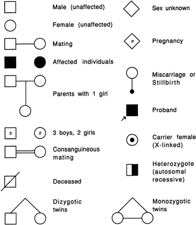
The observation that “like begets like” has been stated throughout recorded history, but current theories describing how genetic traits and illnesses are inherited are just over 100 years old. In the late 1800s, Mendel described how individual genetic traits were passed on from generation to generation. Single gene disorders (i.e., mendelian disorders) are conditions caused by a mutation at a single site in the DNA and inherited in the proportions predicted by Mendel's laws. These disorders can be dominant conditions in which the phenotype is expressed even when only one chromosome of a pair has a defect, or recessive conditions which are expressed only if the defect exists on both chromosomes. Classically, these different modes of inheritance are revealed by pedigree analysis (Fig. 6.1 and Fig. 6.2).
|
|
|
FIG. 6.1. Symbols used to draw a pedigree. |
|
|
|
FIG. 6.2. Patterns of inheritance. |
As we learn more about the tremendous variation that occurs at every locus, the distinctions between dominant and recessive conditions have become blurred. Dominance and recessiveness are attributes of the phenotype, not attributes of the gene or allele. They are empirical terms, and they depend on the sensitivity of the method used to describe the phenotype. The researchers must specify particular phenotypic features when describing inheritance. For instance, sickle cell anemia is a recessive disease only if the full-blown disease is considered, but it is a codominant condition if the hemoglobin is being analyzed by electrophoresis. The ABO blood group is an example of a trait in which both codominant and recessive inheritance is seen. The retinoblastoma gene is a recessive tumor suppressor gene at the cellular level, but abnormalities in the gene are responsible for the autosomal dominant tendency to develop retinoblastomas and osteosarcomas.
In autosomal dominant conditions, the disease is expressed in persons who are heterozygous for the disease-causing mutation. McKusick's catalog of mendelian disorders describes more than 3,000 dominant conditions. Marfan syndrome, myotonic dystrophy, neurofibromatosis, achondroplasia, and Huntington disease are examples of autosomal dominant disorders. The probability of an affected person transmitting the abnormal gene to the children is 50% with each pregnancy. Typically, autosomal dominant conditions have less than 100% penetrance, and fewer than 50% of the offspring show signs of the disorder. Male and female offspring usually are affected with equal frequency and severity. The trait passes through one parental line only, and father-to-son transmission can occur. For highly penetrant, autosomal dominant conditions, the gene is expressed in each generation (i.e., vertical transmission). New mutations are relatively common and, on average, paternal age is advanced when isolated, sporadic, or new mutation cases appear. Autosomal dominant phenotypes often involve isolated or multiple structural defects. They can be extremely variable and the onset of clinical features is often age dependent. Dominant disorders tend to be less severe than recessive diseases, but they are usually lethal in the rare persons who are homozygous for a dominant disease.
Autosomal recessive conditions are expressed only in persons in whom both versions (i.e., alleles) of the involved gene are abnormal. More than 1,500 autosomal recessive conditions have been described. Cystic fibrosis (CF), sickle cell anemia, Tay-Sachs disease, and phenylketonuria are examples of autosomal recessive disorders. Male and female offspring are affected with equal frequency and severity. Each parent is a heterozygous carrier, and abnormal genes are inherited from both parents. Each offspring of two carrier parents has a 25% chance of being affected, a 50% chance of being a carrier, and a 25% chance of being neither a carrier nor affected.
If the recessive phenotype is extremely rare, consanguinity usually is found in the pedigree. Affected persons rarely have affected children; autosomal recessive inheritance shows a “horizontal” pattern in a pedigree, with typically only a single generation of siblings affected. Affected persons who mate with unaffected persons who are not carriers have only unaffected, carrier offspring. Most autosomal recessive phenotypes are biochemical or enzymatic in nature, and they tend to be less variable and more severe than dominant conditions.
X-linked inheritance occurs when a trait is carried on the X chromosome. Boys are hemizygous for X chromosome genes, but girls can be homozygous or heterozygous. Of the 300 X-linked recessive diseases that are recognized, the hemophilias and Duchenne muscular dystrophy are the best known. Characteristics of X-linked recessive inheritance include a higher incidence of the disorder in male than in female offspring. The mutant gene or disease never is transmitted directly from father to son, and all the daughters of an affected man are carriers. The trait is transmitted through carrier females, and affected males in the same kindred are related to one another through the females. X-linked dominant diseases are much rarer; examples include Alport syndrome, vitamin D-resistant rickets, and incontinentia pigmenti. They appear twice as often in female as in male offspring. All daughters of an affected man have the disorder, but no sons are affected. Heterozygous affected women transmit the mutant allele at a rate of 50% to progeny of both sexes. If the affected woman is homozygous, all of her children will be affected.
Y-linked or holandric inheritance occurs when a trait is carried on the Y chromosome. Only male offspring are affected, and there is only male-to-male transmission. No known disease genes are inherited in this fashion, but genes for gender determination, tooth size, and height occur on the Y chromosome.
Mitochondrial inheritance is a more recently described mode of inheritance, causing traits and disorders that are inherited through the mitochondrial chromosome. Unique patterns are seen in affected families. Mitochondria are inherited exclusively with the cytoplasm of the egg; a woman who carries a disease will pass the disease to 100% of her offspring. Male carriers will pass the disorder to none of their offspring. Leber optic atrophy and certain rare myopathies are inherited in this fashion.
Polygenic, Multifactorial Disorders
Multifactorial or polygenic inheritance is the most common form of inheritance. Even in the classic mendelian disorders described above, there can be tremendous quantitative and qualitative differences in the phenotype among persons who have the same allele or the same genetic mutation. This variability can be evident as nonpenetrance of certain features (or the entire phenotype) and as differences in the severity of features, the frequency of cyclic or episodic events, or the age of onset of the first clinical sign of the disorder. Genetic variability can be caused by the underlying genetic background of the affected person, including gender influences and limitations. The phenotype may be influenced further by maternal factors such as cytoplasmic inheritance, the intrauterine environment, or imprinting. X-linked disorders can be altered by variations in X inactivation or lionization. Each genotype undergoes subtle changes through somatic mutation, gene amplification, or transpositions and positional effects over time. Exogenous factors such as the environment, teratogens, medical intervention, and chance also influence variability.
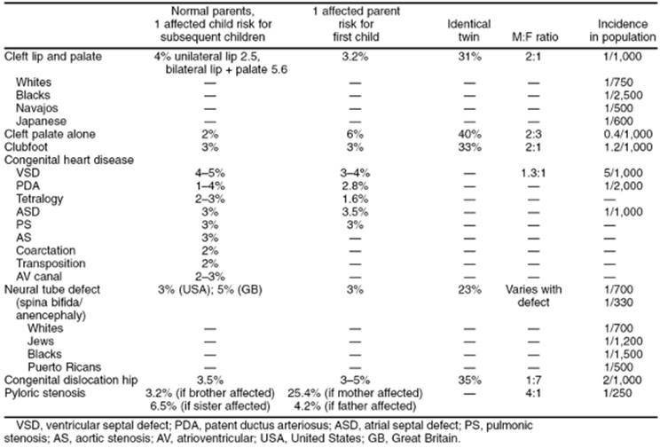
Most congenital anomalies reflect multifactorial inheritance (Table 6.2). A common error some obstetricians make is to counsel a patient that rare conditions will not occur repetitively in her family. If the birth defect in question has a strong genetic component or if there is an identifiable environmental or teratogenic component which would recur in subsequent pregnancy, the risks may remain high for that patient (Table 6.3). The rates may be even higher if a mendelian or chromosomal condition has gone unrecognized in the affected child. Before counseling patients about the recurrence risk of any birth defect, it is pertinent to review which syndromes are associated with that birth defect and ask whether any member of the family has those syndromes. When this requires skill or knowledge beyond the usual expertise of an obstetrician-gynecologist, referral to a medical geneticist is appropriate.
|
|
|
TABLE 6.2. Types of birth defects |
|
|
|
TABLE 6.3. Empiric recurrence risks for common congenital anomalies |
Multifactorial inheritance usually works according to a threshold model. Several factors must collaborate to cause a bodily function to go awry, and only after these factors reach some critical point is the phenotypic effect seen. Many different factors can affect the observed recurrence risk. The intrinsic heritability “or geneticness” of the condition is frequently the most important factor. This usually is determined by examining whether monozygotic twins are concordant for a particular condition relative to dizygotic twins. For instance, neonatal seizures show a very high heritability rate, with 85% to 90% concordance in monozygotic twins verses 10% to 15% concordance in dizygotic twins. The population incidence of the condition is another important variable. The recurrence risk is higher for common disorders or within populations with a high incidence of the disorder. In disorders with a relatively high heritability, the recurrence risk of the disorder approximates the square root of the population incidence. There can be marked variation in the population frequency of different disorders and different ethnic groups. For instance, cleft lip occurs commonly in Native Americans, but African Americans have a nine-fold lower incidence of cleft lip than the general population.
If the incidence of a congenital anomaly shows a sex bias, the recurrence risk is higher in the offspring (and other relatives) if the parent is of the less frequently affected sex. For example, pyloric stenosis affects 5 times as many male as female offspring, and empirical data show that there is a 25% chance of producing an affected child if the mother had pyloric stenosis at birth and only a 4% risk if the father is the affected parent. Similarly, the recurrence risk is higher when the gender of the affected child is the less frequently affected. Again, the risk of having another child with pyloric stenosis is 3.2% if the first-affected sibling is a boy and 6.5% if the affected sibling is a girl. Hirschsprung disease, clubfoot, and cleft lip are examples of anomalies which are more common in male infants. Cleft palate, anencephaly, hip dysplasia, and scoliosis are more common in female infants.
The number of affected individuals in a kindred can affect the recurrence risk. The greater the number of family members who have already been affected with a multifactorial condition, the more likely it is that the genetic background is favorable for expression of this condition. After a couple has one affected child with cleft lip and palate, there is a 4% empirical recurrence risk. After two affected children, the risk rises to 10%. Consanguinity also increases the risk of recurrence because of the greater likelihood of deleterious genes being shared, but a more distant relationship from an affected person decreases the risk of recurrence.
The severity of the disorder often predicts the recurrence risk. An illustration of this is found in Hirschsprung disease. The recurrence risk is proportional to the length of the aganglionic segment of the colon. Neural tube defects are the most notable exception to this rule, because the recurrence risk for any neural tube defect appears to be the same whether the first-affected child had anencephaly or a small spina bifida lesion.
Cytogenetic Disorders
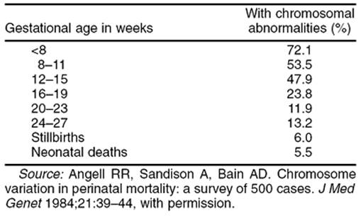
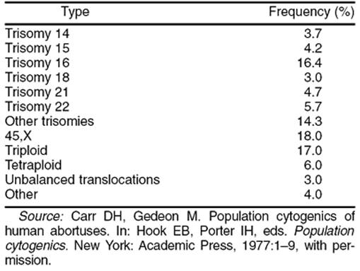
Cytogenetic disorders are changes in the genome visible under a light microscope. These gross lesions involve the loss or duplication of a large number of genes; multiple malformations and dysfunctions usually are observed clinically. Diagnostic clues for a cytogenetic disorder can range from subtle dysmorphic features to major structural malformations, particularly craniofacial, skeletal, cardiac, and genitourinary malformations. No individual anomaly is pathognomonic for a particular chromosomal syndrome, rather it is the pattern that can be distinctive. There is tremendous overlap between patterns and because nonchromosomal syndromes can mimic chromosomal abnormalities, obtaining a karyotype is always necessary to confirm the diagnosis. Cytogenetic disorders usually are associated with some degree of mental retardation and growth deficiency. Most have an increased rate of perinatal loss and premature mortality of live-born neonates. The rate of chromosome abnormalities is at least 40% to 60% in first-trimester abortuses, and the rates of abnormalities also are elevated in fetal deaths and preterm and post-term deliveries (Table 6.4 and Table 6.5). About 1 in 160 babies is born with a genetic defect detectable by ordinary cytogenetic means (Table 6.6).
|
|
|
TABLE 6.4. Incidence of chromosomal aberrations in pregnancy losses at various gestational ages |
|
|
|
TABLE 6.5. Types of chromosomal abnormalities in spontaneous abortuses |
|
|
|
TABLE 6.6. Incidence of chromosomal aberrations seen in newborn surveys |
Cytogenetic studies have been used clinically for approximately 40 years. In the late 1950s, it was determined that humans have 46 chromosomes and that many of the recognized birth defect syndromes, such as Down syndrome, Turner syndrome, and Klinefelter syndrome, have abnormalities of chromosome number or structure. Normally, the nucleus of most human cells contains two sets of chromosomes, with one set contributed by each parent. Each set has 22 autosomes and either an X or a Y sex chromosome (Fig. 6.3).
|
|
|
FIG. 6.3. Normal human karyotype (46,XY). |
Metaphase chromosome preparations can be prepared from any cell undergoing mitosis. Typically, in order to obtain adequate numbers of cells, mitosis is induced artificially using a mitogenic chemical such as phytohemagglutinin. The cells are then incubated in a dilute solution of an agent that poisons the mitotic spindle. The chromosomes are swollen using a hypotonic salt solution, fixed on a slide, and dried for staining. The stained chromosomes can be observed by light microscopy.
The dyes used to stain chromosome preparations reveal patterns of light and dark bands that reflect regional variations in the molecular composition of each chromosome. Giemsa is the most commonly used dye. Q-banding is a fluorescence technique that gives results similar to the G-banding (i.e., Giemsa banding), while R-banding (i.e., reverse banding) gives a pattern opposite to that with G- or Q-banding. T-banding specifically stains the telemetric regions of chromosomes, which can help to screen for missing regions at the ends of the chromosomes, and C-banding primarily stains the centromeric region of the chromosomes.
Differences in the size of the chromosomes, the banding pattern, and the centromere position allow the 24 chromosomes to be differentiated from each other in an analysis called a karyotype. The most common features looked for on a karyotype include aneuploidy (i.e., abnormal number of chromosomes) or structural chromosome abnormalities such as deletions, inversions, insertions, or translocations (Table 6.7) (Fig. 6.4).
|
|
|
TABLE 6.7. Chromosomal nomenclature |
|
|
|
FIG. 6.4. Types of chromosomal abnormalities. A, terminal deletion; B, interstitial deletion; C, paracentric inversion; D, pericentric inversion; E, translocation (additional material from a different chromosome); F, isochromosome; G, dicentric chromosome; H, ring chromosome. The arrows indicate the sites of chromosomal breaks where rearrangement occurs. |
Because the technology to study cytogenetic disorders is well established, there is greater clinical experience with this than with other types of genetic testing. There are several well-defined indications for obtaining a fetal karyotype analysis (Table 6.8). Pregnant women who are 35 years or older routinely are offered a fetal karyotype analysis, because trisomy tends to occur more commonly with advancing maternal age (Table 6.9). Other indications for obtaining a fetal karyotype include having a previous child with an abnormal karyotype, parental chromosomal rearrangements, unexplained intrauterine growth retardation, and an abnormally low level of maternal serum α-fetoprotein. In addition, many fetal anomalies associated with karyotypic abnormalities can now be visualized using high-resolution ultrasonography (Table 6.10). A fetal structural abnormality detected by ultrasonography is another frequent indication for obtaining a fetal karyotype.
|
|
|
TABLE 6.8. Indications for a fetal karyotype |
|
|
|
TABLE 6.9. The risk of karyotypic abnormalities related to maternal age at delivery |
|
|
|
TABLE 6.10. Risk of a chromosomal abnormality with selected sonographic findings |
A variety of genetic defects, including the common trisomies and many chromosomal translocations, can be detected by routine karyotype analysis. Recent modifications, including chromosome painting that allows a particular chromosome to be identified directly, or fluorescent in situ hybridization (FISH) that allows specific sites along the chromosome to be identified, have greatly extended the capabilities of the cytogenetics laboratory. However, there are molecular and single gene rearrangements that cannot be observed by light microscopy and require molecular genetic technology for evaluation.
FlSH is a cytogenetic technique in which a specific DNA probe with a fluorescent label is bound to homologous DNA in a clinical sample. FISH can be performed either on a metaphase chromosome spread to detect microdeletions and microduplications, and during interphase to detect a larger chromosomal region in a nondividing cell. Interphase FISH can be performed on cultured cells, tissue sections, and on cytologic smears. FISH has been used to detect common aneuploidies, such as trisomy 21, trisomy 18, trisomy 13, and the sex chromosome aneuploidies, in prenatal diagnosis.
Because uncultured amniocytes can be used, the FISH technique can offer more rapid detection of chromosome aneuploidies. In one of the first large studies, FISH was performed as an adjunct to conventional cytogenetics in 4,500 patients. Region-specific DNA probes to chromosomes 13, 18, 21,X, and Y were used to determine ploidy by analysis of signal number in hybridized nuclei. A sample was considered to be euploid when all autosomal probes generated two hybridization signals and when a normal sex chromosome pattern was observed in greater than or equal to 80% of hybridized nuclei. A sample was considered to be aneuploid when 70% or more of hybridized nuclei displayed the same abnormal hybridization pattern for a specific probe. The accuracy of all informativeFISH results, euploid and aneuploid, was 99.8%, and the specificity was 99.9%.
Current prenatal FISH protocols are not designed to detect all chromosome abnormalities and should be used only as an adjunctive test to cytogenetics. FISH can provide rapid and accurate clinical information in pregnancies when fetal abnormalities have been observed by ultrasonography.
Future improvements are likely. FISH protocols, which would allow the simultaneous and unequivocal discernment of all human chromosomes, are under development. Each chromosome is labeled in a unique way using several different colors. A “spectral karyotype” can be generated, which allows visualization of a unique, defined emission spectra for each human chromosome. Computerized analysis may allow automatic and rapid analysis, with resolution approaching routine G-banding.
Parent of Origin Effects
Genomic imprinting refers to the differential expression of genes based on the parent of origin of the gene. Imprinting usually is mediated by differential methylation of the alleles involved. Most experimental evidence regarding imprinting comes from animal studies, but some naturally occurring human analogs exist. The paternal genetic contribution appears to be essential for the development and function of the placenta and extraembryonic tissues, but the maternal contribution is required for embryonic development. Ovarian teratoma, the most common benign pelvic tumor in women of reproductive age, is characterized by a diploid karyotype, in which both haploid sets of chromosomes are maternal in origin. Complete hydatidiform moles, which show failure of normal embryonic and fetal development, are usually diploid with two paternal haploid chromosome sets and no maternally derived chromosomes.
The differential function of parental chromosomal contributions in development is also evident when studying human triploidy. In an android conception (i.e., two paternal, one maternal chromosome set), the fetus is severely growth retarded, with a disproportionately large head and syndactyly of digits of the hand. The placenta is usually very large and hydropic. Survival into the second trimester or occasionally into the third trimester is possible but usually requires the presence of mosaicism with a diploid cell line. When the chromosomal constitution is gynoid (i.e., two maternal, one paternal set), the conceptus is underdeveloped and the placenta is small and cystic; such pregnancies rarely continue beyond the first trimester.
In some persons with an apparently normal karyotype, both versions of a pair of homologous chromosomes were inherited from one parent, a phenomenon called uniparental disomy. This can give rise to abnormalities if genomic imprinting causes regions of both chromosomes to be inactivated or overexpressed.
THE MOLECULAR GENETICS REVOLUTION
In the mid 1950s, there were only two “facts” known about the human genome. It was thought that humans had 48 chromosomes and that X-chromosome inactivation in humans occurred by the same mechanism as had been observed in fruit flies. Both of these observations have been proven to be in error. In the past few decades, there has been an explosion of knowledge about the human genome, largely attributed to advances in molecular biology.
Deoxyribonucleic Acid
Genes are the instructions required for building structural proteins and enzymes and peptide hormones, and the complete set of genetic instructions for any organism is called its genome (Table 6.11). The human genome has 46 chromosomes, including 22 pairs of autosomes and two sex chromosomes. The genome is made up of three billion base pairs, somewhere between 40,000 and 100,000 genes. The functions of approximately 10,000 human genes have been characterized, and in 2003 the first draft of the human genome sequence will be completed.
|
|
|
TABLE 6.11. The human genome |
In 1944, Avery and colleagues demonstrated that DNA is the chemical that carries genetic instructions. Roughly equal parts of DNA and its supporting proteins make up the 46 chromosomes. If the strands of DNA in the nucleus of a single cell could be unwound and spliced together, the resulting DNA molecule would stretch more than 1.5 meters long, but it would be only 20 trillionths of a centimeter wide.
The genetic code is spelled out with the four nitrogenous bases: adenine, thiamine, cytosine, and guanine (Fig. 6.5). The purine and pyrimidine bases are arranged in a ladderlike, double helix arrangement that is very stable (i.e., theoretic dissociation constant = 10 - 23). During cell division, DNA is duplicated with extremely high fidelity by synthesis of a new strand of one side of the molecular ladder.
|
|
|
FIG. 6.5. Genetic code. The DNA code consists of four characters and is read three characters at a time. It is translated into an RNA message, which instructs cells in how to assemble proteins from amino acid building blocks. |
The human genome consists of at least 40,000 genes, but the genes comprise only one tenth of the encoded information. Most of the genome is of unknown function, but it probably codes for the proper spacing, alignment, and punctuation of the genetic instructions. About 99.8% of the DNA sequence is identical from one person to the next. Stated another way, there are many minor differences between any two persons; on average, there is a variation of one nucleotide for every 200 to 500 base pairs. When these sequence differences occur within genes, they can lead to genetic diseases or genetic variation. Most of the minor differences have no observable effect because they occur in the noncoding regions of the genome, regions of DNA that do not contain genes. These otherwise unimportant differences have been the basis of the current explosion of genetic knowledge, because much of our ability to study genes or diagnose genetic illness exploits differences (i.e., DNA sequence polymorphisms) in these regions to track or find neighboring genes.
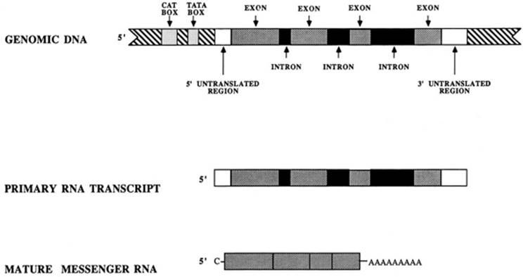
The DNA sequence is read by cellular enzymes three bases at a time, and each triplet directs the positioning of a particular amino acid within the structure of a protein (see Fig. 6.5). The protein coding instructions are transmitted to the cellular machinery through messenger RNA, a transient, intermediary molecule that is similar to a single strand of DNA (Fig. 6.6). The RNA strand is transcribed from the DNA template in the nucleus and has an opposite or complementary genetic sequence. Messenger RNA moves from the nucleus into the cytoplasm, where the protein manufacturing organelles build a protein. Analysis of messenger RNA molecules is extremely useful in the laboratory for detecting genes.
|
|
|
FIG. 6.6. Anatomy of a gene. Regulatory regions are present in the 5′ region. Introns are spliced out of the final messenger RNA. |
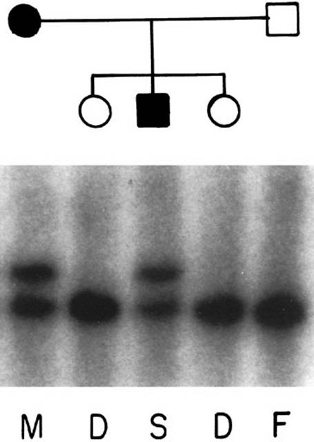
Several advances in molecular biology have enabled the molecular genetics revolution to take place. The first was the discovery of restriction enzymes, which are bacterial proteins that can cut DNA molecules at specific sites by recognizing the DNA sequence at those sites. Over 400 restriction enzymes have been discovered, many are commercially available, and about 25 are used commonly. Restriction fragment length polymorphisms (RFLPs) occur because of minor sequence changes (usually single base substitutions) that abolish or create a recognition site, altering the length of a digestion fragment. Restriction sites occur frequently, and several restriction sites can occur in the vicinity of any given gene. When these RFLPs are polymorphic, they become useful markers for linkage studies, diagnostic testing, and paternity testing (Fig. 6.7). RFLPs and other DNA polymorphisms provide the landmarks for genetic maps.
|
|
|
FIG. 6.7. Linkage study using restriction fragment length polymorphisms. Each lane represents the genotype of one family member. M, mother; F, father; D, daughter; S, son. In this example, the disease allele is associated with the upper band passed from the mother to the son. |
Scientists have gained a greater understanding of how to manipulate the physical conditions, such as pH, salt concentration, and temperature, of in vitro DNA reactions. These skills—combined with the use of restrictions enzymes—allowed the development of recombinant DNA or new combinations of DNA engineered in the laboratory. Recombinant DNA technology has made possible the development of gene probes (pieces of DNA usually radioactively labeled) that recognize and bind specifically to a homologous sequence in another sample of DNA. These technologies also underlie cloning—the copying of DNA segments in lower animals and the manufacturing of human proteins using bacteria or cell cultures.
Various blotting technologies are used commonly to study DNA. With blotting, biologically relevant molecules undergo electrophoresis and are transferred to a stable membrane for repeated experiments. Blots are called Southern blots if DNA is being analyzed, Northern blots if RNA is being analyzed, and Western blots if proteins are being analyzed.
DNA testing is clinically applicable to many disorders and can be performed in one of several ways (Table 6.12). When the molecular basis of a disease is known, direct mutation testing can provide a yes or no answer on any DNA sample. For instance, in CF, hundreds of mutations have been discovered. A battery of mutations can be tested for using various methods such as dot blots, which are simple to interpret (Fig. 6.8).
|
|
|
TABLE 6.12. Common conditions for which DNA testing is available |
|
|
|
FIG. 6.8. Direct mutation diagram. Direct detection of cystic fibrosis mutations using reverse dot blots. In this example, five mutations in exon 11 of the cystic fibrosis gene (G542X, S549N, G551D, R553X, R560T), are tested for using a simple YES/NO assay. Exon 11 is amplified using the polymerase chain reaction. The product of the reaction is labeled to allow its detection and is placed on a membrane. The membrane has been prepared with oligonucleotide probes, which detect either the normal or the abnormal sequence. A: results from a known cystic fibrosis carrier. B: results from a child with cystic fibrosis. |
Similarly, fragile X syndrome is usually the result of an expansion of a triplet sequence within the gene. Normal persons usually have only 5 to 50 copies of this triplet repeat, but affected patients have hundreds or thousands of copies of the triplet repeat. Similar triplet expansions cause myotonic dystrophy, Huntington disease, and Kennedy disease. The region containing the triplet can be amplified using the PCR, which produces millions of copies of the small region of DNA from the X chromosome that contains the fragile X repeat. Specificity is achieved by directing the reaction using two complementary primers on either side of the region of interest. Once amplified, the size of the product can be measured to evaluate the number of triplets, determining whether the mutation exists (Fig. 6.9).
|
|
|
FIG. 6.9. Fragile X mutation detection. In patient A, a shorter polymerase chain reaction product corresponds with a smaller number of triplet repeats. Patient B exhibits an expanded number of repeats. Affected patients typically have hundreds or even thousands of copies of the triplet. |
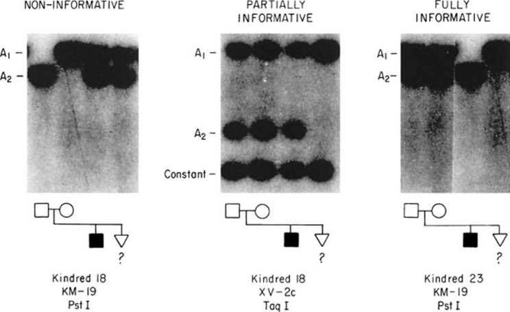
For families with unusual mutations or with diseases for which the molecular basis is unknown, linkage testing can be performed. Linkage tests compare DNA polymorphisms close to the disease-causing gene in family members known to have or carry the disease with those of unaffected and at-risk family members. Indirect assessments can be made about whether at-risk persons have the disease allele. The accuracy of these predictions depends on correct diagnosis and relationships of the family members, and the genetic distance between the polymorphism tested and the disease allele. For some families, linkage testing can be uninformative (Fig. 6.10).
|
|
|
FIG. 6.10. Informativeness of linkage testing for cystic fibrosis. Marker KM-19 in kindred 18 is “not informative”-the disease alleles can not be distinguished in the parents. |
The Genome Project
The Human Genome Project promises to be the single most important project in biology; genetics and genomics are now the central sciences of medicine. An understanding of the relationship between genetic variation and disease risk will alter the future prevention and treatment of common illnesses.
The full human sequence will be completed in 2003, but a very accurate draft is available now for over 95% of the genome. Disease gene identifications that formerly required years of chromosome walking and jumping, cloning, physical mapping, sequencing, and sequence assembly can now be completed in weeks. Industrialized sequencing technologies using capillary electrophoresis, micro arrays, and others developed for the genome project are now widely used for genotyping and sequencing.
We also now have a tremendous catalog of individual sequence variation in humans. Tens of thousands of micro-satellite markers are available for linkage analysis and hundreds of thousands of SNPs (single nucleotide polymorphisms) for genetic association studies. The tools are now well developed for doing these functional studies to find which variations and mutations cause individuals to be at risk for numerous medically important, genetically complex human diseases. The genome project has delivered improved cDNA resources, better predictive software, and additional knowledge about the non-protein coding regions of the genome. Remarkable technologies are commercially available for comprehensive analysis of gene expression in single cells, tissues, or whole organisms.
Advances in gene knockout technology, antisense technology, gene transfer, and gene transfection allow greater in vitro insights using appropriate model systems, including both cell culture and whole organisms. The complete sequence of the Escherichia coli, yeast, nematode, fruit fly, and mouse genomes provide important evolutionary clues to gene function and extend the range of experiments possible.
At the same time there have been parallel improvements in the technology for global protein analysis. Gene expression is played out at the protein level—elegant techniques are now available to examine spatial and temporal patterns of protein expression, protein-ligand interactions, and protein modifications.
Finally, the genome project occurred at the same time as the information technology revolution. Tremendous bioinformatics and computational software is now available for gene discovery, expression profiling, understanding gene-environment interactions, and so on. Suffice it to say, better tools are now available for making advances in women's health care than ever before in human history.
At least 3% of the annual budget of the project is going to the Ethical, Legal, and Social Issues section of the enterprise. This amount of early attention to societal impact is unprecedented for a science and technology project. Grant-funded programs have examined privacy issues, genetic discrimination in insurance and employment, and the role of coercion. Genetic discoveries may challenge long-held beliefs about equality, predetermination, and free will as we learn about genes that have a major role in personality, creativity, intelligence, and mental illness. The safety, efficacy, and utility of new gene tests should be evaluated, especially before treatment is available.
GENETIC EVALUATION
Genetic History and Physical Examination
Important details are being learned about many rare disorders which practitioners may see only once in their careers. In the past, many practitioners have had a “laissez-faire” attitude about genetic disorders, because “you can't do anything about your genes.” However, it is important to detect genetic conditions so that the patient can have adequate counseling about the condition and the risk to offspring. For severe conditions, patients often are interested in prenatal diagnosis, so they can consider pregnancy termination or prepare for the birth of an affected child. Our new predictive powers have led to an expanded medicolegal duty to warn patients of risks of which they may not have been aware. Failure to provide accurate and timely reproductive counseling has resulted in a host of lawsuits. Genetic diagnosis becomes even more critical as more treatment options become available for the child with a severe genetic disease.
In light of these expanding obligations to screen, what is expected of the general obstetrician-gynecologist? As with any medical diagnosis, the history is the most important part of the genetic evaluation. Important aspects of a genetic history, such as the patient's age, menstrual history, and obstetric history, are queried routinely as part of any routine obstetric or gynecologic history. In addition, it is important to ask about the patient and her partner's ethnicity (Table 6.13). The family history should extend to third-degree relatives (i.e., cousins). A minimal familial history can usually be elicited using the following questions:
|
|
|
TABLE 6.13. Single gene disorders with an ethnic predilection |
· Do you have a family history of diabetes, hypertension, cancer, or twins?
· Are there any diseases that seem to run in your family?
· Is there a history of genetic disease like cystic fibrosis, hemophilia, or muscular dystrophy?
· Is there anyone with mental retardation or any kind of birth defect?
· Have any of your sisters, cousins, or other relatives had problems with their pregnancies?
· Are your parents alive? Are they healthy?
· As far as you know, are you and your husband or partner related by blood?
· Do you know your ethnic background? (Do you know where your relatives are originally from?)
· Is there any reason you are especially concerned that you might have trouble with your pregnancy or that your baby may be born with a birth defect or other medical problem?
It takes only a few minutes to ask these questions of a new patient. The final, open-ended question is often the most revealing. Alternatively, many practitioners find it helpful to ask similar questions using a patient-completed questionnaire. I use a form similar to the one suggested by the American College of Obstetricians and Gynecologists (ACOG) (Fig. 6.11). It is designed so that only “yes” responses need to be dealt with further. Including a form such as this one in the patient's chart clearly documents that a genetic history was obtained for medicolegal purposes. More than one fifth of healthy obstetric patients affirmatively answer at least one important question on the form.
|
|
|
FIG. 6.11. Genetic screening questionnaires. |
The physician should suspect genetic factors if a patient has an unusual problem and other people in her family have the same disorder. Similarly, if a patient reports a positive family history, it is important to decide whether the patient unknowingly has the same disease. When a patient reports an unfamiliar genetic condition or a rare illness, the physician should seek information about the genetics of this condition. Is she at risk of passing the condition to her offspring? Is prenatal diagnosis available? Is prenatal treatment available? For instance, consider a patient whose first child died of methylmalonic aciduria. In a subsequent pregnancy, her obstetrician noted this history on his prenatal record but did not inform the patient that there was a one-in-four risk that her current fetus could be affected. The obstetrician was unaware that prenatal diagnosis is available and, more importantly, that a simple treatment (i.e., giving the mother supplemental vitamin B12) could prevent much of the morbidity of this particular form of methylmalonic aciduria. The child was born severely damaged and died at 2 years of age. The patient was very angry that she was not informed about prenatal therapy, and a lawsuit was initiated. Obstetricians cannot be expected to be expert in every rare enzymopathy, but this case illustrates how important it is to seek out additional information about rare conditions through a literature search or by consultation with a genetics center. Fortunately, helpful computerized databases are available on the internet such as OMIM (Online Mendelian Inheritance in Man) and Gene Tests (a directory of DNA diagnostic laboratories).
The general physical examination may reveal dysmorphic features or a distinctive physical finding that frequently is associated with genetic problems. For instance, a dislocated lens found on funduscopic examination of the eye is a distinctive feature consistent with Marfan syndrome or homocystinuria. If the patient has no history of severe ocular trauma, she probably has one of these conditions. Obstetricians cannot be as good at detecting such clues as an expert medical geneticist, because “the eye cannot see what the mind does not know.”
Genetic Counseling
Genetic counseling is a communication process which deals with the occurrence or risk of occurrence of a genetic disorder in a family. As our abilities to learn about the fetus have increased, more couples have an indication for prenatal diagnosis or a need to discuss reproductive options. Although every obstetrician has a role in providing genetic counseling, many practitioners find that genetic counselors—persons with advanced degrees and who are specially trained in the educational, psychological, and administrative aspects of medical genetics—are helpful consultants. Genetic counselors are experienced in obtaining and interpreting a thorough family history; often counselors are involved in the establishment or confirmation of a diagnosis. When presented with a prenatal diagnosis, they can obtain and interpret the history of a current pregnancy, explaining fetal risks and discussing the options available. Genetic counselors can provide the detailed counseling that is necessary regarding fetal chromosomal abnormalities of consanguinity, recurrence risks of multifactorial disorders, fetal abnormalities identified by ultrasonography, or infertility and habitual abortion. They are trained extensively about genetic screening for diseases that are common in various ethnic groups. Genetic counselors play a central role in the discussions regarding the option of aborting a genetically abnormal fetus. This type of counseling is traditionally informational and nondirective.
Pregnancy Termination
Pregnancy termination for genetic reasons can be particularly heart wrenching for a couple because the pregnancy usually is a desired pregnancy. Patients should be encouraged to involve their doctors, genetic counselors, clergy, other support persons, and family in these difficult decisions.
It is the physician's responsibility to explain the fetal diagnosis and prognosis. If a woman decides to have a pregnancy termination, the physician should explain the termination procedure, options if there are any, and the relative risks of the different procedures. The cost of the procedure is discussed and whether the procedure is covered by public funding or insurance. The physician should explain the benefits of diagnostic examination of the fetus by DNA, metabolic, or chromosomal analysis or by dysmorphologic examination. The disposition of the fetal remains should be discussed. The possibility that a fetus may live for a short period after induced labor termination is discussed. With late second-trimester or third-trimester inductions, it is often appropriate to encourage patients to see or hold the baby, and to name their baby. Patients are advised that lactation may occur after the delivery, and they are told about the options available to reduce lactation. With late terminations, the option of having a memorial service or in some way commemorating the baby's existence should be discussed. Physicians and counselors help couples decide what information to tell other children and family members, friends, and acquaintances.
It is important to reinforce that the genetic defect is not caused by the patient. The woman who is carrying the pregnancy and undergoes the termination may grieve in different ways than may the father of the baby. Referral to local support groups and counselors is often appreciated. Six to eight weeks after the procedure, a follow-up visit should be scheduled to summarize the diagnostic findings, review recurrence risks, and discuss prenatal diagnosis or therapy options for future pregnancies.
Laboratory Screening
Laboratory studies play an important role in the diagnosis of genetic disorders. A genetic illness is sometimes first discovered as an incidental finding on blood studies or an ultrasonographic examination. For instance, a low mean corpuscular volume on an automated complete blood count suggests thalassemia. In some instances, a positive family history prompts laboratory studies that clarify a patient's risk. Some programs have evolved to screen entire populations for genetic conditions using laboratory assays. Just as we currently perform a history and physical examination or a cholesterol screen to identify disease risk, soon there will be a DNA screen to detect mutations in dozens of important genes involved in cancer, cardiovascular disorders, and metabolic disease.
Population screening is appropriate when a defined subset of the population is at risk, and an accurate and inexpensive heterozygote test is available (Table 6.14). It is optimal if prenatal diagnosis is available, as well (e.g., sickle cell anemia, Tay–Sachs disease, thalassemia). The goals of screening programs are early diagnosis to allow better treatment of affected persons and identification of at-risk matings between persons who are heterozygotes or carriers of recessive disease. Neonatal screening programs for phenylketonuria, galactosemia, and hypothyroidism are carried out in most states. Successful carrier screening for Tay–Sachs disease has been achieved in several Jewish populations. The cost effectiveness of the screening program is often a primary concern in deciding whether to proceed with population screening. Equally important issues include the ability to manage minor variants which do not require action, stigmatization of carriers, and responsibility for decisions not to screen.
|
|
|
TABLE 6.14. Population screening |
PRENATAL DIAGNOSIS
Limited but important information about the fetus can be gained using the traditional diagnostic techniques of history, auscultation, and palpation. It is important to consider the onset of fetal movement and the assumption of the vertex position as developmental milestones that the fetus does or does not achieve. Experienced examiners can assess fetal size, size or dates discordance, fetal positioning, and fetal heart rate abnormalities.
Maternal Serum Screening
Screening for fetal genetic conditions can be achieved by testing maternal serum. The first such program involved the use of maternal serum α-fetoprotein (MSAFP) levels to test for neural tube defects, an etiologically heterogenous group of conditions characterized by failure of embryonic closure of the neural tube. A cause for a neural tube defect can be identified in only 5% to 20% of cases, and most cases are thought to be polygenic or multifactorial. Between 90% and 95% of all infants with neural tube defects are born to women with no history of a child with neural tube defect.
There is substantial evidence for genetic predispositions to neural tube defects, including racial and ethnic variations in incidence, the increased incidence when a couple is consanguineous, gender bias, and increased monozygotic twin concordance. There is also strong evidence for environmental factors including maternal folate deficiency, previous spontaneous abortion or stillbirth, and the seasonal incidence. Mendelian disorders associated with neural tube defects include Meckel syndrome, in which affected individuals have a posterior encephalocele. Chromosomal syndromes, such as trisomy 18, trisomy 13, and triploidy, and sporadic syndromes such as OEIS complex (i.e., omphalocele, extrophy, imperforate anus, spinal defect) can result in neural tube defects. Other well-described environmental causes of neural tube defects include amniotic band disruption sequence, maternal diabetes, maternal use of valproic acid, and hyperthermia. Forty-five percent of fetuses with neural tube defects have anencephaly, 45% have spina bifida, 5% have an encephalocele, and the remaining 5% have iniencephaly or exencephaly.
The incidence of neural tube defects is high (approximately 1%) in Ireland, Wales, Alexandria, and the Punjab. The rate is between 1 in 1,000 and 2,000 in the United States. Across the United States, the incidence is higher in the east than in the west, and highest in the Appalachian region. In the United States, if a person has previously had one child with neural tube defect, the recurrence risk is 2% to 3%. If there have been two affected children, it is 6.4%, and with three affected children, it may be as high as 25%. For patients at high risk for neural tube defect, prenatal diagnosis can be performed by targeted ultrasonography and an amniocentesis for amniotic fluid α-fetoprotein and acetylcholinesterase at approximately 16 weeks of gestation. The peak concentration of α-fetoprotein in the amniotic fluid occurs between 12 and 14 weeks, the widest margin between abnormal and normal distributions at approximately 16 to 18 weeks. A cutoff of 2.5 multiples of the median again yields a 98% detection rate, with a 0.8% false-positive rate. Acetylcholinesterase level determinations in amniotic fluid do not depend on gestational age. Amniotic fluid α-fetoprotein and acetylcholinesterase levels are normal in the 5% to 10% of cases of neural tube defects that are closed. Other open fetal defects, such as omphalocele and gastroschisis, can cause a rise in amniotic fluid α-fetoprotein.
MSAFP screening was introduced for assessing fetuses of women with no known risk factors for neural tube defects in the 1980s. Like all screening tests, the predictive value of the test depends on the population prevalence and particular cutoff used for setting the limits of normal and abnormal. In the United States, a cutoff of 2.5 multiples of the median frequently is used, meaning that 5% of those tested will have positive results. With this cutoff, more than 95% of anencephalic fetuses, 80% of fetuses with open spina bifida, and approximately 5% of fetuses with closed spina bifida are detected, for an overall detection rate of approximately 64%. MSAFP screening is most accurate from week 16 to 18. MSAFP starts to increase at approximately 13 weeks and peaks at 32 weeks gestation. An inaccurate gestational age determination is the most common reason for an abnormal MSAFP result.
It is important to correct MSAFP values for maternal weight, race, diabetes, and multiple gestation. There is a negative correlation between maternal weight and MSAFP. Blacks have approximately 1.1 times the MSAFP level of Caucasians, and Asians have an intermediate level between blacks and Caucasians. In insulin-dependent diabetics, the MSAFP level is approximately 60% of nondiabetic controls, and it is inversely correlated with the hemoglobin A1C levels. Between 1% and 2% of infants of diabetic mothers have babies with neural tube defects. In multiple gestation, the median twin MSAFP level from 16 to 20 weeks is about 2.5 multiples of the median for a singleton pregnancy.
Low MSAFP levels have been associated with Down syndrome. One fifth to one third of these fetuses' mothers exhibit low MSAFP levels, with a median MSAFP of 0.7. Additional assays, such as unconjugated estriol, can provide more information about risk. Estradiol levels are low in cases of trisomy 21, very low in trisomy 18, and normal with spina bifida. Human chorionic gonadotropin levels are high in trisomy 21, very low in trisomy 18, and low in anencephaly.
For some couples, MSAFP screening raises anxiety, because the results are available around the time they feel that miscarriage is not going to occur and after the pregnant woman already feels fetal movement. When counseling patients about MSAFP screening, it is important to stress that it is a screening rather than a diagnostic test. The physician should explain the possible reasons for a high or low result, discuss the evaluation that would be recommended in that case, and stress that most babies of mothers with an abnormal screening result are normal. Frequently, discussions about α-fetoprotein screening bring out other issues the couple are worried about with respect to birth defects. This discussion is also an opportunity to educate the couple about the background incidence of birth defects.
The search is on for new biochemical markers which would improve the sensitivity and specificity of maternal serum screening. Retrospective studies suggest that maternal serum levels of dimeric inhibin A may be highly predictive. When the fetus is affected by Down syndrome, the maternal serum inhibin A concentrations are 2.1 times the median value in controls. Serum concentrations of inhibin A in Down syndrome pregnancies do not rise above normal until the end of the first trimester. The levels were not significantly different in the women with fetuses affected by trisomy 18.
Small retrospective studies also have examined the feasibility of first-trimester screening for Down syndrome. Earlier screening for Down syndrome would allow more time for intervention in the event of a positive test result. For women who choose pregnancy termination, the procedure can be carried out at a time when it is medically, psychologically, and perhaps morally less problematic. On the other hand, earlier tests will find many Down syndrome fetuses that would have aborted spontaneously. The follow-up diagnostic tests carry a greater risk of miscarriage (of normal pregnancies) at these earlier gestational ages. Serum α-fetoprotein screening for neural tube defects (NTDs) is impossible during the first trimester.
Initial studies show that maternal serum free β-human chorionic gonadotropin and pregnancy-associated plasma protein A are useful markers. The “free-β” chorionic gonadotropin levels are approximately one half the median, and pregnancy-associated plasma protein A levels are twice the median control levels in Down syndrome pregnancies. Urinary markers are being evaluated also. Large, prospective studies are needed to further assess first-trimester screening. Eventually, sorting fetal cells from maternal blood may prove to be the most sensitive and specific screening test.
Fetal Imaging
For many years, the fetus could be seen before birth only by using x-ray films. Radiographic examinations have limited prenatal indications because of concerns about fetal radiation exposure and because the information obtained by radiographs is limited to inspection of the calcified structures. Today, fetal radiography is used mostly for the differential diagnosis of skeletal dysplasias in the third trimester. In the past, attempts to gain information about the fetal soft tissues involved injecting into the amniotic fluid a water-soluble dye to outline the fetal gastrointestinal tract or a fat-soluble dye to outline the fetal skin. Modern high-resolution ultrasonography has revolutionized fetal imaging, giving clinicians a noninvasive way to get information about the internal and external features of the fetus.
Newer imaging methods serve as a useful adjunct to ultrasonographic examination for the prenatal diagnosis of certain conditions. Computed tomography (CT) uses low doses of radiation and computerized processing to obtain cross-sectional images. Magnetic resonance imaging (MRI) is based on detection of moving hydrogen atoms when tissues are subjected to a strong magnetic field. Both methods are expensive, but they are noninvasive and do not exhibit the shadowing phenomena seen with ultrasonography. CT and MRI are most useful for suspected central nervous system anomalies, particularly if ultrasonographic imaging is limited by reverberation artifacts caused by the fetal skull. They also are useful for cases of oligohydramnios; decreased amniotic fluid makes ultrasonographic imaging difficult, but the condition holds the fetus still for CT or MRI.
Fetal movement is a major limiting factor with CT and MRI, but the newer ultrafast scanners can produce an image within fractions of a second. Prenatal studies do not necessarily require maternal sedation or fetal paralysis with an intrauterine, intramuscular injection of a muscle relaxant such as curare. Fast CT scans have slightly lower resolution but an even lower dose of radiation compared with conventional CT. Another advantage of CT is the ability to use contrast agents. For instance, CT amniography can differentiate cyst adenomatoid malformation of the lung and diaphragmatic hernia by demonstrating the location of fetal stomach and small bowel. MRI allows differentiation in tissue densities and is exceptionally useful for differentiating white and gray matter in the central nervous system, fat, and flowing blood. MRI computers can construct images in any plane desired. Although there are no known biologic hazards with MRI, there are also no clear indications for use of MRI in the first trimester. Because the teratogenic risk is unknown, MRI use should be limited to the second and third trimesters.
Direct visualization of the fetus is indicated only in certain clinical situations and can be performed using a small-bore, fiberoptic endoscope. The trocar for the most commonly used fetoscope is 2.2 mm in diameter; the scope, itself, is 1.7 mm in diameter. The narrow field of view and the short focal length give a limited view of a small portion of the fetus. Fetoscopy may reemerge as an important adjunct to amniocentesis and fetal blood sampling as narrower scopes are developed. Scopes small enough to fit through the shaft of a 20-gauge needle have been developed, but it is not yet possible to get enough light inside the uterus to allow visualization with such a narrow scope. Embryoscopy has been used during the first trimester to visualize the embryo or early fetus through the membranes. This is accomplished by passing the endoscope through the cervix and up against the membranes. Neither fetoscopy nor embryoscopy may be possible in many cases because of the placental position or cloudy amniotic fluid.
Fetal Sampling for Prenatal Diagnosis
Amniocentesis
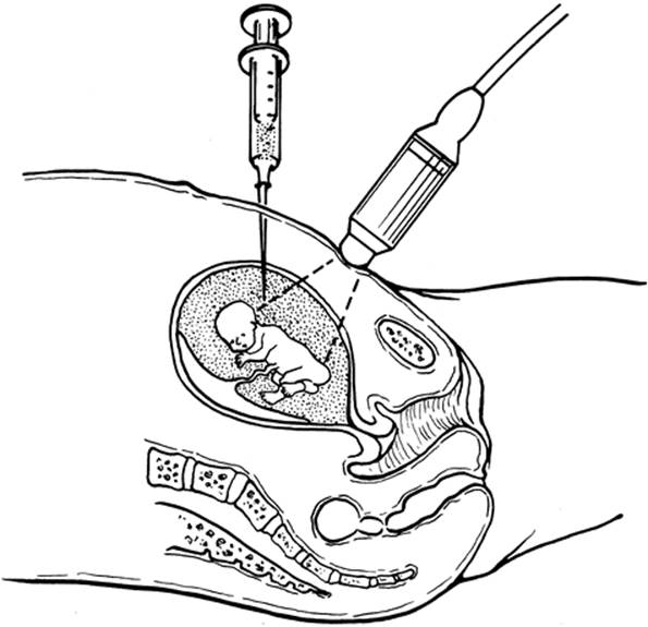
Amniocentesis was introduced to the United States in the 1960s, and it is the most extensively used fetal sampling technique. Genetic amniocentesis is performed routinely at approximately 15 weeks of gestation when the amniotic fluid volume is approximately 200 mL. At this gestational age, ultrasonographic examination cannot detail all of the fetal anatomy, but it can reliably ascertain dates or rule out multiple gestations. Typically, 20 mL of fluid is removed with a 20- to 22-gauge needle using a transabdominal approach with ultrasound guidance (Fig. 6.12). Biochemical testing can be performed on the fluid as indicated. Amniotic fluid α-fetoprotein levels are obtained routinely to screen for open fetal defects, and fetal cells can be grown for karyotype determination or for DNA assays. DNA assays that use the PCR to amplify small amounts of DNA allow direct analysis of amniotic fluid. Roughly one third of the amniocenteses must be performed transplacentally. In most operators' experience, this has not been associated with substantially increased risk if care is taken to avoid major fetal vessels. The transplacental approach is associated with a slightly higher incidence of Rh sensitization. With either approach, it is imperative that Rh-negative women who may be carrying an Rh-positive fetus receive RhoGAM.
|
|
|
FIG. 6.12. Amniocentesis. |
With multiple gestations, it is usually possible to sample each of the gestations. Indigo carmine dye can be placed in the sac after the amniocentesis is completed to prevent tapping the same sac twice. Biochemical assays of amniotic fluid are somewhat harder to interpret in multiple gestations, because many biologic molecules can diffuse from one sac into the other.
Few women describe amniocentesis as terribly painful. Those who do frequently experience a uterine contraction at the time the needle is inserted. It is not unusual to have some cramping or a bruised feeling at the site after the procedure. Vaginal spotting or amniotic fluid leakage occurs in 1% to 2% of cases. After a routine amniocentesis, the fluid usually stops leaking within 2 to 3 days. Even when the amniotic fluid volume becomes markedly decreased, miscarriage is not inevitable, because the membranes usually seal and the amniotic fluid can reaccumulate within a week, allowing the pregnancy to progress normally.
As ultrasonographic equipment has improved, the maternal risk incurred with amniocentesis has decreased. Symptomatic amnionitis occurs in fewer than 1 of 1,000 patients. Serious maternal bowel or vascular injuries are extremely rare. The procedure-related rate of fetal loss after amniocentesis generally is quoted as 0.5% (1 in 200), but many centers are reporting lower rates.
Chorionic Villus Sampling
Chorionic villus sampling (CVS) is a diagnostic technique that was introduced to the United States in the mid-1980s. With this technique, a small sample of the chorionic villi is taken for examination of chromosomal status, biochemical assays, or DNA tests. Assays depending on analysis of amniotic fluid such as α-fetoprotein cannot be performed on a chorionic villus sample.
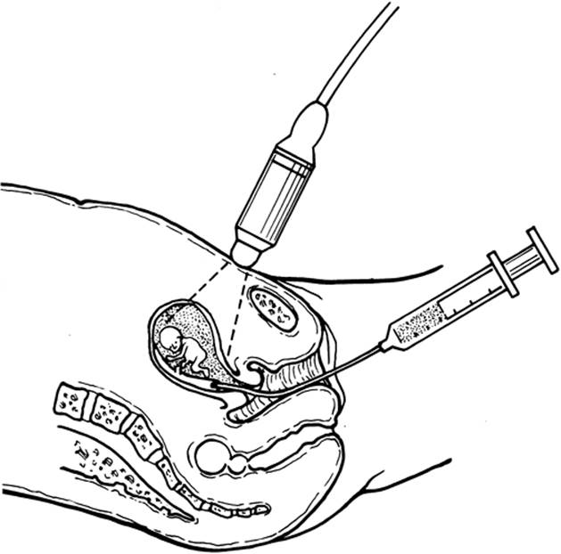
CVS usually is accomplished by the transcervical or transabdominal route. Occasionally a transvaginal CVS is performed with the uterus extremely retroflexed. A transcervical CVS usually is performed between 9 and 12 weeks of gestation, at which time a plastic catheter, approximately 1.5 mm in diameter, is passed through the cervix and then directed toward the placental mass under continuous ultrasound guidance (Fig. 6.13). Between 10 and 20 mg of villi are aspirated through this catheter by negative pressure using a syringe. Transabdominal CVS is performed using an 18- to 20-gauge spinal needle passed into the thickest portion of the placenta that is readily assessable (Fig. 6.14). Villi are aspirated into a syringe. This procedure can be performed throughout gestation.
|
|
|
FIG. 6.13. Transvaginal chorionic villus sampling. |
|
|
|
FIG. 6.14. Transabdominal chorionic villus sampling. |
Transabdominal CVS is considered easier to learn and safer, but patient acceptance appears to be lower. Theoretically, transcervical CVS would have a greater risk of infection, although this has not been borne out by large surveys. In most series, a larger sample is obtained with transcervical CVS, but more passes are required to obtain this sample. Transcervical procedures require more uterine manipulation, and bleeding or leakage of fluid is more common during and after the procedure. Most laboratories report a greater level of maternal cell contamination with transcervical CVS, although this is rarely a clinically important issue. Those performing CVS should be facile with both techniques, because patient anatomy frequently dictates which is the optimal technique. CVS compares favorably with amniocentesis with regard to safety. Two large National Institutes of Health cooperative trials found a procedure-related loss rate of approximately 0.8%.
In approximately 2% of first-trimester chorionic villus samples, a discrepancy is found between the cytogenetic analysis of the placenta and that of the fetus. Frequently, a second invasive procedure, usually amniocentesis or fetal blood sampling, is required to determine whether the fetus is affected. This phenomenon is called confined placental mosaicism. Pregnancies in which confined placental mosaicism is found by CVS may be at risk for spontaneous abortion, perinatal loss, or intrauterine growth retardation. The reported rates of loss have ranged from 3.6% to 16.7%. In chromosomally abnormal conceptuses, a mosaic normal cell line in the placenta may be the factor that allows prolonged survival of aneuploid fetuses. Kalousek studied 14 placentas from live-born or from terminated pregnancies with trisomy 13 or 18, and found the placentas were all mosaic for, or contained only, diploid cells.
There have been several reports of increased incidents of limb anomalies when fetuses have undergone very early CVS using the transabdominal approach. These reports are of concern, because the affected children have a relatively distinctive pattern of malformation, and it is biologically plausible that their anomalies may be related to CVS. The absolute number of fetuses with this problem is small, but the publicity regarding these findings has caused many women to avoid CVS.
The Centers for Disease Control performed a multistate case-control study to assess and quantify the risk for specific limb deficiencies associated with CVS. Between 1988 and 1992, 131 infants with nonsyndromic limb deficiency, born to mothers 34 years of age or older, were reported in seven population-based birth defect surveillance programs. Control subjects were 131 infants with other birth defects. They found that exposure to CVS was associated with a six-fold increase in risk for transverse digital deficiency (odds ratio = 6.4; 95% confidence interval, 1.1 to 38.6). The data showed a significant trend toward increased risk with earlier gestational exposure. The CDC estimates that the absolute risk for transverse digital deficiency in infants after CVS is approximately 1 per 3,000. Most feel that the actual risk is significantly lower and that there is minimal to no risk when CVS is performed after 70 days of gestation. Further studies are needed to determine whether the problem is specific to CVS or whether the same risk affects other invasive first-trimester diagnostic manipulations.
Fetal Blood Sampling and Fetal Biopsy
Originally, fetal blood was sampled by inserting a needle into the placenta; the blood obtained was usually a mixture of fetal and maternal blood, which limited its usefulness. In 1977, fetoscopy came into use, allowing a needle to be placed in a cord vessel under direct sonographic visualization. Pure fetal samples could be obtained, but fetoscopy requires special equipment and expertise, and the procedure-related loss rate was between 3% and 7%. With improvement in ultrasonographic imaging during the mid-1980s, cordocentesis can be performed in a manner similar to amniocentesis (Fig. 6.15).
|
|
|
FIG. 6.15. Cordocentesis (fetal blood sampling). |
A transplacental route usually is preferred, and a spinal needle is advanced under ultrasound guidance into a vessel with cord insertion into the placenta. Cordocentesis usually is not performed until after 17 weeks gestation. Depending on the indication, procedure-related loss rates of as low as 1% have been reported with no observed increase in the rate of preterm delivery. Typically, no maternal sedation or antibiotics are required. In most cases, a fetal sample can be obtained on the first attempt, usually in less than 10 minutes, and any substance measurable in adult blood can be assayed in fetal blood. Most cordocenteses are performed to obtain a fetal karyotype because of fetal anomalies or to determine the fetal hematocrit to assess isoimmunization or severe fetal anemia. Fetal platelet counts, acid-base status, antibody levels, and blood chemistries can be assayed as indicated. Hematologic values are checked routinely (particularly the mean corpuscular volume which is higher in fetus than mother) to be certain that the blood obtained is fetal. A Kleihauer test can be performed to check for maternal blood contamination.
A variety of other tissues, particularly fetal skin, liver, and muscle, have been sampled prenatally to diagnose a genetic disorder using either electron microscopy or biochemical analysis. Such biopsies are necessary if a genetic abnormality is expressed only in certain tissues and the causative gene is unknown. Now that the molecular basis of many of these disorders is known, simpler DNA assays using villi, amniocytes, or blood usually obviate the need for tissue biopsy. Initially, fetal biopsies were performed using fetoscopy, but they now are performed under ultrasound guidance. Typically, local anesthesia, maternal sedation (with or without fetal paralysis), and prophylactic antibiotics are used for tissue biopsy procedures. As with any invasive procedure, RhoGAM is necessary for the nonsensitized Rh-negative mother who may be carrying an Rh-positive fetus.
Early Amniocentesis
Early amniocentesis is similar to amniocentesis at the “traditional” gestational age, except that the procedure is performed at 10 to 12 weeks of gestation. Early amniocentesis was proposed as an alternative first-trimester prenatal diagnostic technique, but a large Canadian prospective study demonstrated an unacceptable rate of procedure-related malformations in the exposed pregnancies.
Preimplantation Diagnosis
Various methods of diagnosis before a pregnancy is formally established are becoming available in the 1990s. Someday these extremely early diagnoses may be necessary to allow initiation of genetic therapy treatments. For many couples, preimplantation diagnosis provides an alternative to selective pregnancy termination, allowing them to avoid the moral issues and the psychological trauma that accompanies termination of a wanted pregnancy.
For most genetic conditions, carriers have both normal and abnormal gametes, and chance determines whether an abnormal gamete is incorporated into the conceptus. Aided by the rapid progress in assisted reproductive technologies and molecular diagnostic techniques, it has become possible to test gametes in vitro and select healthy gametes for fertilization. Sperm sorting has been accomplished using molecular probes tagged with laser-activated dyes and using separation techniques such as flow cytometry, but current approaches usually cause unacceptable damage to the sperm. Greater success has been possible in genotyping oocytes, which are larger and more resistant to damage. Oocyte diagnosis takes advantage of the unique properties of female meiosis; unlike sperm, oocytes conveniently discard their unused genetic material in the form of polar bodies. For a heterozygous woman, the discarded genetic material can be tested to see whether it contains the abnormal allele. If it does, then the oocyte must contain the normal allele. Conversely, if the polar body tests positive for the normal allele, the oocyte must contain the abnormal allele. Only the normal oocytes are then fertilized. Contamination with cumulus cells adherent to the exterior surface of the zona pellucida can lead to errors.
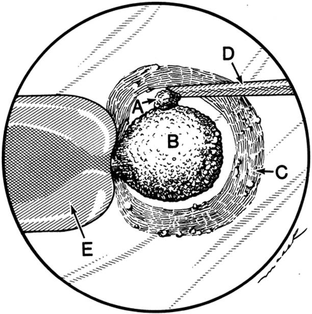
Polar body biopsy can be performed only in conjunction with in vitro fertilization. The first polar body has no essential function and contains no embryonic material; it is small and relatively easy to remove using micromanipulation techniques (Fig. 6.16). Theoretically, DNA testing results can be obtained before fertilization, leaving a long period for confirmatory studies before implantation. Aneuploidy screening can be incorporated as well, at least for maternal meiosis I nondisjunction. The main disadvantages are that polar body biopsy is an indirect assay of the oocyte and that it can be used only for maternal carriers. In addition, the further away from the centromere the gene is located, the more likely recombination is to occur, making polar body biopsy results indeterminate. Polar bodies that are heterozygous need to be discarded or the oocytes biopsied again to remove the second polar body.
|
|
|
FIG. 6.16. Polar body biopsy. |
Initial success has been greater with “selective implantation” protocols in which the diagnosis is made during the first week after fertilization but before implantation. Preimplantation embryos can be grown in vitro and biopsies obtained after the first few cleavage divisions. There is much experience with this technique in animal research, and embryo splitting at this stage has been used extensively for diagnosis in the cattle and sheep industry. The eight-cell preembryo is probably at the ideal stage for biopsy. Cells are still independent and totipotent, they have not developed gap junctions which will make them adherent, and damage is tolerated relatively well. By this stage, some of the embryonic genes have begun to function, and it may become possible to perform some biochemical microassays. This technique is limited by the difficulty in removing blastomeres, which are larger than polar bodies, and the short time to work with the sample before the chance of successful implantation begins to lessen. It has proved difficult to freeze spare embryos after biopsies and, as with other preimplantation techniques, there is the possibility of sperm or cumulus cell contamination.
Attempts have also been made to perform testing 5 days after fertilization, when the preembryo has reached the blastocyst stage. The blastocyst consists of roughly 120 cells that are mostly trophoblastic tissue, but the inner cell mass that eventually becomes the embryo is clearly visible. Only the best laboratories have had any success culturing human preembryos to this developmental stage in vitro. As an alternative, investigators have tried to lavage naturally conceived blastocysts from the uterus during the 2 to 3 days when the conceptus is normally free-floating in the endometrial cavity before implantation. Unfortunately, it has proved exceedingly difficult to obtain multiple blastocysts by lavage after superovulation.
Various methods of sampling blastocyst cells have shown success in animal models including bisection, aspiration of the cavity, and excision of cells herniating through the zona pellucida. The latter is achieved after mechanical disruption of the zona to cause premature herniation or after spontaneous hatching. The advantages of blastocyst biopsy include the relative differentiation of the cells, greater cell number, better transfer efficiency, and self-selection of the healthiest embryos. Lavage of naturally conceived blastocysts offers a potentially “low-tech” approach to preimplantation diagnosis. Perhaps contrary to expectations, disruption of zona pellucida may enhance hatching. Technical limitations include the small number of blastocysts available and their limited incubation time. The cells are very adherent to each other, increasing the risk of damage to the inner cell mass. As with CVS mosaicism, there is the possibility that the trophectoderm does not reflect the fetal karyotype or biochemical status.
Fetal Cells in Maternal Circulation
The newest fetal sampling technique that shows promise involves separating fetal cells that occur naturally in the maternal circulation. Nucleated erythrocytes, fetal leukocytes, and syncytiotrophoblast cells are found in the maternal circulation during most pregnancies, from as early as 6 weeks gestation. It is possible to use a separation technique such as flow cytometry to establish an enriched population of fetal cells and then assay these cells for fetal mutations using a technique such as DNA amplification. Certain cell types have a long life span in the maternal circulation, allowing persistence of cells from prior pregnancies, and confusing results are possible. “Vanished” abnormal co-twins can lead to diagnostic errors. Ultimately, this method may provide a noninvasive, reliable screen for aneuploidy that is inexpensive enough to use in low-risk populations.
Cystic Fibrosis Screening
The CFTR gene was discovered in 1989, and more than 1,000 mutations which can lead to CF have been identified. Couples who both carry the CF gene would have a 1 in 4 chance of delivering a child with CF. Cystic fibrosis causes pulmonary and gastrointestinal disease of varying severity. Most patients with CF have substantial illness and shortened life span and require lifelong medical care.
Screening is now widely available for the most frequent CF mutations. Recently ACOG advised that DNA screening for CF should be made available to allcouples seeking preconception or prenatal care. The ACOG/American College of Medical Genetics publication entitled Cystic Fibrosis Carrier Testing: the Decision is Yours can be helpful for this purpose. Because Caucasians have a higher rate of CF (particularly European or Ashkenazi Jewish), obstetric care providers are advised to offer screening specifically to these couples and to record in the medical record the couple's decision on whether to be screened (Table 6.15.).
|
|
|
TABLE 6.15. Cystic Fibrosis: carrier rates in the United States |
Prenatal Treatment and Gene Therapy
As we begin to understand the molecular mechanisms by which genes cause disease, we will have the opportunity to design and apply preventive, health maintaining measures. Some genetic conditions can be treated by giving patients the protein they are missing or by stimulating a function that is not performing properly (e.g., growth hormone deficiency, diabetes).
More sophisticated gene therapies are being tested. Finding the CF gene enabled the disease to be “cured” in the test tube and improved in animal models. Scientists may be able to design proteins or antisense RNAs that can be used as a drug to block the effect of abnormal genes or kill cancer cells. Modified viruses may be used to insert corrected genetic instructions. Prenatal gene therapy with stem cell transplantation has been attempted for severe combined immunodeficiency with some preliminary success.
Many genetic conditions need to be treated prenatally. Although in utero treatment is experimental, prenatal treatment of disorders with vitamin-dependent or responsive cofactors has been successful. For instance, prenatal treatment of the vitamin B12-responsive form of methylmalonic acidemia by administration of 10 mg per day of vitamin B12 has improved the biochemical defect. Similarly, infants with biotin-responsive multiple carboxylase deficiency have been aided by maternal biotin supplementation. In these rare disorders, prenatal treatment can mean the difference between life and death for affected infants. Dietary restriction of galactose in mothers who are at risk of delivering a galactosemic infant, and dietary restriction of phenylalanine in mothers who themselves have phenylketonuria, are helpful means of preventing the devastating effects of these metabolic conditions. Gene therapy and prenatal tissue transplantation are likely to rapidly expand the therapeutic options for treating and preventing metabolic disease.
GENETICS IN GYNECOLOGIC DISORDERS
Genes play an important role in the pathogenesis of many common gynecologic disorders. Molecular genetic investigations of persons with gonadal dysgenesis and pseudohermaphroditism have defined many aspects of human sexual differentiation. Because it is the smallest chromosome, the Y chromosome became the first human chromosome to be mapped completely.
Genetic testing for susceptibility to ovarian cancer is rapidly becoming part of routine practice. Most cancer is clonal in origin, meaning it arises from a single aberrant cell. Cytogenetic or molecular alterations are observed uniformly in malignant cells. Although some of these changes appear to be random events occurring in rapidly dividing cells, other specific genetic changes play an etiologic role in development of certain cancers. Particular mutations may be either germinal (i.e., inherited) or somatic (i.e., acquired). Either can be seen in familial cancer clusters: germinal because of segregation within the family of a cancer-causing mutation and somatic because of shared environmental exposures to carcinogens. Mendelian transmission of cancer predisposition usually is observed as multifocal and early-onset disease. Typically, cancer-predisposing mutations are found to overexpress protooncogenes that normally drive important cell functions or to inactivate tumor suppressor genes that normally exert a protective effect.
Genetic testing for BRCA1 and BRCA2 mutations is now recommended to most women with invasive ovarian cancer. Approximately 10% of patients with ovarian cancer will have a positive test result, including 4% of women without a family history of ovarian cancer. Women with the BRCA mutation have better ovarian cancer survival rates than women without the mutation, possibly due to enhanced susceptibility to chemotherapy. A variety of strategies for prevention of ovarian cancer in relatives at risk, including chemoprevention and prophylactic oophorectomy, have shown some efficacy.
Common gynecologic diseases such as endometriosis and polycystic ovary syndrome are familial, and genes involved in these conditions are likely to be discovered over the next few years. Age at menopause, susceptibility to hot flushes and osteoporosis, susceptibility to pelvic relaxation, and susceptibility to chronic vaginitis are likely to have genetic components, as well. Disease gene discoveries related to these conditions may suggest novel diagnostic and therapeutic approaches.
TRENDS
The genome project promises to provide us with the most important information in human biology. Technologies developed for the genome project and the genomic sequence, itself, will provide the basis for much of biomedical research in the next century. The possibilities for understanding normal development, disease predisposition, and cancer are staggering. The ability to obtain an accurate prenatal diagnosis will expand exponentially over the next few decades. Gene therapy is becoming a reality faster than anyone thought possible. The challenge is for the obstetrician-gynecologist to stay abreast of all these developments and to educate patients about developments that can influence their care.
SUMMARY POINTS
· Most obstetric and gynecologic diseases show a polygenic, multifactorial pattern of inheritance. One in twenty newborns has a diagnosable genetic disorder.
· A thorough family history is currently the most important part of a genetic evaluation, but laboratory screening of the general population is becoming available for an increasing number of genetic conditions.
· Accurate prenatal diagnosis is now possible for hundreds of genetic conditions through ultrasonographic and genetic testing.
· The Human Genome Project is providing new information each month, making it difficult for care providers to keep abreast of all the new developments, especially for rarer diseases. Reference to current on-line data and liberal referral to genetic counselors and geneticists is necessary when encountering rare conditions.
· Over the next decade, genetic testing will continue to become less invasive, and there will be greater opportunities to prevent the morbidity of genetic disease through prenatal and presymptomatic treatments.
REFERENCES
ACOG Practice Bulletin. Clinical Management Guidelines for Obstetrician-Gynecologists. Prenatal diagnosis of fetal chromosomal abnormalities. Obstet Gynecol 2001;97(5 Pt 1)[Suppl]:1–12.
Aitken DA, Wallace EM, Crossley JA, et al. Dimeric inhibin A as a marker for Down's syndrome in early pregnancy. N Engl J Med 1996;334:1231–1236.
Alfirevic Z. Early amniocentesis versus transabdominal chorion villus sampling for prenatal diagnosis. Cochrane Database Syst Rev 2000;(2):CD000077.
Cohen DP, Layman LC. Relevance of molecular medicine to clinical obstetrics and gynecology. Obstet Gynecol Surv 1997;52:73–80.
Evans MI, Levy HL. The future of newborn screening belongs to obstetricians. Acta Paediatr Suppl 1999;88:55–57.
Forest JC, Masse J, Moutquin JM. Screening for Down syndrome during first trimester: a prospective study using free β-human chorionic gonadotropin and pregnancy-associated plasma protein A. Clin Biochem 1997;30:333–338.
Genetic testing for cystic fibrosis. National Institutes of Health Consensus Development Conference Statement on genetic testing for cystic fibrosis. Arch Intern Med 1999;159:1529–1539.
Gupta GK, Bianchi DW. DNA diagnosis for the practicing obstetrician. Obstet Gynecol Clin North Am 1997;24:123–142.
Hall JG. Genomic imprinting and its clinical implications. New Engl J Med 1992;326:827–829.
Jenkins TM, Wapner RJ. First trimester prenatal diagnosis: chorionic villus sampling. Semin Perinatol 1999;23:403–413.
Krantz DA, Larsen JW, Buchanan PD, et al. First-trimester Down syndrome screening: free β-human chorionic gonadotropin and pregnancy-associated plasma protein A. Am J Obstet Gynecol 1996;174:612–616.
Milunsky JM, Milunsky A. Genetic counseling in perinatal medicine. Obstet Gynecol Clin North Am 1997;24:1–17.
Miny P, Tercanli S, Holzgreve W. Developments in laboratory techniques for prenatal diagnosis. Curr Opin Obstet Gynecol 2002;14:161–168.
Narod SA, Boyd J. Current understanding of the epidemiology and clinical implications of BRCA1 and BRCA2 mutations for ovarian cancer. Curr Opin Obstet Gynecol 2002;14:19–26.
Nyberg DA, Souter VL. Sonographic markers of fetal aneuploidy. Clin Perinatol 2000;27:761–789.
Pergament E. The application of fluorescence in-situ hybridization to prenatal diagnosis. Curr Opin Obstet Gynecol 2000;12:73–76.
Pertl B, Bianchi DW. Fetal DNA in maternal plasma: emerging clinical applications. Obstet Gynecol 2001;98:483–490.
Sanders RC. Prenatal diagnosis of structural abnormalities. Curr Opin Obstet Gynecol 1991;3:259–265.
Schimpf MO, Domino SE. Implications of the Human Genome Project for obstetrics and gynecology. Obstet Gynecol Surv 2001;56:437–443.
Schrock E, du Manoir S, Veldman T, et al. Multicolor spectral karyotyping of human chromosomes. Science 1996;273:494–497.
Simpson JL, Golbus MS. Genetics in obstetrics & gynecology, second ed. Philadelphia: WB Saunders Company, 1992.
Souter VL, Nyberg DA. Sonographic screening for fetal aneuploidy: first trimester. J Ultrasound Med 2001;20:775–790.
Swisher E. Hereditary cancers in obstetrics and gynecology. Clin Obstet Gynecol 2001;44:450–463.
Verlinsky Y, Handyside A, Grifo J, et al. Preimplantation diagnosis of genetic and chromosomal disorders. J Assist Reprod Genet 1994;11:236–243.
Wilson RD. Prenatal evaluation for fetal surgery. Curr Opin Obstet Gynecol 2002;14:187–193.
Yang EY, Flake AW, Adzick NS. Prospects for fetal gene therapy. Semin Perinatol 1999;23:524–534.